A ferroptosis-based panel of prognostic biomarkers for Amyotrophic Lateral sclerosis - Nature
←
→
Page content transcription
If your browser does not render page correctly, please read the page content below
www.nature.com/scientificreports

                                                                                                                              Corrected: Author Correction

                        OPEN            A ferroptosis–based panel
                                        of prognostic biomarkers for
                                        Amyotrophic Lateral Sclerosis
Received: 2 July 2018                   David Devos 1,2, Caroline Moreau1, Maeva Kyheng3, Guillaume Garçon4,
Accepted: 14 January 2019               Anne Sophie Rolland2, Hélène Blasco5, Patrick Gelé6, T. Timothée Lenglet7,
Published online: 27 February 2019      C. Veyrat-Durebex5, Philippe Corcia8, Mary Dutheil2, Peter Bede9,10, Andreas Jeromin11,
                                        Patrick Oeckl12, Markus Otto 12, Vincent Meininger13, Véronique Danel-Brunaud1,
                                        Jean-christophe Devedjian2, James A. Duce14,15 & Pierre François Pradat9,13,16
                                        Accurate patient stratification into prognostic categories and targeting Amyotrophic Lateral Sclerosis
                                        (ALS)-associated pathways may pave the way for promising trials. We evaluated blood-based
                                        prognostic indicators using an array of pathological markers. Plasma samples were collected as part of
                                        a large, phase III clinical trial (Mitotarget/TRO19622) at months 1, 6, 12 and 18. The ALSFRS-r score was
                                        used as a proxy of disease progression to assess the predictive value of candidate biological indicators.
                                        First, established clinical predictors were evaluated in all 512 patients. Subsequently, pathologic
                                        markers, such as proxies of neuronal integrity (Neurofilament light chain and phosphorylated
                                        heavy chain), DNA oxidation (8-oxo-2′-desoxyguanosine), lipid peroxidation (4-hydroxy-2-nonenal,
                                        isoprostane), inflammation (interleukin-6) and iron status (ferritin, hepcidin, transferrin) were assessed
                                        in a subset of 109 patients that represented the whole cohort. Markers of neuronal integrity, DNA
                                        and lipid oxidation, as well as iron status at baseline are accurate predictors of disability at 18-month
                                        follow-up. The composite scores of these markers in association with established clinical predictors
                                        enable the accurate forecasting of functional decline. The identified four biomarkers are all closely
                                        associated with ‘ferroptosis’, a recently discovered form of programmed cell death with promising
                                        therapeutic targets. The predictive potential of these pathophysiology-based indicators may offer
                                        superior patient stratification for future trials, individualised patient care and resource allocation.

                                        Amyotrophic lateral sclerosis (ALS) is a relentlessly progressive neurodegenerative condition with no effective
                                        disease-modifying therapy. Late recruitment to pharmacological trials, clinical heterogeneity, and lack of specific
                                        monitoring markers are some of the main barriers to successful drug development. Accurate patient stratifica-
                                        tion into prognostic categories1 and targeting ALS-associated pathways may pave the way for promising phase

                                        1
                                          Department of Neurology, ALS Center, Lille University, INSERM UMRS_1171, University Hospital Center, LICEND
                                        COEN Center, Lille, France. 2Department of Medical Pharmacology, Lille University, INSERM UMRS_1171, University
                                        Hospital Center, LICEND COEN Center, Lille, France. 3Department of Biostatistics, Lille University, University Hospital
                                        Center, Lille, France. 4Univ. Lille, CHU Lille, Institut Pasteur de Lille, EA4483 IMPECS-IMPact de l’Environnement
                                        Chimique sur la Santé humaine, Lille, France. 5Université François-Rabelais, Inserm U930, Laboratoire de Biochimie,
                                        CHRU de Tours, France. 6CRB/CIC1403, Université de LILLE, Lille, France. 7APHP, Department of Neurophysiology,
                                        Pitié-Salpêtrière Hospital, Paris, France. 8Centre Constitutif SLA, Tours-Fédération des centres SLA Tours-Limoges,
                                        LITORALS, Tours, France. 9Biomedical Imaging Laboratory, CNRS, INSERM, Sorbonne University, Paris, France.
                                        10
                                           Computational Neuroimaging Group, Academic Unit of Neurology, Trinity College Dublin, Dublin, Ireland.
                                        11
                                          Quanterix, Lexington, Massachusetts, USA. 12Department of Neurology, Ulm University Hospital, Oberer Eselsberg
                                        45, 89081 Ulm, Ulm, Germany. 13APHP, Department of Neurology, Paris ALS Center, Pitié Salpêtrière Hospital, Paris,
                                        France. 14ALBORADA Drug Discovery Institute, University of Cambridge, Cambridge Biomedical Campus, Hills Road,
                                        Cambridge, CB2 0AH, UK. 15School of Biomedical Sciences, Faculty of Biological Sciences, University of Leeds,
                                        Leeds, West Yorkshire, United Kingdom. 16Northern Ireland Centre for Stratified Medicine, Biomedical Sciences
                                        Research Institute Ulster University, C-TRIC, Altnagelvin Hospital, Derry/Londonderry, United Kingdom. Caroline
                                        Moreau and Maeva Kyheng contributed equally. Correspondence and requests for materials should be addressed to
                                        D.D. (email: david.devos@chru-lille.fr)

        Scientific Reports | (2019) 9:2918 | https://doi.org/10.1038/s41598-019-39739-5                                                                      1
www.nature.com/scientificreports/ www.nature.com/scientificreports
Unadjusted Adjusted*
Factors at baseline Coefficient β ± SE P-value Coefficient β ± SE P-value
NfLa −0.05 (0.005)www.nature.com/scientificreports/ www.nature.com/scientificreports
Effect on ALSFRS-r progression
Factors at baseline Coefficient β ± SE p
NfLa −0.02 (0.005) 0.004
4-HNEa −0.11 (0.02)www.nature.com/scientificreports/ www.nature.com/scientificreports
value of these biomarkers, they may aid patient stratification for future phase trials. From a clinical perspective,
they may also contribute to precision care planning, resource allocation and management of individual patients.
The nervous system is particularly rich in lipids and products of lipid peroxidation such as 4-HNE may repre-
sent an important and currently under evaluated proxy of disease activity. It is noteworthy that the highly reactive
cytotoxic 4-HNE irreversibly cross-links proteins such as neurofilaments21. Changes in FT, an indicator of brain
iron status, may represent an additional aetiological factor promoting free radical production. Increased lipid
peroxidation and iron accumulation are key components of iron dependent programmed cell death; ferroptosis2.
In conclusion, our findings indicate that markers of ferroptosis in ALS are associated with clinical decline.
Elevated NfL and 8-oxo-dG levels on the other hand are secondary to axonal skeleton disintegration and DNA
fragmentation, likely a downstream effect of ferroptosis. These observations need to be replicated in larger pop-
ulations and the predictive value of these markers need to be examined on survival. The characterisation of
these mechanisms and the development of ferroptosis-based markers is particularly timely, as iron chelation4
and anti-ferroptotic therapy2,22 are currently under investigation for a range of neurodegenerative conditions
including ALS.
Methods
Mitotarget/TRO19622 was a negative, randomized, double-blinded, placebo-controlled phase III trial for ole-
soxime (NCT:00868166) that included 512 ALS patients from 15 European centers20. All experiments were per-
formed in accordance with French and European guidelines and regulations. Following approval from a local
ethics committee at Assistance Publique Hôpital Pitié-Salpêtrière and informed consent from each participant,
data were collected every 3 months during the 18-month trial period. Participants were diagnosed with either
‘probable’ or ‘definite’ ALS according to the revised El Escorial criteria, and only patients with symptom duration
of more than 6 and less than 36 months were enrolled. In addition to riluzole, patients received olesoxime or
placebo.
A subgroup of 109 patients was randomly selected from the 286 patients that completed the 18-month-follow
up assessment. This enabled longitudinal functional assessment (ALSFRS-r)23, but precluded survival analyses.
All recently identified candidate predictors1 (Table e-1) were included in a prediction model with the exception of
frontotemporal dementia (due to a lack of phenotype in this cohort) and the presence of C9orf72 hexanucleotide
repeat expansions (data not available).
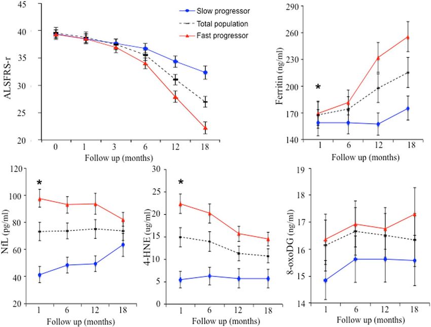
Finally the population was also divided into two groups of disease decline (i.e. slow and fast), according to a
median in the ALSFRS-r score decrease rate from time of inclusion to 18 months.
Plasma samples were obtained at 1, 6, 12 and 18 months after enrolment. Standard ‘Safety parameters’ were
monitored during the trial (Table e-2). The ‘Specific parameters’ were measured in duplicate using commer-
cially available kits for NfL (NF-light Kit Advantage, Quanterix, Lexington, MA, USA), pNfH (Neurofilament
ELISA, Euroimmun AG, Lübeck, Germany), 8-oxo-dG (ELISA Kit, Abcam, Cambridge, UK: ab201734),
™
4-HNE (OxiSelect HNE Adduct Competitive ELISA Kit, Cell Biolabs, Inc., San Diego, CA, USA: STA-838),
8-isoprostane (ELISA Kit, Abcam, Cambridge, UK: ab175819), interleukin-6 (Human Magnetic Luminex
Screening Assay, R&D Systems - Bio-Techne, Lille, France:HUVF4Lrv), ferritin (human ELISA Kit, Abcam,
Cambridge, UK: ab108698), transferrin (human ELISA Kit, Abcam, Cambridge, UK: ab108911) and hepcidin
(Human Quantikine ELISA Kit, R&D Systems - Bio-Techne, Lille, France: DHP250).
Statistical analyses
The predictive value of the clinical and ‘safety parameters’ on the ALSFRS-r score was investigated using bivariate
linear mixed models with randomized coefficients (Table e-2). The fixed effects in the model included time, base-
line characteristics and their interaction. All baseline characteristics that associated either alone (p < 0.05) or in
interaction with time (p < 0.10) were included in a multivariable linear mixed model (Table e-3).
The predictive value of the ‘specific parameters’ on the ALSFRS-r score was investigated using linear mixed
modelling. Random intercept was performed before and after adjustment to the baseline characteristics asso-
ciated with ALSFRS-r progression and allocated treatment group (Table 1). All parameters associated with
ALSFRS-r progression in the adjusted models (interaction with time < 0.10) were included in the multivariable
linear mixed model (Table 2).
Finally, the association between specific parameters at baseline and progression of other parameters (e.g
MMT, SVC, BMI) were investigated by bivariate linear mixed modelling with random intercept.
All statistical tests were performed at the 2-tailed α level of 0.05. Data were analysed using SAS version 9.4
[SAS Institute Inc., Cary, NC 27513, USA].
Data Availability
All the anonymized data and the statistical analyses will be shared by request from any qualified investigator.
References
1. Westeneng, H. J. et al. Prognosis for patients with amyotrophic lateral sclerosis: development and validation of a personalised
prediction model. Lancet Neurol. 17, 423–433 (2018).
2. Stockwell, B. R. et al. Ferroptosis: A Regulated Cell Death Nexus Linking Metabolism, Redox Biology, and Disease. Cell. 171,
273–285 (2017).
3. Chen, L., Hambright, W. S., Na, R. & Ran, Q. Ablation of the Ferroptosis Inhibitor Glutathione Peroxidase 4 in Neurons Results in
Rapid Motor Neuron Degeneration and Paralysis. J Biol Chem. 290, 28097–106 (2015).
4. Moreau, C. et al. Could Conservative Iron Chelation Lead to Neuroprotection in Amyotrophic Lateral Sclerosis? Antioxid Redox
Signal. 29, 742–748 (2018).
5. Feneberg, E. et al. Multicenter evaluation of neurofilaments in early symptom onset amyotrophic lateral sclerosis. Neurology. 90,
e22–e30 (2018).
Scientific Reports | (2019) 9:2918 | https://doi.org/10.1038/s41598-019-39739-5 4www.nature.com/scientificreports/ www.nature.com/scientificreports
6. Skillbäck, T., Mattsson, N., Blennow, K. & Zetterberg, H. Cerebrospinal fluid neurofilament light concentration in motor neuron
disease and frontotemporal dementia predicts survival. Amyotroph Lateral Scler Frontotemporal Degener. 18, 397–403 (2017).
7. Lu, C. H. et al. Neurofilament light chain: A prognostic biomarker in amyotrophic lateral sclerosis. Neurology. 84, 2247–57 (2015).
8. Gaiani, A. et al. Diagnostic and Prognostic Biomarkers in Amyotrophic Lateral Sclerosis: Neurofilament Light Chain Levels in
Definite Subtypes of Disease. JAMA Neurol. 74, 525–532 (2017).
9. Poesen, K. et al. Neurofilament markers for ALS correlate with extent of upper and lower motor neuron disease. Neurology. 88,
2302–2309 (2017).
10. Mitsumoto, H. et al. Oxidative stress biomarkers in sporadic ALS. Amyotroph Lateral Scler. 9, 177–83 (2008).
11. Blasco, H. et al. Panel of Oxidative Stress and Inflammatory Biomarkers in ALS: A Pilot Study. Can J Neurol Sci. 44, 90–95 (2017).
12. Simpson, E. P., Henry, Y. K., Henkel, J. S., Smith, R. G. & Appel, S. H. Increased lipid peroxidation in sera of ALS patients: a potential
biomarker of disease burden. Neurology. 62, 1758–65 (2004).
13. Moreau, C. et al. Elevated IL-6 and TNF-alpha levels in patients with ALS: inflammation or hypoxia? Neurology. 65, 1958–60 (2005).
14. Hu, Y. et al. Increased peripheral blood inflammatory cytokine levels in amyotrophic lateral sclerosis: a meta-analysis study. Sci Rep.
7, 9094 (2017).
15. Su, X. W. et al. Biomarker-based predictive models for prognosis in amyotrophic lateral sclerosis. JAMA Neurol. 70, 1505–11 (2013).
16. Nadjar, Y. et al. Elevated serum ferritin is associated with reduced survival in amyotrophic lateral sclerosis. PLoS One. 7, e45034
(2012).
17. Veyrat-Durebex, C. et al. Iron metabolism disturbance in a French cohort of ALS patients. Biomed Res Int. 2014, 485723 (2014).
18. Ikeda, K., Hirayama, T., Takazawa, T., Kawabe, K. & Iwasaki, Y. Relationships between disease progression and serum levels of lipid,
urate, creatinine and ferritin in Japanese patients with amyotrophic lateral sclerosis: a cross-sectional study. Intern Med. 51, 1501–8
(2012).
19. Patin, F. et al. Biological follow-up in amyotrophic lateral sclerosis: decrease in creatinine levels and increase in ferritin levels predict
poor prognosis. Eur J Neurol. 22, 1385–90 (2015).
20. Lenglet, T. et al. A phase II-III trial of olesoxime in subjects with amyotrophic lateral sclerosis. Eur J Neurol. 21, 529–36 (2014).
21. Perry, E. A. et al. Neurofilaments are the major neuronal target of hydroxynonenal-mediated protein cross-links. Free Radic Res. 47,
507–10 (2013).
22. Do Van, B. et al. Ferroptosis, a newly characterized form of cell death in Parkinson’s disease that is regulated by PKC. Neurobiol Dis.
94, 169–78 (2016).
23. Cedarbaum, J. M. et al. The ALSFRS-R: a revised ALS functional rating scale that incorporates assessments of respiratory function.
BDNF ALS Study Group (Phase III). J Neurol Sci. 169, 13–21 (1999).
Acknowledgements
The authors wish to acknowledge support from the ARSLA charity (Association pour la Recherche sur la Sclérose
Latérale Amyotrophique et autres maladies du motoneurones) and the Fédération de la Recherche Clinique du
CHU de Lille, for Dominique Deplanque, Pauline Guyon, Edouard Millois, Valerie Santraine, Marie Pleuvret
and Bertrand Accart. The authors also thank Andreas Jeromin for providing the neurofilament assays. We thank
Valerie Cuvier for it’s important assistance in the collection of patient samples. The study has been funded by
ARLSA charity. ClinicalTrials.gov: NCT:00868166.
Author Contributions
David Devos MD, PhD, Lille University & CHU, France, Author, Design and conceptualized study; analyzed the
data and interpretation; drafted the manuscript, study supervision, Caroline Moreau, MD, PhD, Lille University
& CHU, France, Author, Conceptualized study; analyzed the data and interpretation; drafted the manuscript,
Maeva Kyheng, CHU of Lille, France, Author, statistician, Statistical analyses, Analyzed the results, drafted
the Tables, Guillaume Garçon, PhD, Lille University & CHU, France, Author, Acquisition of data: Biological
analyses; analysis and interpretation; critical revision, Anne Sophie Rolland, PhD, CHU of Lille, France,
Author, Bibliography; analysis and interpretation critical revision, Hélène Blasco, PharmD, Tours University
& CHU, France, Author, Acquisition of data: Biological analyse; analysis and interpretation; critical revision,
Patrick Gelé, PhD, CHU of Lille, France, Author, Biobanking: storage and quality control; Biological analyse,
Timothée Lenglet T, MD, Pitié-Salpêtrière Hospital, Paris, France, Author, Acquisition of clinical data; critical
revision, Veyrat-Durebex C, PhD, Tours University & CHU, France, Author, Acquisition of data: Biological
analyse; critical revision, Philippe Corcia, MD, PhD, Tours University & CHU, France, Author, Acquisition of
data: Biological analyse; critical revision, Mary Dutheil, Lille University & CHU, France, Author, Acquisition
of data: Biological analyse; critical revision, Peter Bede MD, PhD, Trinity College Dublin, Ireland, analysis and
interpretation; critical revision, Andreas Jeromin PhD11, Quanterix, Lexington, Massachusetts, USA, Author,
Acquisition of data: Biological analyse; critical revision, Patrick Oeckl PhD, Ulm University Hospital, Germany,
Author, Acquisition of data: Biological analyse; analysis and interpretation critical revision, Markus Otto MD,
Ulm University Hospital, Germany, Author, Acquisition of data: Biological analyse; analysis and interpretation
critical revision, Vincent Meininger MD, Hôpital des Peupliers, Paris, France, Author, Acquisition of clinical
data; critical revision, Véronique Danel-Brunaud, MD. Lille University & CHU, France, Author, Acquisition of
clinical data; critical revision, Jean-Christophe Devedjian, PhD, Lille University & CHU, France, Author, Design
and conceptualized study; analyzed the data; analysis and interpretation critical revision, James A. Duce, PhD,
University of Cambridge, UK, Design and conceptualized study; analyzed the data; analysis and interpretation
critical revision, Pierre François Pradat, MD, PhD, Sorbonne Université, Pitié-Salpêtrière Hospital, Paris, France,
Author, Design and conceptualized study; analyzed the data analysis and interpretation; critical revision; study
supervision and fund raising.
Additional Information
Supplementary information accompanies this paper at https://doi.org/10.1038/s41598-019-39739-5.
Competing Interests: The authors have no financial disclosures or potential conflicts of interest in relation to
this academic study. The indicated authors take responsibility for data collection and analysis. The principal
investigator (DD), who had full access to all the study data, takes full responsibility for submitting the final
work for publication. Caroline Moreau has received grants from the France Parkinson charity as well as various
Scientific Reports | (2019) 9:2918 | https://doi.org/10.1038/s41598-019-39739-5 5www.nature.com/scientificreports/ www.nature.com/scientificreports
honoraria from pharmaceutical companies for consultancy and lectures on Parkinson’s disease. Anne Sophie
Rolland, Véronique Danel-Brunaud, Jean-Christophe Devedjian, Mary Dutheil, Charlotte Veyrat-Durebex,
Alain Duhamel, Maeva Kyheng, Hélène Blasco, Guillaume Garçon, Patrick Gelé, Timothée Lenglet, Patrick
Oeckl, Régis Bordet, Vincent Meininger and James Duce have nothing to declare. Philippe Corcia served on the
advisory board and received honoraria from Roche for consultancy and grants from the ARSLA charity. Pierre
François Pradat has received grants from the ARSLA charity, the Association Française contre les Myopathies-
Téléthon (AFM-Téléthon), the Institut pour la Recherche sur la Moelle épinière et l’Encéphale (IRME), the Thierry
Latran Fondation and the Target ALS Fondation. Markus Otto received grants from the EU (Fairpark-II),
German Ministry of Science and Technology (KNDD-FTLDc), Thierry Latran foundation, ALS foundation,
Foundation of the state Baden-Wuerttemberg and the German science foundation. He has served as advisor
for Axon, Biogen and given lectures for Lilly, Fujirebio and Teva. Andreas Jeromin is employed by Quanterix,
Lexington, Massachusetts, USA Peter Bede is supported by the Health Research Board (HRB – Ireland; HRB
EIA-2017–019), the Irish Institute of Clinical Neuroscience IICN − Novartis Ireland Research Grant, and the
Iris O’Brien Foundation Ireland. David Devos has received grants from the French Ministry of Health (PHRC),
the French Ministry of Research (ANR), the European Commission (H2020), the ARSLA charity and the France
Parkinson charity. He served on advisory boards, as a consultant and given lectures on behalf of pharmaceutical
companies such as Orkyn, Boston Scientific, Abbvie and ApoPharma.
Publisher’s note: Springer Nature remains neutral with regard to jurisdictional claims in published maps and
institutional affiliations.
Open Access This article is licensed under a Creative Commons Attribution 4.0 International
License, which permits use, sharing, adaptation, distribution and reproduction in any medium or
format, as long as you give appropriate credit to the original author(s) and the source, provide a link to the Cre-
ative Commons license, and indicate if changes were made. The images or other third party material in this
article are included in the article’s Creative Commons license, unless indicated otherwise in a credit line to the
material. If material is not included in the article’s Creative Commons license and your intended use is not per-
mitted by statutory regulation or exceeds the permitted use, you will need to obtain permission directly from the
copyright holder. To view a copy of this license, visit http://creativecommons.org/licenses/by/4.0/.
© The Author(s) 2019
Scientific Reports | (2019) 9:2918 | https://doi.org/10.1038/s41598-019-39739-5 6You can also read