Riboflavin/Ultraviolet-A-induced Collagen Crosslinking for the Treatment of Keratoconus
←
→
Page content transcription
If your browser does not render page correctly, please read the page content below
Riboflavin/Ultraviolet-A–induced Collagen
 Crosslinking for the Treatment of Keratoconus

    GREGOR WOLLENSAK, MD, EBERHARD SPOERL, PHD, AND THEO SEILER, PHD, MD

● PURPOSE:     In animal eyes, a significant increase in               term side-effects. (Am J Ophthalmol 2003;135:
corneal biomechanical stiffness has been found after                   620 – 627. © 2003 by Elsevier Inc. All rights reserved.)
collagen crosslinking by combined riboflavin/ultravio-

                                                                      K
let-A (UVA) treatment. The aim of the present study                            ERATOCONUS IS A NONINFLAMMATORY CONELIKE
was to evaluate the clinical usefulness of riboflavin/                        ectasia of the cornea, which is usually bilateral and
UVA-induced collagen crosslinking for bringing the                            progresses over time. Its reported frequency is ap-
progression of keratoconus to a halt.                                  proximately 1 in 2,000 in the general population.1 Usually,
● DESIGN: Prospective, nonrandomized clinical pilot                    the condition starts at puberty, progressing in approxi-
study.                                                                 mately 20% to such an extent that penetrating kerato-
● METHODS: Twenty-three eyes of 22 patients with                       plasty becomes necessary.2,3
moderate or advanced progressive keratoconus (maxi-                       Besides penetrating keratoplasty, hard contact lenses are
mum K value, 48 –72 diopters) were included. After                     the major treatment modality for keratoconus. In rare
central corneal abrasion, photosensitizing riboflavin                  cases, epikeratoplasty, photorefractive keratectomy, or in-
drops were applied and the eyes exposed to UVA (370                    tracorneal rings can be considered.1,4 –7 However, all of
nm, 3 mW/cm2) in a 1-cm distance for 30 minutes.                       these techniques only correct the refractive errors of
Postoperative examinations were performed in 6-month                   keratoconus but do not treat the cause underlying the
intervals, including visual acuity testing, corneal topog-             corneal ectasia and therefore cannot stop the progression
raphy, slit-lamp examination, measurement of endothe-                  of keratoconus.
lial cell density, and photographic documentation. The                    A new technique of collagen crosslinking by the pho-
follow-up time was between 3 months and 4 years.                       tosensitzer riboflavin and UVA similar to photopolymer-
● RESULTS: In all treated eyes, the progression of kera-               ization in polymers8 has been developed. In extensive
toconus was at least stopped. In 16 eyes (70%) regres-                 experimental studies in rabbit and porcine eyes, including
sion with a reduction of the maximal keratometry                       biomechanical stress–strain measurements,9 –11 we showed
readings by 2.01 diopters and of the refractive error by               a significant increase in corneal rigidity by approximately
1.14 diopters was found. Corneal and lens transparency,                70% in untreated vs treated corneas9 (Figure 1) after
endothelial cell density, and intraocular pressure re-                 collagen crosslinking by the combined riboflavin/UVA
mained unchanged. Visual acuity improved slightly in 15                treatment.
eyes (65%).                                                               The aim of the present pilot study was to evaluate the
● CONCLUSIONS: Collagen crosslinking may be a new                      effect of the new crosslinking method on the progression of
way for stopping the progression of keratectasia in                    keratectasia in patients with keratoconus and to exclude
patients with keratoconus. The need for penetrating                    possible serious side effects.
keratoplasty might then be significantly reduced in ker-
atoconus. Given the simplicity and minimal costs of the
treatment, it might also be well-suited for developing                                          DESIGN
countries. Long-term results are necessary to evaluate
the duration of the stiffening effect and to exclude long              THIS WAS A PROSPECTIVE, NON-RANDOMIZED PILOT STUDY.

Accepted for publication Dec 2, 2002.
  InternetAdvance publication at ajo.com Feb 26, 2002.
  From the Department of Ophthalmology, Technical University of                                 METHODS
Dresden, Dresden, Germany (G.W., E.S), and the Department of Oph-
thalmology, University of Zurich, Zurich, Switzerland (T.S.).          ● SETTING AND PATIENTS: Starting in 1998, 23 eyes of
  Inquiries to Gregor Wollensak, MD, University Eye Clinic Dresden,
Fetscherstrasse 74, D-01307 Dresden, Germany; fax: (⫹49) 351-458-      22 patients (10 females, 12 males) from the University Eye
4335; e-mail: gwollens@hotmail.com                                     Clinic of Dresden were included in the study. The clinical

620                                        ©   2003 BY   ELSEVIER INC. ALL   RIGHTS RESERVED.                     0002-9394/03/$30.00
                                                                                                    doi:10.1016/S0002-9394(02)02220-1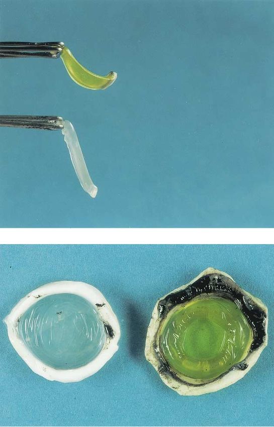
FIGURE 2. Treatment of the central 7 mm of the centrally
abraded cornea with riboflavin drops and two UVA diodes.
ples in the Declaration of Helsinki. Subjects read and
signed an institutional ethics committee–approved con-
sent form before participation in the study.
● OBSERVATION PROCEDURES: The preoperative
screening and the postoperative examinations included
measurement of best-corrected visual acuity, corneal to-
pography using a videokeratoscope (C-scan; Technomed,
Baseweile, Germany), intraocular pressure by Goldmann
applanation tonometry, central endothelial cell density
using an endothelial cell microscope (EM-1200; Tomey,
Erlangen, Germany), corneal photography, and slit-lamp
FIGURE 1. (Top) Stiffening effect of porcine cornea after and fundus examination. Preoperative pachymetry (Pa-
crosslinking with preserved curvature in the treated cornea chette; Technomed, Baseweile, Germany) was performed
(above) and massive bending of the untreated control cornea only in the last eight patients with minimal pachymetry
(below). (Bottom) Wrinkling of untreated porcine cornea (left) values ranging from 460 to 540 m.
and form stability with impressive smoothness of the centrally
crosslinked porcine cornea (right).
● TREATMENT PROCEDURE: The treatment procedure
was conducted under sterile conditions in the operating
room. Proxymetacainhydrochloride 0.5% eyedrops were
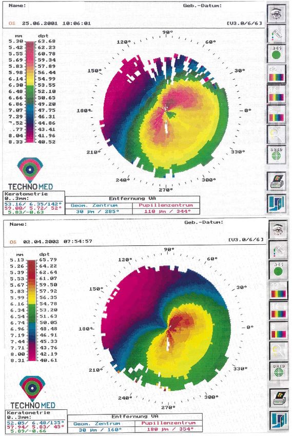
diagnosis of keratoconus was based on corneal topography applied for preoperative local anesthesia. The central 7
(Figure 3) and clinical signs of keratoconus such as stromal mm of the corneal epithelium was cautiously removed
thinning, Fleischer ring, Vogt striae, or apical stromal scar. using a blunt knife. As a photosensitizer, riboflavin 0.1%
The preoperative progression of keratoconus was con- solution (10 mg riboflavin-5-phosphate in 10 ml dextran-
firmed from medical history in all patients, and it was T-500 20% solution) was applied 5 minutes before the
clearly documented by serial corneal topography12 in 12 irradiation and every 5 minutes during the irradiation.
eyes (52%; Figure 4). The average age of the recruited After allowing riboflavin to permeate through the cornea
patients was 31.7 ⫾ 11.9 years and ranged from 13 to 58 for at least 5 minutes, the UVA irradiation was started
years (Tables 1 and 2). Except for patient 1, who had using two UV diodes (370 nm; Roithner Lasertechnik,
congenital Leber amaurosis and acute bilateral keratoco- Vienna, Austria) with a potentiometer in series to regulate
nus, the patients had a moderate to advanced degree of the voltage. Three 1.3-V accumulators were used as a
progressive keratoconus13 with maximum keratometer val- power generator. Before each treatment, the desired irra-
ues between 48 and 72 diopters (Tables 1 and 2). For safety diance of 3 mW/cm2 was controlled with a UVA meter
reasons, in all cases except patient 1, only one eye was (LaserMate-Q; LASER 2000, Wessling, Germany) at a
treated; the fellow eye served as a control eye. Patients 11 1-cm distance and, if necessary, regulated with the poten-
and 22 wore hard contact lenses in the treated eyes before tiometer. The patient’s cornea was irradiated with the
and after the treatment. The prospective, nonrandomized UVA-light diodes (370 nm) at a 1-cm distance for 30
pilot study was conducted in accordance with the princi- minutes using 3 mW/cm2 irradiance, which corresponds to
VOL. 135, NO. 5 TREATMENT OF KERATOCONUS BY CROSSLINKING 621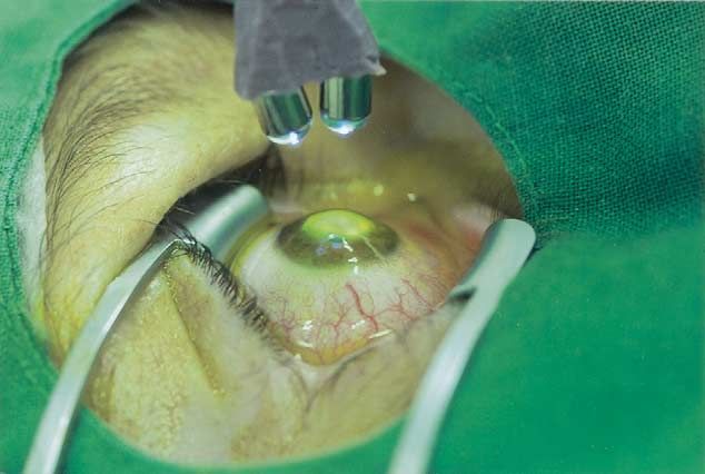
FIGURE 3. Corneal topography of a treated patient (Top) shortly before and (Bottom) 10 months after crosslinking with slight
regression of the maximum K value by 1.06 diopters.

622                                      AMERICAN JOURNAL    OF   OPHTHALMOLOGY                                   MAY 2003
FIGURE 4. (Top) Column diagram demonstrating pretreatment progression of maximum K value in the half year before treatment
and posttreatment regression as measured at the latest follow-up examination for each patient. y axis: difference in maximum K value
in diopters; x axis: patient number; shaded bars ⴝ preoperative change of K value; solid bars ⴝ postoperative change of K value.
(Bottom) Biphasic curve illustrating the mean change over time of the maximum K value relative to the K value on the day of
treatment with mean preoperative progression by 1.4 diopters and postoperative regression by 2.0 diopters (x axis: time in months;
y axis: change of maximum K value in D).

VOL. 135, NO. 5                         TREATMENT    OF   KERATOCONUS    BY   CROSSLINKING                                     623
TABLE 1. Investigation Parameters

                                                                                                                 Endothelial Cell
                                               Visual Acuity       Refractive Correction     Maximum K Value        Density
                                                                                                                                    Corneal and Lens
   Patient   Age   Postoperative Interval   Preop       Postop     Preop          Postop    Preop      Postop   Preop      Postop    Transparency

     1r      13             47              no LP      no LP       —             —           —          —         —          —            ⫽
     1l      13             47              no LP      no LP       —             —           —          —         —          —            ⫽
     2       41             35              HM         20/400      —             —           —          —         —          —            ⫽
     3       32             33              20/50      20/22      ⫺3            ⫺1.5        49.69      48.30    2,300      2,300          ⫽
     4       19             33              20/40      20/33      ⫺1.75         ⫺1.5        57.94      57.90    2,400      2,390          ⫽
     5       38             31              20/25      20/20      ⫺0.75         ⫺0.75       49.36      49.32    2,200      2,250          ⫽
     6       58             31              HM         HM         ⫺3            ⫺2.75       49.10      48.20    1,700      1,700          ⫽
     7       49             30              20/25      20/25       3.5           3.5        50.32      44.45      —          —            ⫽
     8       36             29              20/33      20/28      ⫺8.25         ⫺3.5        50.94      48.20    2,600      2,640          ⫽
     9       29             29              20/66      20/33     ⫺13           ⫺13.125      56.49      53.29    2,580      2,600          ⫽
    10       36             27              20/25      20/22      ⫺4.5          ⫺2.125      53.19      51.11      —        2,700          ⫽
    11       31             27              20/40      20/33      ⫺6            ⫺7          49.85      50.13    2,150      2,130          ⫽
    12       29             24              20/50      20/28      ⫺7            ⫺5.5        55.50      52.61    2,450      2,450          ⫽
    13       39             20              20/66      20/40      ⫺3             0          55.17      54.56    2,290      2,280          ⫽
    14       39             19              20/50      20/66      ⫺2.75         ⫺2.5        52.80      52.00    2,110      2,090          ⫽
    15       31             19              20/40      20/25      ⫺6.75         ⫺2.125      45.88      45.44    2,360      2,400          ⫽
    16       19             12              20/100     20/66     ⫺10.25         ⫺4.25       59.00      57.34    2,060      2,100          ⫽
    17       28              9              20/66      20/33      ⫺0.75         ⫺4          56.00      53.81    2,400      2,420          ⫽
    18       51              8              20/66      20/200      0.375        ⫺0.875      55.60      52.18    2,100      2,050          ⫽
    19       32              7              20/66      20/50      ⫺3.5          ⫺2.5        67.07      62.72    2,700      2,700          ⫽
    20       30              6              20/33      20/20      ⫺1.0           0.5        51.06      47.08    2,050      2,050          ⫽
    21       24              5              20/66      20/50      ⫺3.0          ⫺2.5        72.47      68.60    1,850      1,850          ⫽
    22       22              3              20/50      20/40      ⫺1.25         ⫺0.25       46.07      45.70    1,950      1,900          ⫽

     The preoperative values for visual acuity and maximum K value were determined on the day of treatment. The postoperative values are
   given for the last visits.
     ⫽ indicates unchanged transparency; HM ⫽ hand motion; LP ⫽ light perception; postop ⫽ postoperative; preop ⫽ preoperative.

a dose of 5.4 J/cm2 (Figure 2). After the treatment, an                           val, 1.23 to 3.07 diopters; P ⫽ .001, paired Student t test),
antibiotic ointment was applied until reepithelialization.                        comparing the preoperative values on the day of treatment
                                                                                  vs the postoperative values of the last examination (Figure
                                                                                  3). In five patients the K value remained stable, and in one
                             RESULTS                                              patient a minimal increase of the K value of 0.28 diopters
                                                                                  was present. In the fellow control eyes, however, 5 of 23
THE FOLLOW-UP TIME RANGED FROM 3 TO 47 MONTHS,                                    eyes (22%) showed a continuous progression of the max-
with a mean follow-up time of 23.2 ⫾ 12.9 months (Tables                          imum K value by an average of 1.48 diopters in the first
1 and 2). Best-corrected visual acuity improved statistically                     year after the crosslinking treatment of the contralateral
significantly in 15 patients (65%) by an average of 1.26                          eye.
lines (95% confidence interval, ⫺0.68 to 2.21; P ⫽ .026,                             The postoperative healing process was unremarkable,
paired Student t test), comparing the preoperative values                         except for slight transient stromal edema until reepitheli-
on the day of treatment vs the postoperative values of the                        alization after 3 days. There were no side effects, such as
last examination. The refractive correction improved sig-                         persistent epithelial defects or scarring. During the first
nificantly by an average of 1.14 diopters (95% confidence                         postoperative night, some pain medication was adminis-
interval, 0.12 to 2.17; P ⫽ .03) in spherical equivalent                          tered. The corneal and lens transparency and the endo-
(Table 2).                                                                        thelial cell density (P ⫽ .45) remained unchanged (Tables
   The mean preoperative progression of the maximum K                             1 and 2). Contact lens wear for refractive correction in
value was 1.42 ⫾ 1.18 diopters (Figure 4, bottom) in 12                           patients 19 and 22 could be continued without tear film
eyes (52%). Postoperative regression of keratoconus, as                           stability problems.
measured by the maximum K values (Table 1), was found                                No statistically significant difference was found between
in 16 patients (70%; Figure 4, top and bottom) with an                            the mean preoperative intraocular pressure of 13.6 ⫾ 2.0
average reduction of 2.01 diopters (95% confidence inter-                         mm Hg on the day of treatment and the mean postoper-

624                                                  AMERICAN JOURNAL        OF   OPHTHALMOLOGY                                            MAY 2003
TABLE 2. Summary of Patient Data and Results
Mean ⫾ SD P Value
Mean follow-up 23.2 ⫾ 12.9 months
Preoperative progression in K value 1.42 ⫾ 1.18 D
Postoperative regression K value 2.01 ⫾ 1.74 D .0001
Postoperative regression in refractive error (spherical 1.14 ⫾ 2.18 D .030
equivalent)
Postoperative increase in visual acuity 1.26 ⫾ 1.5 lines .026
Postoperative intraocular pressure Unchanged .612
Postoperative transparency of lens and cornea Unchanged
Postoperative density of endothelial cells Unchanged .45
Number of patients: 22
Number of eyes: 23
Gender: 12 males, 10 females
Age: 31.7 ⫾ 11.9 years
The postoperative values were calculated as the difference between the value on the day of
treatment and at the last follow-up visit. The preoperative progression K value was calculated as the
difference in K value between the value half a year before treatment and the day of treatment.
ative intraocular pressure of 13.8 ⫾ 2.5 mm Hg (P ⫽ .615) riboflavin/UVA using quantitative biomechanical stress-
at the last visit. strain measurements.9 –11 The impressive stiffening effect
after riboflavin/UVA treatment (Figure 1) is similar to
formaldehyde-induced tissue stiffening and fixation in
DISCUSSION pathologic specimens caused by collagen crosslinking.
Pathohistologically, we were able to demonstrate a
THIS STUDY HAS SHOWN THAT COLLAGEN-CROSSLINKING significant increase in collagen fiber diameter as the
appears to be effective in stopping the progression of underlying histopathologic correlate.15 Increased corneal
keratoconus quasi “freezing” the cornea. This effect is collagen fiber diameters and increased collagen rigidity
corroborated by the following data of the study1: Postop- have also been described in diabetes mellitus and aging,
erative regression was observed in 70% of patients with a where collagen crosslinking is also increased.16 –18 In these
decrease of the mean keratometer values by 2.01 diopters conditions, keratoconus rarely occurs.19 Increased resis-
postoperatively despite documented preoperative progres- tance to pepsin digestion after crosslinking has been
sion by 1.42 diopters in 52%.2 The postoperative refractive found,20 which might be important for keratoconus be-
corrections could also be reduced by an average of 1.14 ⫾
cause a significantly elevated activity of collagenases has
2.18 diopters.3 In the untreated fellow control eyes, a
been found.21,22
postoperative progression of keratectasia by 1.48 diopters
The arrest of the progression of keratoconus in our
was found in 22%.4 In biomechanical measurements of
patients could have been spontaneous as a so-called forme
earlier experiments, an increase in biomechanical stability
fruste of keratoconus.23 In epidemiologic studies, however,
of approximately 70% was measured.9
In contrast to other therapeutic measures for treating 21% of patients with keratoconus progress to a state where
keratoconus, such as thermal keratoplasty, intracorneal keratoplasty is required.2,3 In all our cases a progression of
rings, or epikeratoplasty (which basically are only transient keratoconus was known before the beginning of the
refractive corrections),4 –7 the new minimal invasive treatment at least by clinical history and clearly docu-
method presented here seems to be the first approach to mented by corneal topography in 12 eyes (52%; Figure 4,
stop or even reduce the progression of keratoconus. An top). Moreover, postoperative regression was observed in
arrest of keratoconus by contact lenses has been described 16 eyes (70%) after treatment, and this has never been
only in anecdotal reports but has never been confirmed in reported in the natural course of the disease.
a systematic study.1 In the two cases with hard contact lens wear (patients
The success of crosslinking treatment in keratoconus is 11 and 22), the keratometry readings might have been
not surprising, because a significantly reduced tensile influenced by an orthokeratoplastic effect,24 but these two
strength has been measured biomechanically14 in kerato- cases did not show regression. The contact lenses were still
conous and a significant increase in corneal rigidity has tolerated after crosslinking, and their use did not have to
been measured in porcine and rabbit corneas treated by be stopped.
VOL. 135, NO. 5 TREATMENT OF KERATOCONUS BY CROSSLINKING 625In the present study, we treated moderate to advanced might reduce the need for donor material resulting from
keratoconus stages.13 If the good results of the new method keratoconus, which represents approximately 16% of all
are corroborated over time, it would be preferable to treat keratoplasty indications.35 Given the very low costs and
earlier stages of the disease so that a better visual acuity the simplicity of the new method, it could be applied in
might be preserved. Earlier stages were not included in this developing countries where access to keratoplasty or con-
study, because the possible risks involved were not yet tact lenses is a problem.
known.
We did not find an increase of the mean intraocular ACKNOWLEDGMENT
pressure values postoperatively. Applanation tonometry is The authors thank Prof. Josef Wollensak (Berlin) for
not sensitive enough to reflect the increase in corneal continuous support and stimulation in the development of
rigidity. A slight increase in intraocular pressure measure- the new method.
ment might also be masked by the normal slight variability
in intraocular pressure.
We have not observed any complications or adverse REFERENCES
events of the new method, especially no decrease in
endothelial cell density or cataract formation. We have 1. Rabinowitz YS. Keratoconus. Surv Ophthalmol 1998;42:
previously measured the amount of irradiation intensity 297–319.
transmitted by the porcine cornea using a UVA photo- 2. Tuft SJ, Moodaley LC, Gregory WM, Davison CR, Buckley
meter. With 3 mW/cm2 of UVA irradiance at the surface RJ. Prognostic factors for the progression of keratoconus.
Ophthalmology 1994;101:439 –447.
of the cornea and a riboflavin concentration of 0.1%, there 3. Kennedy RH, Bourne WM, Dyer JA. A 48-year clinical and
is a massive reduction of the UVA light by 95%, resulting epidemiologic study of keratoconus. Am J Ophthalmol 1986;
in an irradiance of 0.15 mW/cm2 (⫽ an irradiation dose, 101:267–273.
0.27 J/cm2) at the endothelial level in a 500-m-thick 4. Sekundo W, Stevens JD. Surgical treatment of keratoconus
cornea.9 Without riboflavin, the UVA light would be at the turn of the 20th century. J Refract Surg 2001;17:69 –
73.
reduced in the cornea only by approximately 30%, with
5. Colin J, Cochener B, Savary G, Malet F. Correcting kerato-
approximately 50% UVA absorption in the lens.25 conus with intracorneal rings. J Cataract Refract Surg 2000;
In experiments with rabbit eyes, the cytotoxic threshold 26:1117–1122.
irradiance for the endothelial cells after combined ribofla- 6. Dana MR, Putz JL, Viana MAG, Sugar J, McMahon TT.
vin/UVA treatment is 0.36 mW/cm2 (⫽0.65 J/cm2), Contact lens failure in keratoconus management. Ophthal-
which may be reached with a corneal thickness of under mology 1992;99:1187–1192.
7. Jaeger MJ, Berson P, Kaufman HE, Green WR. Epikerato-
400 m using 3 mW/cm2 irradiance (⫽5.4 J/cm2) at the plasty for keratoconus. A clinicopathologic case report.
epithelial level.26 Preoperative pachymetry is essential and Cornea 1987;6:131–139.
was included in the last eight patients of the study. The 8. Hettlich HJ, Lucke K, Kreiner CF. Light induced endocap-
central corneal thickness in keratoconus usually is not sular polymerization of injectable lens refilling materials. Ger
reduced to less than 400 m.27 If so, however, the cross- J Ophthalmol 1992;1:346 –349.
linking treatment should be avoided. 9. Spoerl E, Schreiber J, Hellmund K, Seiler T, Knuschke P.
Untersuchungen zur Verfestigung der Hornhaut am Kan-
The UVA dose of 0.65 J/cm2 (0.36 mW/cm2) is far inchen. Ophthalmologe 2000;97:203–206.
below a cataractogenous level of 70 J/cm2.28 In addition, 10. Spoerl E, Huhle M, Seiler T. Induction of cross-links in
lens damage is usually induced by UVB light in the corneal tissue. Exp Eye Res 1998;66:97–103.
wavelength range of 290 to 320 nm, which has a higher 11. Spoerl E, Huhle M, Seiler T. Erhöhung der Festigkeit der
energy because of a shorter wavelength than UVA.28,29 Hornhaut durch Vernetzung. Ophthalmologe 1997;94:902–
906.
The durability of the stiffening effect is unknown.
12. Maguire LJ, Lowry JC. Identifying progression of subclinical
Because the collagen turnover in the cornea is estimated to keratoconus by serial topography analysis. Am J Ophthalmol
be between 2 to 3 years,30,31 a repeat treatment may 1991;112:41–45.
become necessary in the long run. 13. Zadnik K, Barr JT, Gordon MO, Edrington TB. Biomicro-
Collagen crosslinking could also be useful for the treat- scopic signs and disease severity in keratoconus. Cornea
ment of iatrogenic keratectasia resulting from laser in situ 1996;15:139 –146.
14. Andreassen TT, Simonsen AH, Oxlund H. Biomechanical
keratomileusis32,33 either as prophylaxis or as postoperative properties of keratoconus and normal corneas. Exp Eye Res
treatment. The new treatment can also be used for treating 1980;31:435–441.
corneal melting lesions or superficial ulcers.34 The residual 15. Wollensak G, Seiler T, Wilsch M, Spörl E. Collagen fiber
corneal thickness should be at least 400 m to spare the diameter after riboflavin/UVA induced collagen-crosslinking
endothelium. in the rabbit cornea. Cornea. Forthcoming.
16. Malik NS, Moss SJ, Ahmed N, Furth AJ, Wall RS, Meek
We believe that collagen crosslinking might become a
KM. Ageing of the human corneal stroma: structural and
standard treatment for progressive keratoconus. Long-term biochemical changes. Biochim Biophys Acta 1992;1138:
studies must exclude serious late complications and con- 222–228.
firm the durability of the stiffening effect. The new method 17. Sady C, Khosrot S, Nagaraj R. Advanced Maillard reaction
626 AMERICAN JOURNAL OF OPHTHALMOLOGY MAY 2003and crosslinking of corneal collagen in diabetes. Biochem advanced keratoconus using ultrasound pachymetry and the
Biophys Res Com 1995;214:793–797. EyeSys videokeratoscope. Optom Vis Sci 1998;75:640 –646.
18. Bailey AJ, Paul RG, Knott L. Mechanisms of maturation and 28. Pitts DG, Cullen AP, Hacker PD. Ocular effects of ultravi-
ageing of collagen. Mech Ageing Dev 1998;106:1–56. olet radiation from 295–365 nm. Invest Ophthalmol Vis Sci
19. Seiler T, Huhle S, Spoerl E, Kunath H. Manifest diabetes 1977;16:932–939.
and keratoconus: a retrospective case-control study. Graefe’s 29. Jose JG, Pitts DG. Wavelength dependency of cataracts in
Arch Clin Exp Ophthalmol 2000;238:822–825. albino mice following chronic exposure. Exp Eye Res 1985;
20. Spoerl E, Seiler T, Wollensak G. Increased resistance of 41:545–563.
crosslinked cornea against enzymatic digestion. Br J Oph- 30. Smelser GK, Polack FM, Ozanics V. Persistence of donor
thalmol, forthcoming. collagen in corneal transplants. Exp Eye Res 1965;4:349 –
21. Abalain J. Levels of collagen degradation products (telopep- 354.
tides) in the tear film of patients with keratoconus. Cornea 31. Nishida T. Basic Science: Cornea. In: Krachmer JH, Mannis
2000;19:474 –476. MJ, Holland EJ, editors. Cornea, vol. 1. St. Louis: Mosby,
22. Zhou L, Sawaguchi S, Twining SS, Sugar J, Feder RS, Yue 1997:13.
BY. Expression of degradative enzymes and protease inhibi- 32. Seiler T, Koufala K, Richter G. Iatrogenic keratectasia after
tors in corneas with keratoconus. Invest Ophthalmol Vis Sci laser in situ keratomileusis. J Refract Surg 1998;14:312–317.
1998;39:1117–1124. 33. Argento C, Cosentino MJ, Tytiun A, Rapetti G, Zarate J.
23. Amsler M. Le kératocône fruste au Javal. Ophthalmologica Corneal ectasia after laser in situ keratomileusis. J Cataract
1938;96:77–83. Refract Surg 2001;27:1440 –1448.
24. Swarbrick HA, Wong G, O’Leary DJ. Corneal response to 34. Schnitzler E, Spörl E, Seiler Th. Bestrahlung der Hornhaut
orthokeratology. Optom Vis Sci 1998;75:791–799. mit UV-Licht und Riboflavingabe als neuer Behandlungsver-
25. Michael R. Development and repair of cataract induced by such bei einschmelzenden Hornhautprozessen, erste Erge-
ultraviolet radiation. Ophthalmic Res 2000;32(S1):1–44. bnisse mit Patienten. Klin Mbl Augenheilk 2000;217:190 –
26. Wollensak G, Spoerl E, Seiler T, Wilsch M. Endothelial 193.
cell damage after riboflavin-UVA treatment in the rabbit. 35. Maeno A, Naor J, Hunter WS, Rootman DS. Three decades
J Cataract Refract Surg, forthcoming. of corneal transplantation: indications and patient charac-
27. Watters G, Owens H. Evaluation of mild, moderate, and teristics. Cornea 2000;19:7–11.
VOL. 135, NO. 5 TREATMENT OF KERATOCONUS BY CROSSLINKING 627You can also read