Eye Care in the Intensive Care Unit - (ICU) - the Royal College of ...
←
→
Page content transcription
If your browser does not render page correctly, please read the page content below
Ophthalmic Services Guidance

Eye Care in the Intensive Care Unit
(ICU)

April 2020

18 Stephenson Way, London, NW1 2HD T. 020 7935 0702
contact@rcophth.ac.uk rcophth.ac.uk @RCOphth
© The Royal College of Ophthalmologists 2020 All rights reserved
For permission to reproduce any of the content contained herein please contact contact@rcophth.ac.uk
Contents

     Section                                                                page
     1   Summary                                                               3
     2   Introduction                                                          3
     3   Protecting the eye                                                    4
     4   Identifying disease of the eye                                        6
         Exposure keratopathy and corneal abrasion                             6
         Microbial infections                                                  8
     5 Rare eye conditions in ICU                                             10
         Red eye in a septic patient: possible endogenous endophthalmitis     10
         Other problems                                                       10
     6 Delivering treatment to the eye when it is prescribed                  10
     7. Systemic fungal infection and the eye                                 11
     8. Tips for ophthalmologists seeing patients in ICU                      11
     9. Authors                                                               12
     10. References                                                           13

Date of review: April 2023

2020/PROF/409                                                                      2
1 Summary
This document aims to provide advice and information for clinical staff who are involved in
eye care in the ICU. It is primarily intended to help non-ophthalmic ICU staff to:
1. protect the eye in vulnerable patients, thus preventing ICU-related eye problems
2. identify disease affecting the eye in ICU patients, and specifically those which might need
ophthalmic referral
3. deliver treatment to the eye when it is prescribed.
It concentrates primarily on the common problems of the eye surface but also touches on
other less common conditions.
2 Introduction
The health of the front surface of the eye, particularly the cornea (the clear front window of
the eye) depends on the ability to produce tears, to blink, and to close the eyes with rest or
sleep. These can be impaired on the intensive care unit (ICU) whether by disease (e.g. facial
oedema, reduced conscious level, peripheral or central neurological injury) or treatments
(e.g. the drying effects of gas flows from CPAP or oxygen masks, muscle relaxants reducing
the strength of lid closure, sedation reducing the blink reflex and the effects of prolonged
prone positioning). Whatever the cause, those unable to close the eye for themselves, or in
whom blinking rates are substantially reduced, are at increased risk of damage to the front
of the eye, and this risk is higher in those mechanically ventilated, due to greater length of
stay, use of sedative/paralysing drugs and the effects of positive pressure ventilation (see
below).
The main possible problems affecting the front of the eye in ICU are:
Direct injury to the cornea, most often a superficial corneal abrasion (scratch)1
Exposure keratopathy
Chemosis (conjunctival swelling)
Microbial conjunctivitis and keratitis.
ICU eye care protocols are sometimes haphazardly followed, and documentation of eye care
is often poor3. However, having a clear protocol for assessment and intervention, which is
applied rigorously and correctly, will prevent the majority of corneal problems 4, 6, 10.
2020/PROF/409 3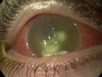
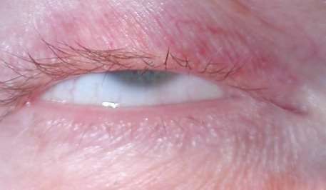
3    Protecting the eye

The eyelid closure and the appearance of the eye should be checked regularly with a bright
light at least once per shift throughout the patient’s stay.
If the eye is red, sticky, chemosed, or there are corneal abnormalities, the medical staff
should be alerted (and consideration of referral for ophthalmological opinion given) and 2
hourly increased lubrication given.

Assessment of lid closure and risk of corneal damage
Incomplete closure of the eyelids is called lagophthalmos. If the eyes do not close properly
the grade of severity must be assessed.
 Grade 0: Lids completely closed

 Grade 1: Any conjunctival exposure
 (visible white of the eye) but no
 corneal exposure

 Grade 2: Any corneal exposure, even a
 very tiny amount

2020/PROF/409                                                                                  4
Protective measures
The action required is based on the grading of exposure:
Grade 0 exposure (i.e. no exposure) requires no action.
Grade 1 exposure requires lubrication
Grade 2 exposure needs lubrication and taping of the lids or other method of lid
closure.
The methods to protect the eyes are:
Lubrication.
o Liberal use of ointment lubricants into the eye four times daily e.g.
simple eye ointment, Lacrilube , Xailin Night, and VitA-POS). Drops do
not last as long. This needs to be applied correctly into the eye and not,
as is sometimes found, over closed eyelids. Such action is superior to
manual eye closure alone3.
o If a patient is conscious an alternative is 2 hourly lubricant drops (e.g.
Hylotears) and ointment before sleep.
Closing the eyelids.
o Manual closure of the eyes
o Taping the eyes shut. Lid taping is not always necessary and can be
distressing to relatives and to conscious patients, and repeated removal
may lead to facial skin or eyelid injury or irritation. It should therefore
only be undertaken when necessary. It is crucial when using taping that
the lids are completely shut and the tape not touching the eye surface
as more damage will be done than prevented.
o Cling film can be used as a safe alternative to tape to protect the eye –
it does not cause damage if in contact with the eyeball. Apply a
10x10cm square over each eye and change every shift. Never share a
cling film roll between patients.
o Hydrogel or silicone dressings or pads (eg Kerrapro, Gelliperm) may be
used instead of taping, if oedema prevents manual lid closure. They
should be changed once per shift and must not be allowed to dry out or
position poorly as this can damage the eye. Only use with great care to
avoid causing eye damage.
What order to do this:
Every 4 hours:
Bathe the eyes with warm water first to remove dried ointment.
Before the next lubricant application, examine the eye for abnormalities with a bright
light.
2020/PROF/409 5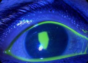
Apply new ointment to the eye surface: pull the lower eyelid down with a finger and
insert the ointment over the top of the lower lid into the gap between the lid and the
conjunctiva.
If taping is also performed, ointment is put in first and the eyes are closed 7, 8, 9. The
position of the lashes is then checked as the lashes must be clear of the cornea if
iatrogenic corneal abrasion is to be avoided). The outside of the eye must be free of
the lubricant ointment for tape to stick properly. Micropore tape is then applied
horizontally across the lids to seal them shut as below:
Prone unconscious patients. In those patients nursed prone and unconscious, the eyelids
and face can become oedematous and conjunctival swelling (chemosis) is common. As in all
ventilated patients, exposure keratopathy (a drying of the corneal surface, see below) can
occur10, 11. Direct eye compression can occur and can be avoided using a 3-pin head holder
as is used for prone spinal surgery12, gel rings or similar devices. The eyes should always be
re-lubricated every 4 hours, and taped or cling-filmed shut as above. Where there is severe
oedema and the swollen conjunctiva prolapses through the closed eyelids, the medical staff
should be contacted as the eyelids may need to be temporarily closed with sutures.
4 Identifying disease of the eye
Exposure keratopathy and corneal abrasion
The corneal can be accidentally injured and nearly always in ICU this is in the form of a
corneal abrasion (a superficial scratch removing the surface epithelium). It will cause the eye
to become red and is best seen using fluorescein dye eye drops and a blue light, where the
2020/PROF/409 6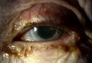
epithelial defect glows bright yellow; a white light will also work but the injury is less
obvious. Exposure keratopathy represents a dryness of the cornea due to incomplete lid
closure allowing excessive tear evaporation and a consequent failure of the tears to spread
adequately across the eye surface. It manifests as a red eye and fluorescein dye drops reveal
smaller or larger epithelial defects which can look identical to corneal abrasions. It affects
20-42% of ICU patients3, and 60% of those sedated for >48hours develop corneal epithelial
defects (42% within the first week) as a result1, 2. Prolonged epithelial defects can cause
scarring or even, in severe cases, perforation of the cornea. Secondary infection (microbial
keratitis: see below) can occur.
Treatment of a simple corneal abrasion without secondary infection can be with
chloramphenicol ointment four times daily for 5 to 7 days and increased lubrication and lid
taping if there is significant unwanted corneal exposure.
Corneal abrasion: A) Eye without fluorescein B) stained with fluorescein with blue light
showing abrasion on cornea
A B
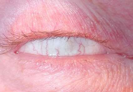
Chemosis
Conjunctival oedema which causes the conjunctiva to bulge out (chemosis) is common in ICU
patients. Risk factors include those which compromise venous return from the ocular
structures (positive pressure ventilation, escalating positive end expiratory pressures or tight
endotracheal tube taping); those states associated with generalised oedema (such as fluid
overload or hypalbuminaemia); gravitational causes of increased hydrostatic pressure
(prolonged recumbency or prone ventilation); or states which increase capillary leak (such as
systemic inflammatory response syndromes)2. Chemosis can cause impaired eyelid closure,
whilst incomplete eyelid closure can also predispose to chemosis.
Swollen conjunctiva
(chemosis)
2020/PROF/409 7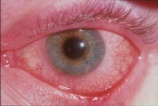
Microbial infections
The eye commonly becomes colonized with bacteria (in a time-dependent fashion) on ICU:
as many as 77% of ventilated medical patients being colonised by at least one abnormal
bacterial species in 7-42 days, 40% of those with prolonged ventilation and sedation with
multiple bacteria. The most common isolated organisms are Pseudomonas aeruginosa,
Acinetobacter spp. and Staphylococcus epidermidis3.
Respiratory secretions are thought to be the major source of ocular surface infection, with
aerosols from tracheal suctioning and direct contact from suction catheters both being
implicated. Pseudomonas infection rates can thus be reduced if endotracheal suctioning is
done from the side (rather than head) of the patient & with their eyes covered4, 5, 6.
Conjunctivitis: ICU staff should look for a sticky eye which is usually (but not always in ICU)
red. Note that if the eye is very red but not sticky, this might not be conjunctivitis and staff
must seek expert ophthalmological help.
RED AND STICKY RED BUT NOT STICKY
Conjunctivitis in this setting is usually bacterial and can be very infectious and virulent.
Without due care it can be spread to other patients and staff.
Management of conjunctivitis: It is wise to take a swab of the eye discharge and send it for
microbial culture because of the increased possibility of infection with unusual organisms.
The discharge can be removed by bathing the eyelids with warm water, using separate gauze
for each eye.
Chloramphenicol ointment (rather than drops to utilise the continued good lubrication from
the ointment) is applied in the eye four times a day for 5-7 days.
If the microbial results suggest that the organism is not sensitive to
chloramphenicol, but the eye is better, leave alone and do not change this. If the
eye is still sticky or red, then the ointment can be changed to one containing an
antibiotic to which the organism is sensitive, or other antibiotic drops can be
used in addition to simple lubricant ointment.
If the discharge and redness have not markedly improved in 48 hours, the
medical staff must be informed and ophthalmic help sought.
If the cornea becomes dull or a white patch appears, an urgent
ophthalmological opinion sought.
2020/PROF/409 8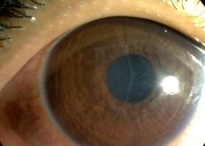
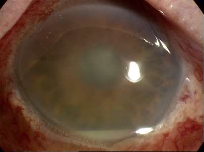
Microbial keratitis: The damaged cornea (for instance, that affected by exposure
keratopathy) is especially vulnerable to bacterial invasion which can occur very rapidly.
Whilst superficial infection can result, deeper infection can lead to permanent and severe
damage, and loss of vision.
Most cases are due to bacteria and appear as a red eye, which may be watery or sticky, with
a corneal ulcer (an epithelial defect -which stains with fluorescein dye- on top of an
underlying white/grey/yellowish opacity). Less commonly, debilitated patients may develop
herpes simplex keratitis which takes the form of typical “dendrites” in the corneal
epithelium and/or ulcers which stain yellow with fluorescein dye, but which can also appear
as non-staining grey areas in the cornea. If any of these corneal problems are seen, urgent
ophthalmic help must be sought.
RED EYE WITH WHITE PATCH ON CORNEA - LIKELY MICROBIAL KERATITIS

RED EYE STAINED WITH FLUORECEIN DYE SHOWING DENDRITE ON CORNEA – LIKELY
HERPES SIMPLEX KERATITIS

2020/PROF/409                                                                                9
5 Rare eye conditions in ICU
Red eye in a septic patient: possible endogenous endophthalmitis
This is a very serious problem caused by spread of systemic infection in the blood stream to
the inside of the eye. The eye may be red, although sometimes much less red than might be
expected. Endophthalmitis is to be suspected if a white line is visible in the eye in front of
the iris, which represents a level of pus in the chamber of the eye (hypopyon). Immediate
ophthalmic help needs to be sought as this is a sight threatening emergency and it also
indicates systemic active sepsis.
Hypopyon – pus in
the eye
Other problems
Other eye problems can complicate ICU care. Severe or recurrent hypotension can cause
blindness from ischaemic optic neuropathy12,13. In those ventilated prone, increased intra-
ocular pressures or intraorbital pressure with marked periocular swelling can decrease
ocular perfusion pressures (worse with concurrent systemic hypotension), leading to
ischaemic optic neuropathy, central retinal artery occlusion and permanent visual loss 12.
Rarely, those nursed prone can develop bilateral acute glaucoma in which there is a sudden
rise in intraocular pressure which can causes visual loss very quickly because of retinal or
optic nerve ischaemia. In this condition, the cornea becomes cloudy and grey and the pupil
becomes fixed at a mid-dilated position and unresponsive to light. This needs immediate
ophthalmic treatment.
6 Delivering treatment to the eye when it is prescribed
This is usually given in the form of drops or ointment. Sometimes several different drops are
required.
When giving several different drops, do not give them at the same time as one
drop may wash out another, thereby reducing its effectiveness. Allow ideally 5
minutes and at least 1 minute between each medication
2020/PROF/409 10 Always put drops in before ointment. The ointment is water repellent and
prevents the drops from getting into the eye tissues.
When putting in ointment in poor lid closure, after instilling ointment manually
shut eyelids to ensure ointment is spread over whole eye surface.
7. Systemic fungal infection and the eye
It is important to recognise that the eye may be involved in any patient who has a systemic
fungal infection. This is of concern in ICU where the patient is unlikely to be able to report
any problems with their eyesight and not all systemic antifungal agents penetrate the eye
sufficiently to treat intraocular disease. Previously, many guidelines recommended referral
of all patients with a positive blood culture or line tip for candida, aspergillus or any other
fungal organism should be referred for urgent ophthalmological assessment. More recent
evidence has emerged that the prevalence of eye involvement and intraocular disease
requiring or amenable to treatment, or requiring extra intraocular treatment or vitrectomy,
is very low and routine screening of all culture positive patients is not indicated 14, 15. We
recommend screening of fungal culture positive patients is done as an exception on a case
by case basis, taking into account risks for that patient, symptoms and abnormal appearance
of the eye taking these principles into account:
No examination:
o Awake and asymptomatic
May need examination:
o Awake and symptomatic
o Unable to report symptoms
Must be examined:
o Very abnormal eye appearance e.g. hypopyon, cloudy pupil, possible ocular
perforation etc.
Case by case assessment for both decision to examine and timing of examination based on
factors such as:
Risk
Prognosis
Microbiology results
Ability of current/planned treatment to penetrate eye
Patient position
Ability to examine.
If there is intraocular infection, liaise with microbiologists and ophthalmologists to ensure an
appropriate antifungal which has good ocular penetration is used.
8. Tips for ophthalmologists seeing patients in ICU
1. If corneal exposure and taping requested, ensure taping done correctly to avoid
lashes rubbing on the cornea.
2. If keratitis present, it is most likely to be virulent bacteria – especially pseudomonas.
Therefore, do a corneal scape and start appropriate intensive topical fluoroquinolone
2020/PROF/409 11antibiotic therapy immediately. Try to avoid lid taping (which can encourage bacterial
growth) and keep lubricated with plenty of ointment.
3. Dilating the eyes: it is safe to use G tropicamide 1% and G. phenylephrine 2.5%, but
write clearly in the notes that the pupils have been dilated, stating the time drops
were given and the time over which pupils are likely to be unresponsive to light
(about 4 hours).
4. Endogenous bacterial endophthalmitis – most likely if a hypopyon is present. Be
guided by systemic infection if known, otherwise urgently tap and inject using
protocol of amikacin and vancomycin. If platelet count is low (10. References
1. Werli-Alvarenga A, Ercole FF, Botoni FA, Oliveira JA, Chianca TC. Corneal
injuries: incidence and risk factors in the intensive Care Unit. Rev Lat Am
Enfermagem 2011;19:1088-95.
2. Grixti A, Sadri M, Watts MT. Corneal protection during general anesthesia for
nonocular surgery. Ocul Surf. 2013;11:109-18.
3. Ezra DG, Lewis G, Healy M, Coombes A. Preventing exposure keratopathy in the
critically ill: a prospective study comparing eye care regimes. Br J Ophthalmol.
2005; 89:1068-9
4. Rosenberg JB and Eisen, MD. Eye care in the intensive care unit: Narrative
review and meta-analysis Crit Care Med 2008; 36:3151-3155
5. Suresh P, Mercieca F, Morton A, Tullo AB Eye care for the critically ill. Intensive
Care medicine 2000 26: 162-166
6. 6 .Mela EK, Drimtzias EG, Christofidou MK, Filos KS, Anastassiou ED, Gartaganis
SP. Ocular surface bacterial colonisation in sedated intensive care unit patients.
Anaesth Intensive Care. 2010; 38:190-3.
7. Parkin B, Turner A, Moore E, et al: Bacterial keratitis in the critically ill. Br J
Ophthalmol 1997; 12:1060–1063
8. Hilton E, Adams AA, Uliss A, Lesser ML, Samuels S, Lowy FD. Nosocomial
bacterial eye infections in intensive-care units. Lancet. 1983; 1:1318-20.
9. Lenart SB, Garrity JA: Eye care for patients receiving neuromuscular blocking
agents or propofol during mechanical ventilation. Am J Crit Care 2000; 9:188–
191
10. Ezra DG, Goyal S, Moosavi R, Millar M, Laganowski HC, Moore AT. Microbial
keratitis in ITU staff: an occupational hazard? Anaesthesia. 2004 59:1221-3.
11. Mercieca F, Suresh P, Morton A, Tullo AB . Ocular surface disease in intensive
care unit patients. Eye 1999 13:231-236
12. Panchabhai TS, Bandyopadhyay D, Kapoor A, Akindipe O, Lane C, Krishnan S.
Acute ischemic optic neuropathy with extended prone position ventilation in a
lung transplant recipient. Int J Crit Illn Inj Sci. 2016; 6(1):45-7.
13. Bansal S, Ansons A, Vishwanath M. Hypotension-induced blindness in
haemodialysis patients. Clin Kidney J. 2014; 7(4):387-90
14. El-Abiary, Jones B, Williiams G, Lockington D. Fundoscopy screening for
intraocular candida in patients with positive blood cultures—is it justified? Eye
2018 32:1697-1702.
15. Breazzano MP, Day HR, Tanaka S, Cherney EF, Stemberg P, Donahue SP, Bond
JB. Utility of ophthalmologic screening for patients with candida bloodstream
infections: a systematic review. JAMA Ophthalmol 2019 137:698-710.
16. Grixti A, Sandri M, Datta AV, Uncomon ophthalmic disorders in intensive care
unit patients. J Crit Care 2012;27:746-e.9-22.
2020/PROF/409 13Patient nursed supine and unconscious
Grade Action
Grade 0 – eyelids close well No action required
Grade 1 – some conjunctival EYES NEED LUBRICATING EVERY 4 HOURS
exposure Check corneal clarity with bright light: IF NOT
CLEAR – ALERT MEDICAL STAFF
Clean off old ointment before putting in new
Pull lower lid down and instil ointment onto eye
between lower lid and conjunctiva
Grade 2 – conjunctival and EYES NEED LUBRICATING AND LID TAPING/CLING FILM
some corneal exposure – Check corneal clarity with bright light: IF NOT
MAJOR RISK CLEAR – ALERT MEDICAL STAFF
Apply ointment as for Grade 1
Close lids, ensure lashes outside eye and lids free
of ointment
Tape upper lid down with micropore tape
horizontally or use 10x10cm clingfilm
Patient nursed prone and unconscious: Major risk to eye in all cases
EYES NEED LUBRICATING AND LIDS TAPING
Check corneal clarity with bright light: IF NOT CLEAR – ALERT MEDICAL STAFF
Apply ointment as for Grade 2
Close lids, ensures lashes outside eye and lids free of ointment
Apply micropore tape horizontally
Red sticky eye Red not sticky eye
Take swab Is the cornea clear or does it stain
Use chloramphenicol ointment with fluorescein?
X 4/DAY to eye If clear cornea, or simple
Condition is contagious and can abrasion, check lubrication
be transmitted to other patients schedule and consider lid taping
IF NO BETTER IN 24 HOURS – ALERT If there is a corneal opacity, or abrasion does
MEDICAL STAFF not improve in 24hrs, or unsure of cause –
ALERT MEDICAL STAFF
Eye opacities on or inside eye: ALERT MEDICAL STAFF IMMEDIATELY
Exclude harmless adherent mucus by rinsing gently with saline. Mucus will move.
Any corneal opacity which does not move on rinsing, or a white line at the bottom of the
iris: suspect sight-threatening infection. ALERT MEDICAL STAFF IMMEDIATELY
2020/PROF/409 14You can also read