Urinary microRNAs as non invasive biomarkers for toxic acute kidney injury in humans
←
→
Page content transcription
If your browser does not render page correctly, please read the page content below
www.nature.com/scientificreports
OPEN Urinary microRNAs as non‑invasive
biomarkers for toxic acute kidney
injury in humans
Fathima Shihana1,2,3*, Wilson K. M. Wong4, Mugdha V. Joglekar4, Fahim Mohamed1,2,5,6,
Indika B. Gawarammana2, Geoffrey K. Isbister7, Anandwardhan A. Hardikar4,8,11,
Devanshi Seth3,9,10,11 & Nicholas A. Buckley1,2,10,11*
MicroRNAs in biofluids are potential biomarkers for detecting kidney and other organ injuries. We
profiled microRNAs in urine samples from patients with Russell’s viper envenoming or acute self-
poisoning following paraquat, glyphosate, or oxalic acid [with and without acute kidney injury (AKI)]
and on healthy controls. Discovery analysis profiled for 754 microRNAs using TaqMan OpenArray
qPCR with three patients per group (12 samples in each toxic agent). From these, 53 microRNAs were
selected and validated in a larger cohort of patients (Russell’s viper envenoming = 53, paraquat = 51,
glyphosate = 51, oxalic acid = 40) and 27 healthy controls. Urinary microRNAs had significantly higher
expression in patients poisoned/envenomed by different nephrotoxic agents in both discovery and
validation cohorts. Seven microRNAs discriminated severe AKI patients from no AKI for all four
nephrotoxic agents. Four microRNAs (miR-30a-3p, miR-30a-5p, miR-92a, and miR-204) had > 17 fold
change (p < 0.0001) and receiver operator characteristics area-under-curve (ROC-AUC) > 0.72. Pathway
analysis of target mRNAs of these differentially expressed microRNAs showed association with the
regulation of different nephrotoxic signaling pathways. In conclusion, human urinary microRNAs
could identify toxic AKI early after acute injury. These urinary microRNAs have potential clinical
application as early non-invasive diagnostic AKI biomarkers.
Main
MicroRNAs are involved in all cellular functions, including development, differentiation, growth, metabolism and
regulate numerous physiological and pathophysiological processes1,2. MicroRNAs are endogenous, non-coding
RNAs, 21–25 nucleotides in size that regulate gene expression by binding to target messenger RNAs (mRNAs).
Cells secrete microRNAs into the extracellular environment to function as regulators, carrying genetic informa-
tion from one cell to another, or following cell injury3. These extracellular microRNAs are potential biomarkers
for a variety of diseases including acute kidney injury (AKI)4 and can also be mediators of disease.
Acute kidney injury is associated with changes in the expression of specific microRNAs. While some micro-
RNAs contribute to the pathogenesis and progression of AKI, others may serve as protective m olecules5. As
signalling molecules, microRNAs mediate cell–cell communications, and form a complex regulatory network2,6.
They play a critical role as anti-apoptotic, pro-apoptotic and inflammatory molecules to facilitate the development
of renal injury in nephrotoxic A KI7,8. Thus, microRNAs have not only emerged as novel biomarkers for AKI,
1
Clinical Pharmacology and Toxicology Research Group, Biomedical Informatics and Digital Health, Faculty of
Medicine and Health, The University of Sydney, Sydney, NSW, Australia. 2South Asian Clinical Toxicology Research
Collaboration, Faculty of Medicine, University of Peradeniya, Peradeniya, Sri Lanka. 3Centenary Institute of
Cancer Medicine and Cell Biology, The University of Sydney, Sydney, NSW, Australia. 4Diabetes and Islet Biology
Group, School of Medicine, Western Sydney University, Campbelltown, NSW, Australia. 5Allied Health Sciences,
Department of Pharmacy, University of Peradeniya, Peradeniya, Sri Lanka. 6Australian Kidney Biomarker
Reference Laboratory, Department of Nephrology, Prince of Wales Hospital and Clinical School, University of
New South Wales, Sydney, Australia. 7Clinical Toxicology Research Group, University of Newcastle, Newcastle,
NSW, Australia. 8Department of Science and Environment, Roskilde University, Roskilde, Denmark. 9Discipline of
Clinical Medicine and Addiction Medicine, Faculty of Medicine and Health, The University of Sydney, Sydney, NSW,
Australia. 10Drug Health Services, Royal Prince Alfred Hospital, Sydney, NSW, Australia. 11These authors jointly
supervised this work: Anandwardhan A. Hardikar, Devanshi Seth and Nicholas A. Buckley. *email: fhan0023@
sydney.edu.au; nicholas.buckley@sydney.edu.au
Scientific Reports | (2021) 11:9165 | https://doi.org/10.1038/s41598-021-87918-0 1
Vol.:(0123456789)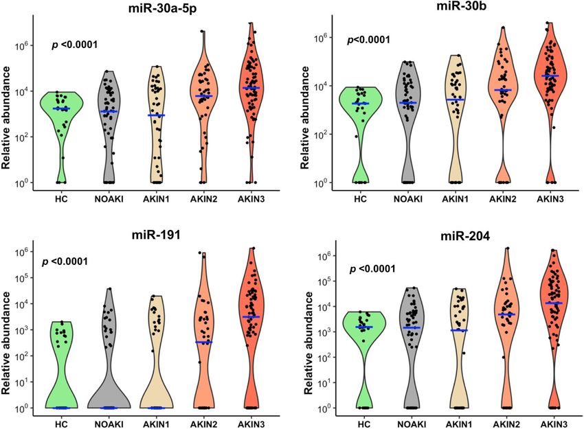
www.nature.com/scientificreports/
but they also hold promise as potential therapeutic targets.Various microRNAs regulate different nephrotoxic
pathways, for example NF-κB dependent inflammatory responses, TGF-β signalling, and oxidative stress9–11.
Thus, different mechanisms of nephrotoxicity including renal tubular toxicity, inflammation, glomerular damage,
crystal nephropathy, and thrombotic microangiopathy from different toxic agents would be expected to lead to
variable microRNA expression.
Traditional biomarkers of AKI such as serum creatinine (SCr) or urine output are both late indicators of
kidney injury12,13. Further, the rapid rise of SCr after oxalic acid, paraquat, and glyphosate surfactant herbi-
cide (glyphosate) poisoning suggests other mechanisms occur beyond loss of renal function14–16. Protein bio-
markers which reflect the underlying renal glomerular and/or tubular damage during nephrotoxicity such as
β2-microglobulin, cystatin C (Cys C), albumin, neutrophil gelatinase-associated lipocalin (NGAL), kidney
injury molecule-1 (KIM-1), interleukin-18 (IL-18) have highly variable diagnostic performance in these three
poisonings14–16. New biomarkers that are consistent for early prediction and/or detection of diverse causes of
nephrotoxic AKI are needed, whilst those specific to particular toxins may provide further understanding of
the mechanisms of nephrotoxicity.
Acute kidney injury is a common clinical complication following envenoming and poisoning14,16–18. Our
main aim was to explore if urinary microRNAs can be used as early renal biomarkers following nephrotoxic-AKI
(snakebite envenoming, oxalic acid, paraquat, and glyphosate poisoning). We also investigated whether micro-
RNA signatures identify some common mechanisms of nephrotoxicity using target mRNAs and pathway analysis.
Results
Global profiling of urinary microRNAs detects AKI after different causes of poisoning. We
identified 112 differentially expressed urinary microRNAs from the discovery cohort of 48 patients with Rus-
sell’s viper envenoming or acute self-poisoning following paraquat, glyphosate or oxalic acid. The cohort
included those who developed no injury (NOAKI), mild injury (AKIN1), moderate injury (AKIN2) or severe
injury (AKIN3) (n = 3 per group and n = 12 per each toxins) as well as healthy controls (n = 3). The expression
of microRNAs was significantly upregulated in urine samples of patients who developed moderate to severe
injury (AKIN2/3) compared to the NOAKI and healthy controls in the discovery cohort. Supervised hierarchi-
cal clustering heat map demonstrates the differentially expressed microRNAs between patients with moderate to
severe AKI (AKIN2/3) from NOAKI and healthy controls (Fig. 1). Based on our discovery analysis or from past
literature, we selected a set of 53 microRNAs (> 4 fold change and p < 0.01; Supplementary Table 1) for further
validation in independent cohorts for each toxins (Russell’s viper bite = 53, paraquat = 51, glyphosate = 51, oxalic
acid = 40, and 27 healthy controls) (see “Methods” section and Supplementary Table 2 for details).
Clustering of microRNA in severe cases from different causes of AKI in the validation
cohort. Forty four microRNAs were significantly upregulated in AKI in one or more nephrotoxic agents
(oxalic acid = 25, glyphosate = 10, paraquat = 34 and Russell’s viper = 25) compared to NOAKI. Thirty two micro-
RNAs were upregulated for two or more agents (Supplementary Table 3). Different sets of microRNAs for dif-
ferent toxins distinguished patients with AKI from NOAKI in the validation cohort (Fig. 2). Twelve microRNAs
distinguished AKI from NOAKI for only one specific toxin [oxalic acid (n = 2), paraquat poisoning (n = 5), Rus-
sell’s viper envenoming (n = 5)] (Supplementary Table 3).
Urinary microRNAs provide signals distinguishing both kidney injury severity and toxic
agents. Differentially expressed microRNAs from all four nephrotoxic agents were examined together to
visualize separation of patients with AKI from NOAKI using tSNE clustering analysis (Fig. 3a). Despite the
selection of differentially expressed microRNAs based on comparisons across all causes of kidney injury for
validation, there was still apparent glyphosate clustering in the tSNE plot (Fig. 3b). Seven common microRNAs
discriminated AKIN2/3 patients from NOAKI for all four nephrotoxic agents (Fig. 3c, Supplementary Table 3).
Of these seven microRNAs, four (miR-30a-5p, miR-30b, miR-191, and miR-204, Fig. 3d) had a greater than
17-fold change (p < 0.0001) and ROC-AUC = 0.72 (Table 1). Logistic regression combination of four and all
seven microRNAs in NOAKI versus AKIN2/3 patients showed AUC-ROC values of 0.75 and 0.77 respectively
(Supplementary Figure 1). The four microRNAs showed an association with increasing disease severity in all
poisoning (Jonckheere–Terpstra nonparametric ordered test, p < 0.0001, Fig. 4). A schematic diagram shows
the initial discovery, through validation and selection of four common microRNAs in all four toxic agents (Sup-
plementary Figure 2).
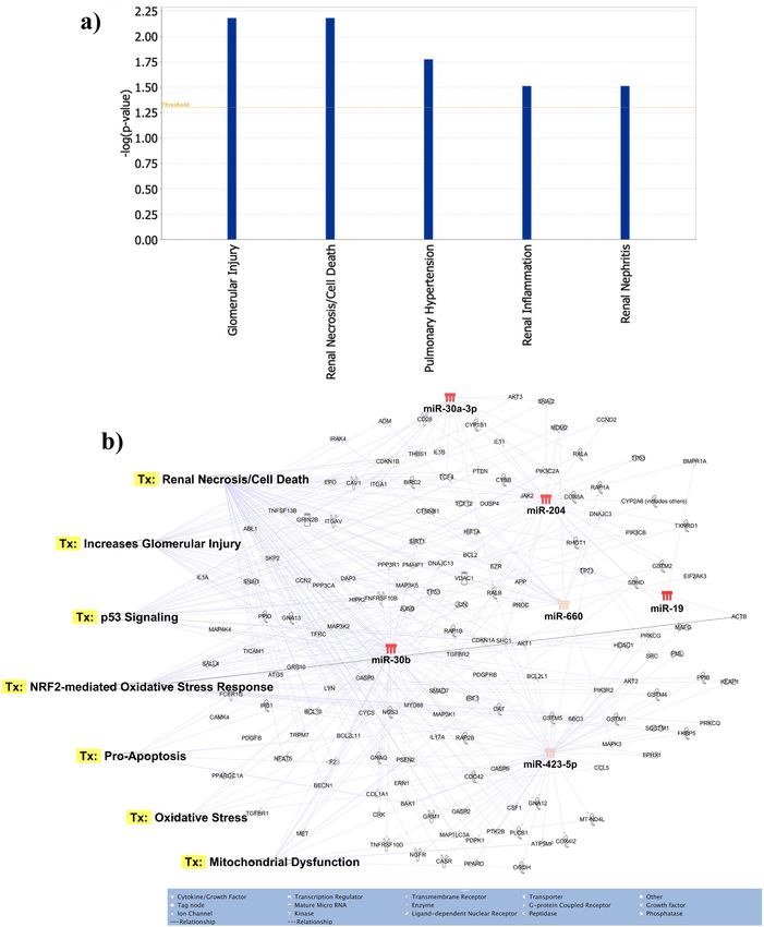
Identification of target genes and pathway analysis. The seven most differentially expressed micro-
RNAs target glomerular injury, renal cell death/necrosis, renal inflammation and renal nephritis as major dis-
ease/functions (Fig. 5a). For the seven differentially expressed microRNAs a total of 586 potential target mRNAs
specific to kidney disease/functions were identified via Ingenuity Pathway Analysis (data not shown). These
differentially expressed microRNAs targeted many genes associated with the regulation of different nephrotoxic
signalling pathways (Fig. 5b). The key pathways targeted by these microRNAs were oxidative stress, renal cell
death/necrosis, oxidative stress, apoptosis and mitochondrial dysfunction (Fig. 5b).
Discussion
We report urinary microRNA signatures for nephrotoxicity in patients with four very different toxic agents
(snake envenoming, pesticide and chemical poisoning) in humans. We identified seven common microRNAs
that correlated with kidney injury across all four nephrotoxic agents. Another 36 microRNAs provided signals
Scientific Reports | (2021) 11:9165 | https://doi.org/10.1038/s41598-021-87918-0 2
Vol:.(1234567890)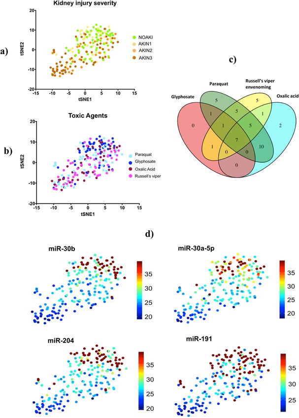
www.nature.com/scientificreports/
Figure 1. Urinary microRNAs detect AKI after different causes of poisoning. Supervised hierarchical
clustering heat map of differentially expressed microRNAs (n=112) distinguished patients with severe AKI
from NOAKI and healthy controls. Green, cyan, maroon, dark blue and purple indicates healthy controls,
paraquat, Glyphosate, Oxalic acid and Russell’s viper bite respectively. AKI stages are numbered as 0, 1, 2 and 3
for NOAKI, AKIN1, AKIN2 and AKIN3 respectivly. Rows represent microRNAs (normalised Ct) and columns
represent the individual samples from healthy controls and patients.
for certain nephrotoxic causes, but not for all agents. We found many of the target mRNAs of these seven com-
mon microRNAs are involved in cellular pathways that promote inflammation, apoptosis and oxidative stress.
Very few studies report on multiple agents in nephrotoxic AKI. A recent report on drug induced-AKI in
rats using three very different nephrotoxic compounds, cisplatin (proximal tubule) puromycin (glomerulus)
and N-phenylanthranylic acid (collecting ducts), identified microRNAs specific to each agent, but did not find
any commonly expressed microRNAs across all three19. Other studies reported on single agents, for example,
cisplatin-induced AKI in rats showed higher expression of urinary miR-191 and miR-30a-5p20, and urinary levels
of miR-423 were significantly higher in both paracetamol overdose patients with AKI compared with healthy
controls and in paracetamol overdose patients without A KI21.
We also noted some urinary microRNAs we identified are also observed in non-toxic AKI and other kidney
diseases as AKI biomarkers. Urinary miR-423 expression increased in critically ill patients (intensive care unit-
AKI)22. Patients with glomerular disease such as idiopathic nephrotic syndrome and focal segmental glomerulo-
sclerosis had high levels of miR-30a-5p in urine compared to c ontrols23–25. MiR-204 was differentially expressed
in urine of patients with chronic acute cellular rejection, characterized by interstitial fibrosis and tubular atrophy,
compared with patients with normal histology and functioning a llografts26.
We studied four very different toxins covering a broad range of mechanisms including ischemia–reperfusion
(Russell’s viper bite), uncoupling of oxidative phosphorylation (glyphosate), free radical generation (paraquat),
Scientific Reports | (2021) 11:9165 | https://doi.org/10.1038/s41598-021-87918-0 3
Vol.:(0123456789)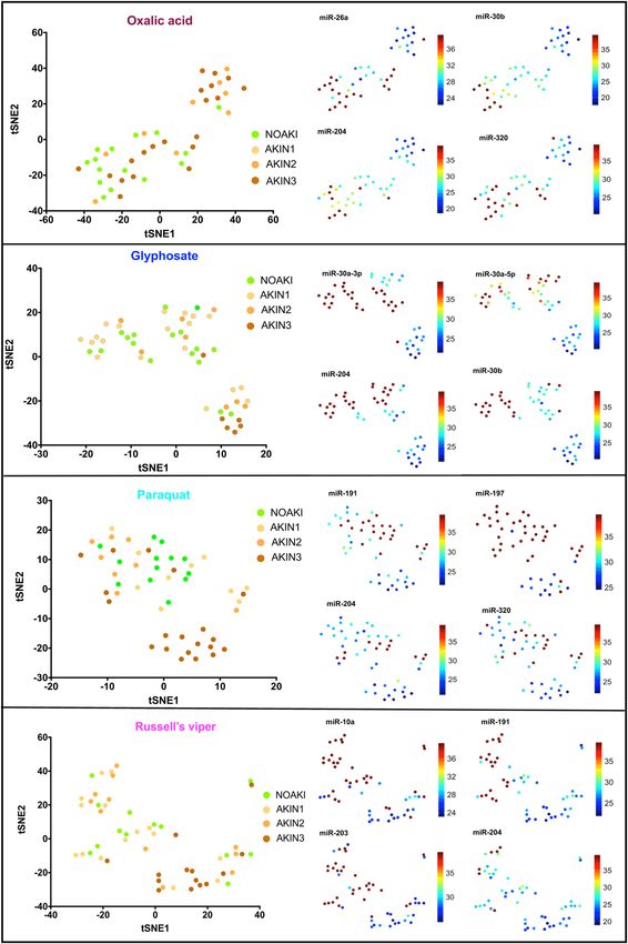
www.nature.com/scientificreports/

                                            Figure 2.  tSNE plots show the clusters of individual microRNAs with NOAKI, AKIN1, AKIN2 and AKIN3
                                            caused by different poisons. Left panels show plots of the non-linear tSNE map of samples in validation cohort
                                            using each specific set of differentially expressed microRNAs for different poisons (oxalic acid, glyphosate,
                                            paraquat) and Russell’s viper bite, (Supplementary Table 3). Plots were generated using 4-dimensional principal
                                            component space into two dimensions showing tSNE analysis. Each point represents a single sample. The right
                                            panel shows the expression of the top four most significantly altered microRNAs for each poison presented as
                                            individual tSNE-projections. Blue to red presented in the color bar indicate high to low microRNA expression
                                            (data shown in normalized Ct values, therefore a lower value represents a higher expression) respectively.

                                            and crystal formation (oxalic acid). However, all these poisonings or envenoming cause mitochondrial damage

          Scientific Reports |   (2021) 11:9165 |               https://doi.org/10.1038/s41598-021-87918-0                                                    4

Vol:.(1234567890)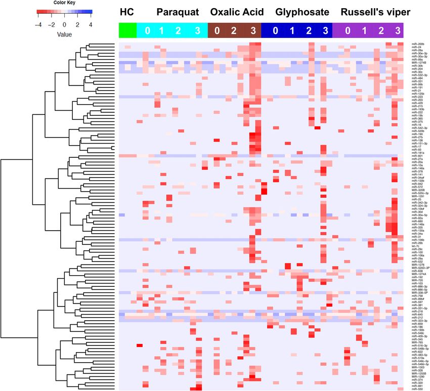
www.nature.com/scientificreports/
Figure 3. Plot of the non-linear tSNE map of samples from 4-dimensional principal component space into two
dimensions showing tSNE analysis applied using all (n=43) differentially expressed microRNAs (Supplementary
Table 3). (a) tSNE plots show the clusters of all different microRNAs in different AKI stages: NOAKI, AKIN1,
AKIN2 and AKIN3 caused by all poisons. (b) tSNE plots show the clusters for different poisons: oxalic acid,
glyphosate, paraquat and Russell’s viper envenoming in validation cohort. (c) Venn Diagram demonstrating
overlap of 43 altered microRNAs in AKI (> 2.8 fold change, p < 0.05) amongst the nephrotoxic agents.
microRNAs altered in different agents: Oxalic acid = 25, glyphosate = 10, paraquat = 34 and Russell’s viper = 25.
(d) tSNE plots show best four microRNAs clusters in AKIN2/3 patients in all four nephrotoxic agents. Blue to
red presented in the color bar indicates high to low microRNA expression (data shown in normalized Ct values,
therefore a lower value represents a higher expression) respectively.
and inflammation triggering oxidative s tress27. Oxidative stress, both directly and indirectly affects all facets of
the kidney, including vascular reactivity and renal hemodynamics, glomerular filtration and tubular reabsorption
and secretion in all nephron s egments28. During poisoning, oxidative stress signalling alters all these processes
and promotes damage pathways that lead to cellular apoptosis, necrosis, altered gene expression, progression of
tissue damage, promotion of fibrosis and abnormal kidney function27,28. We identified several target mRNAs of
Scientific Reports | (2021) 11:9165 | https://doi.org/10.1038/s41598-021-87918-0 5
Vol.:(0123456789)www.nature.com/scientificreports/
miR Fold change ( 2^ΔCt) p value AUC (95% CI)
miR-30a-5p 19.7 < 0.00001 0.72 (0.66–0.79)
miR-30b 17.1 < 0.00001 0.72 (0.66–0.79)
miR-191 32.0 < 0.00001 0.72 (0.66–0.78)
miR-204 17.1 < 0.00001 0.72 (0.65–0.78)
miR-30a-3p 22.6 < 0.00001 0.70 (0.64–0.77)
miR-660 8.0 < 0.00001 0.64 (0.59 0.69)
miR-423-5p 5.7 < 0.00001 0.60 (0.56–0.64)
Table 1. Diagnostic performance of the seven microRNAs in all causes of AKI (NOAKI vs. AKIN2/3).
Diagnostic performance of the seven microRNAs that distinguished AKI in all poisoning was assessed using
normalized Ct values. p values represent Mann–Whitney-U between NOAKI (n = 57) versus AKIN2/3 (n = 94).
Figure 4. Top four microRNAs associated with AKI for all toxic agents. Violin plots show these microRNAs
having increased abundance with increasing AKI severity in all four toxins. Blue lines indicate the medians
and each black dot presents a different sample from validation cohort. The y-axis is relative abundance from
the detectable limit (2(39-Ct), Ct of 39 is the threshold of detectability). MicroRNA expressions were normalised
using two spike control ath-microRNAs (ath-miR-159a and ath-miR-172a). p is the alternative p value from
Jonckheere–Terpstra nonparametric ordered test.
the altered microRNAs were associated with mitochondrial dysfunction, apoptosis, oxidative stress and necrosis/
cell death.
Most nephrotoxic agents induce proximal tubular injury. The increased concentration of microRNAs in
urine may be due to leakage of microRNAs from proximal tubular cells with corresponding decreased levels of
microRNAs in the injured tissue20, but we did not test this due to unavailability of kidney tissue. Other studies
in cisplatin and gentamicin-induced proximal tubular injury showed increased miR-191 and miR-30 family
in urine with corresponding decreased expression in tissue20,29,30. In human miR-30a-3p, miR-30b, miR-30c,
miR-30e-3p had lower expression in acute kidney rejection biopsies compared to normal allograft b iopsies31.
Interestingly, there are no studies highlighting urinary miR-204 in AKI, despite its renal specific e xpression32,33.
MiR-204, that we found common across the four nephrotoxic agents, is involved in the regulation of epithelial-
mesenchymal transition by targeting specificity protein 1 (SP1) in the tubular epithelial cells after ischemia–reper-
fusion injury34, and controls local inflammation by regulating interleukin-6 (IL-6) receptor expression35. MiR-204
protects interstitial tissue of renal tubules from chronic fibrotic change34.
Scientific Reports | (2021) 11:9165 | https://doi.org/10.1038/s41598-021-87918-0 6
Vol:.(1234567890)www.nature.com/scientificreports/
Figure 5. Functions and nephrotoxic pathways associated with target mRNAs of seven differentially expressed
microRNAs in toxic-AKI. We analyzed the expression of the seven microRNAs using co-analysis in Ingenuity
Pathway Analysis (IPA), (QIAGEN, Inc., https:// targetexplorer.ingenuity.com/) and a) shows the main disease
and functions of the differentially expressed microRNAs. We used IPA to identify target mRNA and microRNA-
mRNA interactions. Target mRNAs were overlaid on ‘Toxic’ functions derived from select microRNAs using
‘Pathway design’ option in IPA. The top seven toxic function pathway (Tx) networks identified for microRNA-
mRNA interactions are presented here.
Scientific Reports | (2021) 11:9165 | https://doi.org/10.1038/s41598-021-87918-0 7
Vol.:(0123456789)www.nature.com/scientificreports/
One of our study limitations is that we had a relatively small sample size in the discovery cohort, but we vali-
dated the most differentially expressed microRNAs obtained from the discovery results in a much larger cohort.
Furthermore, rather than only measuring previously studied microRNAs, we globally profiled 754 microRNAs
without ‘a priori’ approach and hence successfully discovered novel microRNAs associated with nephrotoxic
agents-induced AKI. We were unable to measure microRNA expression in kidney tissue as biopsies were not
done on our patients limiting the study to some extent. As for most studies in patients, we did not have baseline
serum creatinine measurement prior to injury and had to use the lowest level measured at any time which may
underestimate the degree of injury.
The major strengths of this study are the comprehensive microRNA analysis of kidney injury with four differ-
ent toxins, global profiling of microRNAs and validation in independent sets of samples using the same analysis
platform. Use of multi-centre prospective study samples from five different geographic regions of Sri Lanka is
also a strength of the study for generalisability of results. Another strength of the study is that most patients’
samples were taken within 8 h of the injury, a timeframe important for clinical prediction of AKI and guiding
treatment plans. Therefore, microRNAs identified in our study can readily serve as potential biomarkers for AKI.
In conclusion, urinary microRNA profiling shows promise in identifying nephrotoxicity after severe AKI in
humans. Signature microRNAs including miR-30a-5p, miR-30b, miR-191 and miR-204, could be promising early
biomarkers for the detection of toxic AKI. Urinary microRNAs have potential clinical applications as early non-
invasive biomarkers for AKI, toxic-AKI, and also for snake envenoming.
Methods
We included patients with Russell’s viper envenoming or who ingested oxalic acid, paraquat, or glyphosate from
a multicenter cohort study in Sri Lanka16,17,36. These patients presented to general hospitals between October
2010 and February 2014. Participants completed a written informed consent prior to inclusion in this study. We
included patients who provided admission urine samples (collected within 8 h of ingestion). Details of sample
collection and storage are d escribed16,17,36. AKI was defined by serum creatinine (SCr) and categorized based on
Acute Kidney Injury Network (AKIN) criteria37. In brief, definition of AKIN stage 1 (mild): an absolute increase
in SCr of more than or equal to 0.3 mg/dl or a relative increase by more than or equal to 150 to 200% (1.5 to 2
fold) from baseline, AKIN stage 2 (moderate): a relative increase in SCr of > 200 to 300% (> 2 to 3 fold) from
baseline and AKIN stage 3 (severe): a relative increase in SCr of > 300% (> 3 fold) from baseline (or an absolute
increase of SCr by more than or equal to 4.0 mg/dl). We used baseline SCr as the lowest level measured at any
time (hospital stay or follow-up) because information on pre-exposure was not available.
This study was approved by the Human Research Ethics Committees of the University of New South Wales
(Sydney), Australia and the Faculty of Medicine, University of Peradeniya, Sri Lanka. We confirm that all methods
were performed in accordance with the relevant guidelines and regulations.
Sample selection for global profiling and independent validation phase. We used urine samples
from patients with acute self-poisoning following paraquat, oxalic acid and glyphosate, and patients who were
envenomed by Russell’s vipers. We grouped patients into those who developed no kidney injury (NOAKI), mild
injury (AKIN1) moderate injury (AKIN2) or severe injury (AKIN3) (n = 3 per group, n = 12 per each toxin) as
well as healthy controls (n = 3) and profiled for microRNAs using the TaqMan OpenArray quantitative real-time
PCR (qPCR) platform. We selected a panel of 53 microRNAs (46 significantly different microRNAs between
AKI and NOAKI in all toxins in discovery cohort with fold change > 4.0, p < 0.01; seven from literature and two
were stage-specific spike-in controls and one A. thaliana (ath-miR-394a) negative microRNA control) (Sup-
plementary Table 1). These were validated in a larger independent cohort of patients (Russell’s viper bite = 53,
paraquat = 51, glyphosate = 51, oxalic acid = 40) and 27 healthy controls (Supplementary Table 2).
RNA extraction. Total RNA (including small RNA species) was extracted from 200 µl urine by first mixing
with 500 µl of TRIzol (ThermoFisher Scientific, USA) and 100 µl chloroform (Sigma Aldrich, Hamburg, Ger-
many), followed by using the RNeasy-HT Kit (Qiagen, Hilden, Germany) as per the manufacturer’s instructions
on a QiaCube-HT robotic RNA isolation system. Samples were also spiked in with 2.5 µl of 50 nM synthetic
control microRNA ath-miR-172a (Sigma Aldrich, Hamburg, Germany) during RNA extraction procedure. The
quantity of total RNA was measured using the NanoDrop spectrophotometer.
OpenArray panels—quantitative real‑time PCR. Complementary (c)DNA was synthesised from total
RNA using Megaplex RT Primers, Human Pool A and Pool B (ThermoFisher Scientific, USA) and reagents from
the TaqMan microRNA Reverse Transcription Kit (ThermoFisher Scientific, USA). Each reverse transcription
(RT) reaction contained a final volume of 7.5 μl (100 ng total RNA input). Pre-amplification was performed
using Megaplex PreAmp Primers, Human Pool A and B (ThermoFisher Scientific, USA) and TaqMan PreAmp
Master mix (ThermoFisher Scientific, USA). Pre-amplified product was diluted 1:40 in 0.1X TE buffer (pH
8.0). Diluted product was then combined with TaqMan OpenArray PCR master mix at a 1:1 ratio and loaded
onto TaqMan OpenArray Human microRNA Panel (ThermoFisher Scientific, USA) using the AccuFill system.
RT-qPCR was completed using the QuantStudio 12 K Flex System (Life Technologies, Foster City, CA, USA)38.
Validation PCR was carried out using the Custom TaqMan OpenArray (ThermoFisher Scientific, USA) accord-
ing to the manufacturer’s ‘low-sample-input’ protocol with custom microRNA RT primer pools, RT spike-in
microRNA (ath-miR-159a), custom PreAmp Primer Pools and PCR master mix (supplied with the custom Ope-
nArray panel).
Scientific Reports | (2021) 11:9165 | https://doi.org/10.1038/s41598-021-87918-0 8
Vol:.(1234567890)www.nature.com/scientificreports/
Data analysis. High throughput data generated from TaqMan OpenArray RT-qPCR data were uploaded
into ThermoFisher Connect for global normalisation and to set a threshold point. MicroRNAs that were unde-
tectable or had an Amp Score < 1.24 and Cq confidence < 0.6 were eliminated, as per the pre-defined QC criteria
for this study. A cut-off threshold cycle (Ct) value of 39 was defined. Validation panel data were directly imported
into Excel and normalised using the two spike controls (ath-miR-159a and ath-miR-172a).
Statistical analysis. The relative cycle change (ΔCt = CtHealthy Control − CtPatient Sample) between each group was
calculated using normalised Ct-values and the statistical significance (p value) was calculated by Student t-tests
in Excel. We used semi-supervised (samples-supervised and microRNA expression-unsupervised) cluster heat-
maps to show the microRNA expression in different toxic agents by grouping the samples as toxic agents with
the severity of AKI stages (supervised). That allows clustering of microRNAs according to expression in each
sample. MicroRNAs showing statistically significant changes over ± 4 fold at p < 0.01 were selected and measured
on the custom OpenArray panel. R statistical program was used to plot heatmaps and t-distributed Stochastic
Neighbour Embedding (tSNE) plots to visualise differentially expressed microRNAs, through using R pack-
age Heatplus and Rtsne respectively, along with other supportive packages for graphics and resampling. Area
under the receiver operator characteristics curve (AUC-ROC) analysis was calculated using GraphPad Prism
8 to evaluate the diagnostic value of candidate microRNAs. Logistic AUC-ROC was calculated to evaluate the
diagnostic performance of the seven microRNAs using R statistical program (Proc package). Relative abundance
was calculated from the detectable limit ( 2^(39-Ct)). The Jonckheere-Terpstra non-parametric ordered test (with
ordering of the populations as follows: Healthy controls, NOAKI, AKIN1, AKIN2, AKIN3) was performed to
identify statistically significant trend between independent groups.
Pathway analysis. We analyzed the expression of the seven microRNAs using co-analysis in Ingenuity
Pathway Analysis (IPA), (QIAGEN, Inc., https://targetexplorer.ingenuity.com/) to find the main disease associa-
tion and functions of these differentially expressed microRNAs. Target mRNAs of these differentially expressed
microRNAs were examined using microRNA target filter option in IPA (filter criteria: Renal and urological
disease, all molecular types, Pathways: transport, apoptosis, cellular growth, development and proliferation, cel-
lular stress and injury, ingenuity toxicity list). The target mRNAs were then overlaid on ‘Disease and function’
and examined for significant functions and involvement in signalling pathways using Tox function in pathway
analysis. Finally, the top pathway and the targets were overlayed with the significantly altered microRNAs to plot
the network diagram.
Data availability
The datasets generated during and/or analysed during the current study are available from the corresponding
author on reasonable request.
Received: 3 August 2020; Accepted: 5 April 2021
References:
1. Yang, Z. & Wang, L. Regulation of microRNA expression and function by nuclear receptor signaling. Cell Biosci. 1, 31 (2011).
2. O’Brien, J., Hayder, H., Zayed, Y. & Peng, C. Overview of microRNA biogenesis, mechanisms of actions, and circulation. Front.
Endocrinol. (Lausanne) 9, 402 (2018).
3. Lorenzen, J. M. & Thum, T. Circulating and urinary microRNAs in kidney disease. Clin. J. Am. Soc. Nephrol. 7, 1528–1533 (2012).
4. Shihana, F. et al. Circulating human microRNA biomarkers of oxalic acid-induced acute kidney injury. Arch. Toxicol. 94, 1725–1737
(2020).
5. Liu, Z., Wang, S., Mi, Q. S. & Dong, Z. MicroRNAs in pathogenesis of acute kidney injury. Nephron 134, 149–153 (2016).
6. Huang, W. MicroRNAs: biomarkers, diagnostics, and therapeutics. Methods Mol. Biol. 1617, 57–67 (2017).
7. Wei, Q., Mi, Q. S. & Dong, Z. The regulation and function of microRNAs in kidney diseases. IUBMB Life 65, 602–614 (2013).
8. Mohamed, D. I., Khairy, E., Saad, S. S. T., Habib, E. K. & Hamouda, M. A. Potential protective effects of Dapagliflozin in gentamicin
induced nephrotoxicity rat model via modulation of apoptosis associated miRNAs. Gene 707, 198–204 (2019).
9. Liao, W. et al. MicroRNA-140-5p attenuated oxidative stress in Cisplatin induced acute kidney injury by activating Nrf2/ARE
pathway through a Keap1-independent mechanism. Exp. Cell Res. 360, 292–302 (2017).
10. Bhatt, K. et al. MicroRNA-34a is induced via p53 during cisplatin nephrotoxicity and contributes to cell survival. Mol. Med. 16,
409–416 (2010).
11. Zhu, H. Y. et al. Role of microRNA-181a in the apoptosis of tubular epithelial cell induced by cisplatin. Chin. Med. J. (Engl.) 125,
523–526 (2012).
12. Devarajan, P. Biomarkers for the early detection of acute kidney injury. Curr. Opin. Pediatr. 23, 194–200 (2011).
13. Coca, S. G. & Parikh, C. R. Urinary biomarkers for acute kidney injury: perspectives on translation. Clin. J. Am. Soc. Nephrol. 3,
481–490 (2008).
14. Mohamed, F. et al. Kidney damage biomarkers detect acute kidney injury but only functional markers predict mortality after
paraquat ingestion. Toxicol. Lett. 237, 140–150 (2015).
15. Wijerathna, T. M. et al. Serum creatinine and cystatin C provide conflicting evidence of acute kidney injury following acute inges-
tion of potassium permanganate and oxalic acid. Clin. Toxicol. (Phila) 55, 970–976 (2017).
16. Mohamed, F. et al. Mechanism-specific injury biomarkers predict nephrotoxicity early following glyphosate surfactant herbicide
(GPSH) poisoning. Toxicol. Lett. 258, 1–10 (2016).
17. Ratnayake, I. et al. Early identification of acute kidney injury in Russell’s viper (Daboia russelii) envenoming using renal biomark-
ers. PLoS Negl. Trop. Dis. 13, e0007486 (2019).
18. Wijerathna, T. M. et al. Albuminuria and other renal damage biomarkers detect acute kidney injury soon after acute ingestion of
oxalic acid and potassium permanganate. Toxicol. Lett. 299, 182–190 (2018).
19. Glineur, S. F. et al. Assessment of a urinary kidney microRNA panel as potential nephron segment-specific biomarkers of subacute
renal toxicity in preclinical rat models. Toxicol. Sci. Off. J. Soc. Toxicol. 166, 409–419 (2018).
Scientific Reports | (2021) 11:9165 | https://doi.org/10.1038/s41598-021-87918-0 9
Vol.:(0123456789)www.nature.com/scientificreports/
20. Kanki, M. et al. Identification of urinary miRNA biomarkers for detecting cisplatin-induced proximal tubular injury in rats.
Toxicology 324, 158–168 (2014).
21. Pavkovic, M. et al. Detection of drug-induced acute kidney injury in humans using urinary KIM-1, miR-21, -200c, and -423.
Toxicol. Sci. 152, 205–213 (2016).
22. Ramachandran, K. et al. Human miRNome profiling identifies microRNAs differentially present in the urine after kidney injury.
Clin. Chem. 59, 1742–1752 (2013).
23. Zhang, W. et al. Evaluation of microRNAs miR-196a, miR-30a-5P, and miR-490 as biomarkers of disease activity among patients
with FSGS. Clin. J. Am. Soc. Nephrol. 9, 1545–1552 (2014).
24. Luo, Y. et al. Increased serum and urinary microRNAs in children with idiopathic nephrotic syndrome. Clin. Chem. 59, 658–666
(2013).
25. Lorenzen, J. M. & Thum, T. MicroRNAs in idiopathic childhood nephrotic syndrome. Clin. Chem. 59, 595–597 (2013).
26. Scian, M. J. et al. MicroRNA profiles in allograft tissues and paired urines associate with chronic allograft dysfunction with IF/TA.
Am. J. Transplant. 11, 2110–2122 (2011).
27. Mohamed, F., Endre, Z. H. & Buckley, N. A. Role of biomarkers of nephrotoxic acute kidney injury in deliberate poisoning and
envenomation in less developed countries. Br. J. Clin. Pharmacol. 80, 3–19 (2015).
28. Ratliff, B. B., Abdulmahdi, W., Pawar, R. & Wolin, M. S. Oxidant mechanisms in renal injury and disease. Antioxid. Redox Signal.
25, 119–146 (2016).
29. Nassirpour, R. et al. Identification of promising urinary microRNA biomarkers in two rat models of glomerular injury. Toxicol.
Sci. 148, 35–47 (2015).
30. Nassirpour, R. et al. Identification of tubular injury microRNA biomarkers in urine: comparison of next-generation sequencing
and qPCR-based profiling platforms. BMC Genomics 15, 485 (2014).
31. Anglicheau, D. et al. MicroRNA expression profiles predictive of human renal allograft status. Proc. Natl. Acad. Sci. U. S. A. 106,
5330–5335 (2009).
32. Sun, Y. et al. Development of a micro-array to detect human and mouse microRNAs and characterization of expression in human
organs. Nucleic Acids Res. 32, e188 (2004).
33. Chandrasekaran, K. et al. Role of microRNAs in kidney homeostasis and disease. Kidney Int. 81, 617–627 (2012).
34. Chen, S. J. et al. miR-204 regulates epithelial-mesenchymal transition by targeting SP1 in the tubular epithelial cells after acute
kidney injury induced by ischemia-reperfusion. Oncol. Rep. 37, 1148–1158 (2017).
35. Chen, Y. et al. Long non-coding RNA NEAT1 plays an important role in sepsis-induced acute kidney injury by targeting miR-204
and modulating the NF-kappaB pathway. Int. Immunopharmacol. 59, 252–260 (2018).
36. Mohamed, F. et al. Mechanisms underlying early rapid increases in creatinine in paraquat poisoning. PLoS ONE 10, e0122357
(2015).
37. Mehta, R. L. et al. Acute kidney injury network: report of an initiative to improve outcomes in acute kidney injury. Crit. Care 11,
R31 (2007).
38. Wong, W., Farr, R., Joglekar, M., Januszewski, A. & Hardikar, A. Probe-based real-time PCR approaches for quantitative measure-
ment of microRNAs. J. Vis. Exp. 98, e52586 (2015).
Acknowledgements
This project was funded by the National Health and Medical Research Council (NHMRC) (Grant ID1055176
and ID1011772). We thank Hardikar Laboratory (Islet Biology and Diabetes Group) at NHMRC Clinical Tri-
als Centre, University of Sydney for the infrastructure support to conduct laboratory experiments. FS received
a University of Sydney International Research Scholarship. AAH, MVJ and WKMW were supported through
JDRF Australia Clinical Research Network CDA, JDRF International Advanced Post-doctoral fellowships and
JDRF Australia/Helmsley Charitable Trust funding respectively.
Author contributions
Conceived the study: N.B., D.S., G.I., A.A.H. and F.S. Patient recruitment and sample collections: I.G. and F.M.
Performed experimental work: F.S. and M.J. microRNA data collection and analysis: F.S., M.J., A.A.H., and W.W.
Drafted the first manuscript: F.S. Critically revised and approved manuscript: all authors.
Competing interests
The authors declare no competing interests.
Additional information
Supplementary Information The online version contains supplementary material available at https://doi.org/
10.1038/s41598-021-87918-0.
Correspondence and requests for materials should be addressed to F.S. or N.A.B.
Reprints and permissions information is available at www.nature.com/reprints.
Publisher’s note Springer Nature remains neutral with regard to jurisdictional claims in published maps and
institutional affiliations.
Open Access This article is licensed under a Creative Commons Attribution 4.0 International
License, which permits use, sharing, adaptation, distribution and reproduction in any medium or
format, as long as you give appropriate credit to the original author(s) and the source, provide a link to the
Creative Commons licence, and indicate if changes were made. The images or other third party material in this
article are included in the article’s Creative Commons licence, unless indicated otherwise in a credit line to the
material. If material is not included in the article’s Creative Commons licence and your intended use is not
permitted by statutory regulation or exceeds the permitted use, you will need to obtain permission directly from
the copyright holder. To view a copy of this licence, visit http://creativecommons.org/licenses/by/4.0/.
© The Author(s) 2021
Scientific Reports | (2021) 11:9165 | https://doi.org/10.1038/s41598-021-87918-0 10
Vol:.(1234567890)You can also read