Direct-Acting Antiviral Use for Genotype 1b Hepatitis C Patients with Associated Hematological Disorders from Romania - MDPI
←
→
Page content transcription
If your browser does not render page correctly, please read the page content below
medicina
Article
Direct-Acting Antiviral Use for Genotype 1b Hepatitis C Patients
with Associated Hematological Disorders from Romania
Iosif Marincu, Felix Bratosin * , Manuela Curescu, Oana Suciu, Mirela Turaiche, Bianca Cerbu and Iulia Vidican

                                          Methodological and Infectious Diseases Research Center, Department of Infectious Diseases,
                                          “Victor Babes” University of Medicine and Pharmacy, 300041 Timisoara, Romania; imarincu@umft.ro (I.M.);
                                          manuela.curescu@gmail.com (M.C.); suciu.oana@umft.ro (O.S.); mirela.paliu@gmail.com (M.T.);
                                          ionitabiancaelena@yahoo.com (B.C.); iulia.georgianabogdan@gmail.com (I.V.)
                                          * Correspondence: felix.bratosin7@gmail.com; Tel.: +40-721-919-123

                                          Abstract: Background and objectives: this study assessed variations in the blood parameters of patients
                                          with hematological disorders infected with HCV throughout a 12-week interferon-free treatment
                                          regimen. Materials and methods: We followed a total of 344 patients suffering from chronic hepatitis
                                          C, infected with the 1b genotype and concomitant hematological disorders, who benefited from
                                          the direct-acting antiviral (DAA) therapy in our clinic. Seven of the most routinely checked blood
                                          parameters were analyzed, namely, hemoglobin, leucocyte count, neutrophils, erythrocyte count,
                                          platelet count, ALT, and total bilirubin level. In total, 129 patients received a treatment scheme
                                          comprising ombitasvir, paritaprevir, ritonavir, and dasabuvir, while the 215 other patients received
                                          a sofosbuvir and ledipasvir regimen. Results: Patients enrolled in the study showed remarkably

                                   increased ALT levels in the first four weeks of DAA treatment, normalizing to levels below 40 U/L
Citation: Marincu, I.; Bratosin, F.;      by the end of regimen. There were no other blood parameters that worsened throughout the
Curescu, M.; Suciu, O.; Turaiche, M.;     12-week regimen to levels below our laboratory’s normal range. After 12 weeks of DAA therapy,
Cerbu, B.; Vidican, I. Direct-Acting      309 patients (90%) achieved SVR. Conclusions: Our findings are consistent in evaluating the efficacy
Antiviral Use for Genotype 1b             and tolerability of direct-acting antivirals for 1b genotype HCV infected patients with associated
Hepatitis C Patients with Associated      hematological malignancies under remission, and other hematological disturbances, that were
Hematological Disorders from              previously unsuccessfully treated with a pegylated interferon regimen. Thus, paving a pathway for
Romania. Medicina 2021, 57, 986.          government-funded programs being implemented in this direction.
https://doi.org/10.3390/
medicina57090986
                                          Keywords: hepatitis C; direct-acting antiviral therapy; hematological disorders; Viekirax; Exviera;
                                          Harvoni
Academic Editor: Rinaldo Pellicano

Received: 10 August 2021
Accepted: 17 September 2021
                                          1. Introduction
Published: 18 September 2021
                                                Chronic hepatitis C is a liver infection caused by the hepatitis C virus (HCV), an
Publisher’s Note: MDPI stays neutral
                                          enveloped, single-stranded linear ribonucleic acid (RNA) virus of the Flaviviridae family
with regard to jurisdictional claims in
                                          with six major genotypes. About 180 million people globally are chronically infected with
published maps and institutional affil-   the HCV [1]. According to a 2018 study [2], more than 380,000 Romanians suffer from
iations.                                  chronic hepatitis C (CHC), placing the country among others in the top of the ranking list
                                          of European countries with the highest numbers of patients with CHC. The genotype 1b of
                                          the HCV is endemic to the European region, and the most prevalent in Romania [3]. The
                                          prevalence of genotype 1b in the general population reaches 4.9%, while in Romania, it
Copyright: © 2021 by the authors.
                                          is responsible for more than 50% of all HCV infections [4]. Chronic HCV is characterized
Licensee MDPI, Basel, Switzerland.
                                          by the persistence of HCV RNA in the blood for over six months after acute infection [5].
This article is an open access article
                                          Around 54% to 86% of patients diagnosed with acute HCV infection develop a chronic
distributed under the terms and           illness [6]. With the disease’s progression, at least 20% of chronically infected individuals
conditions of the Creative Commons        develop liver fibrosis and cirrhosis [7], while being at risk for life-threatening complications
Attribution (CC BY) license (https://     such as hepatocellular carcinoma and end-stage liver disease [8].
creativecommons.org/licenses/by/                The liver plays a vital role in the storage of folic acid, iron, and vitamin B12, and
4.0/).                                    the production of inhibitors and clotting factors. Thus, progressive destruction leads to

Medicina 2021, 57, 986. https://doi.org/10.3390/medicina57090986                                          https://www.mdpi.com/journal/medicina
Medicina 2021, 57, 986 2 of 8
modifications in hematological parameters that are dependent on these molecules. The
most common blood abnormalities seen in patients with chronic HCV infections are throm-
bocytopenia, anemia, leukopenia, and neutropenia. These complications can influence
HCV treatment and adherence, which could compromise outcomes. Thrombocytopenia is
defined as a decrease in platelet count below the lower standard limit (i.e.,During a 4-year period, a total of 680 CHC patients with oncohematological disease
in remissionwere initially identified. After excluding noneligible cases, 344 patients ac-
Medicina 2021, 57, 986 cepted to enroll in the study (Figure 1). The hematological disorders included in our study 3 of 8
comprised leukemias and lymphomas under remission, multiple myeloma in remission,
hemophilia under treatment, and previous anemia, while noneligible cases were patients
in their acute phase of leukemia or lymphoma, and those undergoing induction therapy
in their acute phase of leukemia or lymphoma, and those undergoing induction therapy
due to
due todrug–drug
drug–druginteraction
interaction concerns.
concerns. Patients
Patients who
who did
did not
not have
have the
the1b1bgenotype
genotypewere
were
also excluded from the study, regarding a possible different response to the
also excluded from the study, regarding a possible different response to the direct-actingdirect-acting
antiviral treatment,
antiviral treatment, as
as some
some studies
studiessuggest
suggest [16],
[16],and
andpatients
patientscoinfected
coinfectedwith
withaadifferent
different
hepatitis C virus genotype [17]. Another exclusion criterion was patients associated
hepatitis C virus genotype [17]. Another exclusion criterion was patients associated with with
aa coinfection
coinfection with
with another
another hepatitis
hepatitis virus
virus (A,
(A,B,
B,DDororE).
E).Uncompliant
Uncompliantpatients
patientsand
andthose
those
who had died during treatment due to unrelated causes were also excluded
who had died during treatment due to unrelated causes were also excluded from our data from our data
collection. Eventually, chronic kidney disease patients were not excluded from
collection. Eventually, chronic kidney disease patients were not excluded from the study, the study,
considering research
considering research [18]
[18] being
being conducted
conducted on on direct-acting
direct-acting antiviral
antiviral treatment
treatment for
forCHC
CHC
patients showing no safety concerns.
patients showing no safety concerns.
Figure1.
Figure 1. Flowchart
Flowchart of
of patient
patient recruitment.
recruitment.
Datacollected
Data collectedcomprised
compriseddemographic
demographicinformation
information(age,(age,gender,
gender,height,
height,weight,
weight,and and
bodymass
body massindex),
index),blood
bloodtesttest results
results (hemoglobin,
(hemoglobin, leucocyte
leucocyte count,
count, neutrophils,
neutrophils, erythro-
erythrocyte
count, thrombocytes,
cyte count, thrombocytes, alanine aminotransferase,
alanine aminotransferase,and total bilirubin),
and total and the
bilirubin), andtreatment
the treat-
scheme and date of the blood test. The liver fibrosis stage based on ultrasound
ment scheme and date of the blood test. The liver fibrosis stage based on ultrasound elas- elastography
was established
tography for all patients
was established and
for all classified
patients andas: (i) absent
classified as:or(i)mild fibrosis,
absent (ii) fibrosis,
or mild significant(ii)
fibrosis, (iii)fibrosis,
significant severe fibrosis,
(iii) severeor (iv) cirrhosis
fibrosis, [19].
or (iv) cirrhosis [19].
All
All selected
selected patients
patients werewere subjected
subjected toto the
the following:
following: history,
history, clinical
clinical examination,
examination,
and
and laboratory
laboratory tests.
tests. Whole
Whole venousvenous blood
blood waswas drawn
drawn into
into EDTA
EDTAanticoagulated
anticoagulatedtubestubes
and
and immediately sent to the laboratory at ambient temperature. Blood analysis waswas
immediately sent to the laboratory at ambient temperature. Blood analysis per-
performed
formed using using anan automatedhematology
automated hematologyanalyzer
analyzer(Sysmex
(Sysmex XN XN 1000).
1000). Serum
Serum HCV HCV RNA
RNA
was
wasmeasured
measuredutilizing
utilizingthe theCOBAS
COBASAmpliPrep/COBAS
AmpliPrep/COBAS TaqMan TaqMan HCV HCV Quantitative
Quantitative Test,
Test,
version
version2.02.0(Roche
(RocheMolecular
MolecularSystems).
Systems).
Data
Data were
wereanalyzed
analyzed usingusingIBM IBMSPSS
SPSSversion
version26.0.0.0
26.0.0.0for
forWindows.
Windows.AAnormality
normalitytest test
was conducted before performing one-way ANOVA to assess
was conducted before performing one-way ANOVA to assess differences at every four differences at every four
weeks,
weeks,andandtwo-factor
two-factorANOVAANOVAassessed assessedparticular
particulardifferences
differencesby byfactoring
factoringthe
thedependent
dependent
variables
variables on the basis of the dates of the blood test and treatment scheme. A χA
on the basis of the dates of the blood test and treatment scheme. χ2 test
2 test was
was applied
applied to compare
to compare proportions
proportions between
between genders.
genders. Lastly,
Lastly, multivariate
multivariate analysis
analysis waswasper-
performed,
formed, and and linear
linear regressionwas
regression wasrun
runtotoobserve
observehow howthethe treatment
treatment contributed
contributed in in time
time
to the observed changes in blood
to the observed changes in blood parameters. parameters.
The Local Commission of Ethics for Scientific Research from the Victor Babes Clinical
Hospital for Infectious Diseases and Pulmonology in Timisoara operates under article
provisions 167 of Law no. 95/2006, art. 28, Chapter VIII of Order 904/2006, with EU
GCP Directives 2005/28/EC, International Conference of the Harmonisation of Technical
Requirements for Registration of Pharmaceuticals for Human Use (ICH), and with the
Declaration of Helsinki Recommendations Guiding Medical Doctors in Biomedical Re-Medicina 2021, 57, 986 4 of 8
search Involving Human Subjects. The current study was approved on 10 January 2016
with approval number 7792. All study participants agreed to be involved in this study by
signing an informed consent form.
3. Results
From a total of 344 (100%) participants in the study, 129 (37.5%) patients received the
ombitasvir, paritaprevir, ritonavir, and dasabuvir treatment scheme, while the remaining
215 (62.5%) patients received the sofosbuvir and ledipasvir regimen. Our sample included
39 (11.3%) patients with acute myeloid leukemia, 8 (2.3%) patients with acute lymphocytic
leukemia, 51 (14.8%) patients with chronic lymphocytic or myeloid leukemia, 13 (3.7%)
cases of multiple myeloma, 104 (30.2%) cases of iron and vitamin deficiency anemias,
12 (3.4%) patients suffering from sickle-cell anemia, 88 (25.5%) patients with anemia as-
sociated with malignancies other than leukemia and lymphoma, and 29 (8.4%) patients
with hemophilia. Data were normally distributed for all variables involving hematological
changes. At the same time, except for platelet count (p value < 0.0000) and alanine amino-
transferase (p value < 0.0000), the comparison between the two treatment groups (Table 1)
did not show statistically significant differences. Thus, analysis was continued without
stratification by treatment scheme. In the same manner, proportions between genders did
not return any significant differences (p value = 0.5336).
Table 1. Study sample’s general characteristics, with demographics and average laboratory values recorded from the study
participants. Data in this table were collected at 12 weeks, split by treatment options, and compare means and proportions
between the two study groups.
Number (%) or Mean (SD)
Subtypes/Range p Value
Group 1 * (n = 129) Group 2 ** (n = 215)
Male 45 (34.9) 68 (31.6)
Gender 0.5336
Female 84 (65.1) 147 (68.3)
Age (years) 60 (7.4) 59 (11) 0.0684
Weight (kg) 77 (13.7) 74 (15.3) 0.0004
Height (cm) 165 (8.4) 165 (9.1) 0.8485
BMI 28 (4.4) 27 (4.7)Medicina 2021, 57, 986 5 of 8
statistically significant changes at every four weeks, and between the start and finish of
the DAA regimen. There was also no significant difference in liver fibrosis stages between
the two study groups, although there was an alarming percentage of liver cirrhosis cases
(18.6% vs. 19.0%).
Table 2. Average values of blood tests during treatment comparing main hematological parameters evaluated by the
average results obtained before treatment and the follow-up period. All data were compared by ANOVA between groups
and by a multivariate method.
Two-Way
Before Treatment * At 4 Weeks At 8 Weeks At 12 Weeks Multivariate
ANOVA
Group 1 Group 2 Group 1 Group 2 Group 1 Group 2 Group 1 Group 2 p Value p Value
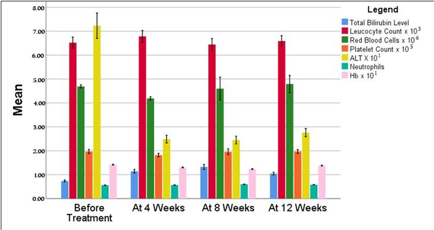
Hemoglobin 14.3 14.1 12.6 13.3 12.4 12.3 13.9 13.7range except for ALT. Patients commenced the new treatment with an average ALT level
around 72 U/L. Still, their hepatic function drastically improved in the first four weeks of
the treatment, normalizing the ALT to levels below 40 U/L, and finishing the regimen on
the same level. At 12 weeks or more after the treatment regimen onset, all 344 patients
Medicina 2021, 57, 986 (100%) who followed the full treatment course were confirmed with an HCV RNA viral 6 of 8
load (Medicina 2021, 57, 986 7 of 8
A limitation of our prospective study was that patients included in the first half,
during 2016–2018, had been treated with an interferon-based regimen before starting DAA
therapy. This probably made them more likely to enroll in the study with worse blood
parameters and general health condition due to the longer exposure to HCV than those of
the second group, treated from 2018 to 2020 with the sofosbuvir and ledipasvir regimen.
Another possible limitation of the current study is the number of blood parameters
that were assessed. Other, more specific analyses were inconsistently checked during the
4-year period, raising the question of which other parameters might have changed during
the course of the 12-week treatment scheme.
In this paper, our findings strengthened the results of previous studies and clinical
trials for the treatment of chronic HCV infection with two treatment schemes (ombitasvir,
paritaprevir, and ritonavir, associated with dasabuvir; and sofosbuvir, associated with
ledipasvir), showing, once again, their efficacy either for patients where previous interferon
regimens had failed or for those recently infected who did not undergo any treatment.
Proving on a medium-scale sample of 344 chronic HCV patients infected with the 1b
genotype suffering from concomitant hematological disturbances that the main blood
parameters mainly improve, leading to a more than 90% achievement of SVR, the study
opens the territory for large-scale government-funded programs aiming to cure one of the
most severely affected countries in Europe from chronic HCV.
5. Conclusions
Current direct-acting antivirals have proven, generally and in our study, to be very
effective in treating the HCV infection. Over 90% of the patients with hematological
malignancies under remission, and hematological disorders included in this prospective
study have achieved SVR by using two different treatment regimens using the association
of ombitasvir, paritaprevir, ritonavir, and dasabuvir, and the single-therapy scheme with
sofosbuvir and ledipasvir association. The above-mentioned treatment regimens are not
significantly different in achieving the desired results. They do not seem to worsen the
patients’ condition throughout the treatment, making them very tolerable and effective for
patients infected with Type 1b HCV compared to the old interferon-based therapies.
Author Contributions: Conceptualization, B.C. and I.M.; methodology, B.C. and M.C.; software, F.B.;
validation, I.M. and O.S.; formal analysis, O.S. and M.T.; investigation, B.C. and I.V.; resources, I.M.;
data curation, F.B.; writing—original draft preparation, I.V. and M.C.; writing—review and editing,
F.B. and M.T.; visualization, F.B.; supervision, I.V.; project administration, I.M. and O.S. All authors
have read and agreed to the published version of the manuscript.
Funding: This research received no external funding.
Institutional Review Board Statement: The study was conducted according to the guidelines of the
Declaration of Helsinki, and approved by the Institutional Review Board of the Victor Babes Hospital
for Infectious Diseases on 10 January 2016 with approval number 7792.
Informed Consent Statement: Informed consent was obtained from all the subjects involved in
the study.
Data Availability Statement: Data available on request.
Conflicts of Interest: The authors declare no conflict of interest.
References
1. Bartenschlager, R.; Bühler, S. Hepatitis C Virus. In Encyclopedia of Virology; Academic Press: San Diego, CA, USA, 2008; pp. 367–374.
[CrossRef]
2. Falla, A.M.; Ahmad, A.A.; Duffell, E.; Noori, T.; Veldhuijzen, I.K. Estimating the scale of chronic hepatitis C virus infection in the
EU/EEA: A focus on migrants from anti-HCV endemic countries. BMC Infect. Dis. 2018, 18, 42. [CrossRef]
3. The Polaris Observatory HCV Collaborators. Global prevalence and genotype distribution of hepatitis C virus infection in 2015:
A modeling study. Lancet Gastroenterol. Hepatol. 2017, 2, 161–176. [CrossRef]Medicina 2021, 57, 986 8 of 8
4. Constantin, M.; Cernea, A.; Dejoianu, T.C.; Lupu, I. Prevalence of Viral Hepatitis B and Viral Hepatitis C Infection among Health Care
Providers from Adult and Children Health Care and Social Assistance Centers; Centrul Medical de Diagnostic si Tratament “Dr. Victor
Babes”: Bucures, ti, Romania, 2020. Available online: https://www.cdt-babes.ro/cercetare/prevalence-of-hbv-and-hcv-infection-
among-health-care-providers.pdf (accessed on 13 November 2020).
5. Lingala, S.; Ghany, M.G. Natural History of Hepatitis C. Gastroenterol. Clin. N. Am. 2015, 44, 717–734. [CrossRef] [PubMed]
6. Seef, L.B. The natural history of chronic hepatitis C. Hepatology 2002, 36, S35–S46.
7. Strader, D.B.; Seeff, L.B. Hepatitis C: A brief clinical overview. ILAR J. 2001, 42, 107–116. [CrossRef] [PubMed]
8. Hancox, S.H.; Smith, B.C. Liver disease as a cause of thrombocytopenia. QJM 2013, 106, 425–431. [CrossRef] [PubMed]
9. Wang, C.S.; Yao, W.J.; Wang, S.T.; Chang, T.T.; Chou, P. Strong association of hepatitis C virus (HCV) infection and thrombocy-
topenia: Implications from a community survey with hyperendemic HCV infection. Clin. Infect. Dis. 2004, 39, 790–796. [CrossRef]
[PubMed]
10. Krishnan, S.M.; Dixit, N.M. Ribavirin-induced anemia in hepatitis C virus patients undergoing combination therapy. PLoS
Comput. Biol. 2011, 7, e1001072. [CrossRef] [PubMed]
11. Chao, T.C.; Chen, C.Y.; Yang, Y.H.; Chen, P.M.; Chang, F.Y.; Lee, S.D. Chronic hepatitis C virus infection associated with primary
warm-type autoimmune hemolytic anemia. J. Clin. Gastroenterol. 2001, 33, 232–233. [CrossRef]
12. Moccia, F.; Tognoni, E.; Boccaccio, P. Autoimmune hemolytic anemia in chronic hepatitis C virus infection: An unusual
extrahepatic autoimmune manifestation. Ann. Ital. Med. Int. 2001, 16, 256–259.
13. Ming-Lung, Y.; Chun-Hao, C. Evolution of Interferon-Based Therapy for Chronic Hepatitis C. Hepat. Res. Treat. 2010, 2010, 140953.
[CrossRef]
14. Kanda, T.; Yasui, S.; Nakamura, M.; Nakamoto, S.; Takahashi, K.; Wu, S.; Sasaki, R.; Haga, Y.; Ogasawara, S.; Saito, T.; et al.
Interferon-free treatment for patients with chronic hepatitis C and autoimmune liver disease: Higher SVR rates with special
precautions for deterioration of autoimmune hepatitis. Oncotarget 2018, 9, 11631–11637. [CrossRef] [PubMed]
15. Centers for Disease Control and Prevention. Testing for HCV infection: An update of guidance for clinicians and laboratorians.
MMWR Morb. Mortal. Wkly. Rep. 2013, 62, 362–365.
16. Cavalcante, L.N.; Lyra, A.C. Predictive factors associated with hepatitis C antiviral therapy response. World J. Hepatol. 2015, 7,
1617–1631. [CrossRef] [PubMed]
17. Schijman, A.; Colina, R.; Mukomolov, S.; Kalinina, O.; García, L.; Broor, S.; Bhupatiraju, A.V.; Karayiannis, P.; Khan, B.; Mogdasy,
C.; et al. Comparison of hepatitis C viral loads in patients with or without co-infection with different genotypes. Clin. Diagn. Lab.
Immunol. 2004, 11, 433–435. [CrossRef]
18. Dhyani, M.; Anvari, A.; Samir, A.E. Ultrasound elastography: Liver. Abdom. Imaging 2015, 40, 698–708. [CrossRef] [PubMed]
19. Iliescu, E.L.; Mercan-Stanciu, A.; Toma, L. Safety and efficacy of direct-acting antivirals for chronic hepatitis C in patients with
chronic kidney disease. BMC Nephrol. 2020, 21, 21. [CrossRef] [PubMed]
20. American Association for the Study of Liver Diseases and Infectious Diseases Society of America. HCV Guidance: Recommenda-
tions for Testing, Managing, and Treating Hepatitis C. Available online: www.hcvguidelines.org/full-report-view (accessed on
1 October 2020).
21. Feld, J.J.; Jacobson, I.M.; Hézode, C.; Asselah, T.; Ruane, P.J.; Gruener, N.; Abergel, A.; Mangia, A.; Lai, C.-L.; Chan, H.L.; et al.
Sofosbuvir and velpatasvir for HCV genotype 1, 2, 4, 5, and 6 infection. N. Engl. J. Med. 2015, 373, 2599–2607. [CrossRef]
22. Simon, K.A.; Flisiak, R.; Łapiński, T.W.; Janczewska, E.; Wawrzynowicz-Syczewska, M.; Jaroszewicz, J.; Zar˛ebska-Michaluk,
D.; Nazzal, K.; Bolewska, B.; Białkowska, J.; et al. Effect of comedication on ombitasvir/paritaprevir/ritonavir ± dasabuvir ±
ribavirin therapy in chronic hepatitis C—a real-world study. Clin. Exp. Hepatol. 2019, 5, 215–223. [CrossRef]
23. El-Kholy, A.M.; Shoeib, S.A.; Zagla, H.A.; Abdelhafez, M.A.; Abdelhamid, A.E.; Kasemy, Z.A.; Abdelmohsen, E.A. Effect of
direct-acting antiviral agents on hepatitis C virus-associated thrombocytopenia. Menoufia Med. J. 2020, 33, 76–81. [CrossRef]
24. Economides, M.P.; Mahale, P.; Kyvernitakis, A.; Turturo, F.; Kantarjian, H.; Naing, A.; Hosry, J.; Shigle, T.L.; Kaseb, A.; Torres,
H.A. Concomitant use of direct-acting antivirals and chemotherapy in hepatitis C virus-infected patients with cancer. Aliment.
Pharmacol. Ther. 2016, 44, 1235–1241. [CrossRef] [PubMed]
25. Łucejko, M.; Parfieniuk-Kowerda, A.; Flisiak, R. Ombitasvir/paritaprevir/ritonavir plus dasabuvir combination in the treatment
of chronic HCV infection. Expert Opin. Pharmacother. 2016, 17, 1153–1164. [CrossRef] [PubMed]You can also read