Robotic assisted CyberKnife radiosurgery for the treatment of iris melanoma
←
→
Page content transcription
If your browser does not render page correctly, please read the page content below
www.nature.com/scientificreports
OPEN Robotic assisted CyberKnife
radiosurgery for the treatment
of iris melanoma
Valerie Schmelter1, Sarah Heidorn2, Alexander Muacevic2, Siegfried G. Priglinger1,
Paul Foerster1 & Raffael Liegl1*
Iris melanoma is a rare form of uveal melanoma with potential metastic spread. Treatment options
include surgical resection, enucleation or irradiation. We analysed visual outcome, complication
appearance and management in eight patients with iris melanoma following robotic-assisted
CyberKnife treatment. Consecutive patients from the Department of Ophthalmology at University
of Munich were included in the study if they had an iris melanoma that was treated with CyberKnife
and had a minimum follow-up of 12 months. We evaluated tumor thickness, largest diameter, visual
acuity and complications. 8 patients were included in this report. The median age was 74 years (range:
53–86 years). The median follow-up was 23 months (range 12–48 months). Tumor thickness decreased
from 2.1 to 1.4 mm on average. Four out of eight patients showed stable or increased visual acuity
compared to visual acuity at first visit. We did not find a correlation of applied radiation volume or
radiation dose on visual outcome. Radiation keratopathy was the most common complication in
five patients. No recurrences were noted. Robotic-assisted radiosurgery following CyberKnife is a
promising non-invasive, single session treatment option for iris melanoma with comparable results
regarding recurrence rate or complications to brachytherapy and proton beam therapy. All included
patients showed good visual outcome.
Uveal melanoma is the most frequent intraocular malignancy and can be subcategorized into uveal melanoma
developing in the choroid, in the ciliary body, in the iris or a combination of any of these locations. The vast
majority of uveal melanoma is found in the choroid (80–90%), whereas ciliary body and particularly iris affection
is considerably less common with approximately 10% and 4% respectively in the mid-aged to older population.
Younger patients, although less commonly affected by uveal melanoma in general, are more frequently affected
by iris melanoma with around 20% of all uveal m elanoma1.
Several factors have been established that are linked to a higher risk of developing iris melanoma. As with
other uveal melanoma, fair skin, light eye color as well as cutaneous nevi, particularly when atypical, are risk
factors for developing iris melanoma2.
The presence of an iris nevus is quite common, representing 25% of all iris lesions in children and 47% of all
iris lesions in middle-aged and senior adults3. It is also a risk factor for the later development of an iris mela-
noma. The rate of transformation of iris nevus into melanoma is controversial and has been estimated at nearly
5% after 5 years4; higher rates have also been reported with some diagnostic c hallenges5. Several studies found
predictive clinical factors for growth of iris nevus into melanoma. These clinical features include hyphema, 4:00
to 9:00 clock hour tumor location, patient age under 40 years, ectropium uveae, the presence of a feeder vessel,
nodule formation and diffuse m alignancy6,7.
In general, Iris melanomas demonstrate low metastatic potential compared to other uveal melanomas and
is believed to be around 3% after 5 years and 5% after 10 years8. Tumor related death occurs in approximately
5–10% of patients, and increases with tumor thickness of more than 4 mm9.
The most appropriate form of treatment is still topic of an ongoing debate, yet radiation therapy has constantly
supplanted surgical resection of the tumor lesion and particularly removal of the whole e ye10.
Surgical resection of iris melanoma can be limited to an iridectomy or incorporate the removal of large parts
of the iris including parts of the ciliary body. Sometimes this procedure needs additional radiotherapy and recur-
rences are often described, even years later11,12.
Today, most cases are either managed by teletherapy using proton or photon beam r adiotherapy13 or plaque
radiotherapy14.
1
Department of Ophthalmology, Ludwig-Maximilians University Munich, Mathildenstr. 8, 80336 Munich,
Germany. 2European CyberKnife Center Munich, Munich, Germany. *email: rliegl@med.lmu.de
Scientific Reports | (2021) 11:5685 | https://doi.org/10.1038/s41598-021-84290-x 1
Vol.:(0123456789)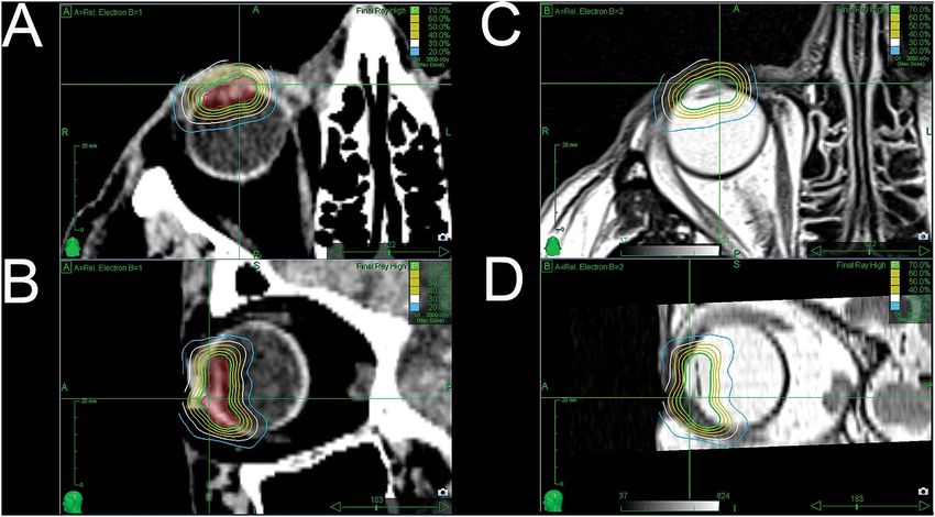
www.nature.com/scientificreports/
We employed photon beam radiotherapy facilitated through the use of a linear accelerator mounted on a
robotic arm, the CyberKnife system, to treat patients with iris melanoma. We report our results on eight patients
that have been treated between 2014 and 2018. We analyzed visual outcome, complications including develop-
ment of cataract and neovascular glaucoma, recurrences as well as overall survival.
To our knowledge, this is the first study evaluating overall outcome of CyberKnife therapy in the manage-
ment of iris melanoma.
Methods
We did a retrospective review of all patients that were diagnosed with iris melanoma and were treated with
robotic assisted radiosurgery (CyberKnife, Accuray Inc., Sunnyvale, CA, USA) at the Department of Oph-
thalmology of the Ludwig-Maximilians-University in Munich, Germany in cooperation with the European
CyberKnife Center in Munich, Germany. The study is approved by the ethics committee´s review board of the
medical faculty at the Ludwig-Maximilians-University (“Ethikkomission der LMU”) for this medical records
review. The study was in accordance with the Declaration of Helsinki. A minimum follow-up of 12 months was
necessary to be included in this study.
We recorded age, gender, laterality, progression of visual acuity (BCVA), tumor thickness and largest diam-
eter using ultrasound-biomicroscopy (UBM) at first visit and follow-up visits. We also recorded central retinal
thickness (CRT) measured via optical coherence tomography (OCT) at each visit. We documented complica-
tions, including cataract progression, glaucoma development, radiation keratopathy, radiation retinopathy and
recurrence rate as well as development of metastases and overall survival. All tumors were categorized following
the updated American Joint Committee on Cancer (AJCC) classification in its eighth e dition15. A correlation of
visual acuity development with different variables, such as radiation dose on fovea, lens and optic disc as well as
with total radiation volume was calculated.
Tumor recurrence was defined as any degree of documented tumor growth (in thickness or base) appreci-
ated by ophthalmoscopy, photographic comparison with earlier visits and ultrasound biomicroscopy (UBM).
Written informed consent was obtained before treatment and risks and chances as well as treatment options
(e.g. brachytherapy and proton beam therapy) were discussed with the patient. CyberKnife radiotherapy was
performed as a standardized outpatient procedure as described previously16. In brief, standard retrobulbar anes-
thesia was performed to achieve akinesia of the globe within the orbit. Target volume was defined by an inter-
disciplinary team composed of ophthalmologists with special knowledge in the treatment of uveal melnaoma,
medical physicists and radiation oncologists using gadolinium-contrast-enhanced MRI, computer tomography
(CT) (1.0 and 1.2 mm slices) as well as all previously obtained clinical data including clinical examination and
ultrasonography as well as ultrasound biomicroscopy results. A non-isocentric inverse algorithm was used in
cooperation with a medical physicist for treatment planning (Multiplan, Accuray Incorporated, Sunnyvale,
California, USA). In all but one case a doughnut shaped pattern incorporating the whole iris was developed and
radiation was delivered according to this plan in a single fraction with a CyberKnife system in a net radiation
time of approximately 20 min.
Data was collected and analyzed in Excel (Microsoft Corporation, Redmond, WA, USA). Statistical analysis
was performed in Graphpad Prism 8.0 (Graphpad, San Diego, CA, USA).
Changes in apical tumor height and largest basal diameter were calculated with a Wilcoxon matched pairs
signed rank test. Correlations were calculated using Spearman´s rho. The significance threshold was set at 0.05.
Visual acuity is displayed as logMAR with averages calculated as mean. Light perception is recorded as 2.70
logMAR.
Ethics statement. A waiver of informed consent was granted and approved by the ethics committee´s
review board of the medical faculty at the Ludwig-Maximilians-University (“Ethikkomission der LMU”) for this
medical records review. The study was in accordance with the Declaration of Helsinki.
Results
A total of 13 patients were treated between 2014 and 2018 in our department. 8 of these patients fulfilled the
inclusion criteria and are described in this report. The median age was 74 years (range: 53–86 years; mean:
71 years). The median follow-up was 23 months [range 12–48 months] and the mean follow-up 26.75 months
[SD ± 12.3 months].
Seven patients received CyberKnife treatment as first treatment option for their iris melanoma. One patient
had a partial iridectomy initially, but showed signs of insufficient resection of the tumor which made a second
treatment approach necessary. CyberKnife was done four months later in the earlier described algorithm. Six
patients were treated solely on clinical signs that allowed for a clear diagnosis of iris melanoma. One patient had
an iris biopsy beforehand, which assured the suspicion of an iris melanoma. This case is depicted in Fig. 1. The
aforementioned patient who was resected as the primary mode of treatment had also an iris melanoma confirmed
by histopathology of the removed specimen.
The overall follow-up time between radiation and last follow-up visit was 27 months (range: 12–48 months).
(Table 1).
Visual Acuity (BCVA). Best-corrected visual acuity (BCVA) before CyberKnife treatment was 0.30 logMAR
(range: 0.70–0.00 logMAR) on average. It decreased to 0.50 logMAR (range: 1.30–0.00 logMAR) one month
after CyberKnife treatment. The main reason for this decrease was development of radiation keratopathy.
One year after radiotherapy five out of eight patients (62.5%) showed stable or even increased visual acuity
compared to visual acuity at first visit (mean: 0.70 logMAR; range: 0.00–2.70 logMAR).
Scientific Reports | (2021) 11:5685 | https://doi.org/10.1038/s41598-021-84290-x 2
Vol:.(1234567890)www.nature.com/scientificreports/
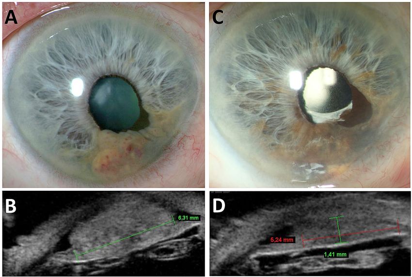
Figure 1. An 85 year old male patient with suspect iris lesion was observed over seven years. When
enlargement of the lesion was suspected, a biopsy was performed, which eventually confirmed the diagnosis of
iris melanoma. (A,B) The patient was subsequently treated with CyberKnife and responded with regression of
tumor at last follow-up two years later (C,D).
Ciliary body
Laterality (RE/ Tumor thickness Tumor diameter Tumor thickness Tumor diameter involvement (yes/ Total follow-up
Pat. no Age Sex (female/male) LE) FD [mm] FD [mm] FU [mm] FU [mm] no) [months]
1 79 Male LE 1.7 6.0 Block excision Block excision Yes 43
2 65 Male LE 2.1 4.6 1.5 4.1 No 48
3 79 Female RE 2.2 5.0 1.32 4.1 No 25
4 79 Female LE 1.5 2.9 1.2 2.8 No 21
5 53 Male LE 1.0 1.9 0.5 3.0 No 22
6 69 Male RE 3.0 8.3 2.5 6.8 No 19
7 86 Male RE 3.6 6.5 1.5 5.3 No 24
8 61 Male RE 2.0 4.9 1.3 4.9 No 12
Table 1. Patient characteristics. FD first diagnosis, FU follow-up, RE right eye, LE = left eye.
There was no statistically significant relationship between tumor thickness or largest basal diameter with
initial visual acuity (Fig. 2).
We noticed no statistically significant impact of applied radiation volume (p = 0.92) or radiation dose delivered
to fovea (p = 0.39), optic disc (p = 0.68) or lens (p = 0.80) on visual outcome at last follow-up (Fig. 3).
Tumor classification according to AJCC classification (8th edition). Tumor thickness at first pres-
entation was 2.1 mm (range: 1.0–3.6 mm) on average with the largest diameter at a mean of 5.0 mm (range:
1.9–8.3 mm). The tumor thickness was reduced at last follow-up to a mean of 1.4 mm (range: 0.50–2.5 mm) and
the diameter decreased to a mean of 4.4 mm (range: 2.8–6.8 mm). The mean reduction was 0.74 mm (range:
0.50–1.10 mm) for thickness and 0.58 mm (range: 0.90–1.50 mm) regarding diameter. The reduction in tumor
thickness was statistically significant (p = 0.01) while the change in largest basal diameter was not (p = 0.16).
4 patients were classified as T1 tumors (one T1a, three T1b) at first presentation whereas 4 patients were T2
tumors with ciliary body affection (three T2a, one T2c).
Treatment modalities. All patients were treated with 21 Gy at a 70% isodose. An inhomogeneous dose
prescription is standard practice for all tumors treated with CyberKnife, and for most other radiosurgery tech-
niques using small photon beams. An example of our treatment plan is shown in Fig. 4. Applied radiation vol-
Scientific Reports | (2021) 11:5685 | https://doi.org/10.1038/s41598-021-84290-x 3
Vol.:(0123456789)www.nature.com/scientificreports/
Figure 2. Although there was a trend for lower best corrected visual acuity [BCVA] over the course of
follow-up with both increased tumor height and larger basal diameter, this was statistically not significant.
Figure 3. There was no correlation between total radiation dose, radiation dose on optic disc, fovea or lens with
the degree of change in visual acuity.
ume was ranging from 0.70 to 3.54 mm3. Mean applied dosage to fovea, lens and optic disc was 4.70gy (range:
0.96–20.7), 23.58gy (range: 0.00–29.97) and 5.15gy (range: 1.20–23.65) respectively. (Table 2).
Complication, recurrence and overall survival. Five out of eight patients (62.5%) were already pseu-
dophakic at first presentation. Two out of three phakic patients developed cataract along the follow-up period
and received cataract surgery six months and 37 months after radiation treatment.
Scientific Reports | (2021) 11:5685 | https://doi.org/10.1038/s41598-021-84290-x 4
Vol:.(1234567890)www.nature.com/scientificreports/
Figure 4. Two treatment plans are depicted in this figure. We used a donut shaped pattern in all our cases in
order to make sure that the entire tumor was within the radiation field. (A,B) as well as (C,D) show the planning
target volume with isodose lines from two different perspectives.
Mean (range)
Radiation dose (Gy), 21
Isodose (%) 70
Radiation volume (mm3) 1.4 (0.7–3.5)
Maxium radiation dose on fovea (Gy) 4.7 (1.0–20.7)
Maxium radiation dose on optic disc (Gy) 5.1 (1.2–23.7)
Treatment form Donut
Table 2. Radiation treatment parameters.
Four patients (50.0%) developed glaucoma after a mean of 14.5 months (range 9.0–22 months) after
CyberKnife treatment. Three of these patients were treated conservatively with intraocular eye pressure reduc-
ing drops, whereas one patient had glaucoma operation nine months after radiation. No patient developed
neovascular glaucoma.
Radiation keratopathy was observed in five patients (62.5%). Out of these, three patients developed radiation
keratopathy immediately after treatment (first follow-up visit after one month). The other two patients developed
first symptoms 2 and 22 months after CyberKnife treatment. All of the patients were treated with lubricating eye
drops and showed response to this treatment.
We did not notice any form of radiation retinopathy or opticoneuropathy—the central retinal thickness (CRT)
showed no significant changes over the whole follow-up time.
No recurrences were appreciated over the time of follow-up and none of the patients needed enucleation
for secondary complications or lack of local control. All patients remained free of metastases over the whole
observation period. (Table 3).
Discussion
We report on 8 patients that have been treated with robotic assisted CyberKnife (Accuray, Inc.) radiosurgery
due to an iris melanoma. We did not see any recurrences over the follow-up time.
Despite the rare appearance of iris melanoma and the lower rate of metastastic development with 5% at
10-year follow-up compared to otherwise located uveal melanomas8, iris melanoma may enlarge in size and
infiltrate other tissues of the eye and may even progress to extraocular extension.
Different treatment modalities are possible and described for iris melanomas although considerably less
is known on the best treatment strategy as compared to uveal melanomas in the choroid or the ciliary body.
CyberKnife treatment is different compared to brachytherapy and proton beam treatment: CyberKnife comprises
a linear accelerator which is mounted on an industry roboter with six degrees of freedom, allowing application
of radiation from every direction. The ability of this system to deliver radiation beams from every possible angle
Scientific Reports | (2021) 11:5685 | https://doi.org/10.1038/s41598-021-84290-x 5
Vol.:(0123456789)www.nature.com/scientificreports/
Number of patients
Lens status
Phakic 5
Cataract progression 2
Surgery 2
Pseudophakic 3
Glaucoma development 4
Medical management 3
Surgery 1
Radiation Retinopathy 0
Radiation Keratopathy 5
Recurrences 0
Metastases 0
Enucleation 0
Table 3. Complications following radiation treatment for iris melanoma.
may be advantageous when trying to save tissue from being exposed to radiation by excluding these structures
during planning of treatment as much as possible. Placement of a radioactive plaques as with brachytherapy
demands surgery. The plaque is directly sutured to the sclera in the region of radiation application to the under-
lying tissue. The time until the plaque can be surgically removed is dependent on the dose calculation of a
medical physicist and radiation oncologist during treatment planning and encompasses usually a few d ays9.
Proton beam therapy on the other hand is, similar to CyberKnife treatment, a teletherapeutic option in which
a radiation beam is delivered in multiple fractions to the target tissue. Since the proton beam cannot be moved
around the patients head, small titanium clips are sutured on the sclera before treatment in order to facilitate
treatment planning and e xecution17.
Popovic et al.18 did a medline search of all existing treatment regimens and found a total of 17 studies with
a total of 761 eyes that met their criteria for further analysis. The main treatment option for iris melanoma is
brachytherapy and many reports on radiotherapy are available. Among these, b rachytherapy14,19,20 and proton
beam therapy (PBT)13,21,22 are the most frequently evaluated treatment approaches. Less can be found on surgi-
cal resection18,23.
As mentioned before, no recurrences were seen in our series of patients. These results compare well to other
treatment approaches, particularly PBT or brachytherapy in which recurrence rates and metastastic development
were also reported to be low. Recurrences occurred in 0–7.5% of patients following proton beam t herapy24–26
and in 0–8%27,28 and up to 15%14 after seven years following brachytherapy. Metastases occur considerably less
frequently in iris melanoma as compared to choroidal melanoma with approximately 5% after five years10.
Surgical resection always goes along with removal of iris tissue which subsequently increases the risk of
photophobia (9–25%), yet rates of recurrences (0–8%) are not lower than for the aforementioned a lternatives29
or the result of our study.
Due to the close proximity of the iris to the anterior chamber angle and intraocular lens, it is almost never
possible to save these structures from being incorporated into the planning target volume when radiation is
planned. This poses a higher risk to the development of cataract with subsequent visual acuity deterioration or
secondary glaucoma caused by radiation induced structural changes in the anterior chamber angle.
Unsurprisingly, the three most commonly reported complications following PBT or plaque radiotherapy
are cataract progression (36–73%), corneal discomfort and defects due to limbal cell deficiency (9–90%)24 and
occurrence of secondary glaucoma (3–92%)18,23.
In all of our patients a donut shaped irradiation pattern was planned to treat the iris melanoma. This rather
aggressive irradiation approach reduces the risk of missed melanoma cells in parts of the iris that are not seen
clinically or with ultrasound but could possibly entail higher incidences of the aforementioned secondary
complications. Notwithstanding our treatment planning, compare our results similar to published data from
brachytherapy and PBT with 50% of patients developing secondary glaucoma and two out for three patients
with progression of cataract and subsequent cataract surgery after 6 and 37 months. The most common finding
after treatment however was keratopathy which occurred in 62.5% of all patients. This complication is often only
temporary and all patients from our cohort could be managed with lubricating eye drops. Keratopathy is often
not mentioned in reports on treatment outcome, so that comparison to other treatment approaches is intricate.
A few reports addressing this complication exist however. Fernandes and associates20 for example reported
mild to moderate keratitis in most of the cases (14 patients) after Idodine-125 brachytherapy for iris melanoma.
In addition, Konstantinidis et al. attribute symptoms of “grittiness” and ocular discomfort observed in 67% of
patients (12 patients) treated with PBT for iris melanoma to ocular surface irregularities21. Three out of eight
patients in our study developed corneal discomfort immediately after radiotherapy and showed subjective and
objective improvement (increase in visual acuity) over time. Our results regarding long-term corneal and scleral
thers20.
affection is in line with reports from o
Scientific Reports | (2021) 11:5685 | https://doi.org/10.1038/s41598-021-84290-x 6
Vol:.(1234567890)www.nature.com/scientificreports/
Half of our patients developed glaucoma over the course of follow-up with a median occurrence after
14.5 months. Three out of four patients could be sufficiently treated with the prescription of intraocular pres-
sure lowering eye drops. One patient needed additional glaucoma surgery to manage the increased eye pressure.
Data regarding visual acuity outcome is inhomogenous. Shields et al.14 reported that 37% of treated patients
(52 out of 141 patients) with plaque brachytherapy would end up with poor visual acuity (< 20/200). Fernandes
et al. reported increased visual acuity in one case, stability in ten cases and worsening in three cases (14 patients
in total)20.
In our study 4 patients (50%) showed decrease in visual acuity from mean 0.3 logMAR (range: 0.10–0.70
logMAR) before treatment to a mean of 0.9 logMAR (range: 0.50–3.00logMAR) at last follow-up. Three patients
(37.5%) remained stable and one patient (12.5%) had an increased visual acuity from 0.7logMAR to 0.3logMAR
at last follow-up visit.
We did not find any correlation of the course of visual acuity and tumor size, neither for tumor height nor
largest basal diameter (Fig. 2). Furthermore was no statistically significant correlation found between visual
acuity and radiation dose on optic disc or fovea. (Fig. 3).
This report has several limitations. The retrospective character, although typical for reports on outcome of
ocular cancers, does not always allow a complete record of data. In addition, is our follow-up period rather short
so that statements regarding metastasis or recurrences must be interpreted with caution as iris melanoma recur-
rences are less common than with other uveal melanomas but may occur later in the course of o bservation12.
Further, complications may also occur later in time and additional reports with a higher patient number and
follow-up time are demanded to better assess this question. The small number of patients is a drawback, which
does not allow general prediction on treatment outcome in linear accelerator treated iris melanoma.
However, to our knowledge there are no other reports reporting clinical outcome after robotic assisted
CyberKnife treatment for iris melanoma. CyberKnife can be facilitated on one day in just over three hours
including treatment planning and execution with an experienced interdisciplinary team comprising radia-
tion oncologists, medical physicists and ophthalmologists. All of our patients showed local control after up to
48 months of post treatment observation and none of the patients had documented metastases. Complications
were comparable to brachytherapy and PBT. We therefore believe that CyberKnife is a safe, effective and a com-
fortable option in selected cases of iris melanomas.
Received: 4 July 2020; Accepted: 16 October 2020
References
1. Shields, C. L., Kaliki, S., Furuta, M., Mashayekhi, A. & Shields, J. A. Clinical spectrum and prognosis of uveal melanoma based
on age at presentation in 8,033 cases. Retina (Philadelphia, Pa.) 32, 1363–1372. https://doi.org/10.1097/IAE.0b013e31824d09a8
(2012).
2. Bataille, V. et al. Risk of ocular melanoma in relation to cutaneous and iris naevi. Int. J. Cancer 60, 622–626. https: //doi.org/10.1002/
ijc.2910600509 (1995).
3. Shields, C. L. et al. Clinical survey of 3680 iris tumors based on patient age at presentation. Ophthalmology 119, 407–414. https://
doi.org/10.1016/j.ophtha.2011.07.059 (2012).
4. Territo, C., Shields, C. L., Shields, J. A., Augsburger, J. J. & Schroeder, R. P. Natural course of melanocytic tumors of the iris. Oph-
thalmology 95, 1251–1255. https://doi.org/10.1016/s0161-6420(88)33022-8 (1988).
5. van Klink, F., de Keizer, R. J., Jager, M. J. & Kakebeeke-Kemme, H. M. Iris nevi and melanomas: a clinical follow-up study. Docu-
mentaophthalmologica. Adv. Ophthalmol. 82, 49–55. https://doi.org/10.1007/bf00156993 (1992).
6. Giuliari, G. P., Krema, H., McGowan, H. D., Pavlin, C. J. & Simpson, E. R. Clinical and ultrasound biomicroscopy features associ-
ated with growth in iris melanocytic lesions. Am. J. Ophthalmol. 153, 1043–1049. https://doi.org/10.1016/j.ajo.2011.11.004 (2012).
7. Shields, C. L. et al. Iris nevus growth into melanoma: analysis of 1611 consecutive eyes: the ABCDEF guide. Ophthalmology 120,
766–772. https://doi.org/10.1016/j.ophtha.2012.09.042 (2013).
8. Shields, C. L. et al. Iris melanoma: risk factors for metastasis in 169 consecutive patients. Ophthalmology 108, 172–178. https://
doi.org/10.1016/s0161-6420(00)00449-8 (2001).
9. Razzaq, L. et al. Ruthenium plaque radiation therapy for iris and iridociliary melanomas. Acta Ophthalmol. 90, 291–296. https://
doi.org/10.1111/j.1755-3768.2010.01967.x (2012).
10. Shields, C. L. et al. Iris melanoma outcomes based on the american joint committee on cancer classification (eighth edition) in
432 patients. Ophthalmology 125, 913–923. https://doi.org/10.1016/j.ophtha.2017.11.040 (2018).
11 Shah, S. U. et al. Plaque radiotherapy for residual or recurrent iris melanoma after surgical resection in 32 cases. Ophthalmology
119, 838–842. https://doi.org/10.1016/j.ophtha.2011.09.039 (2012).
12. Bosello, F., Al-Jamal, R. T. & Cohen, V. M. L. Very late recurrence of iris melanoma: 45 years after treatment. Melanoma Res. 30,
309–312. https://doi.org/10.1097/cmr.0000000000000581 (2020).
13 Rahmi, A. et al. Proton beam therapy for presumed and confirmed iris melanomas: a review of 36 cases. Graefe’s Arch. Clin. Exp.
Ophthalmol. 252, 1515–1521. https://doi.org/10.1007/s00417-014-2735-y (2014).
14. Shields, C. L. et al. Iris melanoma management with iodine-125 plaque radiotherapy in 144 patients: impact of melanoma-related
glaucoma on outcomes. Ophthalmology 120, 55–61. https://doi.org/10.1016/j.ophtha.2012.06.053 (2013).
15. Kivelä, T. et al. AJCC Cancer Staging Manual 8th edn, 805–817 (Springer, Berlin, 2017).
16. Muacevic, A. et al. Development of a streamlined, non-invasive robotic radiosurgery method for treatment of uveal melanoma.
Technol. Cancer Res. Treat. 7, 369–374. https://doi.org/10.1177/153303460800700503 (2008).
17. Amstutz, C. A., Bechrakis, N. E., Foerster, M. H., Heufelder, J. & Kowal, J. H. Intraoperative localization of tantalum markers for
proton beam radiation of choroidal melanoma by an opto-electronic navigation system: a novel technique. Int. J. Radiat. Oncol.
Biol. Phys. 82, 1361–1366. https://doi.org/10.1016/j.ijrobp.2011.04.049 (2012).
18. Popovic, M., Ahmed, I. I. K., DiGiovanni, J. & Shields, C. L. Radiotherapeutic and surgical management of iris melanoma: a review.
Surv. Ophthalmol. 62, 302–311. https://doi.org/10.1016/j.survophthal.2016.12.012 (2017).
19. Tsimpida, M., Hungerford, J., Arora, A. & Cohen, V. Plaque radiotherapy treatment with ruthenium-106 for iris malignant mela-
noma. Eye (London, England) 25, 1607–1611. https://doi.org/10.1038/eye.2011.222 (2011).
20. Fernandes, B. F. et al. Management of iris melanomas with 125Iodine plaque radiotherapy. Am. J. Ophthalmol. 149, 70–76. https
://doi.org/10.1016/j.ajo.2009.08.007 (2010).
Scientific Reports | (2021) 11:5685 | https://doi.org/10.1038/s41598-021-84290-x 7
Vol.:(0123456789)www.nature.com/scientificreports/
21. Konstantinidis, L. et al. Management of patients with uveal metastases at the Liverpool Ocular Oncology Centre. Br. J. Ophthalmol.
98, 92–98. https://doi.org/10.1136/bjophthalmol-2013-303519 (2014).
22. Sandinha, M. T., Kacperek, A., Errington, R. D., Coupland, S. E. & Damato, B. Recurrence of iris melanoma after proton beam
therapy. Br. J. Ophthalmol. 98, 484–487. https://doi.org/10.1136/bjophthalmol-2013-303321 (2014).
23. Pe’er, J. & Frenkel, S. Sector iridectomy of iris melanoma: a novel technique for excising the melanoma extraocularly. Br. J. Oph-
thalmol. 95, 1474–1476. https://doi.org/10.1136/bjo.2011.204958 (2011).
24. Willerding, G. D. et al. Proton beam radiotherapy of diffuse iris melanoma in 54 patients. Br. J. Ophthalmol. 99, 812–816. https://
doi.org/10.1136/bjophthalmol-2014-305174 (2015).
25. Thariat, J. et al. Proton beam therapy for iris melanomas in 107 patients. Ophthalmology 125, 606–614. https://doi.org/10.1016/j.
ophtha.2017.10.009 (2018).
26 Riechardt, A. I. et al. Proton therapy of iris melanoma with 50 CGE: influence of target volume on clinical outcome. Strahlentherapie
und Onkologie : Organ der DeutschenRontgengesellschaft ... [et al] 193, 943–950. https: //doi.org/10.1007/s00066 -017-1166-1 (2017).
27. Agraval, U. et al. Use of ruthenium-106 brachytherapy for iris melanoma: the Scottish experience. Br. J. Ophthalmol. 102, 74–78.
https://doi.org/10.1136/bjophthalmol-2017-310278 (2018).
28. Marinkovic, M. et al. Ruthenium-106 brachytherapy for iris and iridociliary melanomas. Br. J. Ophthalmol. 102, 1154–1159. https
://doi.org/10.1136/bjophthalmol-2017-310688 (2018).
29. Klauber, S., Jensen, P. K., Prause, J. U. & Kessing, S. V. Surgical treatment of iris and ciliary body melanoma: follow-up of a 25-year
series of patients. Acta Ophthalmol. 90, 122–126. https://doi.org/10.1111/j.1755-3768.2010.01889.x (2012).
Author contributions
V.S.: Concept of study, Data collection, Data analysis, Preparation of manuscript. S.H.: Data collection. A.M.:
Data analysis, Concept of study. S.P.: Preparation of manuscript. P.F.: Concept of study, Preparation of manuscript.
R.L.: Concept of study, Data analysis, Preparation of manuscript.
Funding
Open Access funding enabled and organized by Projekt DEAL. AM has previously received speaker fees by
Accuray, Inc.
Competing interests
The authors declare no competing interests.
Additional information
Correspondence and requests for materials should be addressed to R.L.
Reprints and permissions information is available at www.nature.com/reprints.
Publisher’s note Springer Nature remains neutral with regard to jurisdictional claims in published maps and
institutional affiliations.
Open Access This article is licensed under a Creative Commons Attribution 4.0 International
License, which permits use, sharing, adaptation, distribution and reproduction in any medium or
format, as long as you give appropriate credit to the original author(s) and the source, provide a link to the
Creative Commons licence, and indicate if changes were made. The images or other third party material in this
article are included in the article’s Creative Commons licence, unless indicated otherwise in a credit line to the
material. If material is not included in the article’s Creative Commons licence and your intended use is not
permitted by statutory regulation or exceeds the permitted use, you will need to obtain permission directly from
the copyright holder. To view a copy of this licence, visit http://creativecommons.org/licenses/by/4.0/.
© The Author(s) 2021
Scientific Reports | (2021) 11:5685 | https://doi.org/10.1038/s41598-021-84290-x 8
Vol:.(1234567890)You can also read