ECommons@AKU - The Aga Khan University
←
→
Page content transcription
If your browser does not render page correctly, please read the page content below
eCommons@AKU

Department of Radiology                                                   Medical College, Pakistan

7-11-2021

Value of periappendiceal fat sign on ultrasound in acute
appendicitis
Ayesha Walid

Muhammad Azeemuddin

Zainab Hussain

Follow this and additional works at: https://ecommons.aku.edu/pakistan_fhs_mc_radiol

   Part of the Radiology Commons
Open Access Original
                              Article                                                  DOI: 10.7759/cureus.16321

                                              Value of Periappendiceal Fat Sign on Ultrasound
                                              in Acute Appendicitis
                                              Ayesha Walid 1 , Azeemuddin Muhammad 2 , Zainab Hussain 2

                                              1. Radiology, Dow University of Health Sciences, Civil Hospital Karachi, Karachi, PAK 2. Radiology, Aga Khan
                                              University Hospital, Karachi, PAK

                                              Corresponding author: Zainab Hussain, zainab.hussain@aku.edu

                                              Abstract
                                              Introduction
                                              Acute right lower quadrant abdominal pain is one of the most common surgical presentations to the
                                              emergency department with acute appendicitis being the topmost differential diagnosis. Although computed
                                              tomography (CT) is the gold standard in diagnosing appendicitis, in our setup ultrasound is often the initial
                                              imaging modality available in urgent care settings especially for children and pregnant females. On
                                              ultrasound, an inflamed appendix has a diameter of 6 mm or more and is non-compressible. Increased
                                              periappendiceal fat echogenicity is an important ancillary sign of acute appendicitis that supports the
                                              sonographic diagnosis of acute appendicitis. To determine the association of periappendiceal fat echo sign
                                              (PFES) on ultrasound in surgically proven cases of acute appendicitis.

                                              Methods
                                              This cross-sectional study was held at the Department of Radiology at the Aga Khan University Hospital in
                                              Karachi, Pakistan. Periappendiceal fat echogenicity was assessed and prospectively graded in 59 patients.
                                              These patients had sonographic features of acute appendicitis which was later confirmed by surgery. Data
                                              were collected on a proforma and later analyzed. Frequency of increased periappendiceal fat echogenicity in
                                              acute appendicitis was calculated. Association of PFES with gender and ascites was evaluated with Fischer's
                                              exact test and with patient's age and appendiceal diameter was assessed using analysis of variance
                                              (ANOVA).

                                              Results
                                              Increased periappendiceal fat echogenicity was seen in 89.8% of patients with acute appendicitis. 10.2% of
                                              patients had acute appendicitis with normal surrounding fat. Mean appendiceal diameter in patients with
                                              grade 3 PFES was significantly more than those with grade 2 or grade 1 PFES. PFES had no association with
                                              age and gender of the patient or with ascites.

                                              Conclusion
Review began 06/01/2021                       Increased periappendiceal fat echogenicity is an important ancillary sign of acute appendicitis that helps
Review ended 07/02/2021                       support its sonographic diagnosis.
Published 07/11/2021

© Copyright 2021
Walid et al. This is an open access article
distributed under the terms of the            Categories: Emergency Medicine, Radiology, General Surgery
Creative Commons Attribution License          Keywords: ultrasound, acute appendicitis, periappendiceal fat echo, right lower quadrant pain, multiple detector
CC-BY 4.0., which permits unrestricted        computed tomography
use, distribution, and reproduction in any
medium, provided the original author and
source are credited.                          Introduction
                                              Acute appendicitis is the most common surgical emergency affecting individuals of all ages [1]. The life-time
                                              risk of developing acute appendicitis has been reported as 9% for males and 7% for females [2].

                                              Patients presenting with characteristic clinical signs and symptoms of acute appendicitis go through instant
                                              surgery without radiological workup. In patients with atypical or confusing clinical findings radiological
                                              workup is requested. The selection of modality whether US or CT in this clinical scenario is mainly reliant
                                              on institutional preference and availability of skilled experts, although patient demographics and BMI are
                                              important influencing factors [3].

                                              The stated sensitivities for abdominal CT are 90%-100%, specificities ranging from 91%-99%, 94%-98%
                                              accuracy, positive predictive value (PPV) and negative predictive value (NPV) of 92%-98% and 95%-100%,
                                              respectively, for the identification of acute appendicitis [3-6]. The most important imaging study in the
                                              evaluation of patients with atypical presentations of appendicitis is abdominal CT. In selected patients with
                                              suspected appendicitis, studies have reported a decrease in negative laparotomy rate and appendiceal
                                              perforation rate when pelvic CT was used [7-9].

                              How to cite this article
                              Walid A, Muhammad A, Hussain Z (July 11, 2021) Value of Periappendiceal Fat Sign on Ultrasound in Acute Appendicitis. Cureus 13(7): e16321.
                              DOI 10.7759/cureus.16321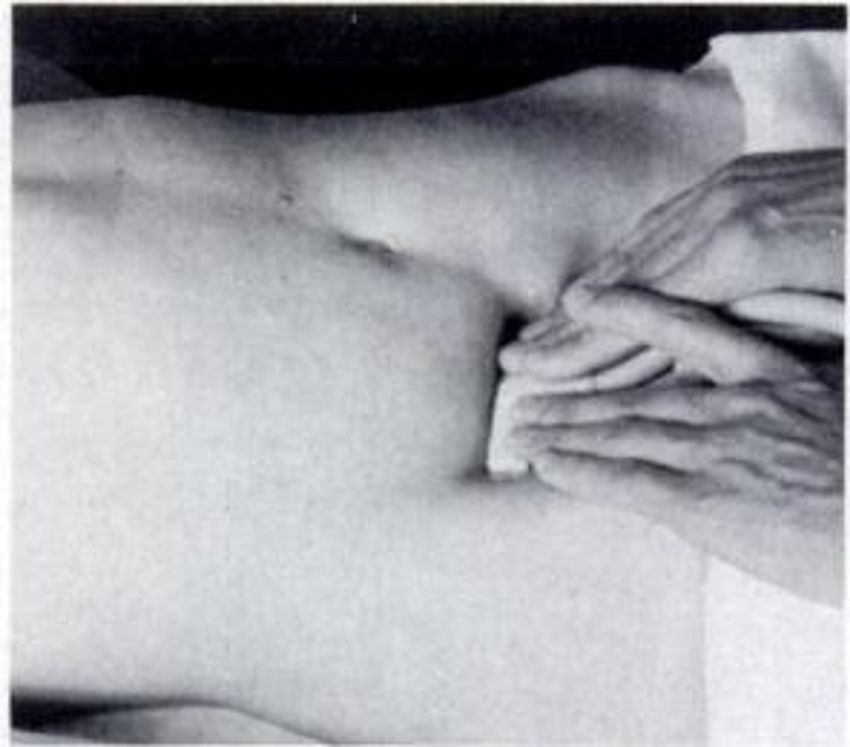
The two most predictive signs, that is, the signs with the highest probability of a correct diagnosis are peri-
                                                  appendiceal fat stranding and appendiceal diameter [10]. No other sign is needed to increase the level of
                                                  confidence for diagnosing appendicitis. In a prospective study, comparing US and CT, Balthazar et al.
                                                  indicated CT to be superior to graded compression US in the diagnosis of acute appendicitis with similar
                                                  specificities (89% vs. 91%, respectively) and PPV (96% vs. 95%, respectively) [11].

                                                  Despite the superior diagnostic parameters of CT scan, US is often the first line of investigation as it is
                                                  simple, rapid, easily available, inexpensive and not associated with ionizing radiation [1-4]. Due to the lack
                                                  of non-ionising radiation and dynamic ability ultrasound is the imaging modality of choice in the evaluation
                                                  of suspected acute appendicitis and allows the radiologist to clinically assess the patient. It is the initial
                                                  imaging examination of choice, particularly in women of childbearing age and children [12]. As sonography
                                                  involves a short acquisition time, not using ionizing radiation, and other causes of abdominal pain such as
                                                  ovarian cysts, tubo-ovarian abscesses and mesenteric adenitis may be diagnosed, it can be performed at the
                                                  bedside [2,10]. The course of the appendix is variable hence making the visualization of the structure slightly
                                                  challenging including both retrocecal and pelvic locations. A retrocecal appendix can be best visualised on
                                                  scans acquired with the transducer position next to the caecum or to the ascending colon, with an oblique
                                                  plane of insonation [13]. A target appearance, characterized by a fluid-filled center and surrounded by an
                                                  echogenic mucosa and submucosa and hypoechoic muscularis, is visualised when imaging in the axial plane
                                                  [14]. Endovaginal scanning is the best method for visualization of the pelvic appendix in women. The ability
                                                  to see a pelvic appendix will be influenced by different degrees of bladder filling [15].

                                                  The ultrasound criteria for acute appendicitis include visualization of a non-peristaltic, non-compressible,
                                                  tubular, blind-ending structure with a diameter of 6 mm or more in the right iliac fossa [13,16]. However,
                                                  there are many ancillary signs of acute appendicitis that can help in its sonographic diagnosis. These include
                                                  prominent hyperechoic mesoappendix or pericecal fat, localized periappendiceal fluid collection,
                                                  aperistaltic bowel loops, enlarged lymph nodes and presence of free fluid [1,6].

                                                  US with graded compression has a sensitivity of 89% and specificity of 100% and is a widely used technique
                                                  within the diagnosis of acute appendicitis [5]. In graded compression technique, pressure is applied through
                                                  a high-resolution linear transducer for displacement and compression of the underlying bowel loops,
                                                  thereby making the appendix visible. The visualization of the psoas muscle and iliac vessels should be part
                                                  of a proficient exam.

                                                  The objective of this study is to evaluate whether periappendiceal fat echo sign (PFES) is an important
                                                  ancillary sign that would be helpful for the sonographic diagnosis of acute appendicitis and to determine its
                                                  frequency in surgically proven cases of acute appendicitis.

                                                  Materials And Methods
                                                  The study was carried out at the Aga Khan University hospital in Karachi, Pakistan. The study design was
                                                  prospective and cross-sectional. This was a residents dissertation and the research training and medical
                                                  center of the College of physicians and surgeons Pakistan had supervised the research ensuring that all
                                                  ethical guidelines were met against this study. The sample size estimation was done on the World Health
                                                  Organization software determinations of sample size. The prevalence of increased intrabdominal fat echo
                                                  has been reported to be 89% [17]. Thus at a confidence level of 95% and significance estimated at a p-value
                                                  of 0.05, 59 patients were enrolled for the study with margin of error calculated within 6%. Non-probability
                                                  purposive sampling technique was deployed for recruiting patients

                                                  The study enrolled all consecutive patients of all ages and both genders who were referred to our department
                                                  for US examination with clinical suspicion of acute appendicitis (periumbilical/right lower abdominal pain,
                                                  vomiting and fever) followed by surgery and histopathology at the same institution. Patients in whom
                                                  adequate ultrasound examination could not be performed or the appendix was not visualized (retrocecal
                                                  location) were excluded as well as those patients who were treated conservatively or did not have surgical
                                                  findings of appendicitis.

                                                  Informed consent was obtained by the principal investigator. US was performed on Toshiba Nemio using a
                                                  high frequency (4-8 MHz ) linear array and convex (3-5 MHz ) array transducers. Graded compression
                                                  technique described by Puylaert et al. was followed for US examination [18]. Uniform pressure was applied
                                                  over the right iliac fossa via the handheld ultrasound transducer. Resultantly normal bowel loops were not
                                                  visualized due to flattening between the layers of the abdominal wall musculature or displacement from the
                                                  image (Figure 1).

2021 Walid et al. Cureus 13(7): e16321. DOI 10.7759/cureus.16321                                                                                                    2 of 9
FIGURE 1: Ultrasound graded compression technique by Puylaert.
                                                    Puylaert JB. Acute appendicitis: US evaluation using graded compression. Radiology. 1986 Feb;158(2):355-
                                                    60.

                                                  Ultrasound examination for acute appendicitis was performed by a consultant radiologist with five-year
                                                  clinical experience. Any abnormal loop of gut or the obstructed appendix was non-compressible and
                                                  adequately visualized on the graded compression image. An enlarged appendix with an outer diameter equal
                                                  to or greater than 6 mm showing lack of compressibility is defined as acute appendicitis. The periapendiceal
                                                  fat echo sign in the right iliac fossa was assessed and classified on the scale developed by Lee et al. [7]. Grade
                                                  1 was described as normal-appearing fat which is hypoechoic to the adjacent appendix with grade 2 showing
                                                  increased echogenicity of the periappendiceal fat; however, underlying muscles and vessels still visualized
                                                  and grade 3 with the periappendiceal fat appearing hyperechoic with obscuration of the underlying muscles
                                                  and vessels

                                                  Comparison was made with the contralateral abdominal fat echo during the sonographic examination.
                                                  Surgical and histopathology findings regarding final diagnosis were obtained from medical records of
                                                  patients by the principal investigator. All findings were recorded on standard Performa.

                                                  Statistical analysis was performed with Statistical Package for the Social Sciences (SPSS) for Windows,
                                                  Version 16 (SPSS Inc., Chicago, IL). Descriptive analysis was conducted with frequencies and percentages for
                                                  categorical variables such as gender while mean and standard deviation were estimated for continuous
                                                  variables like age and appendicular diameter. The presence of the PFES on ultrasound was assessed for
                                                  surgically proven cases of acute appendicitis. Association of PFES with categorical variables like sex and
                                                  ascites was further assessed with Fischer's exact test. Two-sided ANOVA was applied to see the association
                                                  of PFES with continuous variables like age and appendicular diameter. A p-value of < 0.05 was considered
                                                  statistically significant.

                                                  Results
                                                  A total of 59 patients were included in this study. All these patients were diagnosed with acute appendicitis
                                                  on ultrasound and were followed up with surgery and histopathology at our institution which was positive
                                                  for appendicitis. There were 36 (61%) male and 23 (39%) female patients. Age range was seen between 5 and
                                                  62 years with mean age of 19.5 years with standard deviation of 12.3. PFES was assessed and graded into
                                                  three categories in all patients. Six out of 59 patients (10.2%) showed normal periappendiceal fat
                                                  echogenicity, corresponding to grade 1 PFES. Thirty-four patients (57.6%) showed grade 2 PFES.
                                                  Nineteen patients (32.2%) had grade 3 PFES. Thus increased periappendiceal fat echogenicity was seen in a

2021 Walid et al. Cureus 13(7): e16321. DOI 10.7759/cureus.16321                                                                                                       3 of 9
total of 53/59 patients (89.8%). Presence and absence of ascites with acute appendicitis was also taken into
account. Ascites was seen in 25/59 patients (42.4%). Thirty-four patients (57.6%) had no ascites as seen in
Table 1.
Study variables N = 59 (%, ±SD)
Gender
Male 36 61%
Female 23 39%
Age (5-62 years) 19.5 ±12.3
PFES
Grade 1 6 10.2%
Grade 2 34 57.6%
Grade 3 19 32.2%
Ascites
Present 25 42.4%
Absent 34 57.6%
TABLE 1: Baseline data.
PFES: periappendiceal fat echo sign; N: total patients; SD: standard deviation.
Association between appendiceal diameter in acute appendicitis and PFES was assessed with ANOVA. Our
results showed that the mean appendiceal diameter in patients with grade 3 PFES was 1.1 cm. This was
significantly more than patients with normal periappendiceal fat echo and those with increased
periappendiceal fat echo with visualization of underlying structures with a p-value of 0.002 which rendered
it significant.
Mean appendiceal diameter was not significantly different in patients with grade 1 PFES (0.82 cm) and those
with grade 2 PFES (0.9 cm) as seen in Table 2.
PFES Appendix diameter p-value
Grade 1 0.82 cm >0.05
Grade 2 0.90 cm >0.05
Grade 3 1.1 cm 0.02
TABLE 2: Association between appendiceal diameter and periappendiceal fat echo sign.
PFES: periappendiceal fat echo sign.
Association of PFES with gender and ascites was evaluated with Fischer exact test. A p-value of 2.7 and 2.5
was obtained, respectively, indicating no significant association of gender and the presence of ascites in the
sample cohort.
Two-way ANOVA was applied to see how PFES varied with the age of the patient. Mean age of patients with
grade 3 PFES (23 years) was more than those with grade 2 (19 years) and grade 1 PFES (12 years). This
difference was, however, not statistically significant (p = 0.16).
Discussion
Acute appendicitis is one of the commonest causes of acute abdomen presenting to the emergency
2021 Walid et al. Cureus 13(7): e16321. DOI 10.7759/cureus.16321 4 of 9department that often warrants immediate surgical intervention. In most of the cases, the diagnosis is
straight forward based on typical clinical and laboratory findings. However, 20%-30% of patients may
present with atypical/subtle clinical picture [19]. Also there is a vast list of differential diagnosis in patients
presenting with acute RLQ pain. Imaging is therefore crucial to establish or negate the diagnosis of acute
appendicitis and to rule out other causes of right-lower quadrant (RLQ) pain.
The most specific sign of acute appendicitis on US is a dilated non compressible appendix with a diameter
more than 6 mm [16]. There are also a few ancillary signs of acute appendicitis that further support its
sonographic diagnosis.
In a study by Lee et al. 89% of patients with acute appendicitis revealed inflammatory changes on ultrasound
examination [7]. They assessed the importance of increased intra-abdominal fat echo in the sonographic
evaluation of acute right lower quadrant pain. The echogenicity of the intra-abdominal fat was graded in
three categories :
1. Normal,
2. Slight increase with visualization of the underlying muscles and vessels, and
3. Marked and diffuse increase with obscuration of the underlying structures.
They reported the diagnostic accuracy as 81% with a sensitivity of 73%, specificity of 98%, and positive and
negative predictive value of 9% and 64% respectively for increased intraabdominal fat echo for surgically
proven appendicitis [7].
Intra-abdominal fat is packed between the abdominal viscera and consists of the greater omentum and
small bowel mesentery in the RLQ. It serves both as a pathway and a barrier for disease spread. Inflammatory
conditions as acute appendicitis will therefore be associated with infiltration of the adjacent fat leading to an
increase in its echogenicity [7]. These findings were also reported by Noguchi et al. who reported that
progressive inflammation is signified by the presence of a hyperechoic periappendiceal surrounding fat
possibly representing omental or mesenteric spread of inflammation around the appendix [3].
In some clinical situations when the appendix is not directly visualized on US, as retrocecal position or
appendiceal perforation, increased RLQ fat echogenicity can also serve as a warning sign, necessitating
further imaging.
Our study showed that PFES is an important ancillary sign that helps in the sonographic diagnosis of acute
appendicitis. Periappendiceal fat echogenicity was increased in 53/59 (89.8%) of our patients which is
similar to the results of the western literature [7,20].
In a study by Lee et al. diagnostic utility of increased intra-abdominal fat echo during the sonographic
evaluation of 328 patients with acute RLQ pain was assessed [7]. Multiple etiologies (inflammatory) such as
acute appendicitis, right colonic diverticulitis, pelvic inflammatory disease, enteritis and others were
evaluated and a definite diagnosis was established with surgical/pathological findings or clinical follow-up.
They observed that increased intra-abdominal fat echogenicity was seen more commonly in patients with a
positive final diagnosis (73%) as compared to patients with a negative one. They calculated the sensitivity,
specificity, accuracy, PPV and NPV of increased intra-abdominal fat echogenicity for a positive
histopathology outcome as 73%, 98%, 81%, 99% and 64% respectively. They also concluded that increased
intra-abdominal fat echo was seen in 100% cases of right colonic diverticulitis and 89% of acute
appendicitis. In this study, we assessed the frequency of increased periappendiceal fat echogenicity in
patients with sonographic diagnosis of acute appendicitis which was later confirmed with
surgery/histopathology. Our results of 89.8% are comparable with those of Lee et al.
Another study conducted by Kessler et al. prospectively evaluated 125 patients for appendiceal and
periappendiceal US features of appendicitis. They concluded that a diameter of 6 mm or larger, was the most
accurate radiological finding for appendicitis having a sensitivity, specificity, NPV, and PPV of 98%.
Inflammatory fat changes were present in 91% of patients and were the most accurate finding of appendicitis
[20]. This is also similar to our result of 89.8%.
In a retrospective study by Noguchi et al. also concluded that the presence of a periappendicular
hyperechoic structure is suggestive of advanced appendicular inflammation and its accompanying
complications [3]. They studied a small set of 25 patients and correlated the prevalence of periappendicular
hyperechoic structure with the pathologic severity of appendicitis.
Their results showed that periappendiceal hyperechoic structure was seen with 100%, 29% and 0% cases of
gangrenous, phlegmonous and early appendicitis respectively. They also went on to conclude that the
incidence rates of perforation, infected exudate or abscess formation and prominent adhesions to the
2021 Walid et al. Cureus 13(7): e16321. DOI 10.7759/cureus.16321 5 of 9surrounding tissue was higher in patients with positive periappendiceal hyperechoic structure.
In our study, we have evaluated the effect of appendiceal diameter on PFES. Our results show that the mean
appendiceal diameter in patients with grade 3 PFES (1.1 cm) is significantly more than those with grade 1
(0.82 cm) and grade 2 (0.9 cm) PFES. This supports the fact that with severe inflammation in which we see
increased appendiceal diameters there is significant surrounding fat inflammation and the periappendiceal
fat echogenicity is so much increased that even the underlying normal structures cannot be visualized on
ultrasound (Figures 2, 3, 4).
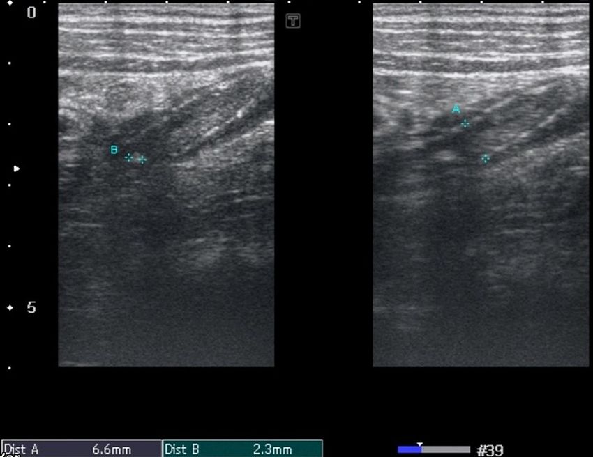
FIGURE 2: Grade 1 PFES.
High-frequency probe demonstrates acute appendicitis measuring 6.6 millimeters (A) with appendicolith
measuring 2.3 millimeters (B) showing grade 1 PFES (periappendiceal fat echo sign).
2021 Walid et al. Cureus 13(7): e16321. DOI 10.7759/cureus.16321 6 of 9FIGURE 3: Grade 2 PFES.
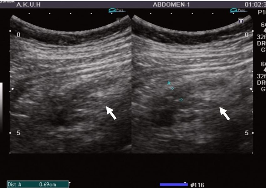
13 years with history of lower abdominal pain. Ultrasound of the right iliac fossa demonstrates acute
appendicitis with transverse dimension of 0.69 centimeters (A). Periappendiceal fat echo sign (PFES) is seen
(white arrow); however, the underlying vessels and muscles can be visualized.
FIGURE 4: Grade 3 PFES.
13 years male presented with lower abdominal pain and vomiting. Ultrasound of the right iliac fossa reveals a
dilated blind-ending bowel loop in RIF measuring 5 centimeters in length (A) and 1.2 centimeters in short axis
(B) representing acute appendicitis. An appendicolith also noted within the appendicular lumen.
Periappendiceal fat echo (white arrow) is so echogenic that the underlying structures are not visualized
consistently with grade 3 PFES (periappendiceal fat echo sign).
The effect of other confounding variables on PFES was also evaluated in our study. Our results show that
patient's age, sex and ascites have no significant association with PFES in acute appendicitis. No such
association has been described in the local and the international literature as well [21,22].
There were six patients in our study group who met the sonographic criteria of acute appendicitis, however,
showed normal echogenicity of periappendiceal fat. On histopathology no evidence of inflammatory
2021 Walid et al. Cureus 13(7): e16321. DOI 10.7759/cureus.16321 7 of 9changes was identified in the surrounding mesentery. This signifies that periappendiceal fat echogenicity is
not increased in all patients of acute appendicitis as these patients likely present with early/mild
inflammation and have a deep-seated appendix such as retrocecal location or inflammation confined to a
segment such as tip appendicitis. Inflammatory fat changes are also difficult to assess in slim patients due to
paucity of intra-abdominal fat. This finding was also reported in previous studies [7,20].
In our study, we had a relatively high proportion of male patients. Age range was between 5 and 62 years
with mean age of 19.5 years for acute appendicitis. This is in keeping with the local literature which also
provides similar statistics for acute appendicitis [23,24].
There were a few limitations to our study. First, as it was a single-center study. Second, a single radiologist
performed all the US examinations. Considering the subjective nature of the US examination we could have
employed two or more radiologists and calculated their interobserver variability however as this was not one
of the objectives it does not hamper the study results. Third, only those patients were enrolled in the study
in whom the appendix was directly visualized. Those patients in whom there was increased RLQ fat
echogenicity with clinical suspicion of acute appendicitis; however, appendix was not visualized were not
further followed.
Conclusions
Increased periappendiceal fat echogenicity is an important indicator of appendiceal inflammation. It
supports the sonographic finding of acute appendicitis in such patients in whom the inflamed appendix is
directly visualized. In cases where the appendix is not visualized on sonographic examination increased RLQ
fat echogenicity should prompt further investigation with CT scan.
Additional Information
Disclosures
Human subjects: Consent was obtained or waived by all participants in this study. College of Physicians
and Surgeons Pakistan issued approval RAD-2008-175-945. This was a residents dissertation and the College
of physicians and surgeons Pakistan had supervised the research ensuring that all ethical guidelines were
met against this study. The RTMC number stated above is the reference number for this study. . Animal
subjects: All authors have confirmed that this study did not involve animal subjects or tissue. Conflicts of
interest: In compliance with the ICMJE uniform disclosure form, all authors declare the following:
Payment/services info: All authors have declared that no financial support was received from any
organization for the submitted work. Financial relationships: All authors have declared that they have no
financial relationships at present or within the previous three years with any organizations that might have
an interest in the submitted work. Other relationships: All authors have declared that there are no other
relationships or activities that could appear to have influenced the submitted work.
References
1. Gamanagatti S, Vashisht S, Kapoor A, et al.: Comparison of graded compression ultrasonography and
unenhanced spiral computed tomography in the diagnosis of acute appendicitis. Singapore Med J. 2007,
48:80-7.
2. Randen A, Lameris W, Boermester M, et al.: Ultrasonography and computed tomography in patients with
right lower quadrant pain: difficult cases of appendicitis. Rep Med Imaging. 2009, 2:41-7.
10.2147/RMI.S4745
3. Noguchi T, Yoshimitsu K, Yoshida M: Periappendiceal hyperechoic structure on sonography: a sign of severe
appendicitis. J Ultrasound Med. 2005, 24:323-7. 10.7863/jum.2005.24.3.323
4. Doria AS, Moineddin R, Kellenberger CJ, et al.: US or CT for diagnosis of appendicitis in children and adults?
A meta-analysis. Radiology. 2006, 241:83-94. 10.1148/radiol.2411050913
5. Mardan MA, Mufti TS, Khattak IU, Chilkunda N, Alshayeb AA, Mohammad AM, ur Rehman Z: Role of
ultrasound in acute appendicitis. J Ayub Med Coll Abbottabad. 2007, 19:72-9.
6. Sohail S, Siddiqui KJ: Doptaus--a simple criterion for improving sonographic diagnosis of acute
appendicitis. J Pak Med Assoc. 2009, 59:79-82.
7. Lee MW, Kim YJ, Jeon HJ, Park SW, Jung SI, Yi JG: Sonography of acute right lower quadrant pain:
importance of increased intraabdominal fat echo. AJR Am J Roentgenol. 2009, 192:174-9.
10.2214/ajr.07.3330
8. Gaitini D, Beck-Razi N, Mor-Yosef D, Fischer D, Ben Itzhak O, Krausz MM, Engel A: Diagnosing acute
appendicitis in adults: accuracy of color Doppler sonography and MDCT compared with surgery and clinical
follow-up. AJR Am J Roentgenol. 2008, 190:1300-6. 10.2214/AJR.07.2955
9. van Randen A, Bipat S, Zwinderman AH, Ubbink DT, Stoker J, Boermeester MA: Acute appendicitis: meta-
analysis of diagnostic performance of CT and graded compression US related to prevalence of disease.
Radiology. 2008, 249:97-106. 10.1148/radiol.2483071652
10. Park NH, Park CS, Lee EJ, Kim MS, Ryu JA, Bae JM, Song JS: Ultrasonographic findings identifying the
faecal-impacted appendix: differential findings with acute appendicitis. Br J Radiol. 2007, 80:872-7.
10.1259/bjr/80553348
11. Balthazar EJ, Birnbaum BA, Yee J, Megibow AJ, Roshkow J, Gray C: Acute appendicitis: CT and US
correlation in 100 patients. Radiology. 1994, 190:31-5. 10.1148/radiology.190.1.8259423
12. Lim HK, Lee WJ, Kim TH, Namgung S, Lee SJ, Lim JH: Appendicitis: usefulness of color Doppler US .
2021 Walid et al. Cureus 13(7): e16321. DOI 10.7759/cureus.16321 8 of 9Radiology. 1996, 201:221-5. 10.1148/radiology.201.1.8816547
13. Quillin SP, Siegel MJ: Appendicitis: efficacy of color Doppler sonography . Radiology. 1994, 191:557-60.
10.1148/radiology.191.2.8153340
14. Rioux M: Sonographic detection of the normal and abnormal appendix . AJR Am J Roentgenol. 1992,
158:773-8. 10.2214/ajr.158.4.1546592
15. Brown MA: Imaging acute appendicitis. Semin ultrasound CT MR. 2008, 29:293-307.
10.1053/j.sult.2008.06.003
16. Jeffrey RB Jr, Laing FC, Townsend RR: Acute appendicitis: sonographic criteria based on 250 cases .
Radiology. 1988, 167:327-9. 10.1148/radiology.167.2.3282253
17. Park NH, Oh HE, Park HJ, Park JY: Ultrasonography of normal and abnormal appendix in children . World J
Radiol. 2011, 3:85-91. 10.4329/wjr.v3.i4.85
18. Puylaert JB: Acute appendicitis: US evaluation using graded compression . Radiology. 1986, 158:355-60.
10.1148/radiology.158.2.2934762
19. Lane MJ, Mindelzun RE.: Appendicitis and its mimickers. Semin ultrasound CT. 1999, 20:77-85.
10.1016/S0887-2171(99)90039-2
20. Kessler N, Cyteval C, Gallix B, et al.: Appendicitis: evaluation of sensitivity, specificity, and predictive
values of US, Doppler US, and laboratory findings. Radiology. 2004, 230:472-8. 10.1148/radiol.2302021520
21. Körner H, Söndenaa K, Söreide JA, et al.: Incidence of acute non perforated and perforated appendicitis:
age-specific and sex-specific analysis. World J Surg. 1997, 21:313-7.
22. Lim HK, Lee WJ, Lee SJ, Namgung S, Lim JH: Focal appendicitis confined to the tip: diagnosis at US .
Radiology. 1996, 200:799-801. 10.1148/radiology.200.3.8756934
23. Sanei B, Mahmoodieh M, Hosseinpour M: Evaluation of validity of Alvarado scoring system for diagnosis of
acute appendicitis. Pak J Med Sci. 2009, 25:298-301.
24. Ashraf K, Ashraf O, Bari V, Rafique MZ, Usman MU, Chisti I: Role of focused appendiceal computed
tomography in clinically equivocal acute appendicitis. J Pak Med Assoc. 2006, 56:200-3.
2021 Walid et al. Cureus 13(7): e16321. DOI 10.7759/cureus.16321 9 of 9You can also read