Risk factors associated with intra stent restenosis after percutaneous coronary intervention
←
→
Page content transcription
If your browser does not render page correctly, please read the page content below
EXPERIMENTAL AND THERAPEUTIC MEDICINE 22: 1141, 2021

                    Risk factors associated with intra‑stent restenosis
                        after percutaneous coronary intervention
          DAN‑MIHAI ALEXANDRESCU1,2*, OVIDIU MITU1, IRINA IULIANA COSTACHE1, LIVIU MACOVEI1*,
               IVONA MITU3*, ANCA ALEXANDRESCU2 and CATALINA ARSENESCU GEORGESCU1

                   1
                  1st Medical Department, ‘Grigore T. Popa’ University of Medicine and Pharmacy, 700115 Iasi;
         2
          Department of Cardiology‑Internal Medicine, Emergency County Hospital, 610136 Piatra Neamt; 3Department of
          Morpho‑Functional Sciences II, ‘Grigore T. Popa’ University of Medicine and Pharmacy, 700115 Iasi, Romania

                                          Received May 24, 2021; Accepted June 23, 2021

                                                  DOI: 10.3892/etm.2021.10575

Abstract. The present study aimed to explore the correlations      without ISR vs. 25.22 mm in patients with ISR; P=0.311).
between clinical, biological, imagistic and procedural factors     There was an estimated two times higher risk (RR=2.13;
with the risk of intra‑stent restenosis (ISR) in coronary artery   95% CI: 1.17‑3.88) concerning multi‑stenting and restenosis
disease (CAD) patients after percutaneous coronary inter‑          degree >70%. To conclude, smoking, hypertension, diabetes
vention (PCI). An observational cross‑sectional study was          mellitus, high CRP levels, CKD, TIMI score, stent type, low
conducted in a high‑volume PCI center over a period of 2 years.    pressure for stent implantation and multi‑stenting were found
A total of 235 consecutive patients diagnosed with angina or       to be associated with ISR in patients following PCI. Therefore,
acute coronary syndrome treated by PCI were included in            a close follow‑up should be targeted in such patients.
the study. Diagnosis of ISR was documented by coronary
angiography in patients with suggestive coronary symptoms          Introduction
and ischemic changes in non‑invasive or invasive paraclinical
investigations. Thus, they were assigned to two groups: With       Cardiovascular disease (CVD) is the leading cause of death
or without ISR. All patients underwent clinical and laboratory     worldwide while atherosclerotic coronary artery disease
examination, providing clinical and paraclinical variables that    (CAD) is mainly involved (1). After performing percutaneous
could be considered risk factors for ISR. Current smokers [risk    coronary intervention (PCI), patients are still at risk of devel‑
ratio (RR)=1.63; 95% confidence interval (95% CI): 1.25‑2.13],     oping new stenosis, such as intra‑stent restenosis (ISR). The
arterial hypertension (RR=1.86; 95% CI: 1.41‑2.45), diabetes       treatment of patients with ISR represents an important clinical
(RR=1.83; 95% CI: 1.42‑2.36), high C‑reactive protein (CRP)        problem and is still considered a challenge (2‑4). Despite the
levels (RR=1.44; 95% CI: 0.93‑2.24), chronic kidney disease        proven safety and efficacy of drug‑eluting stents (DES) in
(CKD) (RR=1.90; 95% CI: 1.53‑2.36) and thrombolysis in             patients undergoing PCI, bare‑metal stents (BMS) are still
myocardial infarction (TIMI) score were found to have a            widely used as well, mainly because of their reduced cost and
significant role in estimating the risk for ISR. Moreover, the     concerns about a debatable increased risk of bleeding associ‑
ISR group (119 patients) presented with a lower stent inflation    ated with prolonged dual antiplatelet therapy after DES (5,6).
pressure when compared to the control group (116 patients)         In addition, neoatherosclerosis is associated more often
(14.47 vs. 16.14 mmHg, P=0.004). An increased mean stent           with 1st generation DES than with BMS and occurs several
diameter used for PCI was not associated with a high ISR           months/years following PCI, while atherosclerosis in native
incidence (P=0.810) as well as complex coronary treated            coronary arteries develops over decades (7).
lesions with longer stents (mean length of 24.98 mm in patients        The incidence of ISR is still significant when considering
                                                                   either DES or BMS for patients following PCI (8,9), mainly
                                                                   because inflammatory responses after PCI lead to abnormal
                                                                   neointimal healing and thus generate a higher risk of unfavor‑
                                                                   able outcomes (7). This suggests that the type of stent is only
Correspondence to: Dr Ovidiu Mitu, 1st Medical Department,         one factor to consider when searching for additional promoters
‘Grigore T. Popa’ University of Medicine and Pharmacy,
                                                                   of ISR. In fact, the results of previous research conclude that
16 Universitatii Street, 700115 Iasi, Romania
E‑mail: mituovidiu@yahoo.co.uk                                     the factors associated with ISR after PCI have not been clearly
                                                                   defined. Thus, the present study aimed to detect the clinical,
*
    Contributed equally                                            biological, imagistic and procedural factors associated with ISR.

Key words: stent restenosis, percutaneous coronary intervention,   Patients and methods
risk factors, coronary artery disease, DES, BMS
                                                                   Patient selection and study design. The design of our study
                                                                   was observational, cross‑sectional, over a 2‑year period, from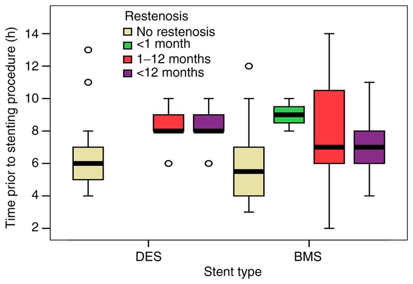
2 ALEXANDRESCU et al: RISK FACTORS ASSOCIATED WITH INTRA‑STENT RESTENOSIS AFTER PCI
a single high‑volume PCI center. A total of 235 consecutive associated with ISR in this group of patients. All these factors
patients who were diagnosed with angina pectoris or acute influenced the risk via a directly proportional relationship.
coronary syndrome (myocardial infarction with or without ST Regarding the endothelial dysfunction markers, the cut‑off
elevation) treated by PCI, were included. Our study population values were calculated in order to establish the risk associ‑
was divided into 2 groups: Experimental group (119 patients) ated with them: ESR=30 mm/h, uric acid=5 mg/dl, creatinine
that presented ISR documented by coronary angiography clearance=5 ml/min, CRP=2 mg/dl, fibrinogen=400 mg%.
(>50% stenosis of a previously stented segment) and the Analyzing both study groups, approximately 44% of the
control group (116 patients) without angiographic ISR, but with patients had an LDL value >100 mg/dl.
different other culprit lesions or no significant angiographic Our study showed a 60.9% use of BMS with a more signifi‑
stenosis. cant frequency in patients with ISR (73.1 vs. 48.3%, P=0.001),
Patients were eligible for the study if they were ≥18 years suggesting the high importance that should be given to the type
and presented with a diagnosis of angina pectoris or acute of stent used for PCI. Comparing the 2 types of stent, the data
myocardial infarction previously treated by stent implantation. for DES implantation were more consistent and well‑described.
Patients were not eligible for the study if they refused or aban‑ Assessing the presence of restenosis events in patients that
doned treatment, if they did not report for the control visit or if suffered stent implantation in the first 8 h after myocardial
they were part of a vulnerable category (e.g., pregnant women, infarction, BMS implantation was directly linked to restenosis
patients in coma). The study was approved by the University events over a period of 1‑12 months and also >12 months, while
of Medicine and Pharmacy ‘Grigore T. Popa’ Iasi Research DES implantation showed no significant restenosis events over
Ethics Committee, and all subjects had initially agreed and the follow‑up period (Fig. 1). Furthermore, early ISR (during
signed an informed consent in order to take part in this study. the first month after PCI) was observed only in patients
with BMS regardless of the time the stent was implemented,
Clinical and paraclinical characteristics. Diagnosis of followed by a higher incidence of ISR in patients with BMS
ISR was documented by coronary angiography in patients vs. DES after a 1‑month period (1‑12 months: 27.3 vs. 14.1%;
with suggestive coronary symptoms and ischemic changes >12 months: 31.5 vs. 20.7%) (Table III).
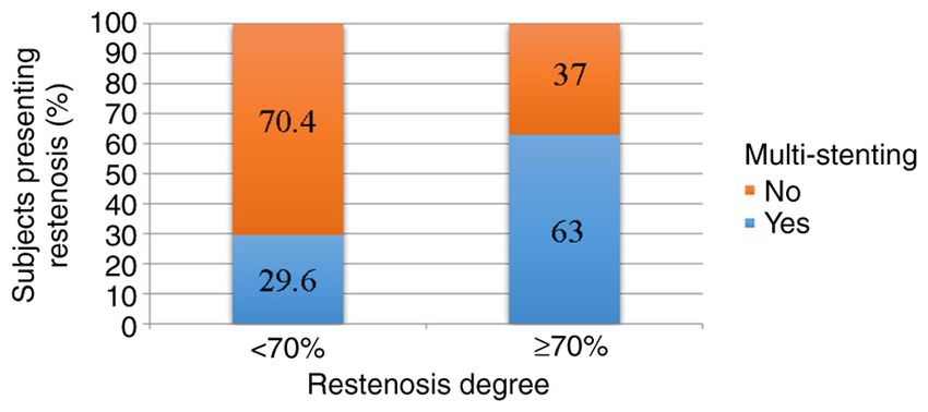
in non‑invasive or invasive paraclinical investigations. All Furthermore, our study aimed to identify if there is a
patients underwent clinical and laboratory examination, correlation between restenosis and various specific variables:
providing a large number of variables that could be considered The pressure under which the stent was deployed, the diameter
risk factors for ISR: i) clinical variables: age, sex, smoking, of the stent or the length of the stent. The mean stent infla‑
hypertension, diabetes, obesity, chronic kidney disease (CKD; tion pressure was significantly lower in patients with BMS
creatinine clearance 70% was
one‑way ANOVA and Student's t‑test for continuous data. statistically significant (63 vs. 29.6%; P=0.004), indicating an
When a normal distribution was not present for continuous estimated 2 times higher risk (RR=2.13; 95% CI: 1.17‑3.88)
variables, Mann Whitney/Kruskal‑Wallis tests were used. In (Fig. 5).
order to estimate the strength of the association between risk
factors and outcome (with or without ISR) relative risk (RR) Discussion
with 95% confidence interval (CI) was used in the statistical
analysis of the data. A P‑valueEXPERIMENTAL AND THERAPEUTIC MEDICINE 22: 1141, 2021 3
Table I. Summary of the general factors associated with ISR.
ISR group Non‑ISR group
Variable (n=119) n (%) (n=116) n (%) P‑value RR 95% CI
Sex
Male 83 (70) 81 (70) 0.989
Female 36 (30) 34 (30) 0.522
Age ≥60 years 77 (64.7) 64 (55.2) 0.125
Current smokers 74 (62.2) 44 (37.9) 0.001 1.63 1.25‑2.13
Hypertension 72 (62.1) 38 (31.9) 0.001 1.86 1.41‑2.45
Diabetes mellitus 68 (57.1) 31 (26.7) 0.001 1.83 1.42‑2.36
Obesity 35 (29.4) 27 (23.3) 0.285 1.16 0.89‑1.52
LDL cholesterol >70 mg/dl 91 (76.5) 93 (80.2) 0.596 1.05 0.95‑1.20
ESR >30 mm/h 48 (40.3) 37 (31.9) 0.178 1.19 0.93‑1.53
Uric acid >5 mg/dl 30 (71.4) 23 (60.5) 0.303 1.27 0.79‑2.07
Creatinine clearance 2 mg/dl 94 (87.0) 83 (77.6) 0.050 1.44 0.93‑2.24
Fibrinogen >400 mg/dl 84 (70.6) 72 (62.6) 0.195 1.20 0.90‑1.60
Albuminuria 16 (13.4) 19 (16.4) 0.528 0.89 0.60‑1.31
CKD 43 (36.1) 11 (9.5) 0.001 1.90 1.53‑2.36
Acute renal failure 7 (5.9) 5 (4.3) 0.583 1.16 0.71‑1.91
ISR, intra‑stent restenosis; RR, risk ratio; CI, confidence interval; LDL, low density lipoprotein; ESR, erythrocyte sedimentation rate; CRP,
C‑reactive protein; CKD, chronic kidney disease. Significant P‑values are indicated in bold print.
hypertension as a risk factor associated with ISR in patients
following PCI (11). Moreover, Mohan and Dhall found a
significant and positive correlation between hypertension and
ISR (12).
There is still a lack of clarity in describing the exact mech‑
anism that promotes the risk of ISR in patients with diabetes,
but a recent animal laboratory study revealed that insulin and,
moreover, insulin receptors are primarily responsible for the
accelerated intimal hyperplasia in diabetes which is directly
linked to the restenosis phenomenon. These results are
surprising, considering multiple previous studies that imply
a more important effect of another factor, the insulin‑like
growth factor‑1 (13). The physiopathological mechanism
presented in the literature and the higher incidence of diabetes
Figure 1. Correlation between type of stent, time prior to stenting procedure in our patients with ISR compared to those without confirm
and time course of restenosis. DES, drug‑eluting stents; BMS, bare‑metal the inclusion of diabetes mellitus in the group of risk factors
stents.
for ISR.
The endothelial dysfunction responsible for ISR is
determined by an inflammatory status in patients with CAD.
mortality. A recent meta‑analysis performed on 141 cohort C‑reactive protein (CRP) is recognized as an important
studies and 55 study reports concluded that smoking one marker for systemic inflammation and for predicting cardio‑
cigarette per day carries around half of the risk than for those vascular events, therefore it can be used in primary and
smoking 20 cigarettes per day (10). In our study, smoking secondary prevention. The cut‑off value for CRP in our study
represents an important risk factor that leads to CAD and also was set at 2 mg/dl. A study on 1, 234 patients undergoing DES
to post‑PCI ISR. implantation showed that high levels of CRP (>2 mg/dl) were
Our results demonstrated that the estimated risk induced detected in 38% of patients at baseline and in 23.6% during
by arterial hypertension was higher in patients without ISR late phase, both stages associated with a higher risk for major
(RR=1.86; 95% CI: 1.41‑2.45; P=0.001). Our findings are in cardiac adverse events (MACE). Moreover, high CRP level
accordance with other studies that have identified a positive in the late phase was a better predictor of MACE compared
correlation between hypertension and ISR. In a retrospective to the CRP level at baseline (14). Our findings are relatively
study that included 289 patients, Wihanda et al identified similar with the current literature data. High CRP levels4 ALEXANDRESCU et al: RISK FACTORS ASSOCIATED WITH INTRA‑STENT RESTENOSIS AFTER PCI
Table II. Summary table of the specific factors associated with ISR.
Variable ISR group (n=119) (%) Non‑ISR group (n=116) (%) P‑value
Site of lesion
LAD 47.9 50.9 NS
RCA 28.6 35.3 NS
LCX 22.7 13.8 NS
LM 0.8 0.0 NS
Clinical diagnosis before stent implantation
MI right ventricle 0.8 3.4 NS
MI posterior‑inferior‑lateral 5.0 3.4 NS
MI antero‑lateral 8.4 6.9 NS
MI anterior 31.1 29.3 NS
MI inferior 21.0 16.4 NS
Angina pectoris 33.6 40.5 NS
TIMI score
TIMI 1 2.5 0.0 0.001
TIMI 2 19.3 3.4 0.001
TIMI 3 78.2 96.6 0.001
Ejection fraction (EF)
EF 50% 15.1 23.3 NS
ISR, intra‑stent restenosis; LAD, left anterior descending; RCA, right coronary artery; LCX, left circumflex artery; LM, left main; MI, myocar‑
dial infarction; TIMI, thrombolysis in myocardial infarction; NS, not significant. Significant P‑values are presented in bold print.
Table III. Correlation between the type of stent and restenosis.
Restenosis
‑‑‑‑‑‑‑‑‑‑‑‑‑‑‑‑‑‑‑‑‑‑‑‑‑‑‑‑‑‑‑‑‑‑‑‑‑‑‑‑‑‑‑‑‑‑‑‑‑‑‑‑‑‑‑‑‑‑‑‑‑‑‑‑‑‑‑‑‑‑‑‑‑‑‑‑‑‑‑‑‑‑‑‑‑‑‑‑‑‑‑‑‑‑‑‑‑‑‑‑‑‑‑‑‑‑‑‑‑‑‑‑‑‑‑‑‑‑‑‑‑‑‑‑‑‑‑‑‑‑‑‑‑‑‑‑‑‑‑‑‑‑‑‑‑‑‑‑‑‑‑‑‑‑‑‑‑‑‑‑
No restenosis 2 months Total
Stent type
DES, N 60 0 13 19 92
% stent type 65.2% 0% 14.1% 20.7% 100%
BMS, N 56 3 39 45 143
% stent type 39.1% 2.1% 27.3% 31.5% 100%
Total
N 116 3 52 64 235
% stent type 49.4% 1.3% 22.1% 27.2% 100%
DES, drug‑eluting stent; BMS, bare‑metal stent.
suggest a chronic inflammation that persists even after revas‑ A recent analysis of 21,386 individuals identified a prognostic
cularization. cut‑off value of 5.34 mg/dl for all heart failure and 4.89 mg/dl for
Another known marker of endothelial dysfunction that fatal heart failure (16). Therefore, the values remain debatable.
registered high levels in our patients with ISR is the uric In our study, the cut‑off value was established at 5 mg/dl.
acid level. Hyperuricemia might inhibit endothelial nitric Even though we observed an increased incidence of elevated
oxide synthesis and stimulate the secretion of inflammatory uric acid levels in patients with ISR, the correlation was
cytokines, leading to neointimal hyperplasia associated with not statistically significant. However, the value of uric acid
a high risk of restenosis (15). The cut‑off value for uric acid correlated well with the ISR incidence, in accordance with the
is different among studies, usually between 6 and 10 mg/dl. literature data (17,18).EXPERIMENTAL AND THERAPEUTIC MEDICINE 22: 1141, 2021 5
Figure 5. Correlation between multi‑stenting and restenosis degree.
Another important factor in the result of the initial proce‑
dure and in the evaluation of the risk of unfavorable events in the
future is the TIMI score. Results after the initial angiography
Figure 2. Correlation between mean stent inflation pressure and restenosis. showed a higher incidence of patients with suboptimal results,
especially in patients that presented ISR afterwards (P=0.001).
Restenosis at 1‑12 months was predominantly represented
by patients with TIMI 2 score (52.2%), while restenosis in a
period >12 months was predominantly represented by patients
with TIMI 3 score (66.7%), but the results were not statistically
significant (P=0.184). These results suggest that late restenosis
occurs without an association with initial coronarography and
initial stent angioplasty.
The advantages of DES over BMS in preventing ISR have
been presented in the literature and are also confirmed in our
study. Zbinden et al showed a significant higher risk of ISR
in segments with a BMS compared to segments with a DES
(5.4 vs. 0.76% after 2 years) in 2,278 patients (21). In addi‑
tion, a systematic review concerning the treatment of coronary
ISR confirmed a higher rate of ISR after BMS implantation
(20‑35%) vs. DES implantation (5‑10%) (22). In our analysis,
BMS presented an associated risk of ISR approximately
2 times higher as compared to DES.
Figure 3. Correlation between stent diameter and restenosis.
Inflation pressure during stent implantation is correlated to
angiographic lumen improvement and stent extension, but the
direction of this correlation has not been yet established. In a
non‑randomized study on 136 patients undergoing PCI with BMS,
a high inflation pressure was associated with unfavorable results
on long term that included higher rates of MACE and target lesion
revascularization (TLR). Higher inflation pressure was associated
with an increased risk of ISR and TLR vs. low inflation pressure
(71 vs. 16%, respectively 27 vs. 8%) (23). However, in a random‑
ized study, there were no significant differences concerning the
risk of ISR when using low or high inflation pressure during stent
implementation (24). Another study analyzed moderate to high
balloon inflation pressure during PCI and found no measurable
improvement in late outcome (25). Finally, a recent retrospective
study on over 90,000 stent implementations suggests that a low
and a very high pressure elevates the risk of ISR (26). Our results
support the findings of this last study. Low pressure was reported
Figure 4. Correlation between stent length and restenosis. in our group of patients with ISR as well as in the group with
BMS. Furthermore, our study described a positive correlation
between multi‑stenting usage and ISR risk. A total of 20.2% of
Endothelial dysfunction is also considered a complication patients from the group with ISR presented 2 or more stents at the
in patients with CKD. Modifications at the vascular level influ‑ region where restenosis was documented vs. 3.4% in the group
ence the evolution after coronary revascularization. Our study without ISR (P=0.001).
defines a statistically significant correlation between CKD Our retrospective study has a number of limitations. Firstly,
and ISR. Data in the literature have also identified a causal the small sample size analyzed may overestimate the magni‑
relationship between these two variables (19,20). tude of an association or even induce false‑positive results. The6 ALEXANDRESCU et al: RISK FACTORS ASSOCIATED WITH INTRA‑STENT RESTENOSIS AFTER PCI
patients were not divided into ischemic or angina subgroups. 2. Alfonso F, Byrne RA, Rivero F and Kastrati A: Current treat‑
ment of in‑stent restenosis. J Am Coll Cardiol 63: 2659‑2673,
However, considering the large amount of data gathered for the 2014.
analysis, this study also offers an overview of variables that 3. Alfonso F: Treatment of drug‑eluting stent restenosis the new
need to be taken into account when establishing correlations pilgrimage: Quo vadis? J Am Coll Cardiol 55: 2717‑2720,
2010.
with ISR. Secondly, the economical factor has a high influence 4. Rao G, Sheth S and Grines C: Percutaneous coronary interven‑
in the treatment decision of the baseline CAD diagnosis, the tion: 2017 in review. J interv Cardiol 31: 117‑128, 2018.
study reporting an increased number of BMS implantations. 5. Baschet L, Bourguignon S, Marque S, Durand‑Zaleski I, Teiger E,
Wilquin F and Levesque K: Cost‑effectiveness of drug‑eluting
In conclusion, smoking, hypertension, diabetes mellitus, stents versus bare‑metal stents in patients undergoing percuta‑
high CRP levels, CKD, TIMI score, stent type, low pressure neous coronary intervention. Open Heart 3: e000445, 2016.
for stent implantation and multi‑stenting are factors associated 6. Neupane S, Khawaja O, Edla S, Singh H, Othman H, Bossone E,
Yamasaki H, Rosman HS, Eggebrecht H and Mehta RH:
with ISR in patients following PCI. Thus, a close follow‑up Meta-analysis of drug eluting stents compared with bare metal
should be targeted in these patients. stents in high bleeding risk patients undergoing percutaneous
coronary interventions. Catheter Cardiovasc Interv 94: 98‑104,
2019.
Acknowledgements 7. Ochijewicz D, Tomaniak M, Opolski G and Kochman J:
Inflammation as a determinant of healing response after coro‑
We would like to thank the Cardiac Catheterisation Laboratory nary stent implantation. Int J Cardiovasc Imaging 37: 791‑801,
2021.
and the Intensive Coronary Care Unit of ‘George I.M. 8. Stone GW and Kirtane AJ: Bare metal and drug‑eluting coronary
Georgescu’ Institute of Cardiovascular Diseases, Iasi. stents. In: Topol EJ, Teristein PS (ed) Textbook of interventional
cardiology 6th ed., Philadelphia: Saunders Elsevier pp171‑196,
2012.
Funding 9. Her AY and Shin ES: Current management of in‑stent restenosis.
Korean Circ J 48: 337‑349, 2018.
No funding was received. 10. Hackshaw A, Morris JK, Boniface S, Tang JL and Milenković D:
Low cigarette consumption and risk of coronary heart disease
and stroke: Meta‑analysis of 141 cohort studies in 55 study
Availability of data and materials reports. BMJ 360: j5855, 2018.
11. Wihanda D, Alwi I, Yamin M, Shatri H and Mudjaddid E: Factors
associated with In‑stent restenosis in patients following percu‑
The datasets used and/or analyzed during the current study are taneous coronary intervention. Acta Med Indones 47: 209‑215,
available from the corresponding author on reasonable request. 2015.
12. Mohan S and Dhall A: A comparative study of restenosis rates
in bare metal and drug‑eluting stents. Int J Angiol 19: e66‑e72,
Authors' contributions 2010.
13. Li Q, Fu J, Xia Y, Qi W, Ishikado A, Park K, Yokomizo H,
DMA and CAG developed the study concept and design. DMA Huang Q, Cai W, Rask‑Madsen C, et al: Homozygous recep‑
tors for insulin and not IGF‑1 accelerate intimal hyperplasia in
collected the data and created the database. IIC, LM, AA and insulin resistance and diabetes. Nat Commun 10: 4427, 2019.
CAG performed the literature research and contributed to 14. Shiba M, Itaya H, Iijima R and Nakamura M: Influence of
the introduction, results and discussion sections. OM and IM late vascular inflammation on long‑term outcomes among
patients undergoing implantation of drug eluting stents: Role of
conducted the statistical analysis and created the images and C‑Reactive protein. J Am Heart Assoc 5: e003354, 2016.
tables, with assistance from DMA and CAG. All authors were 15. Kim SY, Guevara JP, Kim KM, Choi HK, Heitjan DF and
involved in drafting and finalizing the manuscript. All authors Albert DA: Hyperuricemia and coronary heart disease: A system‑
atic review and meta‑analysis. Arthritis Care Res (Hoboken) 62:
read and approved the final manuscript for publication. 170‑180, 2010.
16. Muiesan ML, Salvetti M, Virdis A, Masi S, Casiglia E,
Ethics approval and consent to participate Tikhonoff V, Barbagallo CM, Bombelli M, Cicero AFG,
Cirillo M, et al: Serum uric acid, predicts heart failure in a large
Italian cohort: Search for a cut‑off value the uric acid right for
The study was approved by the University of Medicine and heart health study. J Hypertens 39: 62‑69, 2021.
Pharmacy ‘Grigore T. Popa’ Iasi Research Ethics Committee 17. Wu Y and Fu X: Comprehensive analysis of predictive factors
for rapid angiographic stenotic progression and restenosis risk in
and all subjects had initially agreed and signed an informed coronary artery disease patients underwent percutaneous coro‑
consent in order to take part in this study. nary intervention with drug‑eluting stents implantation. J Clin
Lab Anal 33: e22666, 2019.
18. Joo HJ, Jeong HS, Kook H, Lee SH, Park JH, Hong SJ, Yu CW
Patient consent for publication and Lim DS: Impact of hyperuricemia on clinical outcomes after
percutaneous coronary intervention for in‑stent restenosis. BMC
Not applicable. Cardiovasc Disord 18: 114, 2018.
19. Lambert ND, Sacrinty MT, Ketch TR, Turner SJ, Santos RM,
Daniel KR, Applegate RJ, Kutcher MA and Sane DC: Chronic
Competing interests kidney disease and dipstick proteinuria are risk factors for
stent thrombosis in patients with myocardial infarction. Am
Heart J 157: 688‑694, 2009.
The authors declare that they have no competing interests. 20. Gao WD, Ma M, Zhang GX, Zhang XF and Sun G: First‑generation
versus second‑generation drug‑eluting stents in patients with
References chronic kidney disease: A systematic review and meta‑analysis.
Postgrad Med 131: 43‑51, 2019.
21. Zbinden R, von Felten S, Wein B, Tueller D, Kurz DJ, Reho I,
1. Lozano R, Naghavi M, Foreman K, Lim S, Shibuya K, Aboyans V, Galatius S, Alber H, Conen D, Pfisterer M, et al: Impact of stent
Abraham J, Adair T, Aggarwal R, Ahn SY, et al: Global and diameter and length on in‑stent restenosis after DES vs BMS
regional mortality from 235 causes of death for 20 age groups implantation in patients needing large coronary stents‑A clinical
in 1990 and 2010: A systematic analysis for the global burden of and health‑economic evaluation. Cardiovasc Ther 35: 19‑25,
disease study. Lancet 380: 2095‑2128, 2012. 2017.EXPERIMENTAL AND THERAPEUTIC MEDICINE 22: 1141, 2021 7
22. Pleva L, Kukla P and Hlinomaz O: Treatment of coronary 25. Mattos LA, Sousa AG, Chaves A, Feres F, Pinto I, Tanajura L,
in‑stent restenosis: A systematic review. J Geriatr Cardiol 15: Centemero M, Abizaid A, Seixas AC, Abizaid A, et al: Influence
173‑184, 2018. of balloon pressure inflation in patients undergoing primary
23. Uretsky BF, Rosanio S, Lerakis S, Wang FW, Smiley M, coronary stent implantation during acute myocardial infarc‑
Stouffer GA, Tocchi M and Estella P: A prospective evaluation of tion: A quantitative coronary angiography analysis. Arq Bras
angiography‑guided coronary stent implantation with high versus Cardiol 80: 250‑268, 2003.
very high balloon inflation pressure. Am Heart J 140: 804‑812, 26. Fröbert O, Sarno G, James SK, Saleh N and Lagerqvist B: Effect
2000. of stent inflation pressure and Post‑Dilatation on the outcome of
24. Dirschinger J, Kastrati A, Neumann FJ, Boekstegers P, Elezi S, coronary artery intervention. A report of more than 90,000 stent
Mehilli J, Schühlen H, Pache J, Alt E, Blasini R, et al: Influence implantations. PLoS One 8: e56348, 2013.
of balloon pressure during stent placement in native coronary
arteries on early and late angiographic and clinical outcome: A This work is licensed under a Creative Commons
randomized evaluation of high‑pressure inflation. Circulation 100: Attribution-NonCommercial-NoDerivatives 4.0
918‑923, 1999. International (CC BY-NC-ND 4.0) License.You can also read