A Prospective Study Comparing Laparoscopic vs. Conventional Stomach Pull Up in Total Pharyngo-Laryngo-Esophagectomy for Post Cricoid Cancer - MDPI
←
→
Page content transcription
If your browser does not render page correctly, please read the page content below
Brief Report
A Prospective Study Comparing Laparoscopic vs. Conventional
Stomach Pull Up in Total Pharyngo-Laryngo-Esophagectomy
for Post Cricoid Cancer
Basavegowda Vinod Prakash, Ali Zaid Anwar *, Mahadev Abhishek, Shivaji Sharma and Saseendran Shruthi

                                          Department of Surgical Oncology, Kidwai Memorial Institute of Oncology, M.H. Marigowda Road,
                                          Bangalore 560029, India; bvprakashms@gmail.com (B.V.P.); mahadevabhishek@gmail.com (M.A.);
                                          shivajisharma6@gmail.com (S.S.); shruthi0908@gmail.com (S.S.)
                                          * Correspondence: zaidanwar08@gmail.com; Tel.: +95-60-418-944

                                          Abstract: The aim of this study is to compare laparoscopic and conventional techniques following To-
                                          tal Pharyngo-laryngo-esophagectomy (TPLE) with respect to perioperative morbidity and mortality
                                          and postoperative recovery in post cricoid cancer patients. This is a prospective study, which was un-
                                          dertaken in Gujrat Cancer Research Institute (GCRI) in the period of July 2007 to March 2010. Fifteen
                                          consecutive patients who underwent laparoscopic TPLE were compared to that of 18 consecutive
                                          patients who underwent open TPLE. Laparoscopic and open TPLE procedure were compared with
                                          respect to patient characteristics, intra operative and complications present. The average duration
                                          was observed to be 3.5 h in the MIS (Minimally Invasive Group) group and was 5.3 h in the open
                                group. The average blood loss was 300 mL in the MIS group and 500 mL in the open group. Average

                                          duration of the hospital stay in the MIS group was 13 days and 16 days in the open group. In the
Citation: Prakash, B.V.; Anwar, A.Z.;
                                          MIS group, one patient (6.7%) had a pneumonic complication and two patients (13%) had wound
Abhishek, M.; Sharma, S.; Shruthi, S.
                                          complications. In the open group, six patients (33%) had pneumonic consolidation and four patients
A Prospective Study Comparing
                                          (22%) had wound infections. In both groups, one patient each suffered mortality. Laparoscopic TPLE
Laparoscopic vs. Conventional
                                          has been found to be much safer with less morbidity as compared with open surgery.
Stomach Pull Up in Total
Pharyngo-Laryngo-Esophagectomy
for Post Cricoid Cancer. Clin. Pract.
                                          Keywords: post cricoid cancer; laparoscopy; TPLE; stomach pull through
2021, 11, 178–184. https://doi.org/
10.3390/clinpract11020025

Academic Editor: Camillo Porta            1. Introduction
                                                Post cricoid cancer is more common in women worldwide and usually associated
Received: 4 December 2020                 with iron deficiency anemia and the Plummer-Vinson syndrome. India as well as France
Accepted: 16 December 2020                have the highest rates of post cricoid cancers throughout the world (annual incidence
Published: 29 March 2021
                                          of 8–15 per 10,000) [1]. Hypopharyngeal cancers usually occur in low socioeconomic
                                          classes. Most of the patients usually present late with advanced disease (T3–T4). Generally,
Publisher’s Note: MDPI stays neutral      prognosis of advanced hypopharyngeal cancer is poor. The prognosis is generally dismal
with regard to jurisdictional claims in
                                          with a mean five-year survival rate of 18–35% (Pingree & Axon) [2,3]. The standard of care
published maps and institutional affil-
                                          is surgery followed by adjuvant radiotherapy. This treatment helps us achieve relief of
iations.
                                          dysphagia, which is the main symptom, and requires attaining the best possible survival.
                                          The quality of life is important in these patients because prognosis is poor. Thus, if the
                                          disease can be extirpated with low morbidity, mortality, and a shorter hospital stay, that
                                          should be the preferred method of treatment.
Copyright: © 2021 by the authors.               Patients requiring circumferential ablative surgery for hypopharynx and cervical
Licensee MDPI, Basel, Switzerland.        esophagus has a poor prognosis and significant morbidity. Hence, the method of recon-
This article is an open access article
                                          struction should be chosen so as to give rise to minimal morbidity, a minimal hospital stay,
distributed under the terms and
                                          and allow early return of satisfactory swallowing function. The main goal of treatment is
conditions of the Creative Commons
                                          relief of dysphagia and loco-regional control of disease.
Attribution (CC BY) license (https://
                                                We prefer reconstruction by the stomach pull up because it is easy, reliable, and has
creativecommons.org/licenses/by/
                                          acceptable levels of morbidity and mortality. The conventional technique of resection and
4.0/).

Clin. Pract. 2021, 11, 178–184. https://doi.org/10.3390/clinpract11020025                               https://www.mdpi.com/journal/clinpract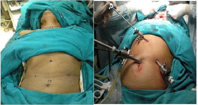
We prefer reconstruction by the stomach pull up because it is easy, reliable, and has
acceptable levels of morbidity and mortality. The conventional technique of resection and
Clin. Pract. 2021, 11 179
reconstruction carries significant morbidity because of neck dissection, laparotomy, and
trans-hiatal dissection of esophagus, which includes pleural or lung injury, as it is a blind
dissection and, thus, causes respiratory morbidity.
reconstruction carries
In this context, wesignificant morbidity
contemplated because of neck
the application dissection,oflaparotomy,
of principles minimallyand invasive
trans-hiatal dissection of esophagus, which includes pleural or lung injury,
surgery for these particular patient populations. The minimally invasive approach isas it is a blind
dissection and, thus, causes respiratory morbidity.
used to mobilize the stomach and to perform the trans-hiatal dissection of the esophagus,
In this context, we contemplated the application of principles of minimally invasive
thus, avoiding laparotomy and reducing the morbidity of pain, respiratory complica-
surgery for these particular patient populations. The minimally invasive approach is used
tions, and resulting
to mobilize in a better
the stomach and tooutcome with
perform the good quality
trans-hiatal of life.
dissection Theesophagus,
of the minimallythus,invasive
surgery is employed
avoiding laparotomywithout violating
and reducing the principles
the morbidity of respiratory
of pain, oncologic surgery.
complications, and
resulting in a better outcome with good quality of life. The minimally invasive surgery is
2. Materials and Methods
employed without violating the principles of oncologic surgery.
This prospective study compared all patients who underwent laparoscopic Total
2. Materials and Methods
Pharyngo-laryngo-esophagectomy (TPLE) with patients who underwent open TPLE in
This prospective study compared all patients who underwent laparoscopic Total
thePharyngo-laryngo-esophagectomy
Gujrat Cancer Research Institute (GCRI) operated from July 2007 to March 2010. Fif-
(TPLE) with patients who underwent open TPLE in the
teen consecutive patients underwent
Gujrat Cancer Research Institute (GCRI) laparoscopic
operated from TPLE
July(Group
2007 to A) were
March compared
2010. Fifteen with
18 patients
consecutivewho underwent
patients open laparoscopic
underwent TPLE (Group TPLEB) from JulyA)2007
(Group weretocompared
March 2010, withi.e.,
18 same
duration in a single institution. Patients were explained the nature
patients who underwent open TPLE (Group B) from July 2007 to March 2010, i.e., same of disease, type of
durationsurgery
extensive in a singletheyinstitution. Patientsto,were
were subjected andexplained the nature
the necessity of disease,tracheostoma
of permanent type of
andextensive surgery
loss of voice. they were subjected
Preoperatively, to, and
we built thethe necessity status
nutritional of permanent
throughtracheostoma
nasogastric tube
and loss
feeding. Allofpatients
voice. Preoperatively, we built
for surgery were the antibiotic
given nutritionalprophylaxis.
status through nasogastric tube
feeding. All patients for surgery were given antibiotic prophylaxis.
The neck is hyper extended while the patient is in a supine position (Figure 1), sub-
The neck is hyper extended while the patient is in a supine position (Figure 1), sub-
platysmal flaps were raised, the involvement of internal jugular vein, carotid artery, pre
platysmal flaps were raised, the involvement of internal jugular vein, carotid artery, pre
vertebral fascia
vertebral fasciawaswasexcluded, and the
excluded, and themodified
modifiedneck neck dissection
dissection waswas carried
carried out inout
N0in N0
disease. In case of palpable nodes, a complete neck dissection was done,
disease. In case of palpable nodes, a complete neck dissection was done, the post cricoid the post cricoid
tumor
tumorwaswas excised
excisedinferiorly
inferiorly to the
the base
baseofoftongue,
tongue, andand
thethe distal
distal levellevel of transection
of transection is is
determined
determined bybythetheextent
extent of the disease.
of the disease.IfIfititisislimited
limited to above
to above the the thoracic
thoracic inlet,inlet,
then then
resection
resection ofofthe thelarynx,
larynx,pharynx,
pharynx, and
andcervical
cervicalesophagus
esophagus would
would be sufficient. If extent
be sufficient. of
If extent of
the tumor is below the thoracic inlet, then the entire esophagus
the tumor is below the thoracic inlet, then the entire esophagus is removed. is removed.
Figure 1. Port placement.
Figure 1. Port placement.
In Inananopen
open method,
method,anan
upper
uppermidline supra-umbilical
midline incision was
supra-umbilical taken. was
incision The greater
taken. The
omentum was mobilized as the left gastro epiploic vessels was ligated and even left gastric
greater omentum was mobilized as the left gastro epiploic vessels was ligated and even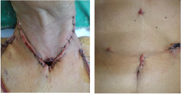
Clin. Pract. 2021, 11 180
vessels were ligated and divided. The preservation of the right gastroepiploic vessels were
assured and blind dissection was carried out in the mediastinum through the esoph.1tus.
In the laparoscopic technique, the peritoneal cavity was insufflated with CO2 , two
10-mm ports, two 5-mm ports were utilized, and the gastric dissection was completed
using a 30-degree telescope in which right gastroepiploic vessels were preserved. The
mediastinal dissection was carried out using a 0-degree telescope and carried meticulously
Clin. Pract. 2021, 12, FOR PEER REVIEW 3
to avoid unnecessary injury.
The size of pharyngostoma was estimated and adequate fundal gastrostomy was
made and single layer anastomosis was made using interrupted polyglactin suture 2-0.
left gastric vesselstube
A nasogastric werewas ligated
placed andanddivided.
feedingThe preservation
jejunostomy of the in
was made right gastroepiploic
all cases, through
vesselsenteral
which were assured
nutritionand wasblind dissection
carried out on was carriedpostoperative
the second out in the mediastinum
day. Each through
patient
the esophageal
was given trial ofhiatus.
clear liquids to swallow and observed for anastomotic leakage until
In the laparoscopic
the stomach became deflated technique, the peritoneal cavity
by an intraoperatively placedwas insufflated
nasogastric with
tube. If aCO 2, two
leak or
10-mm ports, two 5-mm ports were utilized, and the gastric
dehiscence occurred, it was managed conservatively. The jejunostomy tube was removeddissection was completed
usinga apatient
when 30-degree telescope
was able in which
to swallow and right
retaingastroepiploic
a full diet. Once vessels were preserved.
histopathology report was The
mediastinal
available, anddissection
standard was carried
adjuvant out using
treatment wasa given
0-degree telescope
according to aand
unitcarried
protocol meticu-
after
lously to avoid
postoperative unnecessary
recovery (Figureinjury.
2). With respect to pathology, patients with invasive cancer
were The size of
analyzed to pharyngostoma
assess the gradewas of theestimated and adequate
tumor, nodal status, and fundal gastrostomy
margins of resection was
(Figure
made and 3). Patients were
single layer staged using
anastomosis was the AJCC
made (American
using Jointpolyglactin
interrupted Committeesutureon Cancer)2-0.
stagingA system.
nasogastric tube was placed and feeding jejunostomy was made in all cases,
Afterwhich
through discharged
enteralpatients
nutrition werewasfollowed
carried up,
out during which they
on the second were asked
postoperative for Each
day. any
complaints,
patient wasthey were
given examined
trial loco-regionally
of clear liquids to swallowandand
systemically.
observed They were subjected
for anastomotic for
leakage
routine blood
until the investigation
stomach became(including
deflated by TFT,anserum calcium), and
intraoperatively X-raynasogastric
placed chest. The mortality
tube. If a
rate
leakrelated to surgery
or dehiscence (within it
occurred, 30was
daysmanaged
of surgery) and long-term
conservatively. Thesurvival were studied.
jejunostomy tube was
All cases were advised for follow-up every two months for the
removed when a patient was able to swallow and retain a full diet. Once histopathology first year, once every
three
report was available, and standard adjuvant treatment was given according to aonce
months during the second year, once every four months during the third year, unit
every six after
protocol months for the next
postoperative two years,
recovery (Figureand2).then
Withannually.
respect toData collected
pathology, included
patients with
patient
invasivecharacteristics,
cancer were tumor
analyzed site,toand morphology.
assess the gradeOperative information
of the tumor, included
nodal status, andblood
mar-
loss
ginsand duration (Figure
of resection of surgery. The complications
3). Patients were stagedwere usingstudied
the AJCCincluding both major
(American Joint Com-and
minor
mitteefactors.
on Cancer) staging system.
Figure2.2.Postoperative.
Figure Postoperative.Clin. Pract. 2021, 11 181
Clin. Pract. 2021, 12, FOR PEER REVIEW 4
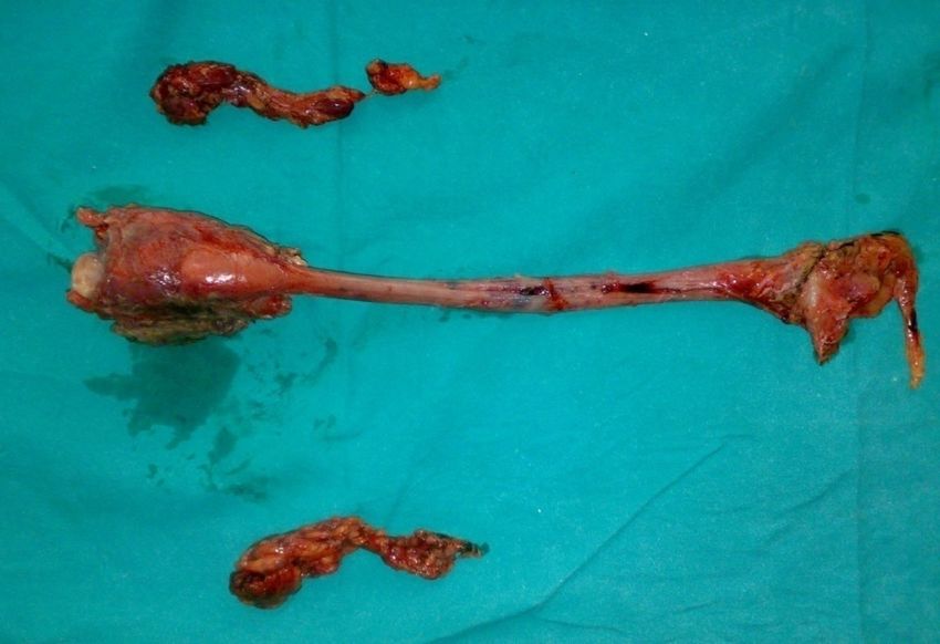
Figure3.3.Laryngopharyngoesophagectomy
Figure Laryngopharyngoesophagectomyand
andneck
neckdissection
dissectionspecimen.
specimen.
After discharged patients were followed up, during which they were asked for any
3. Results
complaints, they were
In the minimal examined
invasive surgeryloco-regionally
group, a total of and systemically.
15 patients They surgery,
underwent were subjected
out of
for routine blood investigation (including TFT, serum calcium),
which two were male and 13 were female. In the open group, a total 18 patients underwent and X-ray chest. The
mortality rate related to surgery (within 30 days of surgery) and
surgery, out of which 5 were male and 13 were female. The median age was 40 (range 28 long-term survival were
tostudied.
65) years in laparoscopic TPLE group where in the open group, the median age was 40
(rangeAll 21 cases
to 60)were
years.advised for follow-up every two months for the first year, once every
threeInmonths
the MISduring the second
(Minimally Invasiveyear, once every
Group) group,four
themonths during site
most common the third year, once
was limited to
every
the six months
post-cricoid for the
region (27%)next twoinyears,
and, andgroup,
the open then annually.
the most Data
common collected
site ofincluded pa-
lesion was
tient
the characteristics,
hypopharynx andtumor site, and
supraglottis inmorphology.
28% of patients.Operative information
The average durationincluded blood
of surgery
lossobserved
was and durationto be of3.86surgery.
(range of The complications
3 to 6) hours in the were
MISstudied
Group.including
Initially, itboth
was major
longerand
in
minorsurgeries
earlier factors. but, later, it reduces to an average of 3.5 h. The average duration of surgery
was observed to be 5.33 (range 4 to 6.5) hours in the Open Group. The difference in the
3. Results
post-operative variables was average blood loss. In the MIS group, it was was 200–500 mL
and, in the Open
In the minimal group, it wassurgery
invasive 300–600group,
mL. a total of 15 patients underwent surgery, out
The average time taken until
of which two were male and 13 were female. In postoperative oral
theintake
openwasgroup,10 (range
a total618 to patients
18) days un-
in
the MIS group and 10.92 (range 6–19) days in the open group.
derwent surgery, out of which 5 were male and 13 were female. The median age was 40 It took around an average
of(range
seven28 days (4 to
to 65) 15 days)
years to remove all
in laparoscopic drains
TPLE in both
group where groups.
in theTheopenaverage
group,duration of
the median
the
ageICU
was(Intensive
40 (range Care 21 toUnit) stay in the MIS group was 3.4 (1–12) days, and the average
60) years.
duration of the ICU stay
In the MIS (Minimally Invasive in the openGroup)
group group,
was 4 (2–13)
the most days.
commonThe average
site was duration
limited to
ofthe
hospitalization was 13.1 (7 to 37) days in the MIS group
post-cricoid region (27%) and, in the open group, the most common site of and the average duration of
lesion was
hospitalization in the open group was 16.88 (11–32) days.
the hypopharynx and supraglottis in 28% of patients. The average duration of surgery
wasIn the MISto
observed group,
be 3.86 one patient
(range of 3(6.7%) had pneumonic
to 6) hours in the MIS consolidation
Group. Initially, and it two
was patients
longer in
(13%) had a wound infection. In the open group, six patients (33%)
earlier surgeries but, later, it reduces to an average of 3.5 hours. The average duration of had pneumonic
consolidation and four patients (22%) had a wound infection.
surgery was observed to be 5.33 (range 4 to 6.5) hours in the Open Group. The difference
In the MIS group, one perioperative death occurred. This patient had a thoracic duct
in the post-operative variables was average blood loss. In the MIS group, it was was
injury with chyle leak and she expired during re-exploration due to cardiac shock. Whereas
200–500 mL and, in the Open group, it was 300–600 mL.
in the open group, one patient had wound infection and IJV bleed leading to hemorrhagic
The average time taken until postoperative oral intake was 10 (range 6 to 18) days in
shock on the 11th postoperative day.
the MIS group and 10.92 (range 6–19) days in the open group. It took around an average
Regarding the adjuvant therapy, in the MIS group, 10 patients (67%) received post-
of seven days (4 to 15 days) to remove all drains in both groups. The average duration of
operative radiotherapy. In the open group, eight patients (44%) received postoperative
the ICU (Intensive Care Unit) stay in the MIS group was 3.4 (1–12) days, and the average
radiotherapy and five patients (28%) received preoperative radiotherapy while one patient
duration of the ICU stay in the open group was 4 (2–13) days. The average duration of
(4%) had received preoperative chemoradiation.
hospitalization was 13.1 (7 to 37) days in the MIS group and the average duration of
In the MIS group, during the study period, two patients developed cervical LN
hospitalization in the open group was 16.88 (11–32) days.
recurrence and one patient developed lung metastasis. In the open group, one patient
developed malignant pleural effusion (Table 1).Clin. Pract. 2021, 11 182
Table 1. Post-operative complication.
Complication MIS Group (15) Open Group (18)
Pulmonary 1 (6.7%) 6 (33%)
Wound infection 2 (13%) 4 (22%)
Hypothyroidism 4 (27%) 6 (33%)
Hypoparathyroidism 7 (47%) 4 (22%)
Anastomotic Leak 1 (6.7%) 1 (5.5%)
Anastomotic stricture 0 (0%) 1 (5.5%)
IJV bleed 1 (6.7%) 2 (11%)
Chyle leak 1 (6.7%) 1 (5.5%)
4. Discussion
Post cricoid cancer is an aggressive cancer. In 1960, Ong and Lee [4] described the
use of the transposed stomach to restore gastrointestinal continuity after circumferen-
tial pharyngectomy. This method was re-popularized by Orringer [5] after trans-hiatal
esophagectomy, which was followed by anastomosis in the neck. Today, a gastric pull-up
operation is a preferred technique, especially when a significant tumor extends into the
proximal esophagus. The advantage includes dealing with potential skip lesions by total
esophagectomy, which is performed as part of the gastric pull-up. The main advantages of
the procedure are single stage surgery, and a single well vascularized anastomosis with
the stomach. Additionally, stomach pull-up is thought to have less complications than
free jejunal transfer (33% vs. 47%, p < 0.05). Spiro et al., in 1991 [6], reviewed 120 patients
who had gastric transposition from 1973 to 1990 at Memorial Sloan Kettering Cancer
Center. There was an 11% operative mortality. Fifty-five percent had intraoperative or
perioperative complications. In addition, 13% had anastomotic leaks, and there were three
instances of stomach necrosis. This translates to a median of eight days of a hospital stay
when recovery was uneventful and 11 days when there were associated complications.
They concluded that gastric pull-up is a reliable, reconstructive method, but advocates
careful patient selection to minimize morbidity. According to another study, there was no
significant difference with regard to the survival between gastric transposition and free
jejunal autograft, but there were fewer respiratory complications (33% vs. 47%, p < 0.05),
significantly low local recurrences (15.8% vs. 33.8%, p = 0.004), and higher survival without
dysphagia (76% vs. 89%, p < 0.05) [6,7].
According to Schusterman et al. [8], in patients with advanced cancer, extensive
esophageal resection into the chest is often required, and gastric pull-up seems to be an
easier and more direct form of reconstruction. Wong et al. [9] have, until now, reported the
largest experience on a minimal invasive approach of gastric mobilization and esophageal
dissection on 13 TPLE of which nine were done totally laparoscopically and four were
performed hand assisted. This series confirmed the feasibility of the laparoscopic approach
and demonstrated its safety. The mean operative time was 8.5 h (range: 5–11), there
was no 30-days mortality, and the morbidity rate was 42%, which was more favorable in
comparison with their open approach. The mean hospital stay was 41 (range: 18–75) days
in this Wong et al. study.
One series by Rossi et al. [10] have also reported this technique on four patients with
recurrent disease after primary chemo-radiotherapy. The mean operative time was 345 min
(range: 300–384). The hospital stay was 20 days (18–20).
In our series, the mean duration of surgery was 4 h, of which the mean time taken for
laparoscopy was 3 h (180 min). However, as the experience increased in the last two cases,
it was 2 h (120 min). This observation is comparable to what was reported by Wong et al. [9]
in which the median total operative time was 8.5 h (range, 5–11 h) and the laparoscopic
time was less than 4 hours. The mean duration of hospital stay in our patients was 16 days
(7–37 days) which is comparable to what was reported by Rossi et al. [10] and less than
Wong et al. [9] where the mean hospital stay was 41 (range: 18–75) days.Clin. Pract. 2021, 11 183
We have noticed that the operative time was less in the patients who were operated
later on in the series because of a learning curve associated with this procedure in the
earlier cases.
The laparoscopic TPLE group lost less blood and, hence, had fewer requirements
for transfusion during the admission than the open approach group. The estimated
average blood loss in the laparoscopic TPLE was 300 mL (200–500), which is less than the
conventional open procedure (i.e., 460 mL (300–600)) in our series. Similar observations
were made by Wong et al. [9], which had less operative blood loss. In a report summarizing
nationwide statistics, the mortality rates from TPLE ranged from 3.4% in high-volume
centers to as high as 17.3% in low-volume centers. Others have also reported high rates
of morbidity (60%–84%) and mortality (1%–4%) [11,12]. Much of the morbidity of the
procedure including lung complications, cardiopulmonary failure, complications from
delayed mobilization, and wound complications are due to the method of access. Most
patients experience less pain, fewer wound complications, less blood loss, and a quicker
return to normal activity with a laparoscopic group.
In our laparoscopic TPLE (Total Pharyngo Laryngo Oesophagectomy) group, 73%
of patients had postoperative morbidity. Among them, one patient (6.7%) developed
pneumonic consolidation and wound infection was seen in two patients (13%). Similarly to
an open group, 89% of the patients developed morbidities, which included wound infection
in four (22%) patients, leak in one patient (5.5%), and consolidation in six patients (33%).
This suggests that a significant reduction in respiratory complications with a laparoscopic
TPLE group (6.7%) occurred as compared to an open group (33%). The wound complication
rate was also reduced in the laparoscopic TPLE group. Other studies have also shown
higher incidence of pneumonia in an open group as compared to laparoscopic TPLE.
The quality of life of patients undergoing laparoscopic TPLE improved, as shown by
Wong et al. [9]. Additional theoretical advantages of the use of laparoscopy in cancer treat-
ments result from a decreased surgical stress response, which may minimize immunologic
suppression. Once again, this is a possible consideration for long-term survival.
By comparing laparoscopic TPLE with an open approach in our experience, we
observed that laparoscopic TPLE for post-cricoid cancer was a safe option in experienced
hands. Since this procedure is associated with a learning curve, our results will improve
with time, as has been observed by others. Laparoscopic TPLE has the potential to replace
conventional techniques.
5. Conclusions
A minimally invasive technique for pulling up the stomach in patients suffering from
post-cricoid cancer and is considerably safe as compared to an open technique. With
regard to morbidity and mortality showing favorable results with the laparoscopic group
since it has less operative blood loss, decreased respiration complication, and decreased
wound infection.
Hence, the laparoscopic technique is superior when compared to an open technique
and has clear advantages in spite of the difficulty in the learning curve present and can be
collectively encouraged.
Author Contributions: All Authors have Contributed Equally. All authors have read and agreed to
the published version of the manuscript.
Funding: This research received no external funding.
Institutional Review Board Statement: Not applicable.
Informed Consent Statement: Informed consent was obtained from all subjects involved in the
study. Written informed consent has been obtained from the patient(s) to publish this paper.
Conflicts of Interest: The authors declare no conflict of interest.Clin. Pract. 2021, 11 184
References
1. Parkin, D.M.; Muir, C.S.; Whelan, S.L. Cancer Incidence in Five Continents, Vol. VI; IARC Scientific Publications: Lyon, France, 1992.
2. Pingree, T.F.; Davis, R.K.; Reichman, O.; Derrick, L. Treatment of hypopharyngeal carcinoma: A 10-year review of 1362 cases.
Laryngoscope 1987, 97, 901–904. [CrossRef] [PubMed]
3. Axon, P.R.; Woolford, T.J.; Hargreaves, S.P.; Yates, P.; Birzgalis, A.R.; Farrington, W.T. A Comparison of surgery and Radiotherapy
in management of postcricoid carcinoma. Clin. Otolaryngol. 1997, 22, 370–374. [CrossRef] [PubMed]
4. Ong, G.B.; Lee, T.C. Pharyngo-gastric anastomosis after oesopharyngectomy for carcinoma of the hypopharynx and cervical
esophagus. Br. J. Surg. 1960, 48, 193. [CrossRef] [PubMed]
5. Orringer, M.B. Technical aids in performing transhiatal esophagectomy without thoracotomy. Ann. Thorac. Surg. 1984, 38,
128–132. [CrossRef]
6. Spiro, R.H.; Shah, J.P.; Strong, E.W.; Gerold, F.P.; Bains, M.S. Gastric transposition in head and neck surgery. Am. J. Surg. 1983, 146,
483–487. [CrossRef]
7. Triboulet, J.P.; Mariette, C.; Chevalier, D.; Amrouni, H. Surgical management of carcinoma of the hypopharynx and cervical
esophagus analysis of 209 cases. Arch. Surg. 2001, 136, 1164–1170. [CrossRef] [PubMed]
8. Schusterman, M.A.; Shestak, K.; de Vries, E.J.; Swartz, W.; Jones, N.; Johnson, J.; Myers, E.; Reilly, J., Jr. Reconstruction of the
cervical esophagus: Free jejunal transfer versus gastric pull-up. Plast. Reconstr. Surg. 1990, 85, 16–21. [CrossRef] [PubMed]
9. Wong, S.K.H.; To, E.W.H.; Ng, E.K.W.; Chung, S.; Chan, A.C.W.; Lee, D.W.H. Minimal invasive approach of gastric and esophageal
mobilization in total pharyngolaryngoesophagectomy. Surg. Endosc. 2003, 17, 798–802. [CrossRef] [PubMed]
10. Rossi, M.; Santi, S.; Barreca, M.; Anselmino, M.; Solito, B. Minimally invasive pharyngo-laryngo-esophagectomy: A salvage
procedure for recurrent postcricoid esophageal cancer. Dis. Esophagus 2005, 18, 304–310. [CrossRef] [PubMed]
11. Birkmeyer, J.D.; Siewers, A.E.; Finlayson, E.V.A. Hospital volume and surgical mortality in the United States. N. Engl. J. Med.
2002, 346, 1128–1137. [CrossRef] [PubMed]
12. Ries, L.A.G.; Eisner, M.P.; Kosary, C. SEER Cancer Statistics Review, 1973–1999; National Cancer Institute: Bethesda, MD, USA,
2002.You can also read