Q-Switched Ruby Laser Treatment of Tattoos and Benign Pigmented Skin Lesions: A Critical Review
←
→
Page content transcription
If your browser does not render page correctly, please read the page content below
Q-Switched Ruby Laser Treatment of Tattoos and Benign
Pigmented Skin Lesions: A Critical Review
Christian Raulin, MD(1)
Matthias P. Schönermark, MD(2,3)
Bärbel Greve, MD(1)
Saskia Werner, MD(1)

From the Laserklinik, Karlsruhe, Germany (1); the Department of Medicine, Dartmouth Medical School, Hanover, NH
(2); and the department of Otolaryngology/Head and Neck Surgery, Hannover Medical School, Germany (3).

The Q-switched ruby laser (694 nm, 25-40 nsec) is an effective and safe therapeutic device for the
treatment of tattoos and well-defined, benign, pigmented epidermal and dermal lesions. Because of
its selective mode of action, dermal pigments of natural and artificial origin are destroyed
photothermically and removed without scar. This method is exceptionally suited for the elimination
of lay and professional tattoos, traumatic tattoos, and permanent makeup. Other frequent indications
include benign pigmented lesions such as lentigines, freckles, café-au-lait spots, seborrheic keratosis,
and Becker nevi. As a dermal pigmented lesion, the nevus of Ota is perfectly treatable. However,
chloasma can no longer be considered an indication for ruby laser treatment due to unsatisfactory
results. Melanocytic nevi and congenital nevi should be treated only in clinical studies. The
effectiveness of the long-term epilation of dark hair with this laser device has to be verified in future
investigations. Particularly attractive is the nonproblematic and straightforward removal of pigmented
lesions in precarious anatomic regions like the lips, eyelids, and genitals (e.g., benign melanosis of
the lips or of the penis, seborrheic keratosis of the lid angle).

Raulin C, Schönermark MP, Greve B, Werner S. Q-switched ruby laser treatment of tattoos and
benign pigmented skin lesions: a critical review. Ann Plast Surg 1998;41:555-565

The ruby laser, which was invented more than 30 years ago, is the oldest laser system in plastic
surgery and dermatology. The impact of ruby laser beams on human skin was described by Goldman
and colleagues in the early 1960s for the first time. The laser was used primarily for the removal of
tattoos, although the relatively long impulse duration (as long as 500 µsec) led to frequent scarring.
The development of the Q (quality)-switched technique allowed Reid and associates' to ablate a tattoo
without any scarring side effect. Since the early 1990s, the Q-switched laser has been experiencing a
Renaissance in dermatology. For the treatment of tattoos and well-defined, benign, pigmented
lesions, this device has proved to be effective, safe, and easy to handle (Table).

Mode of Action

The ruby laser emits red light (694 nm), which is absorbed selectively by pigmented structures
(melanosomes and melanin-containing cells) and exogenously introduced pigments (metallic and
organic color particles, dirt, and other erogenous substances). Oxyhemoglobin as well as hemoglobin
show only a marginal absorption at this wavelength. The main absorption spectrum for these
chromophores lies between 400 and 600 nm, leading to minimal thermal injury.

The quality-switched mechanism, which was investigated with guinea pig and human skin, is based
on the principle of selective photothermolysis. Because the duration of exposure to the energy is
between 20 and 50 nsec, which is well below the thermal relaxation time of the target tissue
(approximately 0.5-1 µsec for melanosomes), the damage to the surrounding tissue is minimal.

The applied ruby laser beam causes extreme heat (as high as 1000°C) in a very short interval (25-40
nsec). This "flash of heat" leads to a rupture of melanosome-carrying cells (melanocytes and
keratinocytes) in a depth of as much as 2 mm. Histological studies have shown that tattoo particles
are located mainly in the macrophages and fibroblasts of the upper dermis. Only a very small portion
may be found in the deeper interstitium. The laser beam, which is absorbed by dark pigments, leads
to a fast, thermally induced expansion of the pigment-carrying cells (so-called "shock waves") and
thus to the loss of their integrity. These released pigments are phagocytosed by invading
inflammatory cells and removed via the lymphatic system. Some smaller portion of the pigmented
fragments are ablated transdermally by a crusting process. A chemical alteration of nonphysiological
pigments by the affecting heat, which alters the optical refraction index, is another mode of action of
the ruby laser that has been discussed recently.

Application of the Q-switched Ruby Laser

The Q-switched ruby laser that we use (Laserase, Lambda Photometrics Ltd., Harpenden, UK)
utilizes a pulse duration of 25 nsec. The fluence may be adjusted between 0.5 and 40 J per square
centimeter, and the spot size can be between 2 and 4 mm . The laser pulses are applied with a
frequency of one pulse per second in a slightly overlapping fashion. Therefore, large areas can be
treated within one treatment session. Immediately after the application, the skin shows a whitish
discoloration (Fig 1B), which is transient and usually disappears within 5 to 20 minutes. The cause of
this phenomenon is the induction of "steam" through the consecutive heating of intra- and
extracellular fluid. The expansive tendency of the steam leads to a rise of the epidermis, which in turn
causes an alteration of the epidermal refractory index and thus a whitish color (see Fig 1B).
Consequently, erythema appears in the treated area, which tapers off within 24 hours. If higher
energies are used or facial areas are treated, a transient swelling may occur that lasts no longer than
36 hours.

Within the next few days, delicate crusts and sometimes small blisters evolve that heal within the
following 2 weeks. Patients should be taught not to manipulate the lesions to prohibit infections and
scarring. As a prophylaxis against infection, dressings with sulfadiazine-silver (Flammazine) or
polyvidon-iodide (Betaisodona) may be applied.
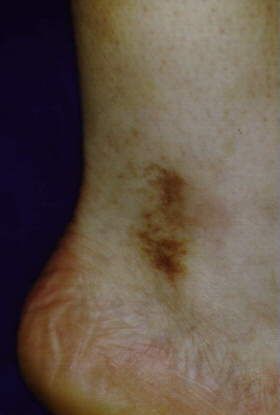
Fig 1. (A) Amateur tattoos on both thighs. (B) Whitish discoloration immediately after the laser
    treatment. (C) Discrete hypopigmentation after five treatments with the Q-switched ruby laser

The typical sensation experienced during lasering is a tingling, somewhat burning, low-range pain
that normally does not need any anesthetic treatment. However, for very sensitive patients and
children, and for most facial applications, a topical anesthetic may be used (e.g., a lidocain-prilocain
cream such as EMLA).

For most patients, several consecutive treatments are necessary. The interval between sessions should
be at least 4 weeks. During the entire therapeutic period and approximately 4 weeks beyond, patients
should be advised to avoid direct sun exposure and tanning booths to minimize the risk of pigmentary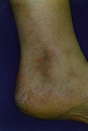
chances. Sun blockers with a high ultraviolet (UV) protection factor should be used if UV exposure is
unavoidable. Hypo- and/or hyperpigmentation may be observed in some patients. Most of these
changes are transient and will fade within several weeks. The risk of scarring or lasting pigmentary
disturbances is very low for this type of laser.

Indications

Tattoos

Techniques for removal of professional and lay tattoos have improved steadily during the past 10
years. The more conventional therapeutic measures include dermabrasion, surgical excision,
salabrasion, and cryotherapy. All of these methods bear a considerable risk for atrophic and
hypertrophic scarring, hypo- and hyperpigmentation, and incomplete removal of the pigment. The
carbon dioxide and the argon lasers, which operate in a continuous mode, led to nonspecific tissue
destruction and thus to posttherapeutic scars.

The introduction of the Q-switched ruby laser, which allows selective destruction of tattoo pigments
with minimal damage to the surrounding tissue, has led to scarless removal of tattoos. Lay and
professional tattoos are often composed of different colors. Black and blue-black pigments can be
removed effectively by the Q-switched ruby laser because these pigments absorb the ruby laser light
well (absorption maximum, 600-800 nm). Green colors respond somewhat variously, and yellow and
red pigments cannot be removed by the ruby laser. These colors absorb light in the range of 500 to
580 nm. Therefore, a combination of different laser systems must be applied to target effectively a
complex, multicolored tattoo. The Q-switched ND:YAG laser seems

to be particularly effective in the freqtienc@1-doubled mode (532 nm) for the treatment of red and
pink tattoos. Yellow and orange pigments respond very well to the pulsed dye laser (510 nm). An
alternative to the ruby laser for the treatment of black tattoos seems to be the Q-switched Nd: YAG
laser (1,064 nm). Finally, black and green pigments also may be treated effectively with the Q-
switched alexandrite laser.

The number of treatment sessions required differs from patient to patient (i.e., from tattoo to tattoo)
and depends on the amount of professionalism used to pierce the tattoo. Lay tattoos are often pierced
with black ink only (Fig 1A) and can be removed with three to five treatments (Fig 1C). Professional
tattoos, however, contain a higher pigment density and more stable pigmentary compounds (heavy
metals, organic pigments), and thus need more treatments (usually more than eight, but sometimes as
many as 20 treatments are required to remove professional tattoos). Older tattoos are more responsive
to ruby laser treatment than newer tattoos. This may be because of the completed phagocytosis of the
pigment and to the more fragile, more easily disrupted pigment crystals. To treat tattoos with the ruby
laser, fluences between 5 and 20 J per square centimeter are applied.

Sun-tanned or dark-colored skin (Fitzpatrick types IV-VI) may respond with some delay because
melanin absorbs the major part of the applied light. Thus the light energy is filtered away by the
pigmented, epidermal cells, which lay above the dermal tattoo pigments. To surmount this effect, the
Q-switched ND:YAG laser may be used, which-due to its longer wavelength (1,064 nm)-penetrates
deeper into the skin and interferes only minimally with the melanin-containing structures.

Because of the interaction with melanin, approximately 50% of all patients show some
posttherapeutic hypopigmentation, which is almost always transient and tapers off within 2 to 6
months. Hyperpigmentation, however, is a rare side effect of ruby laser treatment. A predilection site
for pigmentary changes is the ventral part of the forearms (personal observation). Patients who have
dark pigment seem to be especially susceptible to posttherapeutic pigmentary changes. The highly
selective mode of action of the ruby laser leads to a comparably low risk of scarring (
The penetration of artifacts (e.g., dirt, dust, sand, or plastic particles) into the skin during accidents or
explosions leads to so-called "traumatic tattoos." After wound healing and reepithelialization is
complete, it is difficult to remove the foreign particles from the dermis. Several techniques (e.g.,
surgical excision, dermabrasion, electrosurgery, and carbon dioxide or argon lasers) have been
described. Most of these measures, however, bear a considerable risk of scarring and may not lead to
complete removal of all pigments. In contrast, the Q-switched ruby laser has proved to be a safe and
very effective therapeutic device for the removal of traumatic tattoos. Depending on the depth and the
structure of the injected pigments, 3 to 20 sessions may be needed for complete removal (Fig 3).
Normally, fluencies between 5 and 10 J per square centimeter are applied. Similar results may be
achieved with the Q-switched ND:YAG laser (1,064 nm) or the Q-switched alexandrite laser.

Fig 3. (A) Traumatic tattoo after a car accident in (B) The lesion is removed completely after five
1971.                                               treatments..

Drug-Induced Hyperpigmentation

After long-time and/or high-close treatment with minocycline, cutaneous hyperpigmentation has been
noted to occur. The exact mechanism for this pigmentary change, however, is not fully understood.
The application of Q-switched lasers (e.g., ruby or ND:YAG) may lead to a complete removal of
these cosmetically disturbing lesions within 5 to 10 sessions.

Benign Pigmented Lesions

Due to the selective absorption of the ruby laser light by melanin, several benign pigmented lesions
can be treated quite effectively with this laser.

Hyperpigmented Scars and Postinflammatory Hyperpigmentations

Sometimes operation and trauma, especially burns and scaldings, result in cosmetically disfiguring
hyperpigmented scars. The conservative treatment options for these lesions include cryotherapy,
dermabrasion, and the application of bleach or scar ointments. However, most of these measures do
not lead to satisfying results. Treatment with the Q-switched ruby laser can lead to a dramatic
improvement of the scar zones. Fluencies between 4 and 10 J per square centimeter and several
treatment sessions (three to nine) may be used. Also, postinflammatory hyperpigmentary lesions (e.
g., purpura jaune d'ocre) are a considerable responsive target for ruby laser therapy and can be
lightened with this treatment (Fig 4). As an alternative, the frequency-doubled Q-switched ND:YAG
laser (532 nm) can be applied. In a direct-comparison trial, however, the ruby laser showed a
somewhat higher clearance rate than the ND:YAG laser.
Fig 4. (A) Purpura jaune d'ocre on the left ankle
(B) Complete removal after five treatments.
of a 44-year old female patient.
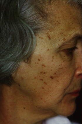
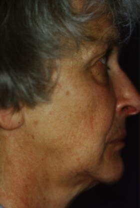
Ephelides and Benign Lentigines
The Q-switched ruby laser can be used safely and effectively to remove cosmetically disturbing
freckles (Fig 5) and benign lentigines of the hands and face (Fig 6). Normally, the lesions are
removed within one to two sessions. Patients should be informed before treatment that ephelides
especially tend to recur at a considerably high rate. Sun exposure and tanning booths should be
avoided for at least 6 weeks, and high-potency sun blockers should be used.Fig 5. Ephelides of the arm. The forearm was treated once with the Q-switched ruby laser; the upper
arm was not treated.
Fig 6. (A) Senile lentigines on the back of the
(B) Clinical picture after two laser treatments.
hand.
Benign Melanosis of the Lip and of the Penis
Before these lesions are treated with the Q-switched ruby laser, it must be made explicitly clear that
they are indeed benign (e.g., by a preceding biopsy). The benign melanosis responds very well to
laser treatment, and disappears within one to three sessions. Fluencies between 6 and 10 J per square
centimeter have been applied. Peutz-Jeghers syndrome-associated lentigines of the lips and the oral
mucosa have also been treated effectively with the ruby laser.
The benign penile melanosis is a rare condition that also has been removed successfully with the ruby
laser. An invasive approach to this lesion (excision, dermabrasion) bears a considerable risk of
cosmetically or even functionally disturbing sequelae. This can be surmounted by application of the
Q-switched ruby laser.
Seborrheic Keratosis
The treatment of choice for pigmented, seborrheic keratotic lesions is still excochleation with a sharp
spoon. Alternatively, cryosurgical measures with liquid nitrogen or the Q-switched ruby laser may beused. Seborrheic keratosis in delicate locations (e.g., at the lid angle) and multiple lesions can be
removed very safely and with excellent cosmetic results (Fig 7). One to three sessions and high
energy fluencies (as high as 40 J per square centimeter) are needed with the Q-switched ruby laser to
remove seborrheic keratotic lesions completely. Another laser system that is used for this indication
is the carbon dioxide laser. However, in a recent study including 32 patients and 690 seborrheic
lesions, 25% of all patients who were treated with the carbon dioxide laser in continuous mode
suffered from posttherapeutic, atrophic scarring. The carbon dioxide laser in the ultrapulse mode and
the Erbium:YAG laser seem to be more gentle and therefore have less side effects (personal
observation).
(B) Cosmetically satisfactory result after two
Fig 7. (A) Seborrheic keratosis of the face.
treatment sessions.
Café-au-lait Spots
These lesions appear in conjunction with genetically determined diseases (e.g., neurofibromatosis) as
often solitary, light- to medium-brown spots. They can be removed with the Q-switched ruby laser
within three to eight treatment sessions (Figs 8). The effective fluencies lie between 7 and 20 J per
square centimeter. The interval between sessions should be 4 to 6 weeks. However, some patients
should be treated in shorter intervals to prohibit early repigmentation. The combined application ofthe Q-switched mode (25-40 nsec) and the normal and long-pulse mode (270 µsec to 1 msec) allows
the delivery of higher fluencies and thus the targeting of more deeply seated pigments (e.g., in
follicles). Thus a higher clearance rate may be achieved (personal observation). Patients with darker
skin types (Fitzpatrick IV and V) can develop posttherapeutic hypopigmentation that taper off within
a few months. Transient Hyperpigmentations have also been described. Therefore, a treatment trial
should be carried out before the entire lesion area is treated. Recurring spots are not uncommon,
especially if the melanocytes are activated due to insufficiently blocked sunlight. Recurrences occur
in 40% to 50% of the patients (own unpublished data).
Resistant lesions can be treated with other laser systems. Tse and colleagues describe a clearance rate
of 30% with the frequency-doubled Q-switched ND:YAG laser (532 nm) compared with a 51%
clearance rate by the Q-switched ruby laser. In contrast, the "normal" Q-switched Nd: YAG laser
(1,064 nm) is, due to its higher wavelength, less effective for the treatment of epidermal pigmented
lesions. The dye laser (510 nm) has been used for the treatment of 14 patients with café-au-lait spots;
35% of the patients showed a 75% lightening whereas 42% of the patients seemed to be resistant.
Somewhat better results could have been achieved by Fitzpatrick and associates.
(B) Complete removal of the lesion after five
Fig 8. (A) Periorbital café-au-lait spot.
laser treatments.
Becker Nevus and Hypertrichosis
The Becker nevus is a hyperpigmented, often hypertrichotic lesion of young, mostly male adults. It
can be treated effectively with the Q-switched ruby laser. Fluencies between 7 and 20 J per square
centimeter are applied. Three to 10 treatment sessions at monthly intervals may be necessary.
Resistant as well as recurring lesions have been described. Hypo- and hyperpigmentation are possible
side effects of the treatment.
For the treatment of cosmetically disturbing hypertrichosis, two highly energetic flashlamps with a
continuous wavelength spectrum have been introduced recently (PhotoDerm VL/PL and EpiLight;
ESC Medical Systems, Yokneam, Israel). Both devices lead to a removal of hair. The Q-switched
ruby laser seems to be ineffective in the long-term removal of hypertrichosis. A delayed hair growth,
however, has been described after the application of a normally pulsed ruby laser (270 µsec). In our
hands, the longpulsed ruby laser (1 msec) also led to a retarded growth of hairs but not to a reductionof total hair number. Preliminary results suggest that the long-pulsed alexandrite laser (755 nm; impulse duration as high as 20 msec) is a very effective therapeutic alternative. Chloasma and Melasma These typically circumscribed, patchy facial hyperpigmentations that appear normally in younger women are difficult to treat. Hydrochinon-containing external therapeutics may have some lightening effect. The etiopathology shows increased melanin synthesis with epidermal and dermal depositions of melanin. These should be responsive to ruby laser treatment. Surprisingly, only 15% to 20% of patients were responsive to the ruby laser. Some lesions became even worse and resulted in considerable additional pigmentation (personal observations). Therefore, we dispense with ruby laser treatment for chloasma. The dye laser (510 nm) seems to be equally ineffective. Grekin and coworkers report a response rate of only 20%. Nevus of Ota This lesion is a rare, congenital oculodermal melanosis. The typical nevus of Ota is a blackblue mole that manifests in the area of the first and second branch of the trigeminal nerve. The Mongolian race has a much higher incidence of nevus of Ota than whites. Conservative therapeutic measures include dermabrasion, surgical excision, and cryotherapy. The results, however, have been mostly unsatisfactory. Argon laser treatment of this lesion has led to changes in the dermal texture and to a permanent loss of pigmentation. In contrast, treatment with the Q-switched ruby laser bears only a minimal risk of scarring and of pigmentary changes, which are almost always transient. To achieve complete removal, 8 to 10 sessions at 1- to 2-month intervals are needed. The applied fluencies range between 5 and 10 j per square. Also, other laser systems have been used quite successfully for the treatment of nevus of Ota. Dye laser therapy has led to an 80% lightening when combined with a flashlamppumped Q-switched alexandrite laser. In a histological study, it has been shown that the dermal melanocytes are targeted and destroyed by both the Q-switched ruby laser and the Q-switched ND:YAG laser. Clinically, however, ruby laser treatment led to a higher clearance rate (57%) than ND:YAG laser treatment (42%). Melanocytic Nevi and Congenital Nevi Laser treatment of congenital melanocytic nevi is a controversial subject. On the one hand, recurrence of lesions after laser treatment is frequent. On the other hand, the effects of laser irradiation on cellular biological behavior and the possible mutagenic responses of nevomelanocytes are still unclear. Congenital and immediate postpartum-appearing nevi have an incidence of approximately 1% among newborns. Without treatment, small- and middle-size nevi bear a risk of malignant transformation of approximately 1% to 5%. Five percent of large nevi may develop into malignant melanoma during a person's lifetime. The therapeutic options for these large lesions include cryotherapy, dermabrasion, successive excision with combined expander techniques, and autologous transplantation.
Fig 9. (A) Superficical melanocytic nevus of the right cheek. (B) Histology of the lesion before
  treatment indicates superficially seated neval cell nests (H&E, original magnification x160 before
     53% reduction). (C) Complete removal after one treatment with the ruby laser (2.5 years after
                                               treatment).

Generally, excision is the therapy of choice in removing suspect congenital and melanocytic nevi.
Only in select patients should ruby laser therapy be considered (Figs 9A, C). As a general rule, nevi
should not be treated uncritically with the Q-switched ruby laser. Clinical and histological studies
show that the Q-switched ruby laser appears to be effective in removing superficial portions of
congenital melanocytic nevi. Deeper dermal melanocytes and nests of nonpigmented melanocytes
remained unaltered. Therefore a therapy of melanocytic nevi and congenital nevi with the Q-switched
ruby laser should only be performed in controlled studies. In these patients careful diagnostic
procedure, especially epiluminescence microscopy and a biopsy, must be carried out before any kind
of laser treatment is begun (Fig 9B). If there is the slightest suspicion of a premalignant alteration, an
excision in toto should be performed. Longterm results of effective ruby laser therapy have to be
evaluated in this situation before recommendations can be made.
Indications and Treatment Parameters for the Q-Switched Ruby Laser
Indication Fluency (J/cm²) No. of Treatments
Tattoos (black, black-blue, green)
Lay tattoos 5-20 3-8
Professional tattoos 5-20 8-20
Permanent makeup 3-20 5-15
Traumatic tattoos 5-10 3-20
Minocycline-induced hyperpigmentations 4-10 3-9
Benign pigmented lessons
Hyperpigmented scars and postinflammatory
4-10 3-9
hyperpigmentations
Ephelides and benign lentigines 6-10 1-2
Labial and penile melanosis 6-10 1-3
Seborrheic keratosis 10-40 2-3
Café-au-lait spots 7-20 3-8
Becker's nevus 7-20 3-10
Nevus of Ota 5-10 8-10
Congenital and immediate postpartum-appearing nevi have an incidence of approximately 1% among
newborns. Without treatment, small- and middle-size nevi bear a risk of malignant transformation of
approximately 1% to 5%. Five percent of large nevi may develop into malignant melanoma during a
person's lifetime. The therapeutic options for these large lesions include cryotherapy, dermabrasion,
successive excision with combined expander techniques, and autologous transplantation.
Generally, excision is the therapy of choice in removing suspect congenital and melanocytic nevi.
Only in select patients should ruby laser therapy be considered (Figs 9A, C). As a general rule, nevi
should not be treated uncritically with the Q-switched ruby laser. Clinical and histological studies
show that the Q-switched ruby laser appears to be effective in removing superficial portions of
congenital melanocytic nevi. Deeper dermal melanocytes and nests of nonpigmented melanocytesremained unaltered. Therefore a therapy of melanocytic nevi and congenital nevi with the Q-switched
ruby laser should only be performed in controlled studies. In these patients careful diagnostic
procedure, especially epiluminescence microscopy and a biopsy, must be carried out before any kind
of laser treatment is begun (Fig 9B). If there is the slightest suspicion of a premalignant alteration, an
excision in toto should be performed. Longterm results of effective ruby laser therapy have to be
evaluated in this situation before recommendations can be made.
(Literatur bei den Verfassern)
Copyright (c) 1997-2002 PD Dr. med. Christian Raulin. Alle Rechte vorbehalten. Homepage
Fragen, Anregungen und Kritik bitte an den Webmaster.
Letzte Änderung: Freitag, 07. Juli 2000
Seitenanfang
Webdesign und Pflege by ISDYou can also read