Potential compression of the musculocutaneous, median and ulnar nerves by a very rare variant of the coracobrachialis longus muscle
←
→
Page content transcription
If your browser does not render page correctly, please read the page content below
Folia Morphol.
                                                                                                                                        Vol. 80, No. 3, pp. 707–713
                                                                                                                                      DOI: 10.5603/FM.a2020.0085
                                                                     CASE REPORT                                                      Copyright © 2021 Via Medica
                                                                                                                                                   ISSN 0015–5659
                                                                                                                                                  eISSN 1644–3284
                                                                                                                                               journals.viamedica.pl

Potential compression of the musculocutaneous,
median and ulnar nerves by a very rare variant
of the coracobrachialis longus muscle
Ł. Olewnik1 , F. Paulsen2, 3, R. Shane Tubbs4, 5, 6, N. Zielińska1, B. Szewczyk1,
P. Karauda1, M. Polguj7
1
 Department of Anatomical Dissection and Donation, Medical University of Lodz, Poland
2
 Institute of Functional and Clinical Anatomy, Friedrich Alexander University Erlangen-Nürnberg, Erlangen, Germany
3
  Department of Topographic Anatomy and Operative Surgery, Sechenov University, Moscow, Russia
4
 Department of Neurosurgery, Tulane University School of Medicine, New Orleans, LA, United States
5
 Department of Neurosurgery and Ochsner Neuroscience Institute, Ochsner Health System, New Orleans,
LA, United States
6
 Department of Anatomical Sciences, St. George’s University, Grenada
7
 Department of Normal and Clinical Anatomy, Medical University of Lodz, Poland

[Received: 18 July 2020; Accepted: 21 July 2020; Early publication date: 22 August 2020]

                                                    The coracobrachialis longus muscle (CBL) is an extremely rare variant of the
                                                    coracobrachialis muscle (CRM). The CBL originates from the apex of the coracoid
                                                    process together with the short head of the biceps brachii and inserts on the
                                                    olecranon of the ulna. The CBL consists of three parts: a superior part (classical
                                                    CRM — length 137.88 mm), a middle fibrous layer (23.41 mm), and an inferior
                                                    part (185.37 mm). A rare relationship between the CBL and median, musculocu-
                                                    taneous and ulnar nerves was observed with potential compression at these three
                                                    parts. In addition, this case report describes a connection between CBL and the
                                                    medial head of the triceps brachii, as well as a third head of the biceps brachii,
                                                    which originate from the fibrous layer. This case report highlights the relationships
                                                    between the CBL and the median, ulnar and musculocutaneous nerves. (Folia
                                                    Morphol 2021; 80, 3: 707–713)

                                                    Key words: anatomical variations, coracobrachialis longus muscle,
                                                    median nerve, musculocutaneous nerve, ulnar nerve

                        INTRODUCTION                                                    fibres from the C5-C7 ventral rami. The MCN passes
    The coracobrachialis muscle (CRM) originates from                                   through the CRM and descends between the biceps
the apex of the coracoid process of the scapula in com-                                 brachii and brachialis muscles both of which it inner-
mon with the short head of the biceps brachii muscle. It                                vates [35]. The median nerve (MN) arises from the
inserts by means of a flat, short tendon into the medial                                medial and lateral cords of the brachial plexus and is
surface of the humerus, between the attachments of                                      innervated by the C6, C7, C8 and T1 ventral rami [35].
the triceps brachii and brachialis muscles [36].                                        The MN provides motor and sensory function to the
    The musculocutaneous nerve (MCN) arises from                                        forearm and hand [35]. The ulnar nerve (UN) is com-
the lateral cord of the brachial plexus and contains                                    prised of C8 and T1 ventral rami. The UN innervates

Address for correspondence: Ł. Olewnik, MD, PhD, Department of Normal and Clinical Anatomy, Interfaculty Chair of Anatomy and Histology,
Medical University of Lodz, ul. Narutowicza 60, 90–136 Łódź, Poland, e-mail: lukasz.olewnik@umed.lodz.pl
This article is available in open access under Creative Common Attribution-Non-Commercial-No Derivatives 4.0 International (CC BY-NC-ND 4.0) license, allowing to down-
load articles and share them with others as long as they credit the authors and the publisher, but without permission to change them in any way or use them commercially.

                                                                                 707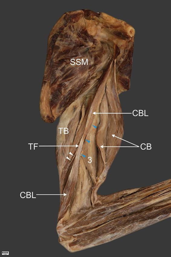
Folia Morphol., 2021, Vol. 80, No. 3
two muscles of the forearm, the flexor carpi ulnaris
and ulnar half of the flexor digitorum profundus. It
has branches extending to the hand over the distal
forearm and wrist [35].
Many earlier works describe the various types of
morphological variations occurring within this CRM.
They mainly concern the morphological variability of
the proximal and distal attachment, but also addi-
tional bands or the occurrence of additional muscle
bellies or heads [4, 16–18, 24, 27, 32]. However, little
is reported for one of its variants, the coracobrachialis
longus muscle (CBL). While Wood [51] was probably
the first to describe such a variant in 1867, a similar
discovery was made by Kyou-Jouffroy et al. [30].
A description of the CBL was more recently made by
Georgiev et al. [18].
Morphological variations have been previously
observed between the CRM and even the MCN or
MN. The presence of an extra muscle head or belly
can place pressure on the MCN or proximal MN.
More importantly, the presence of the CBL can place
pressure on the MCN, MN or UN.
Peripheral neuropathies can be classified as com-
pressive/entrapment and non-compressive forms
[2, 11]. Although peripheral nerve compression or
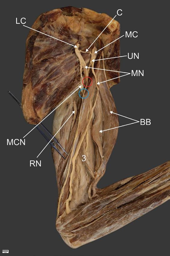
entrapment is possible anywhere along the course Figure 1. The coracobrachialis longus muscle and relation to the
of a nerve, it tends to occur more often where the median and musculocutaneous nerve; LC — lateral cord of the
brachial plexus; MC — medial cord of the brachial plexus; C —
nerve passes through fibro-osseous or fibromuscular communicating branch; UN — ulnar nerve; MN — median nerve;
tunnels or penetrates muscles [11, 26]. MCN — musculocutaneous nerve; RN — radial nerve; BB — bi-
This study describes a very rare and undescribed ceps brachii; 3 — third head of the biceps brachii. The red circle
shows the potential compression site of the median nerve, while
variation of the CBL and its extremely rare relationship the blue circle shows the potential compression site of the muscu-
with the MN, MCN and UN. Knowledge of such a very locutaneous nerve.
rare type can make it easier to understand disease in
this region and improve its treatment.
137.88 mm. The muscle then inserted on the me-
CASE REPORT dial surface of the shaft of the humerus between
The left upper limb from a male cadaver that was the attachments of the triceps brachii and brachi-
78-year-old at death underwent routine anatomical alis muscles. This part continued as a thin fibrous
dissection for research and teaching purposes in the layer (length — 23.41 mm) with the second part
Department of Anatomical Dissection and Donation, of the muscle (Fig. 1). The length of the muscle
Medical University of Lodz, Poland [38, 45, 46]. belly was 185.37 mm; the muscle belly passed the
tendon (13.95 mm length) and inserted on the
Morphology of the coracobrachialis longus muscle olecranon of the ulna. The distal part of the CBL
The proximal part of the CRM corresponded to connected with the brachii triceps tendon (Fig. 2).
the classical description, and originated from the The thin fibrous layer included an accessory band
apex of the coracoid process together with the short (length 25.83 mm) that connected to the medial
head of the biceps brachii. The width of the mus- head of the triceps brachii, and the thin fibrous layer
cle belly origin was 9.94 mm, while the thickness was the origin of the third head of the biceps brachii
was 4.13 mm. The length of the belly muscle was muscle (Figs. 1, 3).
708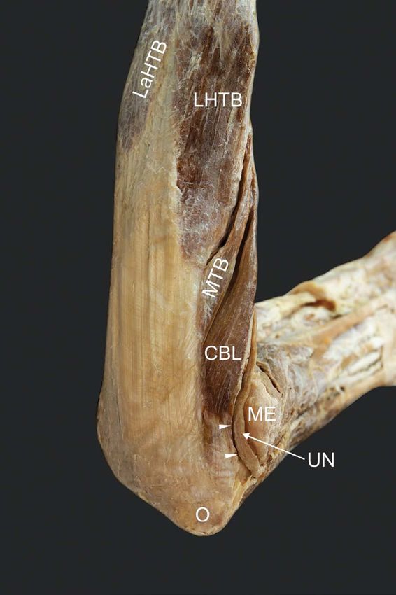
Ł. Olewnik et al., Coracobrachialis longus muscle

Figure 2. Distal part of the arm. Insertion of the coracobrachialis         Figure 3. Coracobrachialis longus muscle. Nerves are removed to
longus muscle; LaHTB — lateral head of the triceps brachii;                 reveal the coracobrachialis longus muscle; SSM — subscapularis
LHTB — long head of the triceps brachii; MTB — medial head                  muscle; CBL — coracobrachialis longus muscle; BB — biceps bra-
of the triceps brachii; CBL — coracobrachialis longus muscle;               chii; 3 — third head of the biceps brachii; TB — triceps brachii;
O — olecranon of the ulna; ME — medial epicondyle; UN — ulnar               TF — tendinous fibrous. The white arrowheads show the slip of
nerve. The white arrowheads show the potential compression site             the coracobrachialis longus muscle which attaches to the medial
of the ulnar nerve.                                                         head of the triceps brachii. Blue arrowheads show the place of
                                                                            origin of the third head of the biceps brachii.

CBL relation to MN, MCN and UN
    Median nerve. The MN arose from both the lat-                               Ulnar nerve. The UN arose from the medial cord
eral and medial cords of the brachial plexus. The                           of the brachial plexus, and ran along the CRM, lying
lateral cord fibres travelled under the CBL and then                        exactly on top of it; in the distal part, it was located
connected with those arising from the medial cord                           between the CBL and medial epicondyle, with a di-
of the brachial plexus. After 100.09 mm, the lateral                        ameter of 2.85 mm, while the CBL at this point was
cord fibres combined with the medial cord fibres.                           8.90 mm wide and was 3.31 thick (Fig. 2).
The medial cord fibres were 97.2 mm in length. The                              Detailed morphometric measurements were tak-
CBL was in the characteristic loop of the MN. The                           en. After photographic documentation, the CRM was
MN passed under the muscle and had a diameter of                            carefully dissected in order to minimise any errors in
5.10 mm, while the CBL at this point was 28.36 mm                           measurement. The measurements were performed
wide and 3.31 mm thick (Fig. 1).                                            using two methods:
    Musculocutaneous nerve. The MCN arose from                              —— with an electronic calliper (Mitutoyo Corporation,
the lateral cord of the brachial plexus and passed                              Kawasaki-shi, Kanagawa, Japan). Each measure-
under the CBL at a point 53.25 mm from the place                                ment was carried out twice with an accuracy of
of origin. The MCN passed under the muscle and had                              up to 0.1 mm;
a diameter of 4.95 mm, while the CBL at this point                          —— an analysis of digital photographic images was
was 21.95 mm wide and 3.19 mm thick (Fig. 1).                                   processed using MultiScanBase 18.03 (Computer

                                                                      709
Folia Morphol., 2021, Vol. 80, No. 3
Scanning System II, Warsaw, Poland). The value and most commonly involve the MN, UN, radial nerve
and precision of this method have been confirmed or MCN [6, 9–11, 19, 21–23, 33, 38, 39, 41, 43].
in previous studies [20, 29, 40, 42]. Musculocutaneous nerve neuropathy is not as
The posterior cord of the brachial plexus was common as MN or UN neuropathy. Most often, it
removed to more accurately visualise the neuromus- is due to muscular compression by the CRM, biceps
cular structures described in this case. The variant brachii, or brachialis muscles [1, 5, 7, 12–14, 28, 37,
muscle was innervated by the MCN. No medical or 49, 50]. The course of the MCN is closely related to
surgical history of the cadaver was available. No sim- that of the CRM. The MCN can pierce or pass deep
ilar variation was observed in the contralateral upper to the CRM [12, 15, 24, 31, 48]. It is believed that
limb. the CRM is the most common site of MCN entrap-
ment and additional heads can place pressure on the
DISCUSSION MCN [4, 14, 16, 17, 27, 32]. A potential site of MCN
Embryologically, the biceps brachii, CRM, and entrapment was also observed in the present case:
brachialis muscle are intimately fused together at at this point, 53.25 mm from its origin, the MCN
a very early stage and probably arise from a common (4.95 mm in diameter) passed under the CBL with
premuscle mass. The origins of the two heads of the the CRM being 21.95 mm in width and 3.19 mm. MN
biceps brachii at this early stage are close together entrapment within the CRM muscle leads to weak-
and only become separated with the later growth of ness and atrophy of the biceps brachii and brachialis
the scapula. The three muscles can be recognized in muscles and a loss of sensation in the lateral fore-
embryos 14 to 16 mm in length, and the tendon of arm. Active young individuals that frequently engage
the long head in embryos of 14 mm in length. The in shoulder and elbow flexion with the forearm in
distal end of the common muscle mass differentiates a pronated position are most susceptible [44]. It also
later than the proximal end [3, 47]. The presence of often occurs following chronic overuse of the CRM
the CBL could be explained as a result of the prema- and consequent hypertrophy. The nerve compressed
ture termination of this regression process. within the CRM has already given off its motor branch
The CRM is characterised by variability in both to the CRM, therefore no loss of CRM muscle function
proximal and distal attachments. Variations regard- will be observed.
ing additional heads of this muscle are uncommon Median nerve compression can occur at various
[8, 12–14, 17, 18, 24, 51] and the CBL itself has sites along its course [2, 33]. The most common type
been described much less often [5, 18, 51, 52]. The of MN neuropathy is carpal tunnel syndrome [11, 33].
CBL might attach to the humerus, to a fibrous band The next most common site of MN compression is
of the medial intramuscular septum, i.e. Struthers’ at the pronator teres i.e., pronator teres syndrome,
ligament, or to the medial epicondyle [5, 18, 51, symptoms of which can be manifested by entrapment
52], it may also insert to the tendinous part of the of MN between the humeral and ulnar heads of the
latissimus dorsi [5, 51]. pronator teres muscle [38]. MN compression in the
The current study describes an extremely rare type arm is much less common and when present, is due
of CBL. The proximal attachment was identical to to compression by Struthers’ ligament i.e., supra-
the normal CRM; however, its distal attachment was condylar process syndrome [25, 38]. Supracondylar
not located on the humerus, medial intramuscular process syndrome is one of the rarest types of MN
septum, Struthers’ ligament, medial epicondyle or neuropathy at about 0.5% [33, 34]. MN compression
latissimus dorsi but only on the olecranon of the can also occur with the presence of a third head of
ulna. In the present study, the thin fibrous layer is the biceps brachii [53]; lastly, MN compression can
characteristic, from which an additional CBL band occur more proximal in the arm, with additional heads
begins, connecting it to the medial head of the triceps of the CRM [13, 31], and the additional head of the
brachii, and gives rise to the origin of the third head CRM causing compression of the lateral cord of the
of the biceps brachii (Figs. 1, 3). brachial plexus [16].
Recent years have seen a growth in the diagnosis Another potential site of MN entrapment was
of neuropathies occurring as a consequence of nerve also identified in the present study. The MN had
entrapment or compression by muscles. These condi- a diameter of 5.10 mm when passing under the mus-
tions are most commonly observed in the upper limb cle, while the CBL at this point was 28.36 mm wide and
710Ł. Olewnik et al., Coracobrachialis longus muscle
3.31 mm thick. Complaints from compression of the 2. Andreisek G, Crook DW, Burg D, et al. Peripheral neuropa-
MN by a CBL include loss of fine motor skills and thies of the median, radial, and ulnar nerves: MR imaging
features. Radiographics. 2006; 26(5): 1267–1287, doi:
a burning sensation or numbness in the palm.
10.1148/rg.265055712, indexed in Pubmed: 16973765.
The second most commonly seen entrapment 3. Bardeen C. Studies of the development of the human
neuropathy of the upper limb after carpal tunnel skeleton. Am J Anat. 1905: 265–302.
syndrome is UN neuropathies. The UN passes over 4. Bauones S, Moraux A. The accessory coracobrachialis
muscle: ultrasound and MR features. Skeletal Radiol. 2015;
the lateral wall of the axilla and passes to the medial
44(9): 1273–1278, doi: 10.1007/s00256-015-2153-1,
side of the arm. It enters into the groove for the UN
indexed in Pubmed: 25924580.
behind the medial epicondyle [5, 35]. This groove 5. Bergman R, Afifi A, Miyauchi R. Illustrated encyclopedia of
is the most common entrapment site of the UN. In- human anatomic variations. Anatomy Atlas 2017.
terestingly, in the current case, the distal part of the 6. Besleaga D, Castellano V, Lutz C, et al. Musculocutaneous
neuropathy: case report and discussion. HSS J. 2010; 6(1):
CBL might also cause UN compression at the level of
112–116, doi: 10.1007/s11420-009-9143-6, indexed in
the medial epicondyle. At this location, the UN was Pubmed: 20013159.
found to have a diameter of 2.85 mm, while the CBL 7. Catli MM, Ozsoy U, Kaya Y, et al. Four-headed biceps
at this point was 8.90 mm wide and 3.31 thick; any brachii, three-headed coracobrachialis muscles associ-
hypertrophy of this muscle could cause compression. ated with arterial and nervous anomalies in the upper
limb. Anat Cell Biol. 2012; 45(2): 136–139, doi: 10.5115/
UN neuropathy at the elbow can be recognized by
acb.2012.45.2.136, indexed in Pubmed: 22822469.
numbness of the 4th and 5th digits, hypoesthesia of 8. Chouke KS. Variation of the coracobrachialis muscle. Anat
the medial palm, atrophy and paraesthesia of the Rec. 1924; 27(3): 157–163, doi: 10.1002/ar.1090270303.
hand muscles innervated by the UN. 9. Claassen H, Schmitt O, Wree A, et al. Variations in bra-
chial plexus with respect to concomitant accompanying
aberrant arm arteries. Ann Anat. 2016; 208: 40–48,
CONCLUSIONS
doi: 10.1016/j.aanat.2016.07.007, indexed in Pubmed:
The present case report describes a very rare 27507152.
variant of the CBL and its relationship between the 10. Davidson JJ, Bassett FH, Nunley JA. Musculocutaneous
MN, MCN and UN. Unfortunately, due to the lack of nerve entrapment revisited. J Shoulder Elbow Surg. 1998;
7(3): 250–255, doi: 10.1016/s1058-2746(98)90053-2,
an adequate number of descriptions of such a rare
indexed in Pubmed: 9658350.
muscle, confusion may occur during surgery, and the 11. Dong Q, Jacobson JA, Jamadar DA, et al. Entrapment
assessment of imaging of this region may be com- neuropathies in the upper and lower limbs: anatomy and
plicated. A greater understanding of the potential MRI features. Radiol Res Pract. 2012; 2012: 230679, doi:
compression sites of individual nerves is needed for 10.1155/2012/230679, indexed in Pubmed: 23125929.
12. El-Naggar MM. A study on the morphology of the cora-
the correct diagnosis of unrecognised compression
cobrachialis muscle and its relationship with the muscu-
sites, and such knowledge of rare variations is an locutaneous nerve. Folia Morphol. 2001; 60(3): 217–224,
essential part of every clinician’s daily practice. indexed in Pubmed: 11552663.
The CBL can have anatomical variants. Along its 13. El-Naggar MM, Al-Saggaf S. Variant of the coracobrachialis
muscle with a tunnel for the median nerve and brachial
course, compression of the MN, MCN and UN can
artery. Clin Anat. 2004; 17(2): 139–143, doi: 10.1002/
occur. Knowledge of such rare muscle variations and ca.10213, indexed in Pubmed: 14974102.
their relation to the MN, UN and MCN is required for 14. El-Naggar MM, Zahir FI. Two bellies of the coracobrachialis
effective daily clinical practice. muscle associated with a third head of the biceps brachii
muscle. Clin Anat. 2001; 14(5): 379–382, doi: 10.1002/
Ethical approval and consent to participate ca.1067, indexed in Pubmed: 11754228.
15. Flatow EL, Bigliani LU, April EW. An anatomic study of
The cadaver belonged to the Department of Ana- the musculocutaneous nerve and its relationship to the
tomical Dissection and Donation, Medical University coracoid process. Clin Orthop Relat Res. 1989; NA(244):
of Lodz, Poland. 166–171, doi: 10.1097/00003086-198907000-00014.
16. Garbelotti SA, Marques SR, Rocha PR, et al. An unusu-
al case of accessory head of coracobrachialis muscle
Conflict of interest: None declared
involving lateral cord of brachial plexus and its clinical
significance. Folia Morphol. 2017; 76(4): 762–765, doi:
REFERENCES 10.5603/FM.a2017.0033, indexed in Pubmed: 28353299.
1. Abu-Hijleh MF. Three-headed biceps brachii muscle associ- 17. Georgiev GP, Landzhov B, Tubbs RS. A novel type of
ated with duplicated musculocutaneous nerve. Clin Anat. coracobrachialis muscle variation and a proposed new
2005; 18(5): 376–379, doi: 10.1002/ca.20100, indexed in classification. Cureus. 2017; 9(7): e1466, doi: 10.7759/
Pubmed: 15971222. cureus.1466, indexed in Pubmed: 28936378.
711Folia Morphol., 2021, Vol. 80, No. 3
18. Georgiev GP, Tubbs RS, Landzhov B. Coracobrachialis lon- cobrachialis muscle. Folia Morphol. 2005; 64(2): 101–108,
gus muscle: humeroepitrochlearis. Cureus. 2018; 10(5): indexed in Pubmed: 16121328.
e2615, doi: 10.7759/cureus.2615, indexed in Pubmed: 32. Mestdagh H, Maynou C, Cassagnaud X. Accessory cora-
30027007. cobrachialis muscle as a cause of anterior impingement
19. Gillingham BL, Mack GR. Compression of the lateral syndrome of the rotator cuff in an athlete. Eur J Orthop
antebrachial cutaneous nerve by the biceps tendon. Surg Traumatol. 2002; 12(2): 96–98, doi: 10.1007/s00590-
J Shoulder Elbow Surg. 1996; 5(4): 330–332, doi: 10.1016/ 002-0021-x, indexed in Pubmed: 24570160.
s1058-2746(96)80062-0, indexed in Pubmed: 8872933. 33. Meyer P, Lintingre PF, Pesquer L, et al. The median nerve
20. Gonera B, Kurtys K, Karauda P, et al. Possible effect of at the carpal tunnel … and elsewhere. J Belg Soc Radiol.
morphological variations of plantaris muscle tendon on 2018; 102(1): 17, doi: 10.5334/jbsr.1354, indexed in
harvesting at reconstruction surgery-case report. Surg Pubmed: 30039031.
Radiol Anat. 2020; 42(10): 1183–1188, doi: 10.1007/ 34. Miller TT, Reinus WR. Nerve entrapment syndromes of
s00276-020-02463-1, indexed in Pubmed: 32248255. the elbow, forearm, and wrist. Am J Roentgenol. 2010;
21. Green JR, Rayan GM. The cubital tunnel: anatomic, histo- 195(3): 585–594, doi: 10.2214/AJR.10.4817, indexed in
logic, and biomechanical study. J Shoulder Elbow Surg. Pubmed: 20729434.
1999; 8(5): 466–470, doi: 10.1016/s1058-2746(99)90078- 35. Moore KL, Dalley AF, Agur AMR. Clinically Oriented Anat-
2, indexed in Pubmed: 10543601. omy. Lippincott Williams&Wilkins, Philadelphia 2013.
22. Halac G, Topaloglu P, Demir S, et al. Ulnar nerve entrap- 36. Moore KL, Dalley AF. Clinical Oriented Anatomy. Lippincott
ment neuropathy at the elbow: relationship between Williams&Wilkins, Philadeplphia 1999.
the electrophysiological findings and neuropathic pain. 37. Nakatani T, Tanaka S, Mizukami S. Bilateral four-headed
J Phys Ther Sci. 2015; 27(7): 2213–2216, doi: 10.1589/ biceps brachii muscles: the median nerve and brachial
jpts.27.2213, indexed in Pubmed: 26311956. artery passing through a tunnel formed by a muscle slip
23. Hayashi M, Shionoya K, Hayashi S, et al. A novel clas- from the accessory head. Clin Anat. 1998; 11(3): 209–212,
sification of musculocutaneous nerve variations: The doi: 10.1002/(SICI)1098-2353(1998)11:33.0.CO;2-N, indexed in Pubmed: 9579595.
transposed innervation of the brachial flexors to the me- 38. Olewnik Ł, Podgórski M, Polguj M, et al. Anatomical vari-
dian nerve. Ann Anat. 2017; 209: 45–50, doi: 10.1016/j. ations of the pronator teres muscle in a Central European
aanat.2016.08.004, indexed in Pubmed: 27765675. population and its clinical significance. Anat Sci Int. 2018;
24. Ilayperuma I, Nanayakkara BG, Hasan R, et al. Coracobra- 93: 299–306.
chialis muscle: morphology, morphometry and gender 39. Olewnik Ł, Waśniewska A, Polguj M, et al. Morphological
differences. Surg Radiol Anat. 2016; 38(3): 335–340, variability of the palmaris longus muscle in human fetuses.
doi: 10.1007/s00276-015-1564-y, indexed in Pubmed: Surg Radiol Anat. 2018; 40(11): 1283–1291, doi: 10.1007/
26464302. s00276-018-2069-2, indexed in Pubmed: 30022223.
25. De Jesus R, Dellon AL. Historic origin of the “Arcade of 40. Olewnik Ł, Waśniewska A, Polguj M, et al. Rare combined
Struthers”. J Hand Surg Am. 2003; 28(3): 528–531, doi: variations of renal, suprarenal, phrenic and accessory
10.1053/jhsu.2003.50071, indexed in Pubmed: 12772116. hepatic arteries. Surg Radiol Anat. 2018; 40(7): 743–748,
26. Kim SJ, Hong SH, Jun WS, et al. MR imaging mapping doi: 10.1007/s00276-018-2026-0, indexed in Pubmed:
of skeletal muscle denervation in entrapment and 29667030.
compressive neuropathies. Radiographics. 2011; 31(2): 41. Olewnik Ł, Wysiadecki G, Polguj M, et al. Anatomical varia-
319–332, doi: 10.1148/rg.312105122, indexed in Pubmed: tions of the palmaris longus muscle including its relation to
21415181. the median nerve - a proposal for a new classification. BMC
27. Kopuz C, Içten N, Yildirim M. A rare accessory coracobra- Musculoskelet Disord. 2017; 18(1): 539, doi: 10.1186/
chialis muscle: a review of the literature. Surg Radiol Anat. s12891-017-1901-x, indexed in Pubmed: 29258498.
2003; 24(6): 406–410, doi: 10.1007/s00276-002-0079-5, 42. Olewnik Ł, Wysiadecki G, Polguj M, et al. A rare anastomo-
indexed in Pubmed: 12652369. sis between the common hepatic artery and the superior
28. Kosugi K, Shibata S, Yamashita H. Supernumerary head mesenteric artery: a case report. Surg Radiol Anat. 2017;
of biceps brachii and branching pattern of the muscu- 39(10): 1175–1179, doi: 10.1007/s00276-017-1859-2,
locutaneus nerve in Japanese. Surg Radiol Anat. 1992; indexed in Pubmed: 28432408.
14(2): 175–185, doi: 10.1007/BF01794898, indexed in 43. Pai MM, Nayak SR, Krishnamurthy A, et al. The accessory
Pubmed: 1641744. heads of flexor pollicis longus and flexor digitorum pro-
29. Kurtys K, Gonera B, Olewnik Ł, et al. A highly complex fundus: Incidence and morphology. Clin Anat. 2008; 21(3):
variant of the plantaris tendon insertion and its potential 252–258, doi: 10.1002/ca.20612, indexed in Pubmed:
clinical relevance. Anat Sci Int. 2020; 95(4): 553–558, 18351652.
doi: 10.1007/s12565-020-00540-4, indexed in Pubmed: 44. Pećina M, Bojanić I. Musculocutaneous nerve entrapment
32248353. in the upper arm. Int Orthop. 1993; 17(4): 232–234, doi:
30. Kyo-Jouffroy M, Lessertisseur J, Saban R, Souteyrand-Bou- 10.1007/BF00194185, indexed in Pubmed: 8407039.
lenger J. Musculature des membres, membre pectoral, 45. Podgórski M, Olewnik Ł, Rusinek M, et al. ‘Superior biceps
groupe branchial ventral. In: Traite de zoologie, mam- aponeurosis’: Morphological characteristics of the origin
miferes. Masson, Paris 1971. of the short head of the biceps brachii muscle. Ann Anat.
31. Loukas M, Aqueelah H. Musculocutaneous and median 2019; 223: 85–89, doi: 10.1016/j.aanat.2019.01.014,
nerve connections within, proximal and distal to the cora- indexed in Pubmed: 30797975.
712Ł. Olewnik et al., Coracobrachialis longus muscle
46. Podgórski MT, Olewnik Ł, Grzelak P, et al. Rotator cable in revisited. Clin Anat. 2003; 16(3): 197–203, doi: 10.1002/
pathological shoulders: comparison with normal anatomy ca.10060, indexed in Pubmed: 12673814.
in a cadaveric study. Anat Sci Int. 2019; 94(1): 53–57, 50. Sargon MF, Tuncali D, Celik HH. An unusual origin
doi: 10.1007/s12565-018-0447-9, indexed in Pubmed: for the accessory head of biceps brachii muscle. Clin
29987440. Anat. 1996; 9(3): 160–162, doi: 10.1002/(SICI)1098-
47. R.Bardeen C. Development and variation of the muscula- 2353(1996)9:33.0.CO;2-K, indexed in
ture of the inferior extremity and the neighboring regions Pubmed: 8740475.
of the trunk in man. Am J Anat. 1907; 6(1): 259–390, doi: 51. Wood J. On human muscular variations and their relation
10.1002/aja.1000060108. to comperative anatomy. J Anat Physiol. 1867; 1: 44–59.
48. Remerand F, Laulan J, Couvret C, et al. Is the musculocu- 52. Wood J. On some varieties in human myology. Proc R Soc,
taneous nerve really in the coracobrachialis muscle when London 1864: 299–303.
performing an axillary block? An ultrasound study. Anesth 53. Yershov D, Hudák R. Unusual Variation of the Biceps Brachii
Analg. 2010; 110: 1729–1734. with Possible Median Nerve Entrapment. Prague Med Rep.
49. Rodríguez-Niedenführ M, Vázquez T, Choi D, et al. Su- 2015; 116(2): 167–172, doi: 10.14712/23362936.2015.55,
pernumerary humeral heads of the biceps brachii muscle indexed in Pubmed: 26093671.
713You can also read


