Surgical Anatomy and Approach to the Abdominal Wall of Domestic Animals
←
→
Page content transcription
If your browser does not render page correctly, please read the page content below
Asian Journal of Medicine and Health

                           19(5): 11-21, 2021; Article no.AJMAH.68843
                           ISSN: 2456-8414

     Surgical Anatomy and Approach to the Abdominal
                           Wall of Domestic Animals
         Kabkia Dieudoné1*, Sahidi Adamou1, Bilkiss V. N. Assani1, Mireille Kadja1
                                                               and Agba Kondi2
                    1
                     Ecole Inter-Etats des Sciences et Médecine Vétérinaires (EISMV), Dakar, Sénégal.
                                      2
                                        Université de Lomé, Ecole Supérieure d’Agronomie, Lomé, Togo.

                                                                                            Authors’ contributions

         This work was carried out in collaboration among all authors. Authors KD, SA, BA reviewed the
     literature and wrote the first draft of the manuscript under the direction of the authors KM, AK who
           made their corrections for the validation of the final document submitted. All authors read and
                                                                            approved the final manuscript.

                                                                                                 Article Information
                                                                                  DOI: 10.9734/AJMAH/2021/v19i530325
                                                                                                                 Editor(s):
                                                (1) Dr. Mohamed Salem Nasr Allah, Weill Cornell Medical College, Qatar.
                                                                                                               Reviewers:
                                                 (1) Tomasz Z. Zuzak, Fryderyk Chopin University Hospital No 1, Poland.
                                                              (2) Konstantinos Zagorakis, University of Thessaly, Greece.
                                            Complete Peer review History: http://www.sdiarticle4.com/review-history/68843

                                                                                          Received 20 March 2021
  Review Article                                                                           Accepted 28 May 2021
                                                                                          Published 01 June 2021

ABSTRACT
 The variety of procedures that can be performed on the organs contained in the abdominal cavity is
 such that there are many different ways to approach them. The different laparotomies available are
 adapted to both the type of organ to be approached and the type of procedure to be performed;
 they must also take into account the anatomy of the abdominal wall, so as to be as minimally
 disruptive as possible. This article successively describes the surgical anatomy of the abdominal
 wall and the different types of laparotomies used in scheduled surgery.

Keywords: Abdomen; abdominal; anatomy; animals; surgery; domestic; way.

1. INTRODUCTION                                              numerous and varied and affect the digestive
                                                             system      (foreign     bodies,    dilatation/torsion
Abdominal diseases are a frequent reason for                 syndromes, intestinal tumors...), urogenital
consultation, taking into account the frequency of           (hydronephrosis,        pyelonephritis,     polycystic
diseases in animals received at the EISMV clinic             nephritis, urolithiasis, ureteral ectopia, congenital
in Dakar in general and in domestic carnivores in            malformation, cystitis, ureteral calculi, prostatitis.
particular [1,2]. Indeed, these conditions are
_____________________________________________________________________________________________________

*Corresponding author: E-mail: dieudone.kabkia@yahoo.com;
Dieudoné et al.; AJMAH, 19(5): 11-21, 2021; Article no.AJMAH.68843

At the EISMV veterinary clinic in Dakar,                   intertwine, on the median plane, the fibers of
abdominal      pathologies,   particularly  those          their aponeuroses with those of the opposite
affecting the digestive system, are predominant.           muscles by forming a solid raphe: the white line,
                                                           extending from the pubis to the sternum and
Generally speaking, among abdominal wall                   bearing the umbilical scar. This white line
pathologies, ventration is the most frequent               thickens and widens considerably at its pubic
postoperative complication in general surgery [3].         insertion where it merges with a large tendon
L’éventration est une complication post-                   common to all abdominal muscles: the prepubic
opératoire fréquente puisqu’elle survient dans             tendon. Finally, on either side of the latter, there
2% à 10% des laparotomies [4,5,6,7,1]. Venting             is a remarkable interstice between the two
is the protrusion of viscera under the skin                oblique muscles or their dependencies: the
through an unnatural opening in the abdominal              inguinal space or "canal", of great surgical
wall. This condition usually occurs after a                interest [8].
laparotomy. The eventration may result from a
failure to heal due to infection of the wound or           2.1 Abdominal Tunic (Fig. 2)
more commonly when chronic tension is placed
on the abdominal muscles. Large ventures can               The abdominal tunic is a yellow fibroelastic
lead to severe pain, skin ulcers and respiratory           expansion that lines the surface of the two
problems [7,1]. Different risk factors affect the          external oblique muscles of the abdomen. In
development of a ventricle. The factors due to             domestic mammals, it is developed in proportion
the surgical technique are the material used for           to the mass of the abdominal viscera. Very
the suture and the method of suture used but               reduced in carnivores and pigs, it reaches its
especially the type of incision (median, oblique or        maximum extent in large herbivores. Formed of
transverse).                                               elastic fibers mixed with a few collagenous
                                                           bundles, this covering is thick and very adherent
Surgical treatment allows the resolution of a              in the vicinity of the prepubic tendon and the
large number of these pathologies, and it                  white line. It becomes thinner and easier to
requires a good mastery of the approach.                   separate in its lateral and cranial parts. In
                                                           Equidae and Ox, it extends below the sternal
Having a good knowledge of anatomy is                      region and covers the hypochondrium; in the
therefore essential for the veterinary clinician to        former, it even extends beyond the limits of the
ensure anatomical precision and a very good                external oblique muscle of the abdomen. It is
healing after the surgical act.                            much thinner in small ruminants and limited in
                                                           pigs to the caudal half of the abdomen, where it
2. MUSCLES OF THE LATERAL-VENTRAL
                                                           forms a narrow band. In Carnivores, it is even
   WALL OF THE ABDOMEN                                     weaker and almost devoid of elastic bundles.
These muscles are all even, flat and wide, partly          The abdominal tunic is covered by the skin and
fleshy and partly aponeurotic. Arranged in                 the cutaneous muscle of the trunk, which is
superimposed layers, they constitute the totality          separated by a thick layer of loose and mobile
of the regions of the abdomen and the flank.               connective tissue, crossed by numerous vessels
They also play an important role in the                    and nerves. Opposite the prepubic tendon or in
organization of the hypochondrium and                      its vicinity, it attaches: in the male, to the dartos,
epigastrium. The wall that they constitute is also         the subcutaneous envelope of the testicles, as
connected, caudally and on each side, to the root          well as, in domestic species, to the elastic tissue
of the pelvic limb by a very remarkable passage            of the prepuce in the male, to the suspensory
region: the inguinal or groin region. Four muscles         apparatus of the udders in the female. Its deep
are superimposed in the latero-ventral wall of the         face adheres to the external oblique muscle of
abdomen: the external oblique muscle, the                  the abdomen, in a much more intimate way
internal oblique muscle, the rectus muscle, and            opposite the aponeurotic part of this muscle than
the transverse abdominal muscle.                           opposite its fleshy part. This yellow tunic
                                                           strengthens the contraction of the muscles on
Various formations are still attached to these             whose surface it extends. But its most important
muscles. A fibro-elastic coating, the abdominal            role is that of an elastic strap that supports the
tunic, lines their surface and contributes                 mass of the abdominal viscera. It passively
passively, especially in large species, to the             opposes the pressure they exert on the
support of the abdominal viscera. On the other             abdominal wall, both because of their weight and
hand, the oblique and transverse muscles                   because of the way quadrupeds stand [8,10].

                                                      12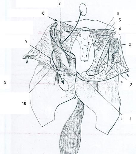
Dieudoné et al.; AJMAH, 19(5): 11-21, 2021;; Article no.AJMAH.68843

 Fig. 1. Cross anatomical section at the level of the cranial part of the epigastric region (10 th
                               thoracic vertebra) of the dog. [9]
 1. 10th thoracic vertebra. 2. Right multifidus thoracis. 3. Right longissimus thoracis muscle. 4. Right iliocostalis
 thoracis muscle. 5. Right levator costorum muscle. 6. Right latisssimus dorsi muscle. 7. Right muscle obliquus
    externus abdominis. 8. Right transverse thoracis muscle. 9. Right deep pectoral muscle. 10. Right rectus
abdominis muscle. 11. Aorta. 12. Caudal lobe of the right lung. 13. Caudal lobe of the left lung. 14. Caudal vena
              cava. 15. Liver. 16. Gall bladder. 17. Falciform ligament
                                                                ligament. 18. Right
                                                                                ght intercostal muscles

Fig. 2. Right view of the lateral abdominal wall showing the muscle transversus abdominis and
                                    muscle rectus abdominis [9]
1. Thoracolumbar fascia. 2. Aponeurotic origin of the muscle transversus abdominis. 3. Costal part of the muscle
   transversus abdominis. 4. Lumbar part of the muscle transversus abdominis. 5. Aponeurosis of the muscle
 transversus abdominis. 6. Transverse ab   abdominal
                                             dominal fascia. 7. Medial ramus of nerve costoabdominalis. 8. Medial
    ramus of the nerve iliohypogastricus cranialis. 9. Medial ramus of the nerve iliohypogastricus caudalis. 10.
  Connecting branch between the lateral ramus of nerve iliohypogastricus caudali
                                                                               caudalis s and the lateral ramus of the
nerve ilioinguinalis. 11. Lateral ramus of the nerve ilioinguinalis. 12. Lateral ramus of the nerve iliohypogastricus
 caudalis. 13. Lateral ramus of the nerve cutaneus femoris lateralis. 14. Medial ramus of the nerve ilioinguilioinguinalis.
 15. First rib. 16. Aponeurotic origin of the muscle rectus abdominis. 17. Serratus ventralis muscle. 18. Serratus
   dorsalis cranialis muscle. 19. Serratus dorsalis caudalis. 20. External intercostal muscles. 21. Intersections
  tendinae.. 22. Ilium. 23. Spermatic cord. 24.Pectin oss pubis. 25. Last rib. I. II. III. IV. V and VI. First, Second.
                 Third, Fourth, Fifth and Sixth segment of the muscl
                                                               muscle e rectus abdominis respectively

                                                           13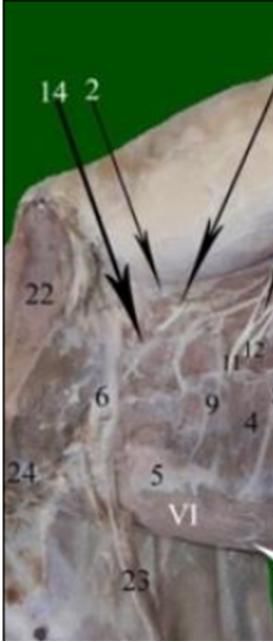
Dieudoné et al.; AJMAH, 19(5): 11-21, 2021; Article no.AJMAH.68843

2.2 External Oblique Muscle                of   the          aponeurosis, the latter directly continuing the
    Abdomen (Figs. 1 and 2)                                  former. The fleshy part is thick, especially at the
                                                             level of the chord of the flank, which contributes
The external oblique muscle of the abdomen is                to drawing the protrusion in domestic Mammals.
the most superficial and largest of the abdominal            It presents a flabelliform disposition, its most
muscles. It extends from the lateral aspect of the           cranial bundles being almost longitudinal and the
thorax and the edge of the lumbar region to the              most caudal ones following the fold of the groin.
linea alba and the groin crease, where its fascia            On the whole, its fascicles run ventro-cranially
connects to specific formations: the inguinal arch           and     their   orientation    is    approximately
and the medial thigh fascia.                                 perpendicular to that of the external oblique.
It is a flat, very wide, irregularly triangular              In carnivores, the rabbit, covers the entire flank
muscle, which comprises a dorsocranial fleshy                region. It is the same in Ruminants and Pigs, but
part and a ventrocaudal aponeurosis. The fleshy              in these species, its cranial part, which responds
part constitutes a wideband spread over the                  to the paralumbar fossa, is thinned and partially
entire surface of the hypochondrium and on the               dissociated. This dissociation is much clearer in
ventral end of the ribs (asternals and last                  Equidae, where the fleshy part, devoid of lumbar
sternals). It is formed of parallel bundles,                 attachment, leaves the para-lumbar fossa (hollow
oriented obliquely in a ventro-caudal direction.             of the flank) uncovered, in the cranial part of
Progressively widened towards its caudal part, it            which we find a small muscle completely
is divided at its cranio-dorsal edge into regular            isolated: the retractor muscle of the last rib.
serrations whose number is equal to that of the              The internal oblique muscle is an auxiliary of the
ribs, minus the first three or four. The opposite            external oblique. Like the latter, it pulls the ribs in
edge, hardly sinuous, is continued by                        a caudal direction and is involved in exhalation; it
aponeurosis. At the dorsocaudal end, the                     also tightens the abdomen and indirectly
bundles, which have become almost longitudinal,              contributes to the flexion of the spine [8,10].
extend directly from the proximal end of the last
rib and the thoracolumbar fascia to the iliac crest,         2.4 Transverse Abdominal Muscle (Figs.
the hip angle, or the inguinal region. The external
                                                                 1 and 2)
oblique muscle of the abdomen supports and
compresses the abdominal viscera. As such, it is
involved in all acts that involve muscular effort. In        The transverse abdominal muscle is flat and
addition, it pulls the ribs in a caudal direction and        wide; it occupies the deepest plane of the
thus contributes powerfully to expiration. Finally,          abdominal wall. Formed by parallel fibers that run
it contributes to the flexion of the spine, directly         dorsoventrally, following the concavity of the
or laterally, depending on whether the action is             abdominal wall (hence its name), this muscle has
bilateral or unilateral. Blood is supplied by the            a fleshy part and an aponeurosis. The fleshy part
dorsal intercostal, lumbar, iliac circumflex, and            is dorsal or dorso-cranial. It occupies the deep
cranial abdominal (Carnivores, Pigs, Rabbits)                surface of the hypochondrium, which it extends
arteries, secondarily by branches of the musculo-            caudally, then widens in the region of the flank,
phrenic, cranial and caudal epigastric arteries.             which it occupies almost entirely. Under the
The innervation is provided by the intercostal               hypochondrium, it is divided at its dorsal edge
nerves of the spaces covered by the insertions of            into serrations that correspond to the asternal
the fleshy part and by the ventral branches of the           ribs or their cartilages. The part which occupies
first two or three lumbar nerves [8,10].                     the flank begins on the contrary, at its dorsal
                                                             edge, by a short aponeurotic blade that connects
2.3 Internal Oblique Muscle                of   the          it to the lumbar region and whose extent is
    Abdomen (Figs. 1 and 2)                                  variable with the species. The most caudal part,
                                                             weakened, dissociated in thin and not very
The internal oblique muscle of the abdomen is                consistent bundles in the domestic Mammals, is
flat, almost as wide as the previous one, which              more solid in Man, as if to ensure better support
covers it completely. It radiates from the ilium             of the viscera in the erect position; it does not
bone and the edge of the lumbar region to the                reach in any case the bottom of the inguinal
last ribs, the Manche line, and down to the groin            region. The aponeurosis is shaped like an
fold.                                                        irregular triangle. Its dorsocranial border follows
                                                             the fleshy part. Its ventral border corresponds to
Molded on the flank and belly regions, this                  the white line. Its caudal border, poorly defined,
muscle comprises a fleshy part and an                        reaches the inguinal arch only near the hip and

                                                        14
Dieudoné et al.; AJMAH, 19(5): 11-21, 2021; Article no.AJMAH.68843
the very loose bundles that constitute it muscle, to which is joined a sheet which is
dissociate ventrally. detached from that of the internal oblique
opposite the lateral edge of the rectus muscle. Its
The transverse muscle tightens the abdomen: it constitution varies with the levels: the deep
lifts the viscera and presses them against the expansion of the aponeurosis of the internal
lumbar region. It also lowers the costal arches to oblique muscle exists only in the epigastric
which it attaches and acts as an auxiliary to the region or its vicinity.
expiratory muscles. Blood is supplied by
divisions of the dorsal intercostal, musculo- Through its sheath, the rectus abdominis muscle
phrenic, cranial abdominal and deep iliac responds on its ventral side to the ascending
circumflex arteries. The nerves originate from the pectoralis, to a small extent of the fleshy part of
last intercostals and the ventral branches of the the external oblique, and in the rest of its surface,
first lumbar nerves [8,10,2]. to the subcutaneous formations. Also through its
sheath, the deep or dorsal side responds to the
2.5 The Rectus Abdominis Muscle costal cartilages, the corresponding intercostal
muscles, and in the rest of its extent to the
The rectus abdominis muscle is flat, in the form transversalis fascia and the subperitoneal
of a wideband extending from the ventral aspect connective tissue. The cranial and caudal deep
of the sternum and costal cartilages to the pubic epigastric arteries and veins run along the lateral
bone. It is enlarged in the middle and much border on this side.
narrower at the caudal end, which is extended by The rectus abdominis muscle lifts and
the prepubic tendon, than at the cranial end, compresses the abdominal viscera. It also brings
which ends in a more or less long aponeurosis; the pelvis closer to the thorax and thus flexes the
this cranial part is narrowed in ungulates, but lumbar region.
enlarged in humans and carnivores. The muscle
is cut by a series of strong, regular and Finally, pulling the ribs in a caudal direction, it
transverse tendon intersections, which give it a contributes to expiration.
polygastric structure and its particular
appearance. These intersections, which evoke its Blood is supplied by the deep epigastric arteries,
metameric nature, are variable according to cranial and caudal. Innervation comes from the
individuals and even more so from one species intercostal nerves (except the first) and the
to another. There are about ten in Equidae, five ventral branches of the first three lumbar nerves.
in cattle, seven in sheep, eight to ten in pigs, four It is remarkable that despite its metamerized
or five in carnivores and three, sometimes four, appearance, the rectus abdominis muscle
in humans. These intersections are very close to receives segmental nerves in much greater
the walls of the fibrous sheath that envelops the number than the fleshy bellies that constitute it
muscle and are more or less symmetrical from [8,10,2].
one side to the other. The most caudal belly of
the muscle is always the longest and the 2.6 Retractor Muscle of the Last Rib
intersections are generally less spaced in the
The retractor muscle of the last rib or costal
part between the umbilicus and the xiphoid
retractor is a flat, triangular muscle, located in the
region.
lumbocostal angle; the orientation of its fibers
could make it related to the internal intercostal
The rectus abdominis muscle is enclosed in a system. It originates at the lateral end of the first
fibrous sheath formed by the terminal lumbar transverse processes and ends at the
aponeuroses of the oblique and transverse caudal edge or the internal face of the last rib.
muscles. This sheath is made up of two blades, Taking a fixed point at the lumbar vertebrae, it
one superficial and the other deep, which meet at pulls the latter in a caudal direction and fixes it in
the lateral edge of the muscle and meet at the this position; it initiates expiration and this action
linea alba. The aponeurotic intersections adhere seems to be transmitted to the more cranial ribs
to them on both sides, but the superficial blade is by the intervention of the internal intercostal
the most difficult to detach. This one is the muscles [3].
thickest. It is formed by the aponeuroses of the
two oblique muscles, to which a part of the 2.7 Fascia Transversalis
aponeurosis of the transversus is added
caudally. The deep blade is weaker; it is formed The transversalis fascia is a vast fibrous
in principle by the aponeurosis of the transverse expansion that covers the deep face of the
15Dieudoné et al.; AJMAH, 19(5): 11-21, 2021; Article no.AJMAH.68843
transverse abdominal muscle and extends to the 2.10 Inguinal Space Or Canal (Fig. 3)
abdominal face of the diaphragm. It begins at the
lateral border of the lumbar region, where it The inguinal space or canal is an interstice
connects to the fascia iliaca, and ends ventrally located on the side of the prepubic region and
at the linea alba. Its fibers are mainly transverse, opens on the one hand in the subcutaneous
but there are also fibers oblique or longitudinal, space of the groin and on the other hand under
especially in its ventral part. Its thickness is not the prepubic peritoneum. Very oblique in lateral
uniform. In domestic mammals and especially in and dorsal direction and rapidly narrowed
large ungulates, whose visceral support is towards its deep end, it gives way in the male to
provided differently, it is especially developed on the spermatic cord and in both sexes to
the lateral walls and opposite the rectus important vessels and nerves. Of great surgical
abdominis muscle [8,10,2]. interest, it presents two walls, two orifices and a
content.
2.8 White Line
These walls are not limited to the inguinal space
The linea alba is a narrow, odd, median fibrous itself. They belong respectively to the external
blade, extending from the xiphoid process of the oblique and internal oblique muscles of the
sternum to the cranial border of the pubic bones, abdomen which, directly leaning everywhere
where it merges with the prepubic tendon. It is a else, move away from each other at the bottom
sort of thick raphe resulting from the medial of the groin fold, near the pubis bone.
crossing of the aponeurotic fibers belonging to Very unequal and of very different constitution,
the right and left broad abdominal muscles. This these orifices are two in number: one is
very strong fibrous cord is bordered on each side subcutaneous or superficial, the other deep,
by the medial edge of the rectus abdominis abdominal.
muscle, which adheres to it and to whose sheath
it attaches. Its thickness varies according to the Superficial inguinal ring : it is an orifice
level and the species; it is generally maximal in in the shape of an elongated slit in the
the pubic region. The width also presents same direction as the groin fold, i.e. in a
variations, but these are not analogous in all the cranio-lateral direction. In the Ungulates, it
species [8,10,2]. takes on an oval shape and widens
considerably when the limb is pulled in
2.9 Prepubic Tendon abduction. It is pierced in the aponeurosis
of the external oblique muscle, slightly
The prepubic tendon is a strong quadrilateral cranial to the pubic bone, on the side of the
fibrous blade inserted at the cranial edge of the prepubic tendon.
two pubic bones, from one iliopubic eminence to
Deep inguinal ring: this is a much smaller
the other. It is complex in structure and directly
orifice than the previous one, a simple slit
extends the linea alba and the tendons of the
between the inguinal arch and the edge of
rectus abdominis muscles and receives part of
the muscle internal oblique. It is real only in
the aponeurotic fibers of the oblique and
the male, after the descent of the testicle;
transverse abdominal muscles on both sides.
in the female or the cryptorchid male, it
remains in principle virtual. Located on the
Excavated on each side, the prepubic tendon is side of the cranial strait of the pelvis,
not placed in direct prolongation of the pubic lateral to the edge of the pubis, not far from
bones, but forms with them an obtuse dihedral the tubercle of the lesser psoas, it is
open ventro-caudally. It is indeed oblique in a separated from that of the opposite side by
ventro-cranial direction, much more so in the entire width of this strait. In the female,
ungulates than in carnivores. This orientation is the fascia transversalis and the peritoneum
so strongly plunging in ungulates that the angle pass tangentially over its surface. In the
formed by the tendon and the pubic bones can male, during migration of the testis, the
reach 90°. In cattle, the insertion even extends peritoneum and transversalis fascia
far into the ventral side of the pubic bones, evaginate at this level in front of the gland,
whose cranial edge thus forms a kind of to form its deepest envelopes.
threshold between the abdominal cavity and the
pelvic floor. Knowledge of these arrangements is The contents of the inguinal space are different
important for obstetrics and surgery. [8,10,2]. in males and females. In the male, the most
16Dieudoné et al.; AJMAH, 19(5): 11-21, 2021; Article no.AJMAH.68843
important part is the spermatic cord surrounded (mammary in the Ungulates) and the branches of
by the deep or intra-inguinal testicular envelopes: the genitofemoral nerve run, moreover arranged
internal spermatic fascia, lined internally by the more or less like their counterparts in the male
parietal lamina of the serosa and covered on its [8,11].
caudo-lateral side by the cremaster muscle.
Among domestic mammals, the rabbit is an 3. REGIONS OF THE ABDOMINAL WALL
exception, the testis only descending into the
scrotum during mating and the cremaster then The ventro-lateral wall of the abdomen, whose
enveloping the fibro-serosa on all sides. The constituents we have just studied, is thus vast
spermatic cord itself includes the vas deferens, and its organization presents great local
the testicular vessels and the mesos that variations. Three major regions must be
suspend them inside the serosa. Around the cord distinguished, whose anatomical structures are
there is a lamellar connective tissue, dependent different. One is ventral and odd: it corresponds
on the external spermatic fascia, in which to the region of the "belly". The other two are
vessels and nerves that reach the subinguinal lateral and each of them is repeated on the right
region and the scrotum run in contact with the and on the left: they are the hypochondrium and
cord itself or in its vicinity: external pudendal the flank; the latter is connected to the pelvis and
artery, its satellite vein and branches of the to the root of the pelvic limb by the inguinal
genitofemoral nerve. In the female, the inguinal region [10].
space, narrower, is only occupied by connective
tissue in which the external pudendal vessels
Fig. 3. Inguinal region [12]
1: Prepubic tendon; 2: Cremaster muscle; 3: Internal oblique muscle; 4: Fascia illiaca; 5: Psoas major muscle; 6:
Tendon of the small psoas muscle; 7: Superficial inguinal ring; 8: Hip angle; 9: Superior inguinal ring; 10: The
arrow indicates the direction of migration of the testicles
17Dieudoné et al.; AJMAH, 19(5): 11-21, 2021; Article no.AJMAH.68843
3.1 Belly Region 4.1.1 Semiological indications
It extends from the xiphoid process of the Exploratory laparotomy is the name given to
sternum to the cranial edge of the pubic bones. It coeliotomy whose sole purpose is to examine the
corresponds to the subregions of the abdominal viscera on a live animal.
epigastrium, the belly proper and the
hypogastrium or prepubic region. The first of This indication is quite exceptional in large
these three subregions covers the ventral parts species. It is, on the other hand, much more
of the two costal arches and between them the frequent in small species, although its relative
xiphoid cartilage; its surface is covered by the importance tends to decrease due to the
most caudal part of the ascending pectoral development of endoscopy (laparoscopy) and
muscles and by the cranial end of the fleshy part radiology techniques.
of the external oblique muscles of the abdomen.
The hypogastric region is supported by the Exploratory laparotomy allows a rapid and
prepubic tendon in domestic mammals [10]. complete examination of all the abdominal
viscera. It very often allows for the decision to
3.2 Hypochondrial Region perform a life-saving curative operation, for
This region is indeed characterized by the example the extraction of a migrating foreign
presence of the ventral ends of the asternal ribs body causing recurrent subocclusions, or the
and all their cartilages, as well as the removal of a tumor. One should never hesitate to
corresponding intercostal muscles. It belongs perform it provided that the instrumentation is
only in appearance to the thorax: because of the available to allow the eventual completion of the
very particular mode of attachment of the specific times. Therefore, in veterinary practice,
diaphragm, it covers only abdominal viscera. It is there should be no technical difference between
remarkably extensive in cattle and pigs [10]. an exploratory laparotomy with a therapeutic aim
[12].
3.3 Flank Region
4.1.2 Therapeutic indications
This region extends from the costal arch to the
The purpose of laparotomies is to allow the
cranial edge of the thigh and from the lumbar
surgeon to intervene on the abdominal organs.
border to the belly, to which its curvature
They are the first and last operative steps in all
connects in an insensitive way. It is very long in
procedures involving the liver, spleen, kidney,
Carnivores and Rabbits, still vast in Ruminants
and urinary tract, as well as the digestive tract.
and Pigs, but very short in Equidae; in the latter,
barely a handbreadth separates the last rib from
In domestic animals, laparotomies are most often
the cranial border of the thigh. The flank is, like
performed for interventions on the reproductive
the belly, characterized by the absence of any
system, (ovariectomies, castration of cryptorchid
bony formation. It is almost exclusively
males), caesarean operations, hysterectomy.
constituted by the fleshy parts of the three flat
The treatment of abdominal wall malformations,
muscles of the abdomen: external oblique,
in particular the surgical cure of hernias, uses
internal oblique and transverse. These muscles
specific laparotomy techniques called kelotomies
are also covered, in the subcutaneous plane, by
[11,12,13].
the cutaneous muscle of the trunk, from which a
thick layer of very mobile connective tissue
separates them [7,10]. 4.2 Criterion for the Choice of the Place
of Election
4. LAPAROTOMIES
The place of choice for abdominal surgery is
Laparotomies are surgical procedures that determined by seven criteria which are based on
consist of opening the abdominal cavity. They three imperatives: safety of the patient, solidity
constitute the first and last operative steps of all and aesthetics.
abdominal surgery operations [11,12].
The chosen site must provide convenient
4.1 Indication for Laparotomies access to the organ to be operated on: this
condition is the most important of all in
It is traditional to contrast semiological terms of surgical safety.
indications and therapeutic indications for The organization of the dissection must
laparotomy. minimize parietal damage, allowing the
18Dieudoné et al.; AJMAH, 19(5): 11-21, 2021; Article no.AJMAH.68843
muscles to be incised in the direction of eventration because the viscera weigh down the
their fibers, reducing to a strict minimum incision. Intraperitoneal fluids can also get in
the sectioning of the motor nerves of the between the sutures and form collections in this
wall, which cause amyotrophy and thus area. Depending on the location of the incision, a
secondary weakening, and reducing to a distinction is made between supra-umbilical,
minimum the sectioning of the vascular umbilical, sub-umbilical, and xipho-pubic
trunks for the same reason. laparotomy (it includes all the first three),
The place of choice must allow the according to the landmarks on the white line
incisions to be enlarged in order to [11,14,15].
increase the possibilities of intervention on
the organ concerned. This criterion is 4.3.1.2 Paramedian laparotomies
particularly important in obstetrical surgery
and carcinology, disciplines where the Paramedian laparotomies are performed on a
volume of the element on which we line parallel to the midline.
intervene can present very important The advantage of this opening is its rapid healing
fluctuations. because the region is better vascularized. The
The repair must be easy to perform. solidity is good because the stitches are
The incisions must not seriously supported by solid fascia.
compromise the solidity of the walls; for It requires a period of hemostasis of the vessels;
example, opening the abdominal cavity in the haemorrhage is generally not very abundant
purely fascial areas must be avoided. In because the incision of the rectus abdominis
particular, in all species, one must refrain muscle is made in the direction of the fibers.
from intervening in the posterior zone of As the thickness of the wall to be crossed may
the flank leak, which is constituted solely be relatively large in this area, abdominal
by the overlap of the two aponeuroses of exploration is more difficult. It is no longer
the two oblique muscles of the abdomen. recommended today in domestic carnivores for
The parietal operative sinus must drain this reason [11,14,15].
easily. The accumulation of serosities in
the areas of muscle detachment facilitates 4.3.2 High laparotomies (Fig. 4)
the formation of abscesses or phlegmons
of the walls. 4.3.2.1 Oblique laparotomies
Finally, the skin wound should, as far as
possible, leave an inconspicuous scar [12]. These laparotomies can be performed along the
axes of the external oblique (sub-costo-
4.3 Anatomical Classification of abdominal or costo-lumbar) or the internal
oblique (sub-costo-alumbar). These techniques
Laparotomies (Fig. 4)
have the same advantages. Cleavage between
the muscle fibers is easy and does not cause
According to their place of choice, it is possible to hemorrhaging. Closure is easy and healing is
classify laparotomies into four groups: rapid. However, since the two muscle planes are
perpendicular, one of them must be cut to access
4.3.1 Low laparotomies (Fig. 4) the abdominal cavity.
4.3.1.1 Midline laparotomies The opening of the abdominal cavity can be
targeted on a particular organ to allow a wide
These are performed through the white line. exploration. In the first case, it is possible to limit
They have the great advantage of allowing the the approach by knowing the topography of the
pathway to be widened. The incision is limited in target organ.
front by the xiphoid process and in the back by
the pubis. In males, the skin incision is not Thus, a subcostal laparotomy allows access to
median in the caudal region but avoids the the ovaries in the bitch, and a sublumbar
sheath, while the puncture of the linea alba is laparotomy allows the removal of an adrenal
median. Post-operative recovery is rapid, the gland or a kidney. In the second case, the
scar is not very visible and solidity is ensured by exploration of the abdomen is always performed
strong fascia. after a median laparotomy along the linea alba.
In conclusion, the vast majority of operations are
On the other hand, healing is slow due to the performed along the white line. This technique is,
lack of irrigation. This route involves risks of in the end, simpler, and its possibilities of
19Dieudoné et al.; AJMAH, 19(5): 11-21, 2021; Article no.AJMAH.68843
enlargement are greater (extension on the linea Ovariectomy by the vaginal route in the
alba, addition of a subcostal laparotomy, a cow used to be very common in the
thoracotomy by sternotomy, a pelvic nineteenth century but very little
symphysiotomy). [11,12,15]. nowadays by experienced practitioners.
Inguinal hernias, consist of an incision at
4.3.2.2 Transverse laparotomies the level of the inguinal region, mostly
Transverse laparotomies are performed along encountered in horses, sheep ….
the axis of the fibres of the transverse muscle. [11,15,16,17,18,20].
They are located either in the hollow of the flank,
or cross it to extend into its recess. Transverse 5. CONCLUSION
laparotomies in the hollow of the flank (Abert's
technique) can also be performed according to The muscles constitute the walls of the
the fiber planes of the different muscles, which abdominal cavity. They contribute to maintain
allows for blunt dissection. The advantage of and protect the main digestive and urogenital
such laparotomies is that they avoid the risk of viscera and are therefore of paramount
postoperative ventrations, but they only allow importance.
very limited exploration (e.g.: oophorectomy for
convenience). In the dorsal region, it is possible The choice of one approach or another depends
to access certain organs such as the kidney or on the clinical and para-clinical assessment, but
the adrenal gland in the retroperitoneal region also largely depends on the anatomical situation
[11,15,16]. of the organ on which the surgical procedure will
take place.
4.3.2.3 Laparotomy of the flank
Laparotomy under ilio-abdominal located below A precise knowledge of the anatomy of the
the flank recess under the wing of the ilium. The abdominal wall is essential for surgeons to
most frequent are those performed at the level of understand its structure and its relationship with
the right flank chord during intestinal surgery in the surrounding organs, to know the necessary
cattle (OSTERMAN), those performed at the precautions to be taken during the laparotomy to
level of the left flank leakage during caesarean ensure a rapid recovery and healing of the
section in cows (GOFFINET-HERNAUX) or animal.
ovariectomy.
CONSENT
It is not applicable.
ETHICAL APPROVAL
It is not applicable.
COMPETING INTERESTS
Authors have declared that no competing
interests exist.
REFERENCES
1. Kapan S, Kapan M, Goksoy E, Karabicak I,
Oktar H. Comparison of ptfe pericardium
Fig. 4. The different sites of choice for bovine and fascia lata for repair of
laparotomies [19]. incisional hernia in rat model, experimental
1: Median laparotomy; 2: Paramedian Laparotomy; 3:
study. Hernia. 2003;7:39-43.
Transversal laparotomy; 4: Sub-costal laparotomy
2. Chatelain E. Arthrology and myology of the
trunk. Handout from the envl anatomy
4.3.3 Laparotomy by natural route (Fig. 4)
laboratory. 1991;53-57.
These are laparotomies performed at very 3. Schumpelick V, Junge K, Klinge U, Conze
specific locations in the abdomen. J. Incisional hernia: pathogenesis,
20Dieudoné et al.; AJMAH, 19(5): 11-21, 2021; Article no.AJMAH.68843
presentation and treatment. Deutsche 12. Sevestre J. Conduct of operating times in
ärtzblatt. 2006:103(39):A2553-8. elements of animal surgery - biological
4. Conze J, Presher A, Klinge U, Saklak M, bases and perioperative anesthesia-
Schumpelick V. Pitfallas in retromuscular resuscitation techniques. Ed. The
mesh for incisional hernia : the importance veterinary point. 1981;39-81.
of the “fatty triangle”. Hernia 2004 ; 8 : 255- 13. Garnier E. Exploratory laparotomy in dogs
9. and cats, le point vétérinaire.
5. Gutiérrez De La Pena C, Medina Achirica 2002;N°223:56.
C, Dominguez-adame E, Medina Diez J. 14. Kouakou Deassath HB. Retrospective
Primary closure of laparotomies with high study of clinical cases of domestic
risk of incisional hernia using prosthetic carnivores seen in medical consultation at
material: analysis of useless. Hernia. the EISMV in Dakar from 2005 to 2010,
2003;7:134-6. Thesis: Med. vet: Dakar; 2010.
6. Hoer J, Klinge U, Schachtrupp A, Tons C, 15. Neu JC. The abdominal wall of the dog:
Schumpelick V. Influence of suture anatomy, topography, access route. Audio-
technique on laparotomy wound healing : visual illustration. Veterinary doctorate
an experimental study in the rat. thesis. Claude Bernard University of Lyon;
Langenbeck’s arch surg 2001;386:218-23. 1994.
7. Hobeika J, Houdart R. EVENTRATIONS. 16. Duhautois B. Practical guide to soft tissue
Notice d’informations medicales du groupe surgery in dogs and cats. Paris, Ed.
hospitalier diaconesses croix saint simon; Med'com. 2005;605.
2005. 17. Hsiao W-C, Young KC, Wang ST, Lin PW.
Available:http://www.hopital- Incisional hernia after laparotomy:
dcss.org/soins-services- Prospective randomized comparison
hopital/informationsmedicales/item/145- between early resorbable and late-
eventrations.html resorbable suture materials. World J. Surg.
8. Ellenberger W, Baum H. Descriptive and 2000;24:747-52.
topographical anatomy of the dog. c. 18. Fossog Tine FX. Assessment of the
Reinwald; 1894. demand and cost of additional analyzes in
9. Hickman J, Houlton J, Edwards B. An atlas private veterinary clinics in the Dakar
of veterinary surgery. 3rd ed: b blackwell region, THESIS: MED. VET: DAKAR;
science. 1995;275. 2008.
10. Barone R. Comparative anatomy of 19. Claude Pavaux. Techniques of dissection
domestic mammals, volume 2 / arthrology of domestic animals. National Veterinary
and myology. Vigot, Paris. 1989;647-671. School of Toulouse; 1975.
11. Dickele G, Cazieux A. Surgery course, 20. Moissonnier P, Degueurce C, Bougault S.
volume 2: Special techniques, organ Exploratory laparotomy in dogs. Ed.
surgery. National veterinary school of Kallanxis. 2008;164.
Toulouse; 1976.
_________________________________________________________________________________
© 2021 Dieudoné et al.; This is an Open Access article distributed under the terms of the Creative Commons Attribution
License (http://creativecommons.org/licenses/by/4.0), which permits unrestricted use, distribution, and reproduction in any
medium, provided the original work is properly cited.
Peer-review history:
The peer review history for this paper can be accessed here:
http://www.sdiarticle4.com/review-history/68843
21You can also read