Case Report Endometriosis Associated Striated Muscle Changes: Simulating Tumor
←
→
Page content transcription
If your browser does not render page correctly, please read the page content below
Hindawi
Case Reports in Pathology
Volume 2021, Article ID 6666283, 4 pages
https://doi.org/10.1155/2021/6666283

Case Report
Endometriosis Associated Striated Muscle Changes:
Simulating Tumor

          Rusella Mirza , Jehan Abdulsattar, and James Cotelingam
          Department of Pathology and Translational Pathology, Louisiana State University Health Science Center, Shreveport, LA 71103, USA

          Correspondence should be addressed to Rusella Mirza; rmirza@lsuhsc.edu

          Received 24 November 2020; Revised 6 January 2021; Accepted 22 January 2021; Published 2 February 2021

          Academic Editor: George Adonakis

          Copyright © 2021 Rusella Mirza et al. This is an open access article distributed under the Creative Commons Attribution License,
          which permits unrestricted use, distribution, and reproduction in any medium, provided the original work is properly cited.

          Endometriosis in the abdominal wall can be a diagnostic challenge, both clinically and pathologically. We are describing a case of
          abdominal wall endometriosis where regenerative muscle cells are florid. The cells are small and polygonal in shape, arranged in
          groups, and intersecting through differentiated skeletal muscle. Cells have pink cytoplasm and a centrally located nucleus.
          Immunohistochemistry demonstrates diffuse positivity with S100 and CD56, and they are negative with Ae1/Ae3, Pax8, inhibin,
          calretinin, and CD68. Cells are also focally positive with actin, vimentin, and myogenin. Electron microscopy reveals the
          presence of intracytoplasmic myofibrils. These groups of small cells are compatible with early to intermediately differentiated
          skeletal muscle cells as demonstrated by histomorphology, immunohistochemical pattern, and ultrastructure analysis. Muscle
          undergoes a regenerative process following injury. Endometriosis associated regenerative changes in muscle are very rare as
          documented in the English literature. The presence of florid regenerative muscle cells mimics a neoplastic process. In this
          report, we describe the histomorphological and immunohistochemical pattern of regenerative muscle groups and discuss
          differential diagnoses. We found electron microscopy to be an important diagnostic tool in this case.

1. Introduction                                                         endometriosis-associated skeletal muscle regeneration posed a
                                                                        diagnostic dilemma [7]. Limited number of articles docu-
Endometriosis is defined as the presence of the functional               mented endometriosis-associated skeletal muscle change. In
endometrial gland and stroma at an ectopic location. Most               this article, we are demonstrating florid regenerative muscle
common locations are the ovary, broad ligament, fallopian               changes mimicking neoplasm. We present the histomorphol-
tube, and nearby pelvic organs. Extrapelvic endometriosis is            ogy, immunohistochemistry, and electron microscopy of endo-
very rare, especially at the anterior abdominal wall location           metriosis associated regenerative muscle cells and some helpful
[1–3]. Patients with endometriosis at the abdominal wall                features to distinguish it from other potential differentials.
usually present with a painful mass which is suspected as
hernia or neoplasm [4]. Most of the time, the patient has a             2. Case Presentation
history of cesarean section and sometimes has a history of
endometriosis at another location. The mass is usually pres-            A 36-year-old female presented with an anterior abdominal
ent for a couple of years, and the patient complains of cyclic          wall mass, present for last six months. She experienced
or periodic pain [4]. Endometriosis at the abdominal wall can           intermittent pain and recent enlargement of the mass. She
be a diagnostic dilemma for both clinicians and pathologists.           had a history of uterine rupture status post hysterectomy 15
Endometriosis is a benign process; however, malignant trans-            years ago. Ultrasonography demonstrated bilateral adnexal
formation may occur [5, 6]. Endometriosis in the skeletal               masses. She has no history of cancer and denied any recent
muscle occasionally is associated with tissue destruction               unintentional weight loss. Core needle biopsy was performed
and repair. The histomorphological and immunohistochem-                 of the abdominal mass which was indeterminate due to scant
ical properties of regenerative muscle cells make the diagno-           amount of tissue retrieved. Due to persistent symptoms and
sis challenging in those cases. Colella et al. demonstrated that        suspicion of neoplasm, resection of the mass was done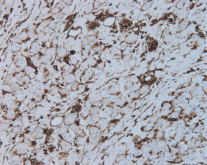
2 Case Reports in Pathology
⁎
(a) (b)
(c) (d)
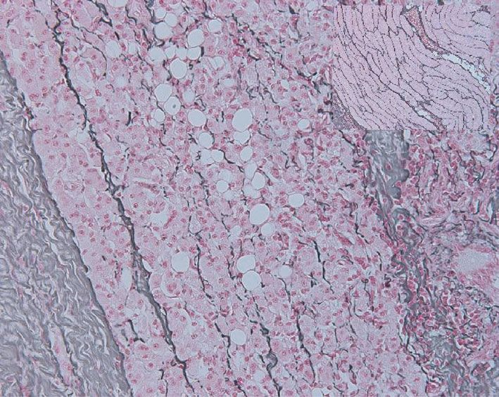
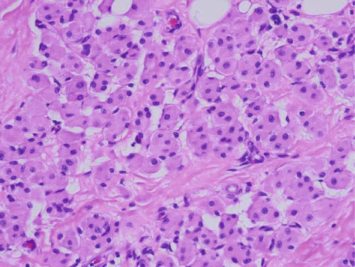
Figure 1: (a) Endometrial glands and stroma on the left (arrow) and multiple groups of small polygonal cells intervening the mass (∗) are
demonstrated. (b) The cells are round to polygonal with distinct cell border and round single to multiple nuclei. (c, d) Those cells are
negative with CD68 and Pax8 antibody, respectively.
followed by reconstruction with mesh. The mass measured expression of actin is noted in nearby differentiated muscle
8 × 7 × 3 cm and weighted 150 grams. Sectioning revealed a cells (Figure 2(c)). Myogenin shows focal cytoplasmic stain-
stellate-shaped tan-white lesion grossly infiltrating the skele- ing without any nuclear positivity (Figure 2(d)). Vimentin
tal muscle and adipose tissue. Representative sections were demonstrates membranous positivity, and no cytoplasmic
examined under a microscope. Immunohistochemical stains positivity is noted in those cells (Figure 2(e)). The reticulin
were performed with AE1/AE3, CD68, EMA, MART-1, stain demonstrated a strong pericellular stain in the
S100, inhibin, PAX8, calretinin, vimentin, myogenin, and differentiated skeletal muscle as shown in the inbox; how-
muscle-specific actin with a VENTANA automated stainer. ever, the small cells lack the staining pattern (Figure 2(f)).
A reticulin stain was also performed. Appropriate controls Figures 3(a) and 3(b) demonstrate the EM pictures of those
were used for each stain. Light microscopy pictures were small cells with cytoplasmic myofibrils.
taken with a Lumera Infinity3 camera. Electron microscopy
(EM) was performed in our facility with a JEOL electron 3. Discussion
microscope.
Hematoxylin and eosin (HE) stain reveals multiple foci of Our patient has bilateral adnexal masses and is now present-
endometrial glands, stroma, macrophages, and lymphocytes. ing with an enlarging abdominal wall mass which is clinically
Besides that, multiple groups of small polygonal pink cells are suspicious for neoplasm, metastatic carcinoma, or endome-
intervening in between skeletal muscles which are strikingly triosis. Grossly, the lesion was infiltrative through the muscle
different from those of surrounding macrophages and skele- and adipose tissue which can be seen in both neoplasm and
tal muscles. These cells are round to oval with distinct cell endometriosis. Microscopically, multiple endometrial glands
borders and have low nuclear to cytoplasmic ratio. The were present suggesting endometriosis. However, the infil-
nucleus is round, single, or multiple and centrally located trating groups of polygonal cells were a dilemma. Macro-
(Figures 1(a) and 1(b)). Immunohistochemistry with CD68 phages and other inflammatory cells are observed around
and PAX8 are negative (Figures 1(c) and 1(d), respectively). the ectopic endometrial tissue. But the suspicious cells were
Figure 2 demonstrates that the cells are strongly positive with different than histiocytes and were negative for CD68. Cells
CD56 and S100. The well-differentiated skeletal muscles are are arranged in groups, round to polygonal with a smooth
negative with both CD56 and S100 as shown in the inbox distinct cell border. The nuclei are small round, single, or
(Figures 2(a) and 2(b)). Actin shows mild positivity in those multiple. Our differential diagnoses were granular cell tumor,
small cells which are shown with black arrows, and stronger steroid cell tumor, luteinized granulosa cell tumor, oxyphilic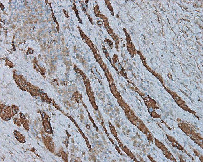
Case Reports in Pathology                                                                                                                   3

                                            (a)                                                (b)

                                            (c)                                                (d)

                                            (e)                                                (f)

Figure 2: The immunohistochemical property of the cell groups is demonstrated: (a) CD56 is strongly positive, and there is progressive loss of
CD56 in the differentiated muscle cells (inbox); (b) S100 detected with red chromogen which is strongly expressed in those small cell groups,
and progressive loss of S100 is noted in the differentiated skeletal muscle (inbox); (c) actin is mildly positive in those cells (arrows) and
strongly positive in nearby intermediate to well-differentiated muscle cells; (d) myogenin shows few cytoplasmic positivity to negative
stain; (e) no strong cytoplasmic positivity is noted with vimentin; (f) reticulin stain shows no pericellular stain which is evident in
well-differentiated skeletal muscle (inbox).

variant of clear cell tumor of ovary, and degenerated muscle             and cells had low nuclear to cytoplasmic ratio which
cells. The granular cell tumor is reported at the anterior               prompted consideration of a benign entity such as regener-
abdominal wall with granular cytoplasm and diffuse positiv-               ative muscle cells.
ity with S100 and CD68 [8]. The cytoplasm of our cells is                     Muscle cells undergo a regenerative process following
pink, but not granular, strongly positive with S100, and                 injury [7, 9]. The myoblast differentiates to intermediate
negative with CD68. Calretinin and EMA are also negative                 and fully differentiated skeletal muscle. Each stage has dis-
in these cells which are sometimes positive with a granular              tinct histomorphology and immunohistochemical proper-
cell tumor. AE1/AE3, MART-1, PAX8, inhibin, and calreti-                 ties. The less differentiated muscle cells react with CD56,
nin were negative in these cells which ruled out other                   S100, vimentin (strong cytoplasmic), desmin, myogenin,
potential differentials. No mitosis or necrosis was noted,                and myo-D1 but not with myoglobin or p21. Intermediately,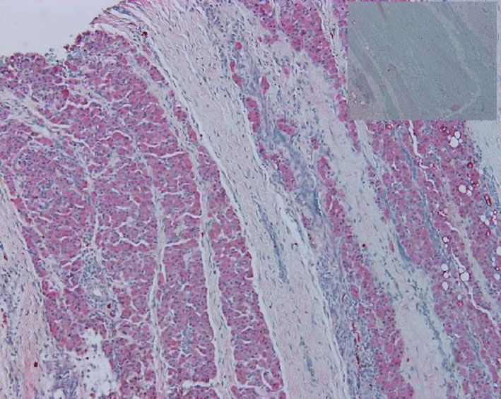
4 Case Reports in Pathology
⁎
⁎
(a) (b)
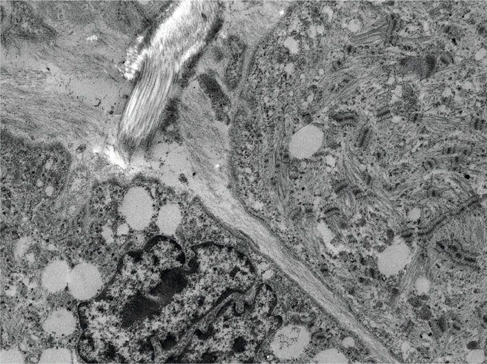
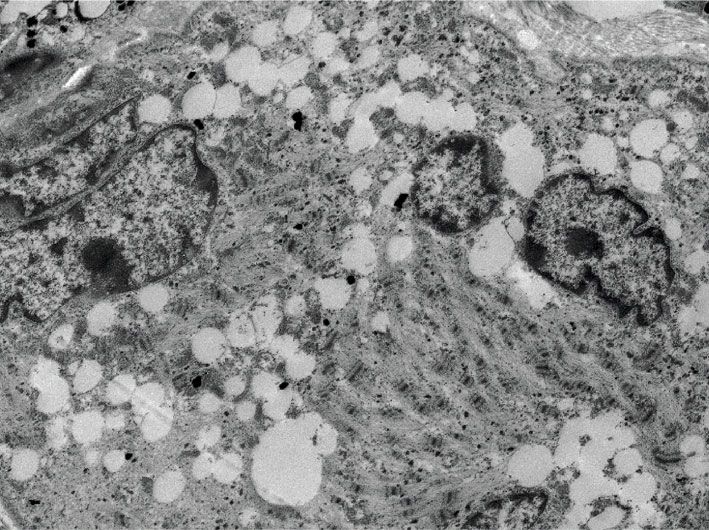
Figure 3: (a, b) Electron microscopy of those small cells reveals multiple nuclei with prominent nucleoli (red asterisks) and myofibrils (yellow
arrows). Scale bar is 2 μM and 1 μM for (a, b), respectively.
differentiated cells progressively lose vimentin, CD56, and Conflicts of Interest
myo-D1, whereas they are positive with S100, myogenin,
myoglobin, and p21. Terminally differentiated cells react The authors declare that they have no conflicts of interest.
with desmin and myoglobin [7]. In our case, skeletal muscles
close to the ectopic endometrial tissue demonstrated a regen-
erative process. These cells express CD56 and S100 strongly References
and focally positive with myogenin and do not have the cyto-
plasmic positivity with vimentin (Figure 2). Electron micros- [1] C. Saliba, H. Jaafoury, M. El Hajj, G. Nicolas, and H. H.
copy demonstrated abundant small myofibrils in the Ahmad, “Abdominal wall endometriosis: a case report,” Cur-
cytoplasm. Myofibrils are short, haphazardly arranged, and eus, vol. 11, no. 2, 2019.
are not arranged as long filaments yet. Multiple euchromatic [2] H. Karaman, F. Bulut, and A. Ozasalamaci, “Endometriosis
nuclei with prominent nucleoli were observed. These features externa within the rectus abdominis muscle,” Turkish Journal
are usually seen in regenerative muscle cells at the early stage of Surgery/Ulusal cerrahi dergis, vol. 30, no. 3, pp. 165–168,
of their differentiation [10]. These groups of small cells are 2014.
early to intermediately differentiated skeletal muscle cells as [3] M. Anand and S. D. Deshmukh, “Massive abdominal wall
demonstrated by histomorphology, immunohistochemical endometriosis masquerading as desmoid tumor,” Journal of
pattern, and ultrastructure analysis. Terminally differentiated cutaneous and aesthetic surgery, vol. 4, pp. 141–143, 2011.
muscle cells were located away from the endometrial tissue [4] H. Bektas, Y. Bilsel, Y. s. Sari et al., “Abdominal wall endome-
and demonstrate progressive loss of CD56 and S100 expres- trioma; a 10-year experience and brief review of the literature,”
Journal of Surgical Research, vol. 164, 2010.
sion (Figure 2).
Our study together with previous report highlights the [5] K. Vagholkar and S. Vagholkar, “Abdominal wall endome-
importance of understanding the histology and immunohis- trioma: a diagnostic enigma—a case report and review of the
literature,” Case report in Obstetrics and Gynecology,
tochemistry of regenerative muscle cells. The regenerative
vol. 2019, pp. 1–4, 2019.
changes can be focal or can be florid [7]. The latter is true
in our case. Endometriosis associated regenerative myoblast [6] A. Kocakusak, E. Arpinar, S. Arikan, N. Demirbag, A. Tarlaci,
and C. Kabaca, “Abdominal wall endometriosis: a diagnostic
proliferation is rare. We have encountered 18 cases of endo-
dilemma for surgeons,” Medical Principles and Practice,
metriosis at the abdominal wall location in our institution in vol. 14, no. 6, pp. 434–437, 2005.
the last five years. And only one case demonstrated florid
[7] R. Colella, M. G. Mameli, G. Bellezza, R. Del Sordo,
regenerative muscle groups. Because of its rarity and confus-
A. Cavaliere, and A. Sidoni, “Endometriosis-associated skeletal
ing immunohistochemical property, this could be a potential muscle regeneration: a hitherto undescribed entity and a
diagnostic pitfall. Clinical history is helpful in most of the potential diagnostic pitfall,” The American journal of surgical
cases. Abdominal wall endometriosis sometimes occurs after pathology, vol. 34, 2010.
a cesarean section or pelvic surgery, and it has an incidence of [8] J. Lee, N. W. McGhan, S. W. Young, J. M. Collins, and A. E.
0.03%-1.5% in women with previous cesarean delivery [1]. McCullough, “Granular cell tumor of anterior abdominal
Endometriosis should be considered when women present wall,” Radiology Case Reports, vol. 7, no. 3, p. 716, 2012.
with intermittent or cyclic abdominal pain. Our patient’s his- [9] S. Bodin-Fowler, “Skeletal muscle regeneration after injury: an
tory of ruptured uterus, bilateral adnexal mass, and intermit- overview,” Journal of voice, vol. 8, no. 1, pp. 53–62, 1994.
tent pain at the abdominal mass fits with symptoms of [10] C. A. Sewry, “Electron microscopy of human skeletal muscle:
endometriosis. role in diagnosis,” Current Diagnostic Pathology, vol. 8, no. 4,
In conclusion, endometriosis associated regeneration of pp. 225–231, 2002.
muscle cells can be florid and mimic neoplastic cells morpho-
logically and immunohistochemically. Electron microscopy
is helpful in challenging cases.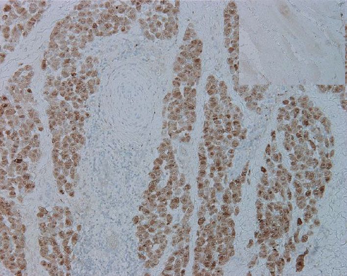
You can also read