Effects of Ultrasound and Stretching on Skeletal Muscle Contusion in Rats: Immunohistochemistry Analysis - SciELO
←
→
Page content transcription
If your browser does not render page correctly, please read the page content below
Int. J. Morphol.,
38(5):1288-1295, 2020.

       Effects of Ultrasound and Stretching on Skeletal Muscle
           Contusion in Rats: Immunohistochemistry Analysis

                                 Efectos de Ultrasonido y Estiramiento en la Contusión
                                 Muscular Esqueletal en Ratas: Análisis Inmunohistoquímico

                      Ana Carolina Brandt de Macedo1; Julye Leiko Ywazaki2; Ana Paula Camargo Martins3;
                        Marina Luise Viola de Azevedo3; Lucia Noronha4 & Anna Raquel Silveira Gomes1,5

DE MACEDO, A. C. B.; YWAZAKI, J. L.; MARTINS, A. P. C.; AZEVEDO, M. L. V.; NORONHA, L. & GOMES, A. R. S. Effects
of ultrasound and stretching on skeletal muscle contusion in rats: immunohistochemistry analysis. Int. J. Morphol., 38(5):1288-1295,
2020.

         SUMMARY: The aim of this study was to evaluate the effects of stretching and therapeutic ultrasound (TUS) on desmin and
laminin contents of rat muscle after contusion. Male Wistar rats (n = 35, 8–9 weeks of age, 271 ± 14g body weight) were divided into five
groups: Control group (CG) (n= 03); Injured group (IG) (n= 8); Injured + ultrasound group (IUSG) (n= 8); Injured+stretching group
(ISG) (n= 8); Injured +ultrasound + stretching group (IUSSG) (n= 8). The application of ultrasound started 72 hours after the contusion,
using the 50 % pulsed mode, 0.5 W/cm2, 5 min, once a day, for five consecutive days. Passive manual stretching was started on the tenth day
after injury, with four repetitions of 30 s each and 30 s rest between repetitions, once a day, five times per week, for a total of ten applications.
After 22 days, the rats were euthanazied and the gastrocnemius of both limbs removed for desmin and laminin immunohistochemistry
morphometric measurement. Analysis was conducted using ANOVA one way post-hoc Tukey to parametric data and Kruskall-Wallis for
non-parametric data. The IUSSG animals showed a larger area of desmin than ISG (p
DE MACEDO, A. C. B.; YWAZAKI, J. L.; MARTINS, A. P. C.; AZEVEDO, M. L. V.; NORONHA, L. & GOMES, A. R. S. Effects of ultrasound and stretching on skeletal muscle contusion in
                                                rats: immunohistochemistry analysis. Int. J. Morphol., 38(5):1288-1295, 2020.

cells and the extracellular matrix, but also the                                          a distinct event (Zar, 1998). To control for unseen
neovascularization and adhesion of the myofibers to the                                   circumstances or possible losses, two additional animals
extracellular matrix, indicating the importance of proteins                               were added to each experimental group. The animals were
involved in this mechanism such as desmin and laminins                                    divided into five groups, housed four per cage and maintained
(Wilkin et al., 2004; Piedade et al.; Rantanen et al.; Shu et                             in standard plastic cages under controlled environmental
al.; Cação-Benedini et al., 2014; Gianello et al., 2016).                                 conditions (luminosity: 12 h light/dark cycle), with free
                                                                                          access to water and pelleted feed, in the vivarium of the
         It has been reported that physiotherapy program was                              Integrated Faculties of Brazil (Unibrasil). The project was
more effective when stretching exercises was added to re-                                 carried out according to the international norms of ethics in
duce time to return to sport after skeletal muscle injury (Pas                            animal experimentation.
et al., 2015).
                                                                                                  The animals (n=35) were divided into five groups: a
        Experimental animal models contribute to                                          control group (CG, n=3); an injured group (IG, n=8); an
understanding the effects of lengthening stimulus, such as                                injured and ultrasound group (IUSG, n=8); an injured and
stretching, in the cellular mechanisms of skeletal muscle                                 stretching group (ISG, n=8); and an injured and ultrasound
healing after contusion (Hwang et al., 2006; de Macedo et                                 and stretching group (IUSSG, n=8) (Fig. 1).
al., 2014, 2016). The involvement of desmin and laminin in
force transmission has been described in the literature (De                                      No samples were lost from any of the experimental
Deyne, 2001). An increase in sarcomerogenesis and an                                      groups. After completing the experimental protocols, on Day
antifibrotic effect after a stretching protocol of injured rat                            22 the animals were anesthetized and the gastrocnemius
gastrocnemius has been reported (de Macedo et al., 2014,                                  muscle was removed from both limbs, and then the muscles
2016). Although the proteins desmin and laminin involved                                  were weighed, and processed for the morphometric and
in the sarcomerogenesis and muscle healing, have not yet                                  immunohistochemistry desmin and laminin analysis.
been investigated (Yu & Thornell, 2002; Cação-Benedini et
al.; Gianelo et al., 2016).                                                               Muscle injury. The animals were anesthetized with an
                                                                                          intraperitoneal injection of Ketamine (95 mg/kg) and
        The markers desmin and laminin are involved in the                                Xylazine (12 mg/kg) and maintained in a prone position
cellular mechanisms of skeletal muscle healing and force                                  with the right hind limb manually immobilized and the knee
transmission in response to TUS or stretching (Piedade et                                 extended. The contusion was produced on the mid belly of
al.; Cação-Benedini et al.; Gianelo et al.). Nevertheless, the                            the right gastrocnemius muscle (RGM) as described by
literature did not explain how desmin and laminin act after                               Minamoto et al. (2001). The leg was shaved, the location
muscle contusion treated with a combination of TUS and                                    for the projectile to fall over the muscle was defined with a
stretching. Thus, our hypotheses were: 1) The TUS would                                   marker, and the ink refreshed daily to guarantee that the
increase desmin and laminin contributing to skeletal muscle                               TUS applications were applied over the same area.
healing; 2) The stretching might strengthen the impact that
TUS could have on the skeletal muscle regeneration. Hence,                                Therapeutic ultrasound protocol. The animal was
the present study aimed to evaluate the effects of ultrasound                             positioned in a supine position with the anterior limbs
combined or not with stretching on injured gastrocnemius                                  immobilized by one physiotherapist. To apply the TUS by
of young rats, in desmin and laminin content through                                      the other physiotherapist, the right hind limb was positioned
immunohistochemistry.                                                                     with the knee extended and ankle dorsiflexed at a 90-degree
                                                                                          angle. Ultrasound was applied over the belly of the medial
                                                                                          part of the RGM, with the treatment area previously shaved
MATERIAL AND METHOD                                                                       and marked with a marker pen. The animal was not under
                                                                                          anesthesia. The effective radiation area of the probe was 1
                                                                                          cm 2. The equipment used was the Sonopulse especial
Animals. After approval by the ethics committee in animal                                 (Ibramed, Brazil), at a frequency of 1MHz, intensity of 0.5
experimentation (CEEA) of Federal University of Paraná                                    W/cm2 spatial average temporal peak (SATP) (Piedade et al.),
(Certificate Number 491/2010), 35 young (8–9 weeks old,                                   a pulsed duration of 50 % duty cycle, which corresponded to
271±14 g weight) albino Wistar rats were selected. To de-                                 0.25 W/cm2 spatial average temporal area (SATA), with an
termine the sample size, a minimum of six animals per ex-                                 application time of 5 minutes and using a total energy of 75J.
perimental group was considered, as in a homogeneous                                      A commercially available gel was used as a coupling agent
population, such as laboratory animals, a minimum sample                                  (São Paulo Institute, Brazil). The equipment calibration was
size of six represents a 16 % opportunity per animal to present                           guaranteed by the manufacturer. The treatment of IUSG and
                                                                                                                                                                      1289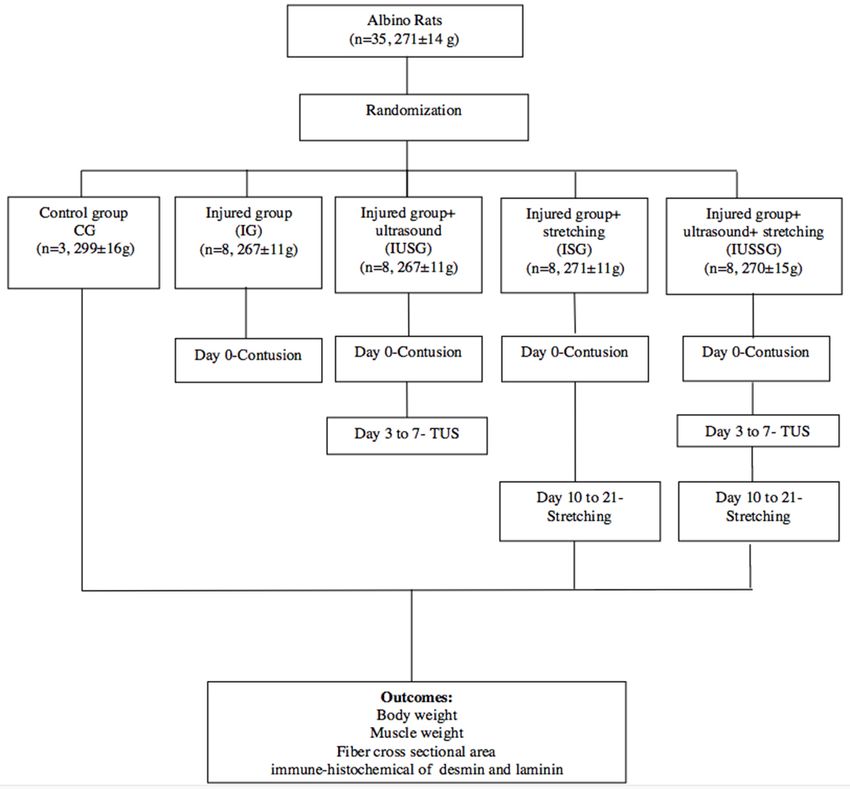
DE MACEDO, A. C. B.; YWAZAKI, J. L.; MARTINS, A. P. C.; AZEVEDO, M. L. V.; NORONHA, L. & GOMES, A. R. S. Effects of ultrasound and stretching on skeletal muscle contusion in
                                                rats: immunohistochemistry analysis. Int. J. Morphol., 38(5):1288-1295, 2020.

                                                                                                            Euthanasia of the animals and removal of
                                                                                                            the muscles. The animals from all the groups
                                                                                                            were weighed and anesthetized with
                                                                                                            intraperitoneal injections of Ketamine (95 mg/
                                                                                                            kg) and Xylazine (12 mg/kg) to excise the
                                                                                                            gastrocnemius muscles. The muscles were
                                                                                                            periodically sprayed with a saline solution (0.9
                                                                                                            % NaCl) to avoid drying out the tissue during
                                                                                                            dissection. Following the muscle excision, and
                                                                                                            while still under anesthesia, the animals were
                                                                                                            euthanized with an overdose of Ketamine and
                                                                                                            Xylazine anesthetics.
                                                                                                                   Each muscle was weighed separately
                                                                                                            using an analytical scale (Bioprecisa, Brazil)
                                                                                                            and divided at the middle into lateral and medial
                                                                                                            portions. The medial portion was then divided
                                                                                                            longitudinally into two parts, right and left, for
                                                                                                            analysis: The right portion was used to other
                                                                                                            analysis and the left part was fixed in a 10 %
                                                                                                            formalin solution for subsequent morphometric
                                                                                                            (muscle fiber cross sectional area) and
                                                                                                            immunohistochemistry analysis.
Fig. 1. Fluxograma of the study.

                                                                                          Fiber cross sectional area (FCSA). The cross-sectional
IUSSG consisted of, one application per day for five                                      areas of 100 muscle fibers were measured for each
consecutive days, at the same time of day, starting at the 3rd                            gastrocnemius muscle, chosen at random from the region of
day after contusion.                                                                      the muscle belly of the histological section, as described by
                                                                                          Coutinho et al. (2004). The photomicrography was done
Gastrocnemius muscle stretching protocol. The animals                                     through a light microscope (BX50-Olympus®, Brazil) (x20
were placed in the supine position to stretch the RGM, and                                objective) and the software Pro Plus 4.0 image was used to
the anterior limbs were immobilized by one physiotherapist                                measure the muscle fiber cross-sectional area.
while the other carried out the passive RGM manual
stretching. The animal was not under anesthesia during this                               Immunohistochemistry. The medial part (left portion) of the
procedure. Maximum ankle dorsal flexion was carried out                                   belly of the gastrocnemius muscle was fixed in 10 % formalin
manually, with the knee extended, as described by                                         for 72 hours and then embedded in paraffin. After fixing, 4
Mattiello-Sverzut et al. (2006), considering the limit of the                             µm slices were cut from the block to make a new multi-sample
joint. The stretching protocol was started on Day 10 and                                  block, also known as the Artisanal Tissue Microarray. These
maintained for 30 seconds per repetition (4 repetitions),                                 blocked samples were cut with a microtome (Leica RM 2145,
with 30 seconds of rest between each repetition (Hwang et                                 Brazil) at 4 µm and affixed on the histological slides
al.), once a day, five times per week from Monday to Friday,                              (76X26mm AutoWrite Green AdesinSakura, Brazil). After
for two weeks, for a total of ten applications up to Day 21                               drying the slides overnight in an incubator at 60 ºC (Orion–
of the experiment. The duration in seconds of each repetition                             model 502, Lynd, Wirral, England) and deparaffinizing, the
and the rest periods were monitored using a stopwatch                                     antigen pre-treatment was started. The endogenous peroxidase
(Technos YP21518P, Technos, Brazil).                                                      (Streptavidin-Peroxidase, Brazil) was blocked with a
                                                                                          hydrogen peroxide solution diluted in 5 % methanol. The
Chronological groups interventions. Pulsed ultrasound was                                 slides were incubated in desmin (Dako, Brazil) and laminin
applied during the inflammatory phase of the lesion, starting                             (Dako, Brazil) antibody solution overnight at 4 ºC. The stain
72 hours after the contusion for 5 consecutive days (Plentz                               DAB (Diaminobenzidine, Brazil) (1:1) was used until the slide
et al., 2008), considering the protocol described by Rantanen                             contents appeared brown in color, and then destained using
et al. The stretching protocol was applied between 10th to                                Harris hematoxiline (Alpha Tec, Brazil). If positive, the
21stdays, because 10 days after muscle injury, cells show                                 desmin and laminin antibodies can stain intermediary filament
high level of regeneration (Hwang et al.).                                                and basal lamina brown, respectively.
1290
DE MACEDO, A. C. B.; YWAZAKI, J. L.; MARTINS, A. P. C.; AZEVEDO, M. L. V.; NORONHA, L. & GOMES, A. R. S. Effects of ultrasound and stretching on skeletal muscle contusion in
rats: immunohistochemistry analysis. Int. J. Morphol., 38(5):1288-1295, 2020.
The Image Pro Plus® program was used to read the considered non-parametric if assumptions of normality and/
laminin and desmin antibodies with the aid of a Dino-eye® or homogeneity of variance were not met.
camera and an optical microscope (BX50-Olympus®,
Brazil). The between groups comparisons were carried out
through one-way ANOVA and post-hoc Tukey tests for the
A photomicrograph of a positive control slide was parametric data and Kruskal-Wallis tests for the non-
made using a magnification of 400x. A sample of the brown parametric data, considering a 5 % significance level (p<
coloration for the program was used as a positive control 0.05, two-tailed). Parametric and non-parametric data are
to allow the quantification of desmin and laminin by the expressed as the mean standard deviation and median,
colorimetric method. This photomicrograph was considered minimum, maximum. Statistical analysis was performed
the “mask” (control slide) as reported by Calvi et al. (2012), using SPSS 25.0.
made from a specific structure demarcated into an image.
After selecting a specific structure, the program identified
the structure’s pixels, and then the software selected RESULTS
adjacent pixels with similar color.
A field 200 x 200 µm2 in size containing only muscle There was an increase in body weight in the IUSG
fibers was selected from the histological slide using the and IG animals compared to the ISG and IUSSG, also in
Image Pro Plus program to quantify the areas of the desmin the CG animals in comparison to the ISG and IUSSG
and laminin proteins. The “mask” was superimposed on each animals (ANOVA, p< 0.05). The gastrocnemius muscle of
histological slide photographed, and the program the IUSG animals was heavier than that of the ISG. The
automatically read the totality of the immune-positive brown IUSSG relative muscle wet weight-to-body weight was
color area previously standardized in the field photographed, greater than IG animals (Kruskall-Wallis, p< 0.05). The IG
thereby quantifying the desmin or laminin areas. samples cross-sectional area (FCSA) was larger than the
ISG samples (12787±995 µm 2 vs 8721±2341 µm 2,
Initially ten photomicrographs were taken of the ANOVA, p = 0.01). The data are described in Table I.
right gastrocnemius muscle of each sample in each block,
that is, from each rat in each group. The five images The area of desmin content in the IUSSG samples
containing the fewest artifacts and greatest number of fibers was larger than that found in the ISG samples (Kruskal-
were selected. Artifacts included the following: reagent Wallis, p< 0.02, Table II). Figure 2 shows the desmin (brown
deposits, dust, bent fibers, torn fibers, or badly focused color) more pronounced in the IUSSG samples.
fibers. After reading the five selected images, the arithmetic
mean of the measured area from each slide was calculated. The results obtained for the laminin area in the
The program automatically read the totality of the immune- muscle showed normal distribution and homogeneity
positive area in µm2 identifying the brown color previously (Shapiro-Wilks and levene respectively, p>0.05). It was
standardized. found a decrease in laminin comparing IUSG to IG.
Nevertheless, laminin area in ISG was higher than all the
Statistical Analysis. The Shapiro-Wilks and Levene tests groups (ANOVA, p0.05). The data were pronounced in the ISG samples.
Table I. Body weight, gastrocnemius muscle weight, relative muscle wet weight-to-body-weight and FCSA in all experi-
mental groups.
Group Final Body we ight (g) Gastrocnemius wet weight (g) MW/BW (mg/g) FCSA (µm2 )
IG 350.0±17.9* 1.7±0.1 5.0 12787±995¥
IUSG 352.2±19.1* 1.8±0.2# 4.9 10819±2612
ISG 323.±17.6§ 1.5±0.3 4.9 8721±2341
IUSSG 317.7±15.8§ 1.7±0.1 5.6† 10300±924
CG 383.3±24.5 1.8±0.1 4.7 13814±2904
IG, injured group; IUSG, injured+ultrasound group; ISG, injured+stretching group; IUSSG, injured+ultrasound+stretching group; CG,
control group; MW, muscle weight; BW, body weight; FCSA, fiber cross sectional area. All data are expressed as the mean±standard
error mean.*compared to ISG and IUSSG (pDE MACEDO, A. C. B.; YWAZAKI, J. L.; MARTINS, A. P. C.; AZEVEDO, M. L. V.; NORONHA, L. & GOMES, A. R. S. Effects of ultrasound and stretching on skeletal muscle contusion in
rats: immunohistochemistry analysis. Int. J. Morphol., 38(5):1288-1295, 2020.
DISCUSSION
The stretching applied isolated enhanced
gastrocnemius regeneration indicated by the
increase in lamina area. The combination of TUS
and stretching strengthened the muscle healing
as shown in the rise of desmin area.
Differences in body weight among expe-
rimental groups were noticed, in particular, in
the stretched muscle. For instance, the animals
subjected to stretching intervention gained
approximately 17–19 % body weight compared
to the injury or ultrasound groups, which gained
approximately 31–32 % body weight. Compared
to the final body weight among the injury groups,
a decrease of approximately 8–10 % was found
Fig. 2. Effects of stretching and/or in the animals receiving the stretching therapy
ultrasound in desmin area of gastrocnemius compared to those in the injury or ultrasound
muscle in rats. A. Injured group (IG); B.
groups. As the CG animals had the highest final
Injured and stretching group (ISG); C.
body weight, suggesting that exposure to a range
Injured and ultrasound group (IUSG); D.
Injured and ultrasound and stretching group of stimuli, including contusion, ultrasound, and
(IUSSG); E. Control group (CG). Arrow stretching, may have negatively impacted the
indicates desmin. Bar 30 µm for all panels. absolute body weight gain.
Regarding FCSA it was found that the IG
was larger than that in the ISG muscle, although
there was no difference with the control group.
The FCSA difference may have been influenced
by the increase in body weight, although no
increase in muscle weight was detected. In this
study, ultrasound and stretching produced no
changes in FCSA. Markert et al. (2005) evaluated
the effects of TUS and low-intensity walking
exercise for 20 minutes on treadmill at a velocity
of 14 m/min for 4 days, starting 24 hours after
mechanical injury of the gastrocnemius, and
found no increase in FCSA. As IG muscle had a
greater FCSA than ISG muscle, stretching may
have interfered with edema reduction and
regeneration, but was not sufficient to stimulate
radial or transverse muscle growth, as reported
in other studies (Rantanen et al.; Market et al.,
Fig. 3. Effects of stretching and/or 2005).
ultrasound in gastrocnemius muscle
laminin area of rats. A. Injured group (IG); In the present study, the injured
B. Injured and stretching group (ISG); C.
gastrocnemius muscle treated by both ultrasound
Injured and ultrasound group (IUSG); D.
Injured and ultrasound and stretching group and stretching showed a larger area of desmin
(IUSSG); E. Control group (CG). than by stretching isolated or just ultrasound.
Rectangle indicates laminin (present in the Other studies found an increase in immune-
whole circumference of the cell). Bar 30 reactivity of the desmin, and in the number of
µm for all panels. myoblasts and myotubes when muscle submitted
1292DE MACEDO, A. C. B.; YWAZAKI, J. L.; MARTINS, A. P. C.; AZEVEDO, M. L. V.; NORONHA, L. & GOMES, A. R. S. Effects of ultrasound and stretching on skeletal muscle contusion in
rats: immunohistochemistry analysis. Int. J. Morphol., 38(5):1288-1295, 2020.
Table II. Effects of stretching and/or therapeutic ultrasound in the desmin and laminin area of rat right gastrocnemius
muscle.
Desmin area (µm_) Laminin area (µm_)
Group Mean±SD (median, minimum, maximum) Mean±SD (median, minimum, maximum)
IG 1746.1±766.7 (1835.7, 802.8, 3154.9) 279.6±76.0 (217.7, 187.4, 378.1)
IUSG 1858.9±781.0 (1686.5, 908.0, 3428.2) 163.4±27.1 (163.3, 115.4, 214.6)§
ISG 1549.5±513.2 (1481.7, 1019.6, 2679.2) 498.5±81.7 (473.5, 362.3, 594.6)#
IUSSG 2960.7±1113.7 (3332.8, 1521.8, 4593.0)* 223.9±39.7 (220.1, 155.2, 276.1)
CG 2585.8±924.5 (2081.9, 1775.8, 3593.0) 216.2±39.0 (199.0, 173.7, 250.6)
IG, injured group; IUSG, injured+ultrasound group; ISG, injured+stretching group; IUSSG, injured+ultrasound+stretching
group; CG, control group; SD, standard deviation. All data were expressed as the mean±standard deviation (median; minimum-
maximum). *pDE MACEDO, A. C. B.; YWAZAKI, J. L.; MARTINS, A. P. C.; AZEVEDO, M. L. V.; NORONHA, L. & GOMES, A. R. S. Effects of ultrasound and stretching on skeletal muscle contusion in
rats: immunohistochemistry analysis. Int. J. Morphol., 38(5):1288-1295, 2020.
population of stem cells that can differentiate into muscle laminin a2, specific for skeletal muscle. Further studies
cells or other types of cell (Lieber). When activated, satellite should investigate the effects of stretching in the acute phase
cells migrate to the injured region, proliferate and of muscle injury, and also evaluate other glycoproteins such
differentiate into myoblasts, and then unite to form myotubes. as integrins, collagen IV, and fibronectin, and use molecular
Studies have shown that both ultrasound (Lieber; Hwang et biology analysis to demonstrate the mechanisms involved
al.) and stretching, by way of its mechanical effect (Tatsumi in the skeletal muscle regeneration process.
et al., 2001), stimulate satellite cells.
The stretching effect may complement TUS as
Thus it can be proposed that the increase in desmin reported by other studies which have shown that TUS applied
resulting from the association of TUS with stretching may to injured muscle tissue displayed a better structural
have enhanced the muscle regeneration process by activating arrangement with a more regular alignment of collagen muscle
the satellite cells. In the present study the satellite cells were fibers, and that stretching induced antifibrotic and regenerative
not marked, although we noticed centralized nucleus in the effects (Market et al., 2005; Hwang et al.; Piedade et al.).
histological analysis, data not shown in this manuscript, but The present study has demonstrated larger areas of desmin in
it could indicate its activation. The present authors suggest IUSSG tissue when compared to ISG tissue and larger laminin
marking the satellite cells in a future study to reveal the area in ISG than other groups suggesting enhanced skeletal
effects of TUS and stretching on the muscle regeneration muscle regeneration. However, better treatment evidence
mechanism. would exist if a statistical difference had been found between
the control or injured groups and treatments in desmin area.
The present study showed that ISG groups present The present data are insufficient to indicate pulsed TUS
higher laminin amount area than other groups. When a application to muscle lesions without restriction, as the optimal
muscle is stretched, it transmits the force to a set of structures repair of striated muscle requires not only morphologic
located in the extra milieu, such as laminin (Cação-Benedini investigation, but functional tests which were not conducted.
et al.). Laminins play important roles in tissue structure and Stretching can be used after 10 days of contusion for
maintenance, cell signaling, adhesion, migration and it is promoting muscle healing.
important for the success of regeneration, for the formation
and spatial orientation of the new myotubes, and for
minimizing the development of fibrosis (Durbeej, 2010). The CONCLUSIONS
laminins can contribute to tensile strength in adults, and serve
as a scaffold to orient regenerating myotubes after muscle
damage. Stretching is a technique capable of inducing The results of our study indicated an improvement
myofibrillogenesis, by lengthening longitudinally the in the muscle regeneration in different ways. When both
costameric structures, intermediate filaments, contractile ultrasound and stretching therapies were applied reinforced
proteins and extracellular matrix elements (Coutinho et al.). the muscle healing by increasing desmin area. Although
De Deyne reported that passive stretch is transmitted from stretching applied isolated improved gastrocnemius
the endomysium (extracellular matrix), across the muscle regeneration detected by rising laminin area.
fiber membrane (sarcolemma), to noncontractile elements
(costamere, like desmin) to the Z. line (laminin, fibronectin)
have the capacity to bind integral membrane proteins DE MACEDO, A. C. B.; YWAZAKI, J. L.; MARTINS, A. P. C.;
(integrins, dystroglycan complex). Although our study found AZEVEDO, M. L. V.; NORONHA, L. & GOMES, A. R. S. Efec-
an increase in laminin area in the group treated with tos del ultrasonido y estiramiento en la contusión muscular esqueletal
stretching, it was not possible to observe the same response en ratas: Análisis inmunohistoquímico. Int. J. Morphol., 38(5):1288-
in the group that associated ultrasound and stretching or that 1295, 2020.
applied only ultrasound. Rantanen et al. study showed that
RESUMEN: El objetivo de este estudio fue evaluar los efec-
ultrasound after muscle injury promoted the satellite cell tos del estiramiento y la ecografía en los contenidos de desmina y
proliferation phase of the myoregeneration but not affected laminina del músculo de rata después de la lesión. Ratas Wistar ma-
myotube production inside the basal lamina, market by cho (n = 35, 8–9 semanas de edad, 271 ± 14 g de peso corporal) se
laminin. Therefore it is possible that the previous application dividieron en cinco grupos: grupo de control (CG) (n = 03); Grupo
of ultrasound and its mechanical effect is not sufficient to lesionado (GL) (n = 8); Lesionado + grupo de ultrasonido (LGU) (n
increase the production of laminin. = 8); Lesionado +grupo de estiramiento (LGE) (n = 8); Lesionado +
ultrasonido + grupo de estiramiento (LUGE) (n = 8). La aplicación
de ultrasonido comenzó 72 horas después de la lesión, usando el modo
Our study was limited by using a general pulsado al 50 %, 0,5W / cm2, 5 min, una vez al día, durante cinco
immunohistochemistry technique for laminin and not using días consecutivos. El estiramiento manual pasivo se inició el décimo
1294DE MACEDO, A. C. B.; YWAZAKI, J. L.; MARTINS, A. P. C.; AZEVEDO, M. L. V.; NORONHA, L. & GOMES, A. R. S. Effects of ultrasound and stretching on skeletal muscle contusion in
rats: immunohistochemistry analysis. Int. J. Morphol., 38(5):1288-1295, 2020.
día después de la lesión, con cuatro repeticiones de 30 seg cada una y Lieber, R. L. Skeletal Muscle Structure, Function and Plasticity. 2nd ed.
30 seg de descanso entre repeticiones, una vez al día, cinco veces por Philadelphia, Lippincott Willians & Wilkins, 2002.
semana, para un total de diez aplicaciones. Las ratas fueron sacrifi- Markert, C. D.; Merrick, M. A.; Kirby, T. E. & Devor, S. T. Nonthermal
ultrasound and exercise in skeletal muscle regeneration. Arch. Phys. Med.
cadas después de 22 días, y se extrajo el músculo gastrocnemio de
Rehabil., 86(7):1304-10, 2005.
ambos miembros para la medición morfométrica de desmina y Mattiello-Sverzut, A. C.; Carvalho, L. C.; Cornachione, A.; Nagashima, M.;
laminina a través de inmunohistoquímica. El análisis se realizó utili- Neder, L. & Shimano, A. C. Morphological effects of electrical stimulation
zando ANOVA unidireccional Tukey post-hoc para datos paramétricos and intermittent muscle stretch after immobilization in soleus muscle.
y Kruskall-Wallis para datos no paramétricos. Los animales LUGE Histol. Histopathol., 21(9):957-64, 2006.
mostraron un área mayor de desmina que LGE (pYou can also read