COSMETIC - The Venkat Center
←
→
Page content transcription
If your browser does not render page correctly, please read the page content below
lww             14/9/18             14:58         4 Color Fig(s): F1-11         Art: PRS-D-18-00447

                                                                                                 COSMETIC

      Clinical Implications of Gluteal Fat Graft
      Migration: A Dynamic Anatomical Study
      Daniel A. Del Vecchio, M.D.
                                               Background: The intraoperative mortality and overall complication rate for
         Nathaniel L. Villanueva,
                                               gluteal augmentation with fat transplantation is unacceptably high. The cur-
                             M.D.
                                               rent controversy among experts regarding safety is whether fat should be
                Raja Mohan, M.D.               placed within the gluteus muscle or limited to only the subcutaneous space.
              Bret Johnson, M.D.               The purpose of the present study was to test the hypothesis that under certain
                 Dinah Wan, M.D.               pressures, fat injected within the gluteal muscle can actually migrate out of the
       Aniketh Venkataram, M.D.                muscle and into a deeper plane containing critical neurovascular structures,
AQ1          Rod J. Rohrich, M.D.              by means of the process of deep intramuscular migration.
          Boston, Mass.; Dallas, Texas; and    Methods: A total of eight human cadaver dissections were performed. Four
                          Bangalore, India     hemibuttocks were selected for intramuscular fat injection. The patterns of
                                               subfascial fat migration were evaluated in three of these hemibuttocks by direct
                                               visual inspection and in one hemibuttock by endoscopic evaluation. Four other
                                               hemibuttocks were selected for subcutaneous or suprafascial fat injection.
                                               Results: Proxy fat was found to migrate through the muscle and into the deep
                                               submuscular space with each intramuscular injection. With subcutaneous in-
                                               jection, no proxy fat was found during dissection in the intramuscular septae
                                               or submuscular space.
                                               Conclusions: The intramuscular insertion of fat, which up to this point has
                                               been considered reasonable to perform in the superficial muscle and even
                                               recommended in the literature, is now deemed to be an inexact and risky sur-
                                               gical technique. This technique, because of the migratory nature of injected
                                               fat, should be avoided from further use in fat transplantation to the gluteal
                                               region. (Plast. Reconstr. Surg. 142: 00, 2018.)

      D
              espite having the highest annual growth                         Surgeons who have experienced complications
              rate of any cosmetic surgical procedure, the               of sciatic nerve injury in their own patients as a result
              intraoperative mortality rate for gluteal aug-
      mentation with fat transplantation (i.e., Brazilian
                                                                           Disclosure: Dr. Rohrich receives instrument royal-
      butt lift) is unacceptably high.1–7 A recent survey
                                                                           ties from Eriem Surgical, Inc., and book royalties
      performed by the American Society of Aesthetic
                                                                           from Thieme Medical Publishing. He is a clinical
      Plastic Surgeons estimates the intraoperative death
                                                                           and research expert for Allergan, Inc., and MTF Bi-
      rate from this operation to be approximately one
                                                                           ologics, and the owner of Medical Seminars of Texas,
      in 2351.1 In addition to these fatal complications,
                                                                           LLC. No funding was received for this article. Dr. Del
      serious nonfatal complications, namely microfat
                                                                           Vecchio is a founder of Surgistem Technologies, LLC,
      embolism and sciatic nerve injury, are also surpris-
                                                                           a device company involved in fat transplantation,
      ingly high.8,9 The root cause of these complications
                                                                           receives royalties from Microaire, and is a founding
      focuses on the depth of fat insertion.1,4–6 The current
                                                                           member of Peninsula Partners, LLC, a consulting
      controversy among experts is whether fat should
                                                                           firm in the plastic surgery sector. The remaining au-
      be placed within the gluteus muscle or whether fat
                                                                           thors have no financial interest to declare in relation
      should be limited to the subcutaneous space only,
                                                                           to the content of this article.                               AQ8
      which lies superficial to the muscle fascia.7,10–31

       From Back Bay Plastic Surgery; the Department of Plastic            Supplemental digital content is available for
       Surgery, University of Texas Southwestern Medical Center;           this article. Direct URL citations appear in the
       the Dallas Plastic Surgery Institute; and the Venkat Center         text; simply type the URL address into any Web
       for Skin and Plastic Surgery.                                       browser to access this content. Clickable links
       Received for publication February 13, 2018; accepted July
                                                                           to the material are provided in the HTML text
       11, 2018.
       Copyright © 2018 by the American Society of Plastic Surgeons        of this article on the Journal’s website (www.
                                                                           PRSJournal.com).
       DOI: 10.1097/PRS.0000000000005020

                                                         www.PRSJournal.com                                                          1
  Copyright © 2018 American Society of Plastic Surgeons. Unauthorized reproduction of this article is prohibited.
lww         14/9/18       14:58           4 Color Fig(s): F1-11         Art: PRS-D-18-00447

                                                       Plastic and Reconstructive Surgery • November 2018

  of gluteal fat transfer insist that fat was injected only   Four other hemibuttocks were selected for subcu-
  within the muscle and not below it.8 Similarly, sur-        taneous or suprafascial fat injection.
  geons who have experienced patient death from
  pulmonary fat embolism insist that their gluteal fat        Intramuscular Fat Injection
  injections remained only in the subcutaneous tis-                Direct Inspection of Subfascial Fat Migration
  sue and superficial muscle. Nevertheless, such dev-              On three hemibuttocks, the posterior superior
  astating complications continue to be seen with the         iliac crest, the sacral hiatus, and the greater tro-
  Brazilian lift, with a reported rate of 1.7 percent for     chanter were palpated. Drawing the letter A from
  sciatic nerve injuries alone.4 The fact that these mor-     the structures, the approximate location of the
  bidities and mortalities continue to be reported at
  unacceptably high rates despite seemingly superfi-
  cial fat injections suggests that another mechanism
  of injury other than direct cannula hits of major glu-
  teal vessels and nerves may be playing a role.
       In this study, we introduce the phenomenon of
  deep intramuscular migration, wherein large vol-
  umes of fat injected into the gluteus muscle have
  the potential to migrate along the path of least resis-
  tance within the muscle and, ultimately, even out of
  the muscle in the setting of exceedingly high pres-
  sures. This process describes subjacent fat migra-
  tion to danger zones deep to the gluteal muscle
  leading to potential pressure-induced injuries of
  the sciatic nerve or traction-induced tears of major
  venous structures in the region. This in turn may
  explain conditions of sciatic nerve impairment and
  pulmonary fat embolism after the Brazilian lift pro-
  cedure despite surgeon insistence that injection was
  performed in a more superficial plane.
       The purpose of this study was to test the
  hypothesis that under certain pressures, fat
  injected in the gluteal muscle can actually migrate
  out of the muscle into a deeper plane containing
  critical neurovascular structures, by means of the
  process of deep intramuscular migration.

            MATERIALS AND METHODS
       A total of eight dissections were performed in
  four fresh human cadavers obtained with permis-
  sion from the Willed Body Program at the Univer-
  sity of Texas Southwestern Medical Center. The
  cadavers were selected randomly to be in one of
  three groups. Four hemibuttocks were selected
  for intramuscular fat injection. The patterns of
  subfascial fat migration were evaluated in three of
  these hemibuttocks by direct visual inspection and
  in one hemibuttock by endoscopic evaluation.

      By reading this article, you are entitled to claim
      one (1) hour of Category 2 Patient Safety Credit.
      ASPS members can claim this credit by logging
      in to PlasticSurgery.org Dashboard, clicking
      “Submit CME,” and completing the form.                  Fig. 1. (Above) Landmarks, (center) dissection zone, and (below)
                                                              posterior gluteus fascia.

  2
Copyright © 2018 American Society of Plastic Surgeons. Unauthorized reproduction of this article is prohibited.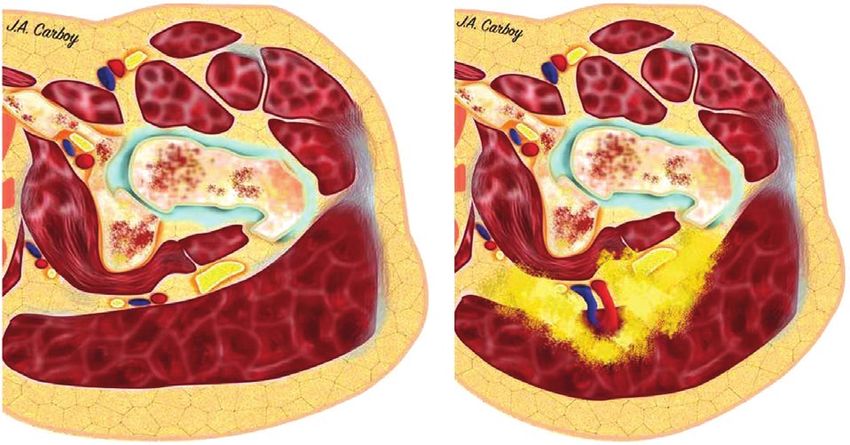
lww         14/9/18       14:58          4 Color Fig(s): F1-11       Art: PRS-D-18-00447

     Volume 142, Number 5 • Gluteal Fat Graft Migration

     superior gluteal and inferior gluteal vascular leashes   used previously was placed beneath the skin, and
F1   were marked (Fig. 1, above). A 10-cm-diameter circle     proxy fat loaded into 60-ml syringes was injected
     encompassing these vascular structures was then          into a subcutaneous space, directly between the
     marked on the skin (Fig. 1, center). Dissection of the   estimated location markings of the superior and
     skin and subcutaneous tissue was performed, reveal-      inferior gluteal vessels, and superficial to the glu-
     ing the superficial gluteal fascia (Fig. 1, below).      teus maximus muscle fascia. With each progressive
          Using a 4-mm Luer-lock multihole injection          injection of 60-cc increments, intramuscular recip-
     cannula (Lipo, Farmingdale, N.Y.), applesauce            ient-site pressures were measured using a Stryker
     (Motts, Inc., a division of Dr. Pepper Snapple,          compartment pressure monitor (Fig. 2, above) and            F2
     Plano, Texas) was stained using blue food color-         the location of the cutaneous contour change was
     ing as a proxy for transplanted fat. This “proxy         recorded as the distance from the visible “migra-
     fat” was injected into the superficial intramuscular     tion front” to the injection epicenter (Fig. 2, below).
     space of the gluteus maximus muscle using 60-ml          At the completion of the subcutaneous injection,
     syringes. Care was taken to avoid cannula passage        gross inspection of subcutaneous fat migration was
     deeper than 2 cm, keeping the cannula tip visibly        performed by dissecting the subcutaneous layer
     in the superficial muscle throughout the course of       from the superficial gluteal fascia. Finally, the glu-
     the injection. With each progressive 60-cc syringe       teus maximus muscle was reflected laterally from
     injection, intramuscular recipient-site pressures        its sacral origin to inspect for the presence of fat
     were measured using a Stryker compartment pres-          migration within the gluteus maximus muscle or
     sure monitor (Stryker, Inc., Kalamazoo, Mich.).          into the deep submuscular space in proximity to
          The gluteus muscle was then reflected later-        the superior and inferior gluteal vascular systems.
     ally off of its sacral origin to expose the subjacent
     musculature and venous plexus of the superior            Anatomical Dissection
     gluteal and inferior gluteal vascular systems, and           Using a traditional “pages of a book” dissec-
     to inspect for the location of fat migration. The        tion, skin and subcutaneous tissue of one hemibut-
     deep surface of the gluteus muscle was examined          tock was reflected inferiorly, followed by gluteus
     for the presence of demonstrable fascia.                 maximus muscle, exposing the submuscular areolar
         Endoscopic Inspection of Intramuscular Fat           space, the gluteal vessels, the sciatic nerve, and the
     Migration                                                sciatic notch. (See Video, Supplemental Digital Con-        V1

         A 3-cm horizontal incision was made in the           tent 1, which demonstrates anatomical dissection.
     upper outer quadrant on one hemibuttock.                 The detailed anatomy deep to the gluteus maximus
     Using digital dissection, the most cephalad por-         muscle is shown. There is a robust posterior fascia
     tion of the gluteus medius was encountered.              overlying the gluteus maximus muscle but there is
     More distally, the cephalad border of the glu-           no clinically apparent anterior fascia. The location
     teus maximus was identified and the submuscu-            of the gluteal vessels is highlighted in this dissection,
     lar space was navigated using a 10-mm 0-degree           available in the “Related Videos” section of the full-
     endoscope (Karl Storz, Charlton, Mass.). An              text article on PRSJournal.com or, for Ovid users,
     endoscopic breast system including a fiberoptic          available at http://links.lww.com/PRS/D46.)
     camera and an endoscopic breast retractor was
     used to expose the superior gluteal and infe-                                  RESULTS
     rior gluteal vascular bundles. The proxy fat was
     again injected into the superficial intramuscular        Direct Inspection of Subfascial Fat Migration
     space of the gluteus maximus muscle using 60-ml              Subfascial insertion of proxy fat volumes
     syringes. Direct real-time inspection of the sub-        ranged from 540 ml to 720 cc. With each 60 ml               AQ2
     muscular space was monitored for the appear-             of proxy fat inserted, visual expansion of the but-
     ance of migrated proxy fat.                              tock volume was observed. As larger volumes were
                                                              inserted, expansion of buttock projection was
     Subcutaneous Fat Injection                               observed from the perisacral area all the way to
         Direct Inspection of Subcutaneous                    the greater trochanter, consistent with the shape
     (Suprafascial) Fat Migration                             of the gluteus maximus muscle (Fig. 3). With each           F3
         On four hemibuttocks, a 10-cm-diameter circle        progressive 60 ml of proxy fat inserted, intramus-
     in the same location as shown in Figure 1, above         cular compartment pressures increased progres-
     was drawn. Using a percutaneous needlestick, the         sively, as shown in Table 1. Of note, during one            T1
     same 4-mm Luer-lock multihole injection cannula          of the injections, there was a sudden drop in

                                                                                                                     3
 Copyright © 2018 American Society of Plastic Surgeons. Unauthorized reproduction of this article is prohibited.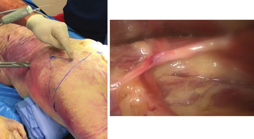
lww        14/9/18       14:58              4 Color Fig(s): F1-11                Art: PRS-D-18-00447

                                                         Plastic and Reconstructive Surgery • November 2018

                            Fig. 2. (Above) Initiation of subcutaneous injection. O represents
                            the epicenter of proxy fat insertion. (Below) After 500 cc of proxy
                            fat, a contour change representing the maximum migration front
                            (MF) is identified and its distance from the epicenter O is measured.

                            Video 1. Supplemental Digital Content 1 demonstrates anatomical
                            dissection. The detailed anatomy deep to the gluteus maximus mus-
                            cle is shown. There is a robust posterior fascia overlying the gluteus
                            maximus muscle but there is no clinically apparent anterior fascia. The
                            location of the gluteal vessels is highlighted in this dissection, avail-
                            able in the “Related Videos” section of the full-text article on PRSJour-
                            nal.com or, for Ovid users, available at http://links.lww.com/PRS/D46.

  4
Copyright © 2018 American Society of Plastic Surgeons. Unauthorized reproduction of this article is prohibited.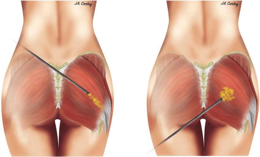
lww         14/9/18         14:58             4 Color Fig(s): F1-11              Art: PRS-D-18-00447

      Volume 142, Number 5 • Gluteal Fat Graft Migration

                                     Fig. 3. Proxy fat placed in the central portion of the gluteus muscle
                                     (green arrow) results in submuscular proxy fat emanating near the
                                     origin of the gluteus maximus muscle.

                                     Fig. 4. Deep intramuscular migration. Proxy fat (upper arrow)
                                     escaping between muscle fibers of the gluteus maximus muscle to
                                     enter the deep muscular space in the area of the superior gluteal
AQ3                                  vessels (lower arrow).

      injection resistance followed by a decrease in pres-                    When the gluteus maximus muscle was released
      sure. On further volume injection, the pressure                     from its sacral origin, a large rush of proxy fat was
      began to rise once more.                                            noted to emanate from the submuscular space, in
                                                                          an area far medial and deep to the original can-
                                                                          nula insertion (Fig. 4). [See Video, Supplemental       F4,V2
      Table 1. Subfascial Migration*                                      Digital Content 2, which demonstrates the migra-
      Volume Injected (cc)                       Pressure (mmHg)          tion of intramuscular fat in gluteal augmentation.
      60                                                  48              The landmarks to identify the location of the glu-
      120                                                 55              teal vessels are first shown. Underneath the subcu-
      180                                                 55              taneous layer, the posterior fascia over the gluteus
      240                                                 65
      300                                                 70              maximus muscle is observed. Applesauce (pseu-
      360                                                 79              dofat) is injected into the superficial fascia of the
      420                                                 87              gluteus maximus muscle. The pressure within the
      480                                                 92
      540                                                 95              submuscular space increased with each injection.
      600                                                117              The pseudofat migrated underneath the muscle
      660                                                121              and spread throughout the submuscular space.
      720                                                128
      780                                                112              Because of the lack of anterior fascia on the glu-
      *Changes in intramuscular pressure with increasing injection        teus maximus muscle, the pseudofat migrated
      volume.                                                             deep to the muscle and surrounded the gluteal

                                                                                                                             5
  Copyright © 2018 American Society of Plastic Surgeons. Unauthorized reproduction of this article is prohibited.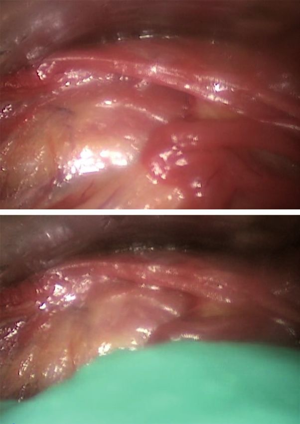
lww         14/9/18        14:58              4 Color Fig(s): F1-11              Art: PRS-D-18-00447

                                                               Plastic and Reconstructive Surgery • November 2018

                                   Video 2. Supplemental Digital Content 2 demonstrates the migra-
                                   tion of intramuscular fat in gluteal augmentation. The landmarks to
                                   identify the location of the gluteal vessels are first shown. Under-
                                   neath the subcutaneous layer, the posterior fascia over the gluteus
                                   maximus muscle is observed. Applesauce (pseudofat) is injected
                                   into the superficial fascia of the gluteus maximus muscle. The
                                   pressure within the submuscular space increased with each injec-
                                   tion. The pseudofat migrated underneath the muscle and spread
                                   throughout the submuscular space. Because of the lack of anterior
                                   fascia on the gluteus maximus muscle, the pseudofat migrated
                                   deep to the muscle and surrounded the gluteal vessels, available
                                   in the “Related Videos” section of the full-text article on PRSJournal.
                                   com or, for Ovid users, available at http://links.lww.com/PRS/D47.

       vessels, available in the “Related Videos” section                was noted to well up in the submuscular space,
       of the full-text article on PRSJournal.com or, for                consistent with an intramuscular septal “blowout”
       Ovid users, available at http://links.lww.com/PRS/                and subsequent entrance of proxy fat into the sub-
       D47.] When the gluteus maximus muscle was                         muscular space (Fig. 8). (See Video, Supplemental         F8
       reflected laterally to expose the gluteal vessels,                Digital Content 3, which demonstrates the migra-
       proxy fat was noted to occupy the space beneath                   tion of intramuscular fat in gluteal augmentation.
       the gluteus maximus muscle, tracking all the way                  After injection of pseudofat into the superficial fas-
       to the greater trochanteric insertion. In addi-                   cia of the gluteus maximus muscle, we show that the
       tion, fat was noted to occupy intermuscular sep-                  pseudofat migrates deep and is deposited within
F5     tal spaces within the muscle itself (Fig. 5). Finally,            the submuscular space. The pseudofat enters deep
       during inspection of the undersurface of all glu-                 to the muscle through multiple areas within the
       teus maximus muscles in the study, there was no                   muscle. Using an endoscope, we demonstrate that
       evidence of a deep fascial layer on the undersur-                 these injections of pseudofat into the superficial fas-
F6     face of the gluteus maximus muscle (Fig. 6).                      cia of the muscle migrated into the space deep the
                                                                         muscle. The posterior fascia of the gluteus maximus
       Endoscopic Inspection of Intramuscular Fat                        muscle acts as a backstop and prevents migration of
       Migration                                                         pseudofat into the subcutaneous space, available in
           During endoscopic inspection of the undersur-                 the “Related Videos” section of the full-text article
       face of the gluteus muscle, it was possible to safely             on PRSJournal.com or, for Ovid users, available at
       enter the deep submuscular space by means of                      http://links.lww.com/PRS/D50.)
       a cephalad approach and to identify the superior
       and inferior gluteal vessels that closely coincided               Direct Inspection of Subcutaneous (Suprafascial)
F7     with the topographic cutaneous landmarks (Fig. 7).                Fat Migration
       During simultaneous endoscopic inspection and                         During subcutaneous insertion of 500 to 1500
       real-time intramuscular injection of proxy fat, fat               cc of proxy fat in 60-cc increments, intramuscular

       6
     Copyright © 2018 American Society of Plastic Surgeons. Unauthorized reproduction of this article is prohibited.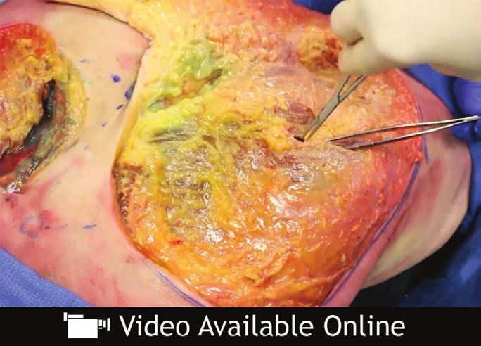
lww         14/9/18        14:58             4 Color Fig(s): F1-11              Art: PRS-D-18-00447

     Volume 142, Number 5 • Gluteal Fat Graft Migration

                              Fig. 5. Intramuscular fat migration. Fat was noted to occupy inter-
                              muscular septal spaces within the muscle itself.

                              Fig. 6. Lack of anterior gluteal fascia. Note that on the deep or ana-
                              tomically “anterior” side of the gluteus maximus muscle there is no
                              fascial layer.

     compartment pressures did not change (remained                  recipient-site connective tissue, internal expan-
     at 0). As the volume of proxy fat increased in                  sion of the subcutaneous space is impossible, as
     the subcutaneous space, topographic contour                     is the case when using traditional syringe-based
     change, as represented by a visible migration                   injection techniques that simply “wedge” fat in as
     front, increased in dimension and subcutane-                    microdroplets.35 Thus, the intramuscular space
     ous pressures increased to as high as 55 mmHg.                  has been traditionally favored as a recipient site
     Postinjection dissection of the subcutaneous                    for gluteal fat transfer. However, since the incep-
     space revealed proxy fat, which freely traversed                tion of expansion vibration lipofilling, which cre-
     throughout the subcutaneous tissue. Neither the                 ates intraoperative expansion of the subcutaneous
     gluteus maximus intramuscular septae nor the                    space by means of mechanical disruption with
     deep submuscular space subjacent to the gluteus                 internal caged reciprocating cannulas, there has
     maximus muscle revealed any evidence of disrup-                 been less reliance on the intramuscular space as
F9   tion or the presence of proxy fat (Fig. 9).                     the only recipient site capable of accepting rela-
                                                                     tively large fat volumes.36 Expansion vibration lipo-
                       DISCUSSION                                    filling can potentially increase the capacity of the
         Intramuscular fat grafting in the gluteal region            subcutaneous recipient site and allow for effective
     has been a mainstay of Brazilian lift surgery for               gluteal lipofilling without the need to resort to the
     the past decade or more.12–16,19,21–34 This is because          muscle.37
     of the theoretically increased volume capacity of                    There has been much discussion about the
     the intramuscular space compared with the sub-                  safety of intramuscular fat grafting to the gluteal
     cutaneous space. Without active disruption of                   region, many with the stipulation that surgeons

                                                                                                                        7
 Copyright © 2018 American Society of Plastic Surgeons. Unauthorized reproduction of this article is prohibited.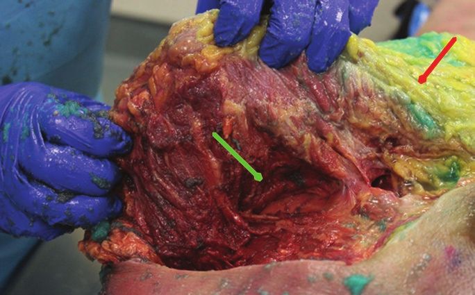
lww          14/9/18           14:58             4 Color Fig(s): F1-11              Art: PRS-D-18-00447

                                                              Plastic and Reconstructive Surgery • November 2018

          Fig. 7. Accuracy of cutaneous markings and navigation of the submuscular space. (Left) The submuscular plane
          can be entered from above (this is the left hemibuttock). The inferior gluteal vascular leash is visible in the loose
          areolar submuscular plane, and its location coincides with the topographic markings. (Right) Endoscopic real-time
          inspection of fat migration. Endoscopic view of the undersurface of the gluteus maximus muscle before subfascial
          fat injection. Note the absence of fascia.

  follow “safety zones” or stay in the “superficial                   alternative explanation, whereby injecting from
  muscle.”3 Although the anatomical basis of the                      the natal cleft directs the cannula parallel to the
  “safety triangle” theoretically makes sense, there                  muscle fibers, depositing fat along longitudinally
  is insufficient clinical evidence to prove that it                  separated fibers without disrupting the muscle. By
  is failsafe in human patients. The opinion that                     keeping the muscle and connective tissues grossly
  intramuscular fat transplantation is “safe” rests on                intact, the grafted fat is more likely to remain
  an important assumption—that fat placed in the                      within the muscle. In contrast, injection from
  intramuscular space remains in the intramuscular                    below results in a cannula course perpendicular
  space. The findings of the present study suggest                    to muscle fibers. By disrupting the muscle fibers
  that fat grafted within the muscle can migrate                      and septae, the cannula creates a perpendicular
  through the deep side of the gluteus muscle into                    passage through the muscle fibers through which
  the underlying submuscular space, implying that                     fat can more easily track down to the submuscular
  there is no zone within the gluteus maximus mus-                    space, along the path of least resistance (Fig. 10).        F10
  cle that can be considered safe.                                         The absence of deep fascia on the gluteus
       Whether one inserts fat in the deep or super-                  muscle has not been described previously. Other
  ficial muscle, given enough volume, it will not                     large truncal muscles, including the latissimus
  remain in the muscle and will spill deep to the                     dorsi and the pectoralis major muscle, exhibit
  submuscular space. There have been reports                          similar anatomy, with a superficial subcutaneous-
  of the direction of cannula insertion as connot-                    facing fascial component that is dense and a deep
  ing some element of safety.10,38 Insertion from                     component that is nonexistent. Similar to the
  the inferior gluteal crease incision (from below)                   gluteus muscle, fat injected into the pectoralis
  has been suggested to be more dangerous than                        muscle can also be presumed to migrate poste-
  injecting from a natal cleft approach (from above,                  riorly into the subpectoral space given the lack
  medial). This has been traditionally explained by                   of deep fascia to serve as a barricade. Yet unlike
  a “direct hit” paradigm, namely that angulating                     gluteal fat injections, fat grafting to the pectoralis
  the cannula in certain directions poses increased                   muscle has not been associated with pulmonary
  risk for penetrating the submuscular space and                      fat embolism.39 The likely reason for this is two-
  hitting a deep vein coming from below. The                          fold. First, the maximum fat volumes injected into
  deep intramuscular migration theory provides an                     the pectoralis muscle are on the order of 100 to

  8
Copyright © 2018 American Society of Plastic Surgeons. Unauthorized reproduction of this article is prohibited.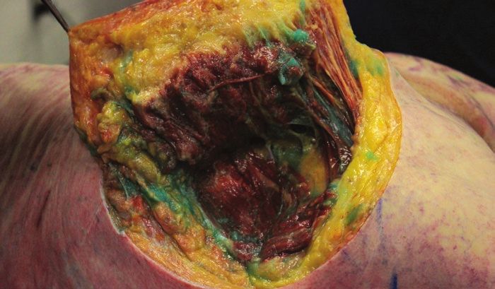
lww           14/9/18          14:58             4 Color Fig(s): F1-11       Art: PRS-D-18-00447

  Volume 142, Number 5 • Gluteal Fat Graft Migration

                                                                     volumes of fat preferentially migrate deep to the
                                                                     muscle because of the lack of deep fascial struc-
                                                                     tures acting as a barricade.
                                                                          The migration of fat parallel to or longitudi-
                                                                     nally between muscle fibers appears to occur both
                                                                     proximally and distally along the gluteal muscle to
                                                                     some extent. However, there is higher resistance
                                                                     in this direction because the fibers must separate
                                                                     longitudinally for fat deposition. This leaves only
                                                                     the deep egress as the path of least resistance. In
                                                                     this scenario, fat dissects between, or perpendicu-
                                                                     lar to, muscle fibers, spreading only a small dis-
                                                                     tance in the anterior direction before egressing
                                                                     into the lower pressure submuscular space. The
                                                                     fat exits the deep surface of the muscle in the area
                                                                     between the gluteal vessels and is deposited in the
                                                                     deep intermuscular space near the sciatic notch.
                                                                     Fat entering the notch causing a wedge can poten-
                                                                     tially lead to sciatic nerve entrapment with subse-
                                                                     quent transient or permanent nerve injury.

                                                                     The Venous Traction Theory
                                                                          A mechanism of venous trauma, without direct
                                                                     cannula contact injury to the vein, can be postu-
                                                                     lated to occur as a result of acute venous traction.
                                                                     This may occur when a volume of grafted fat col-
                                                                     lecting in the submuscular space causes posterior
  Fig. 8. Endoscopic anterior septal blowout. After 180 cc of fat,   projection of the muscle (Fig. 11, left). As the mus-   F11
  proxy fat is noted to burst through muscle fibers and well up in   cle expands posteriorly, it puts traction stretch on
  the subcutaneous space.                                            the fixed venous plexus, potentially causing fail-
                                                                     ure, or venous tear, setting up a pressure gradient
                                                                     for siphoning of fat into the venous system and
  150 cc, much less than volumes reported in glu-                    pulmonary fat embolism (Fig. 11, right).
  teal fat injections. Without high volumes creating                      Vascular surgical studies on the tensile
  a high-pressure effect, fat is less likely to egress.              strength of veins suggest that as low as a 7 per-
  Second, unlike the deep gluteal region, the sub-                   cent increase in axial length by traction on a filled
  pectoral space is devoid of significant large and                  vein can lead to failure of the conduit.40 Assum-
  fragile veins that carry with them the potential for               ing the average length of a superior gluteal vein
  devastating embolisms.                                             is 2.5 cm, a submuscular fat collection secondary
       Because of the lack of deep fascia lining the                 to deep intramuscular migration causing venous
  undersurface of the gluteus muscle, sufficient                     traction of (0.07 × 25 mm) less than 2 mm could
  volumes of fat placed in the muscle can migrate                    potentially lead to avulsion of the superior gluteal
  freely out of the muscle from its deep surface into                vein.41 As such, deep intramuscular migration–
  the submuscular plane, along the path of least                     induced traction injury poses another potential
  resistance. The superficial surface of the gluteus                 mode of venous injury aside from the obvious
  muscle, in contrast, is lined with a dense super-                  direct cannula trauma.
  ficial fascia that acts as a “backstop” to prohibit                     The superficial gluteal fascia, not the muscle,
  intramuscular fat from egressing out of the mus-                   acts as the lynchpin in this polemic. If the fascia
  cle in the opposite direction, into the superficial                acts as a superficial backstop to force fat deeper
  subcutaneous space. Indeed, in the current study,                  during intramuscular injection, pressure gener-
  there was no egress through the superficial fascia                 ated during the intramuscular injection can create
  even with recipient-site pressures exceeding 100                   the danger. If, in contrast, the superficial gluteal
  mmHg. As such, the fascial anatomy of the glu-                     fascia acts as an equally powerful deep backstop
  teus muscle creates the basis for the deep intra-                  during “subcutaneous only Brazilian butt lift” (i.e.,
  muscular migration phenomenon, wherein high                        SAFEBBL), it serves as an effective barricade to

                                                                                                                        9
Copyright © 2018 American Society of Plastic Surgeons. Unauthorized reproduction of this article is prohibited.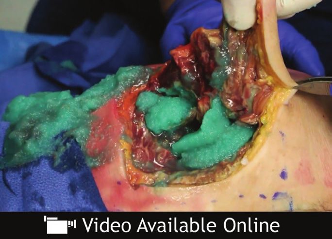
lww       14/9/18       14:58              4 Color Fig(s): F1-11              Art: PRS-D-18-00447

                                                        Plastic and Reconstructive Surgery • November 2018

                             Video 3. Supplemental Digital Content 3 demonstrates the migra-
                             tion of intramuscular fat in gluteal augmentation. After injection of
                             pseudofat into the superficial fascia of the gluteus maximus mus-
                             cle, we show that the pseudofat migrates deep and is deposited
                             within the submuscular space. The pseudofat enters deep to the
                             muscle through multiple areas within the muscle. Using an endo-
                             scope, we demonstrate that these injections of pseudofat into the
                             superficial fascia of the muscle migrated into the space deep the
                             muscle. The posterior fascia of the gluteus maximus muscle acts
                             as a backstop and prevents migration of pseudofat into the sub-
                             cutaneous space, available in the “Related Videos” section of the
                             full-text article on PRSJournal.com or, for Ovid users, available at
                             http://links.lww.com/PRS/D50.

  prevent subcutaneously placed fat from entering                differently, “if pressure beneath the fascia is your
  the muscle. In this scenario, pressure generated               enemy, pressure above the fascia is your friend.”
  from the subcutaneous injection can be used to                     Some reading this may be wary of the concept
  help guide the dispersion of fat within this space,            of creating intentionally high pressures in the
  a concept analogous to “lipotumescence.”42,43 Said             subcutaneous space in gluteal lipoaugmentation.

                            Fig. 9. Inspection of subcutaneous migration. Fat injected into the
                            subcutaneous (suprafascial) space generated high subcutaneous
                            pressures and migration through the subcutaneous tissue (red
                            arrow), whereas the submuscular space pressures remained 0 and
                            were devoid of proxy fat (green arrow). In this setting, the posterior
                            gluteal fascia acted as a protective “backstop.”

  10
Copyright © 2018 American Society of Plastic Surgeons. Unauthorized reproduction of this article is prohibited.
lww 14/9/18 14:58 4 Color Fig(s): F1-11 Art: PRS-D-18-00447
Volume 142, Number 5 • Gluteal Fat Graft Migration
Fig. 10. (Left) Injection from above leads to cannula disruption and graft placement parallel to muscle
fibers. (Right) Injection from below causes cannula disruption and graft placement perpendicular to
muscle fibers. Deep intramuscular migration may occur more readily when grafting perpendicular to
muscle fibers.
Fig. 11. Venous traction theory of pulmonary fat embolism. (Left) Fat from deep intramuscu-
lar migration collecting in the submuscular space separates the gluteus muscle, projecting it
posteriorly. (Right) At some stretch length, a vein fails and ruptures, allowing a pressure gradi-
ent and siphoning of fat into the venous circulation.
However, with postgraft relaxation of connective defect into the muscle. With sufficiently high vol-
tissue, water absorption, and internal recipient- umes and pressure, fat may migrate even deeper
site expansion provided by expansion vibration into the submuscular space.
lipofilling, such high pressures created by lipo- In the Aesthetic Surgery Education and
tumescence are transient after the completion of Research Foundation survey, many surgeons
postgraft shaping, recipient-site equalization, and reporting pulmonary fat embolism mortality
“fat shifting.”44,45 It must be noted, however, that insisted they were in the subcutaneous plane. As
if the superficial gluteal fascia is violated, the sub- a response, the authors of the survey stated that
cutaneous fat can take a path through the fascial “it is also possible that subcutaneous injections
11
Copyright © 2018 American Society of Plastic Surgeons. Unauthorized reproduction of this article is prohibited.lww 14/9/18 14:58 4 Color Fig(s): F1-11 Art: PRS-D-18-00447
Plastic and Reconstructive Surgery • November 2018
may track between a muscle plane or along a vas- REFERENCES
cular pedicle deep and into an area of large veins 1. Mofid MM, Teitelbaum S, Suissa D, et al. Report on mortality
or a venous plexus.”1 We saw no evidence of this from gluteal fat grafting: Recommendations from the ASERF
in the present study. In fact, the anatomical find- Task Force. Aesthet Surg J. 2017;37:796–806.
2. Cárdenas-Camarena L, Bayter JE, Aguirre-Serrano H,
ings derived from this cadaver study speak directly Cuenca-Pardo J. Deaths caused by gluteal lipoinjection: What
against the validity of this statement. Although are we doing wrong? Plast Reconstr Surg. 2015;136:58–66.
we applaud the Aesthetic Surgery Education and 3. Rosique RG, Rosique MJ. Deaths caused by gluteal lipoin-
Research Foundation survey finding that the mor- jection: What are we doing wrong? Plast Reconstr Surg.
tality rate associated with the Brazilian lift is unac- 2016;137:641e–642e.
4. Sinno S, Chang JB, Brownstone ND, Saadeh PB, Wall S Jr.
ceptably high (prompting this research), to our
Determining the safety and efficacy of gluteal augmentation:
knowledge, there has never been a case of fatal A systematic review of outcomes and complications. Plast
pulmonary fat embolism where, at autopsy, fat was Reconstr Surg. 2016;137:1151–1156.
confined only to the subcutaneous or suprafascial 5. Astarita DC, Scheinin LA, Sathyavagiswaran L. Fat transfer
plane. Furthermore, although the numbers are and fatal macroembolization. J Forensic Sci. 2015;60:509–510.
too low, there have been no cases of pulmonary 6. Wall S Jr, Del Vecchio D. Commentary on: Report on mor-
tality from gluteal fat grafting: Recommendations from the
fat embolism reported when subcutaneous only ASERF Task Force. Aesthet Surg J. 2017;37:807–810.
Brazilian lift has been performed. The Aesthetic 7. Villanueva NL, Del Vecchio DA, Afrooz PN, Carboy JA,
Surgery Education and Research Foundation Rohrich RJ. Staying safe during gluteal fat transplantation.
statement that subcutaneous fat insertion can lead Plast Reconstr Surg. 2018;141:79–86.
to pulmonary fat embolism is not substantiated by 8. Cardenas-Mejia A, Martínez JR, León D, Taylor JA, Gutierrez-
Gomez C. Bilateral sciatic nerve axonotmesis after gluteal
the scientific data of this anatomical study. lipoaugmentation. Ann Plast Surg. 2009;63:366–368.
9. Cárdenas-Camarena L, Durán H,Robles-Cervantes JA,
CONCLUSIONS Bayter-Marin JE. Critical differences between microscopic
(MIFE) and macroscopic (MAFE) fat embolism during
Although a great deal of attention has focused liposuction and gluteal lipoinjection. Plast Reconstr Surg.
on the gluteus maximus muscle in fat grafting 2018;141:880–890.
safety, it appears the superficial gluteal fascia is 10. Ramos-Gallardo G, Orozco-Renteria D, Medina-Zamora P, et
the key anatomical structure, forcing intramus- al. Prevention of fat embolism in fat injection for gluteal aug-
cular fat deep, and keeping subcutaneous fat mentation: Anatomic study in fresh cadavers. J Invest Surg.
2018;31:292–297.
superficial, over a wide range of interstitial tissue 11. Cardenas Restrepo JC, Muñoz Ahmed JA. Large-volume lipoin-
pressures. Because of the migratory ability of fat jection for gluteal augmentation. Aesthet Surg J. 2002;22:33–38.
within the gluteus muscle during fat transplanta- 12. Rosique RG, Rosique MJ, De Moraes CG. Gluteoplasty with
tion, deep intramuscular migration is a phenom- autologous fat tissue: Experience with 106 consecutive cases.
enon that may occur when fat is inserted in any Plast Reconstr Surg. 2015;135:1381–1389.
13. Abboud MH, Dibo SA, Abboud NM. Power-assisted gluteal
part of the gluteus maximus muscle. The intra- augmentation: A new technique for sculpting, harvesting,
muscular insertion of fat, which up to this point and transferring fat. Aesthet Surg J. 2015;35:987–994.
has been considered reasonable to perform in 14. Ali A. Contouring of the gluteal region in women: Enhancement
the superficial muscle and even recommended in and augmentation. Ann Plast Surg. 2011;67:209–214.
many articles and textbooks on the subject, is now 15. Cardenas-Camarena L, Lacouture AM, Tobar-Losada A.
Combined gluteoplasty: Liposuction and lipoinjection. Plast
deemed to be an inexact and potentially danger- Reconstr Surg. 1999;104:1524–1531; discussion 1532–1533.
ous technique. This strategy, because of its migra- 16. Condé-Green A, Kotamarti V, Nini KT, et al. Fat grafting for
tory uncertainty, should be discontinued in fat gluteal augmentation: A systematic review of the literature
transplantation to the gluteal region. and meta-analysis. Plast Reconstr Surg. 2016;138:437e–446e.
17. de Pedroza LV. Fat transplantation to the buttocks and legs
Daniel A. Del Vecchio, M.D. for aesthetic enhancement or correction of deformities:
38 Newbury Street Long-term results of large volumes of fat transplant. Dermatol
Boston, Mass. 02116 Surg. 2000;26:1145–1149.
fatvsfiction@gmail.com 18. Hoyos AE, Perez ME, Castillo L. Dynamic definition mini-
lipoabdominoplasty combining multilayer liposculp-
ture, fat grafting, and muscular plication. Aesthet Surg J.
ACKNOWLEDGMENTS 2013;33:545–560.
The authors thank the Dr. Rod Rohrich Research 19. Lewis CM. Correction of deep gluteal depression by autolo-
Fund from the University of Texas Southwestern Depart- gous fat grafting. Aesthetic Plast Surg. 1992;16:247–250.
ment of Plastic Surgery, Garret Adams of Stryker for the 20. Marwah M, Kulkarni A, Godse K, Abhyankar S, Patil S,
Nadkarni N. Fat ful’fill’ment: A review of autologous fat
compartment pressure monitor, Jourdan Carboy for illus- grafting. J Cutan Aesthet Surg. 2013;6:132–138.
trations, and the Willed Body Program of the University 21. Mendieta CG. Gluteal reshaping. Aesthet Surg J.
AQ4 of Texas Southwestern. 2007;27:641–655.
12
Copyright © 2018 American Society of Plastic Surgeons. Unauthorized reproduction of this article is prohibited.lww 14/9/18 14:58 4 Color Fig(s): F1-11 Art: PRS-D-18-00447
Volume 142, Number 5 • Gluteal Fat Graft Migration
22. Murillo WL. Buttock augmentation: Case studies of fat 34. Moscatiello F, Aznar-Benitah S, Grella R, Jover JH. Gluteal
injection monitored by magnetic resonance imaging. Plast augmentation with cryopreserved fat. Aesthet Surg J.
Reconstr Surg. 2004;114:1606–1614; discussion 1615–1616. 2010;30:211–216.
23. Nicareta B, Pereira LH, Sterodimas A, Illouz YG. Autologous 35. Coleman SR. Structural fat grafting: More than a permanent
gluteal lipograft. Aesthetic Plast Surg. 2011;35:216–224. filler. Plast Reconstr Surg. 2006;118(Suppl):108S–120S.
24. Pereira LH, Radwanski HN. Fat grafting of the buttocks and 36. Del Vecchio D, Wall S Jr. Expansion vibration lipofilling:
lower limbs. Aesthetic Plast Surg. 1996;20:409–416. A new technique in large-volume fat transplantation. Plast
25. Perén PA, Gómez JB, Guerrerosantos J, Salazar CA. Reconstr Surg. 2018;141:639e–649e.
Gluteus augmentation with fat grafting. Aesthetic Plast Surg. 37. Del Vecchio DA, Del Vecchio SJ. The graft-to-capacity ratio:
2000;24:412–417. Volumetric planning in large-volume fat transplantation.
26. Roberts TL III, Toledo LS, Badin AZ. Augmentation of the Plast Reconstr Surg. 2014;133:561–569.
buttocks by micro fat grafting. Aesthet Surg J. 2001;21:311–319. 38. Montanana AR. Evolution of my technique. Paper presented
27. Roberts TL III, Weinfeld AB, Bruner TW, Nguyen K. at: American Society for Aesthetic Plastic Surgery Annual
“Universal” and ethnic ideals of beautiful buttocks are best Meeting; April 27–May 2, 2017; San Diego, Calif. AQ5
obtained by autologous micro fat grafting and liposuction. 39. Khouri RK, Rigotti G, Cardoso E, Khouri RK Jr, Biggs TM.
Clin Plast Surg. 2006;33:371–394. Megavolume autologous fat transfer: Part II. Practice and
28. Toledo LS. Gluteal augmentation with fat grafting: The techniques. Plast Reconstr Surg. 2014;133:1369–1377.
Brazilian buttock technique. 30 years’ experience. Clin Plast 40. Donovan DL, Schmidt SP, Townshend SP, Njus GO, Sharp
Surg. 2015;42:253–261. WV. Material and structural characterization of human
29. Valeriani M. GLADI: Gluteal adipose implant. A new tech- saphenous vein. J Vasc Surg. 1990;12:531–537.
nique for the reshaping of the gluteal-trochanteric region. 41. Hamdi M, Gagnon AR. Gluteus flap. In: Wei FC, Mardini
Acta Chir Plast. 2004;46:70–73. S, eds. Flaps and Reconstructive Surgery E-Book. Philadelphia:
30. Willemsen JC, Lindenblatt N, Stevens HP. Results and long- Saunders; 2009:375–394. AQ6
term patient satisfaction after gluteal augmentation with 42. Khouri RK, Rigotti G, Cardoso E, Khouri RK Jr, Biggs TM.
platelet-rich plasma-enriched autologous fat. Eur J Plast Surg. Megavolume autologous fat transfer: Part I. Theory and
2013;36:777–782. principles. Plast Reconstr Surg. 2014;133:550–557.
31. Wolf GA, Gallego S, Patrón AS, et al. Magnetic resonance 43. Bucky L. Getting started with fat grafting. Paper presented
imaging assessment of gluteal fat grafts. Aesthetic Plast Surg. at: The Aesthetic Meeting 2012: Annual Meeting of the
2006;30:460–468. American Society for Aesthetic Plastic Surgery; May 3–8,
32. Avendaño-Valenzuela G, Guerrerosantos J. Contouring the 2012: Vancouver, British Columbia, Canada. AQ7
gluteal region with tumescent liposculpture. Aesthet Surg J. 44. Wall S Jr. SAFE circumferential liposuction with abdomino-
2011;31:200–213. plasty. Clin Plast Surg. 2010;37:485–501.
33. Cárdenas-Camarena L. Various surgical techniques for 45. Wall SH Jr, Lee MR. Separation, aspiration, and fat equal-
improving body contour. Aesthetic Plast Surg. 2005;29:446– ization: SAFE liposuction concepts for comprehensive body
455; discussion 456–459. contouring. Plast Reconstr Surg. 2016;138:1192–1201.
13
Copyright © 2018 American Society of Plastic Surgeons. Unauthorized reproduction of this article is prohibited.AUTHOR QUERIES
AUTHOR PLEASE ANSWER ALL QUERIES
AQ1—For indexing purposes, please confirm that author names have been correctly identified as
given names (blue), surnames (red), and suffixes (black). Color in the byline will not appear
on the final published version.
AQ2—ml and cc both OK, or 540 to 720 cc [or ml]?
AQ3—Please verify that arrows appear in Figure 4 or revise legend as needed.
AQ4—Acknowledgment statement correct as edited?
AQ5—Ref 38: Correct as edited?
AQ6—Ref 41: End page number correct? If not, please provide.
AQ7—Ref 43: Meeting information correct as edited?
AQ8—Please double-check the financial disclosure statement to confirm that it is correct. If it is
incorrect, revise as needed. Please also provide statements for the other authors.You can also read