Effects of applying amoxicillin in juvenile mice on enamel mineralization and the expression of kallikrein related peptidase 4 and tight junction ...
←
→
Page content transcription
If your browser does not render page correctly, please read the page content below
INTERNATIONAL JOURNAL OF MOlecular medicine 46: 179-190, 2020

            Effects of applying amoxicillin in juvenile mice on enamel
             mineralization and the expression of kallikrein‑related
             peptidase 4 and tight junction proteins in ameloblasts
            Jianghong Gao1-3*, Xinmei Li2,4*, liping Gao2,5, Haiyan Chen6, Bashayer H. Baras7,8,
                 Xiaojing Liu2, Hao liu2, Ayesha Rana2, Meili Gao9 and Jianping Ruan1-3

     1
       Key Laboratory of Shaanxi Province for Craniofacial Precision Medicine Research, 2Department of Preventive Dentistry,
            3
              Clinical Research Center of Shaanxi Province for Dental and Maxillofacial Diseases, College of Stomatology,
          Xi'an Jiaotong University, Xi'an, Shaanxi 710004; 4Department of Stomatology, Yan'an People's Hospital, Yan'an,
         Shaanxi 716000; 5Department of Stomatology, Inner Mongolia People's Hospital, Huhhot, Inner Mongolia 010020;
    6
      Core Research Laboratory, The Second Affiliated Hospital of Xi'an Jiaotong University, Xi'an, Shaanxi 710004, P.R. China;
         7
           Department of Advanced Oral Sciences and Therapeutics, University of Maryland School of Dentistry, Baltimore,
              MD 21201, USA; 8Department of Restorative Dental Science, College of Dentistry, King Saud University,
               Riyadh 11545, Saudi Arabia; 9Department of Biological Science and Engineering, The Key Laboratory of
                 Biomedical Information Engineering of Ministry of Education, School of Life Science and Technology,
                                     Xi'an Jiaotong University, Xi'an, Shaanxi 710049, P.R. China

                                       Received November 3, 2019; Accepted April 7, 2020

                                                  DOI: 10.3892/ijmm.2020.4598

Abstract. Amoxicillin is a common pediatric drug. However,          19 days. The surface morphology and calcium (Ca), phospho-
to the best of our knowledge, the role of amoxicillin in enamel     rous (P) and carbon contents of mandibular incisors and first
hypomineralization has not yet been fully elucidated. The aim       molars were examined by scanning electron microscopy and
of the present study was to assess the effects of amoxicillin on    energy dispersive X‑ray spectroscopy. Histological changes
enamel mineralization, the morphology of ameloblasts, as well       in the ameloblasts of mandibular incisors were analyzed by
as the expression of kallikrein‑related peptidase 4 (KLK4),         hematoxylin and eosin staining. The KLK4, CLDN1, CLDN4
and the tight junction proteins, claudin 1 (CLDN1), claudin 4       and OCLN expression levels of ameloblasts were observed by
(CLDN4) and occludin (OCLN), in ameloblasts of juvenile             immunohistochemical staining. The incidence of white patches
mice. A total of 36 3‑day‑old Kunming mice were randomly            in the incisor was 100% in the 100 mg/kg amoxicillin‑treated
divided into three groups. The mice were administered 0, 50         groups. A greater number of enamel defects were observed
or 100 mg/kg amoxicillin by intragastric administration for         in the incisal/occlusal half of mandibular incisors/molars
                                                                    compared with in the cervical half in the amoxicillin‑treated
                                                                    groups. Following phosphoric‑acid treatment, the enamel
                                                                    rod and interrod were aligned in a disorderly manner in
Correspondence to: Professor Meili Gao, Department of               the amoxicillin‑treated groups. Amoxicillin decreased the
Biological Science and Engineering, The Key Laboratory of           Ca/P ratio in the enamel of mandibular incisors and molars.
Biomedical Information Engineering of Ministry of Education,        More intercellular spaces among maturation ameloblasts
School of Life Science and Technology, Xi'an Jiaotong University,   were observed in the amoxicillin‑treated groups. Amoxicillin
28 Xianning West Road, Xi'an, Shaanxi 710049, P.R. China            decreased KLK4 and CLDN1, CLDN4 and OCLN expression
E‑mail: gml.1369@mail.xjtu.edu.cn                                   in mature ameloblasts. The administration of amoxicillin in
                                                                    juvenile mice induced enamel hypomineralization, and the
Professor Jianping Ruan, Key Laboratory of Shaanxi Province for
                                                                    effects of amoxicillin on enamel hypomineralization may be
Craniofacial Precision Medicine Research, College of Stomatology,
                                                                    mediated via multiple pathways.
Xi'an Jiaotong University, 98 Xiwu Road, Xi'an, Shaanxi 710004,
P.R. China
E‑mail: ruanjp@mail.xjtu.edu.cn                                     Introduction

*
    Contributed equally                                             Tooth enamel is a highly mineralized substance that acts as a
                                                                    barrier to protect teeth (1). Ameloblasts, which are epithelial
Key words: amoxicillin, enamel hypomineralization, kallikrein-      cells, are responsible for amelogenesis (1). Secretory amelo-
related peptidase 4, tight junction proteins                        blasts secrete matrix proteins to organize the entire enamel
                                                                    thickness with approximately 30‑35% of the final mineral
                                                                    content (2). During the maturation stage of amelogenesis,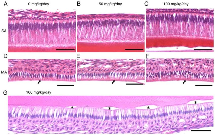
180                             GAO et al: EFFECTS OF AMOXICILLIN ON ENAMEL MINERALIZATION

kallikrein‑related peptidase 4 (KLK4) is secreted to degrade        Materials and methods
enamel proteins. Proteins and water are removed and specific
ions required for the concurrent accretion of mineral are           Animals. A total of 6 female Kunming mice on gestation
deposited in the enamel until the mineral content is >95% in        day 18 with a body weight of 40±3 g were purchased from the
fully formed enamel (3,4).                                          Experimental Animal Center of Xi'an Jiaotong University. The
    Ameloblasts are attached to each other at their lateral         research was conducted in accordance with the Declaration of
membranes by a complex of intercellular junctions during            Helsinki and with the Guide for Care and Use of Laboratory
amelogenesis (5). The most apical complex of the intercellular      Animals as adopted and promulgated by the United National
junctions is tight junctions (TJs) (5). TJs generate the perme-     Institutes of Health. All experimental protocols were approved
ability barrier among neighboring cells and control the flux of     by the Ethics Committee for the Use of Human or Animal
ions and non‑electrolytes through the paracellular space (5‑8).     Subjects of Xi'an Jiaotong University. Mice were kept in cages
In addition, TJs can also act as a ‘fence’ and permit cells to      at room temperature and were provided ad libitum access
act in a polarized manner, which is crucial for morphogenesis,      to food and water. The temperature of the environment was
protein‑membrane trafficking and transportation (5‑8). The          maintained at 25±3˚C, with the relative humidity maintained
TJs of ameloblasts ensure a suitable microenvironment for           at 40‑60%. Artificial lighting was provided for 12 h each day.
enamel deposition and maturation by determining the para-           The water, food and padding were changed every 24 h.
cellular permeability and selectivity of solutes (5,9). Claudins        After birth, the mouse offspring were raised with their
(CLDNs) and occludin (OCLN) are integral membrane                   mothers until they were weaned. A total of 6 mice from each
proteins of TJ strands (9). CLDN1, CLDN4 and OCLN are               mother, resulting in a total of 36 offspring mice, were randomly
expressed in mature ameloblasts. These proteins may differ-         selected and divided into the 0, 50 and 100 mg/kg amoxicillin
entially regulate paracellular permeability against the enamel      treatment groups. Each group included 12 offspring mice,
surface to perform their functions appropriately (9‑13).            which were labeled on their back. Since day 3 after birth,
    Amelogenesis is strictly controlled by genes. However, if       the offspring mice in the control group were intragastrically
enamel organ cells are exposed to environmental stress for a        administered saline at 8 a.m. and 8 p.m, and offspring in the
long period of time during critical periods of amelogenesis,        experimental groups were also intragastrically administered
enamel defects may occur, such as amelogenesis imperfecta,          amoxicillin at 50 or 100 mg/kg twice a day, at 8 a.m. and
dental fluorosis, enamel hypoplasia and molar incisor hypo-         8 p.m. Saline and amoxicillin treatment were administered
mineralization (MIH) (14‑16). Enamel hypomineralization             for 19 days. The mother mice were fed a regular diet. The
poses an increased risk for toothaches, caries and concerns         offspring mice were weighed daily and were weaned on day 21
about appearance (17‑19). Therefore, it is crucial to prevent       after birth. On postnatal day 25, all of the offspring mice were
enamel hypomineralization.                                          anesthetized by an intraperitoneal injection of 10% chloral
    The β‑lactam antibiotic, amoxicillin, is widely prescribed      hydrate at a dose of 400 mg chloral hydrate per kg of animal
as a first‑choice antibiotic for common infections in pediat-       body weight (28). No sign of peritonitis was observed following
rics and pedodontics, such as ear, nose and throat infections,      the intraperitoneal injection. The incisors of the mice were
pneumonia and other infections (20). However, certain studies       photographed with a digital camera (Canon; EOS M5) under
have found that amoxicillin may affect enamel mineraliza-           natural light. The offspring were then sacrificed by cervical
tion (21‑27). Epidemiological studies have demonstrated that        dislocation for the following experiment. On the same day the
amoxicillin administration during pregnancy and/or early            mother mice were administered an intraperitoneal injection
childhood may be associated with MIH (21‑23). Furthermore,          of 10% chloral hydrate at a dose of 400 mg chloral hydrate
hypomineralization in enamel has been found in the offspring        per kg of animal body weight, and no signs of peritonitis were
of pregnant rats administered amoxicillin daily (24). In addi-      observed after intraperitoneal injection. The mice were then
tion, amoxicillin can play a contributing role in the development   sacrificed by cervical dislocation and handled according to the
of tooth fluorosis (25). It has been reported that amoxicillin      Ethics Committee for the Use of Human or Animal Subjects
administered to 20‑ to 24‑month‑old children increases the risk     of Xi'an Jiaotong University. Direct cardiac palpation was used
of dental fluorosis (26), and an in vitro study further suggested   to confirm the death of mice.
that amoxicillin and fluoride exert a potentiation effect on the
developing enamel of mouse molars (27). However, the patho-         Tissue preparation. Complete mandibles were harvested and
logical mechanisms underlying the effects of amoxicillin on         the surrounding tissues of the bone were gently removed. A
enamel hypomineralization have not yet been fully elucidated.       total of 12 right hemimandibles in each group were washed
Furthermore, to the best of our knowledge, no in vivo studies       with distilled water and fixed for 24 h in 5% glutaraldehyde
using laboratory juvenile animals exposed to amoxicillin,           at room temperature prior to performing scanning electron
which is similar to amoxicillin used in early childhood, have       microscopy (SEM) and energy dispersive X‑ray spectroscopy
been performed to date. Therefore, the aim of the present           (EDX) analysis. Subsequently, 6 of the 12 left hemimandibles
study was to assess the effects in juvenile mice produced by        in each group, which were randomly selected, were immersed
amoxicillin administration on enamel mineralization, the            in 1% phosphoric‑acid for 60 sec, and were then washed with
morphology of ameloblasts and the expression of KLK4 and            distilled water three times. In order to perform histological and
TJ proteins, including CLDN1, CLDN4 and OCLN. The find-             immunohistochemistry analyses, the other 6 left hemimandi-
ings of the present study may promote the understanding of          bles in each group were washed with 0.01% phosphate‑buffered
the role of amoxicillin in enamel hypomineralization and the        saline (PBS) solution and were then fixed by immersion in 5%
pathological mechanisms of enamel hypomineralization.               paraformaldehyde in 0.1 M phosphate buffer (pH 7.3) for 48 h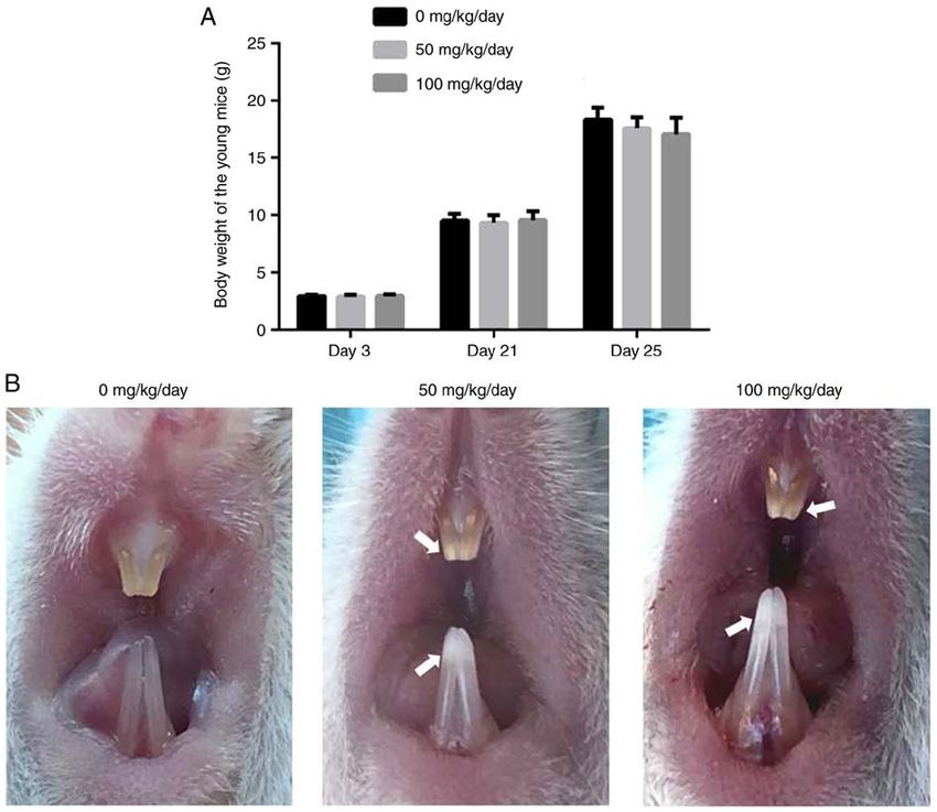
INTERNATIONAL JOURNAL OF MOlecular medicine 46: 179-190, 2020                                     181

at room temperature. Hemimandibles were then decalcified             PBS three times and antibody binding was visualized using
in 10% EDTA (pH 7.3) for 4 weeks at 4˚C and embedded in              the Vectastain ABC Elite kit (cat. no. PK‑6100; Vector
paraffin. Sagittal serial sections at a thickness of 5‑10 µm were    Laboratories, Inc). The slides were then washed with PBS
prepared and mounted on polyline‑coated glass slides.                solution. This was followed by incubation with biotinylated
                                                                     anti‑goat IgG (1:200; cat. no. PK‑6100; Vector Laboratories,
SEM and EDX analyses. For both SEM and EDX analyses,                 Inc.) for 15 min at 37˚C. The reaction was amplified with
the incisal/occlusal half and cervical half of labial/facial         Ultra‑Steptavidin conjugated to horseradish peroxidase (1:50;
surface enamel of the mandibular incisors and the molars             cat. no. PK‑6100; Vector Laboratories, Inc.) and visualized
were assessed, respectively. For the mandibular incisors             with diaminobenzidine. Slides were counterstained with
treated by 1% phosphoric‑acid, the incisal half of distal            hematoxylin for 3 min at room temperature prior to micro-
surface enamel of the mandibular incisors were assessed. The         scopic analysis. Immunohistochemical images were acquired
outer enamel structure of each hemimandible was examined             using a Leica DM 750 microscope (Leica Microsystems
by SEM (JXA‑8100; JEOL, Ltd.) operating in low‑vacuum                GmbH) with an attached digital camera Leica ICC50 HD
mode for secondary electron imaging. The JXA‑8100 SEM                (Leica Microsystems GmbH) a x200 magnification.
was equipped with an EDX system (INCA Energy; Oxford
Instruments) for qualitative and quantitative analyses and           Quantification of staining. Following immunohistochemistry
elemental mapping. Each sample without phosphoric‑acid               staining, quantitative immunostaining was assessed as previ-
was randomly observed at three points in each half of enamel         ously described (30). The positive immunostaining expression
(incisal/occlusal half and cervical half of labial/facial enamel).   of KLK4, CLDN1, CLDN4 and OCLN in mature amelo-
Each sample with phosphoric‑acid was randomly observed at            blasts of mandibular incisors was subjected to microscopic
three points in each incisal half of labial enamel. A total of       analysis. The intensity of the staining signal was measured
6 micrographs of each sample without phosphoric‑acid and             and documented using Image‑Pro Plus 6.0 image analysis
3 micrographs of each sample with phosphoric‑acid were               software (Media Cybernetics, Inc.). The intensity of staining
obtained at each magnification. The images were compared             was expressed as the mean signal density of tissue areas from
among the groups and representative images were selected.            six randomly selected visions. The mean signal densities
The relative amounts of calcium (Ca), phosphorus (P) and             were made relative to the mean signal density of the 0 mg/kg
carbon (C) of samples without phosphoric‑acid were assessed          amoxicillin‑treated group.
as atom percentage (Atom%) by EDX analysis, and the
mean Atom% of Ca, P, C and Ca/P ratios of each group was             Statistical analysis. All assays were conducted with three
calculated.                                                          independent experiments by SPSS 17.0 software (SPSS, Inc.).
                                                                     Data are expressed as the mean ± standard deviation. One‑way
Histological analysis. Deparaffinized sections were stained          analysis of variance followed by Tukey's method was used to
using the haematoxylin and eosin staining technique. Briefly,        compare differences between groups. P0.05; Fig. 1A).
                                                                     However, the color and translucency of incisor enamel was
Immunohistochemistry. Immunohistochemical staining was               altered following amoxicillin exposure (Fig. 1B). In the control
performed on arrayed tissue samples according to a previ-            group, the enamel of maxillary and mandibular incisors was
ously described protocol (29). Briefly, dewaxed sections were        orange in color, and smooth and translucent (Fig. 1B). However,
rinsed in PBS and subjected to antigen retrieval in 10 mM            chalk/white patches in the incisor enamel, particularly the
citrate buffer (pH 6.0) for 20 min at 95˚C in a microwave            incisal half, were observed in the amoxicillin‑exposed groups
oven. Subsequently, endogenous peroxidase was blocked                (Fig. 1B). Furthermore, 8.3, 58 and 100% of mice exhibited
with 3% H2O2 for 10 min, and the sections were then washed           enamel chalk/white patches in the 0, 50 and 100 mg/kg
with PBS three times. Non‑specific staining was blocked for          amoxicillin‑exposed groups, respectively.
30 min at 37˚C with rabbit serum (cat. no. ab7487; Abcam)
in a wet box, followed by overnight incubation at 4˚C with           Amoxicillin induces enamel hypomineralization of incisors
rabbit anti‑KLK4 (1:100; cat. no. Ab197657; Abcam), CLDN1            and molars of juvenile mice. The SEM observations revealed
(1:100; cat. no. 13050‑1‑AP; Proteintech Group, Inc.), CLDN4         that the enamel surface of the incisors (Fig. 2A and D) and
(1:200; cat. no. 16195‑1‑AP; Proteintech Group, Inc.) and            molars (Fig. 3A and D) was smooth and homogeneous with a
OCLN (1:100; cat. no. 13409‑1‑AP; Proteintech Group, Inc.)           few visible cracks in the control group. However, the incisal
antibodies, and non‑immune IgG (1:200; cat. no. ab37415;             half of the mandibular incisors exhibited a rough, irregular and
Abcam) served as a negative control. Following incuba-               scratched enamel pattern in the 50 mg/kg amoxicillin‑exposed
tion with primary antibodies, the slides were washed in              group (Fig. 2B). Deeper scratched enamel defects and foam‑like
182 GAO et al: EFFECTS OF AMOXICILLIN ON ENAMEL MINERALIZATION
Figure 1. Effects of amoxicillin on body weight and morphology of the incisors in young mice. (A) Weight changes, data are presented as mean ± standard
deviation. (B) Representative photographs of the mice incisors in the 0, 50 and 100 mg/kg/day amoxicillin‑exposed groups, respectively. Arrows indicate
chalk/white patches in the enamel of the maxillary and mandibular incisors.
enamel with obvious holes were observed in the 100 mg/kg rods and inter‑rods were indistinct and were hardly visible in
amoxicillin‑exposed group. The length of enamel defect could some sites, where the packing of the crystals was less tight and
be extended to several microns (Fig. 2C). However, there less well organized in the 50 (Fig. 4B) and 100 (Fig. 4C) mg/kg
were no obvious enamel defects in the cervical half enamel amoxicillin‑exposed groups.
of the mandibular incisors in the 50 (Fig. 2E) and 100 mg/kg EDX analysis revealed that, except for the 50 mg/kg
(Fig. 2F) amoxicillin‑exposed groups. Scratched enamel with amoxicillin‑exposed group, amoxicillin significantly reduced
a different depth was also often observed in the occlusal half the mean values of Ca, P and the Ca/P ratio of the incisal half
of molars enamel in the 50 (Fig. 3B) and 100 mg/kg (Fig. 3C) (Fig. 5A and B) and cervical half (Fig. 5C and D) of mandib-
amoxicillin‑exposed groups; however, the enamel defects ular incisor enamel (P0.05). No significant
mandibular incisors. Similar to the mandibular incisors, differences were found for the changes in Ca, P and C in the
enamel defects in the cervical half of the mandibular molars occlusal half (Fig. 5E and F) and cervical half (Fig. 5G and H)
were not obvious in the 50 (Fig. 3E) and 100 mg/kg (Fig. 3F) of the mandibular first molar enamel between the groups;
amoxicillin‑exposed groups. whereas amoxicillin significantly decreased the Ca/P ratio
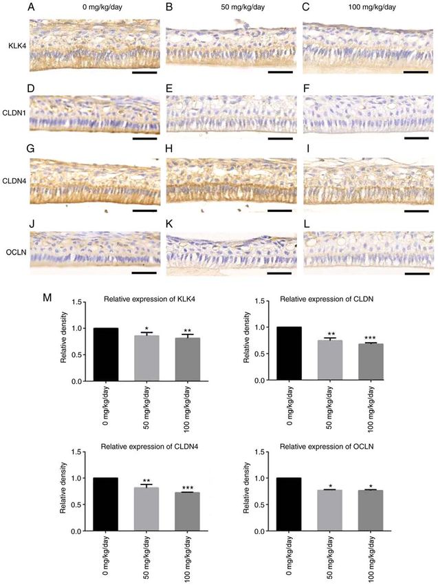
Following phosphoric acid etching, the rods and interrod in the occlusal half (PINTERNATIONAL JOURNAL OF MOlecular medicine 46: 179-190, 2020 183 Figure 2. Effects of amoxicillin on the enamel surface of the mandibular incisors. Representative scanning electron microscopy micrographs of the enamel surface of the incisors. (A) The micrographs of the enamel surface of the incisors incisal half in the 0 mg/kg/day amoxicillin‑exposed group. (B) The micrographs of the enamel surface of the incisors incisal half in the 50 mg/kg/day amoxicillin‑exposed group. (C) The micrographs of the enamel surface of the incisors incisal half in the 100 mg/kg/day amoxicillin‑exposed group. (D) The micrographs of the enamel surface of the incisors cervical half in the 0 mg/kg/day amoxicillin‑exposed group. (E) The micrographs of the enamel surface of the incisors cervical half in the 50 mg/kg/day amoxicillin‑exposed group. (F) The micrographs of the enamel surface of the incisors cervical half in the 100 mg/kg/day amoxicillin‑exposed group. Scale bars, 20 µm (upper images) and 2 µm (lower images). appeared to be similar in the 50 (Fig. 6B) and 100 mg/kg Amoxicillin reduces the expression of KLK4, CLDN1, (Fig. 6C) amoxicillin‑exposed groups. In the control group, CLDN4 and OCLN in mature ameloblasts of juvenile mice. the mature ameloblasts were orderly arranged and obvious Immunohistochemical staining revealed that brown granules gaps among the cells were occasionally observed (Fig. 6D). were found in the cytoplasm of dental epithelial cells of the However, larger intercellular spaces among mature ameloblasts maturation stage in the control group, particularly in mature could be identified in the 50 (Fig. 6E) and 100 mg/kg (Fig. 6F) ameloblasts (Fig. 7A). However, immunohistochemical amoxicillin‑treated groups. Furthermore, it was demonstrated staining of KLK4 in the 50 (Fig. 7B) and 100 mg/kg (Fig. 7C) that some mature ameloblasts separated from the enamel and amoxicillin‑exposed groups exhibited a pale yellow color, formed vesicles in the amoxicillin‑exposed groups (Fig. 6G). which was lighter in color than that in the control group. In total, 8.3, 50 and 75% of mouse samples exhibited vesicles Weak staining for CLDN1 (Fig. 7D) and OCLN (Fig. 7J) was in the 0, 50 and 100 mg/kg amoxicillin‑exposed groups, detected at the distal ends of mature ameloblasts in the control respectively. group. The staining color of CLDN1 in the 50 (Fig. 7E) and
184                                   GAO et al: EFFECTS OF AMOXICILLIN ON ENAMEL MINERALIZATION

Figure 3. Effects of amoxicillin on the enamel surface of the mandibular molars. Representative scanning electron microscopy micrographs of the enamel
surface of the mandibular first molars. (A) The micrographs of the enamel surface of the first molars occlusal half in the 0 mg/kg/day amoxicillin‑exposed
group. (B) The micrographs of the enamel surface of the first molars occlusal half in the 50 mg/kg/day amoxicillin‑exposed group. (C) The micrographs of
the enamel surface of the first molars occlusal half in the 100 mg/kg/day amoxicillin‑exposed group. (D) The micrographs of the enamel surface of the first
molars cervical half in 0 mg/kg/day amoxicillin‑exposed group. (E) The micrographs of the enamel surface of the first molars cervical half in the 50 mg/kg/day
amoxicillin‑exposed group. (F) The micrographs of the enamel surface of the first molars cervical half in the 100 mg/kg/day amoxicillin‑exposed group. Scale
bars, 20 µm (upper images) and 2 µm (lower images).

100 mg/kg (Fig. 7F) amoxicillin‑treated group became a little                    lower than that in the 0 mg/kg group, and the differences were
lighter. The staining color of OCLN in the 50 (Fig. 7K) and                      statistically significant (P
INTERNATIONAL JOURNAL OF MOlecular medicine 46: 179-190, 2020                                                      185

Figure 4. Effects of amoxicillin on the distal enamel surface of the lower incisors after 1% phosphoric‑acid treatment. Representative scanning electron
microscopy micrographs of the distal enamel surface of the lower incisors. (A) The micrographs of the distal enamel surface of the incisors incisal half in
0 mg/kg/day amoxicillin‑exposed group. (B) The micrographs of the distal enamel surface of the incisors incisal half in 50 mg/kg/day amoxicillin‑exposed
group. (C) The micrographs of the distal enamel surface of the incisors incisal half in 100 mg/kg/day amoxicillin‑exposed group. Scale bars, 20 µm (upper
images) and 2 µm (lower images).

that continuous amoxicillin administration (from postnatal                     maxillary incisors (35). However, in the present study, the
days 3 to 21) led to enamel hypomineralization in juvenile                     enamel chalk/white patches were also observed in the enamel
mice. Furthermore, it was found that amoxicillin reduced the                   of maxillary incisors. Adult mice were used in the previous
expression of KLK4, CLDN1, CLDN4 and OCLN in mature                            study, while the present study used juvenile mice whose drug
ameloblasts.                                                                   metabolism was weaker than that of adult mice. Properties of
    The first mandibular molars of 3‑day‑old Kunming mice                      light reflection and the transmission of enamel are dependent
begin to secrete enamel matrix proteins and they erupt approx-                 on its texture, the orientation of enamel rods and histological
imately on postnatal day 20 (31). During postnatal days 3 to 21,               characteristics (36,37). Therefore, chalk/white patches and a
the first mandibular molars of Kunming mice experience both                    less translucent appearance in the enamel surface suggested
secretory and maturation periods of amelogenesis. Therefore,                   that surface smoothness and/or histological characteristics of
the young mice were exposed to amoxicillin from postnatal                      the enamel may be affected by amoxicillin. Furthermore, some
days 3 to 21 to intervene with the whole mineralization period                 enamel defects in the enamel surface were found by SEM,
of the first mandibular enamel.                                                which confirms the aforementioned finding. During chewing
    The dosage of amoxicillin prescribed for children is                       of mice, the dentin of maxillary incisor contacts the enamel of
≥25 mg/kg (32,33). According to the guideline of drug conver-                  mandibular incisors due to enamel formed only on the labial
sion from humans to mice provided by the USA Food and Drug                     surface of the mice incisors (38). Therefore, the enamel of
Administration, the dose of 25 mg/kg in humans is ~310 mg/kg                   maxillary incisors bear less friction than that of mandibular
in mice (34). However, some studies have demonstrated that                     incisors. That may be the reason why the enamel white patches
pregnant rats and adult mice exposed to 50 and 100 mg/kg                       in maxillary incisors appeared less severe than those in the
amoxicillin exhibit enamel defects (24,35), which indicates                    mandibular incisors, even if they were similarly affected by
that the enamel is quite sensitive to amoxicillin. Furthermore,                amoxcillin.
in the present study, 3‑day‑old mice were exposed to amoxi-                        In the present study, amoxicillin reduced the ratio of Ca/P
cillin; thus, the lower doses of 50 and 100 mg/kg amoxicillin                  in the mandibular incisors and first molars, which suggested
were selected. In addition, 50 and 100 mg/kg amoxicillin did                   that amoxicillin indeed caused enamel hypomineralization,
not affect the body weight of young mice, which suggested that                 even where there was no obvious enamel chalk/white patches.
50 and 100 mg/kg amoxicillin per day may be relatively safe                    However, of note, the SEM observations revealed more enamel
for the metabolism of the young body.                                          defects in the incisal/occlusal half of the enamel surface in
    In the present study, the enamel chalk/white patches                       the amoxicillin‑exposed groups, which was a confirmation of
induced by amoxicillin were similar to the clinical manifesta-                 the change in enamel of adult mice in a previous study (35).
tion of enamel hypomineralization (19). It has been reported                   This may be caused by the bite and frictional force during
that enamel chalk/white patches appear in the enamel of                        chewing. The buccal cusps of mandibular first molars occluded
mandibular incisors of adult mice exposed to amoxicillin                       with opposing central fossa areas of maxillary first molars;
for 60 days, while no enamel patches are found in the                          Approximately incisal one third of labial surface of the
186 GAO et al: EFFECTS OF AMOXICILLIN ON ENAMEL MINERALIZATION Figure 5. Effects of amoxicillin on enamel Ca, P and C levels and the Ca/P ratio of the mandibular incisors and molars, as analyzed by X‑ray spectroscopy analysis. Data are presented as mean ± standard deviation. (A) Ca, P and C atom% of the enamel surface of the incisors incisal half. (B) The Ca/P ratio of the enamel surface of the incisors incisal half. (C) Ca, P and C atom% of the enamel surface of the incisors cervical half. (D) The Ca/P ratio of the enamel surface of the incisors cervical half. (E) Ca, P and C atom% of the enamel surface of the first molars occlusal half. (F) The Ca/P ratio of the enamel surface of the occlusal half. (G) Ca, P and C atom% of the enamel surface of the first molars cervical half. (H) The Ca/P ratio of the enamel surface of the cervical half. * P
INTERNATIONAL JOURNAL OF MOlecular medicine 46: 179-190, 2020                                                          187

Figure 6. Effects of amoxicillin on the morphology of the secretory and mature ameloblasts, as analyzed by haematoxylin and eosin staining. (A) The morphology of
the secretory ameloblasts in the 0 mg/kg/day amoxicillin‑exposed group. (B) The morphology of the secretory ameloblasts in the 50 mg/kg/day amoxicillin‑exposed
group. (C) The morphology of the secretory ameloblasts in the 100 mg/kg/day amoxicillin‑exposed group. (D) The morphology of the mature ameloblasts in the
0 mg/kg/day amoxicillin‑exposed group. (E) The morphology of the mature ameloblasts in the 50 mg/kg/day amoxicillin‑exposed group. (F) The morphology of
the mature ameloblasts in the 100 mg/kg/day amoxicillin‑exposed group. (G) Some mature ameloblasts separated from the enamel in the 100 mg/kg/day amoxi-
cillin‑exposed group. There were larger intercellular spaces (black arrow) between mature ameloblasts in the 50 and 100 mg/kg/day amoxicillin‑exposed groups.
Asterisk indicates cyst-like lesions between the mature ameloblasts and the enamel. SA, serectory ameloblasts; MA, mature ameloblasts. Scale bars, 50 µm.

is hypomineralized, the mechanical properties of the enamel,                      due to the limitation of the examination method used in the
such as pressure and friction resistance, are decreased (41‑43).                  present study. The widening intercellular spaces between
Therefore, enamel defects were more obvious in the amoxi-                         mature ameloblasts were observed in the amoxicillin‑exposed
cillin‑exposed groups than in the control group. The enamel                       groups, which suggested that amoxicillin affects the connec-
defects in the occlusal half of the molars tended to not be as                    tion between mature ameloblasts. Several studies have
severe as those in the incisal half of the mandibular incisor                     demonstrated that enamel hypomineralization can be exacer-
enamel. This may result from the shorter masticating experi-                      bated by certain potentiating factors, such as amoxicillin and
ence of the mandibular first molar. The first molars of mice                      fluoride, bisphenol A and fluoride (16,25,27). In the present
often erupt on postnatal day 20 (31). When samples of the mice                    study, similar to the changes induced by an overdose of fluo-
were collected on postnatal day 25, the mandibular first molars                   ride (29,47), amoxicillin induced more cyst‑like lesions and a
experience mastication for only 5 days, which is shorter than                     decrease in the level of KLK4 in mature ameloblasts, which
the mandibular incisors.                                                          indicated that amoxicillin may exacerbate dental fluorosis by
    The rods and interrod enamel are the fundamental organi-                      forming cyst‑like lesions and decreasing KLK4 expression.
zational units of mammalian fully maturation enamel (44‑45).                          TJs are widely distributed at the top of all epithelial and
In the present study, following phosphoric acid etching, the                      endothelial cells (48). Several studies have found that the
featureless and amorphous appearance of enamel induced                            TJ proteins, CLDN1, CLDN4 and OCLN, are expressed in
by amoxicillin was similar to a SEM study of human enamel                         mature ameloblasts. These proteins may regulate paracel-
opacities (43). The different acid response of enamel between                     lular permeability to create a microenvironment suitable for
the control and experiment groups suggested that the histo-                       enamel maturation (9‑13). In the present study, amoxicillin
logical characteristics and mineralization of enamel may be                       reduced CLDN1, CLDN4 and OCLN expression in mature
affected by amoxicillin.                                                          ameloblasts, which indicated that amoxicillin may influence
    It has been reported that amoxicillin interferes with the                     TJs in cells during enamel maturation, thereby defecting the
initial stages of amelogenesis by causing structural changes                      paracellular permeability and microenvironment for enamel
in ameloblasts (46). However, in the present study, no obvious                    mineralization. This may also be associated with the widening
changes in secretory ameloblasts were found among the                             intercellular spaces and cyst‑like lesions in mature ameloblasts;
groups. Some changes in secretory ameloblasts may have                            however, this warrants further investigations. In addition, the
been induced by amoxicillin; however, they were not detected                      decreased KLK4 protein expression induced by amoxicillin
188 GAO et al: EFFECTS OF AMOXICILLIN ON ENAMEL MINERALIZATION Figure 7. Effects of amoxicillin on the expression of KLK4, CLDN1, CLDN4 and OCLN in mature ameloblasts of the incisors, as analyzed by immunohisto- chemical staining. The expression of KLK4 in (A) 0, (B) 50 and (C) 100 mg/kg/day amoxicillin‑exposed groups. The expression of CLDN1 in (D) 0, (E) 50 and (F) 100 mg/kg/day amoxicillin‑exposed groups. The expression of CLDN4 in (G) 0, (H) 50 and (I) 100 mg/kg/day amoxicillin‑exposed groups. The expression of OLCN in (J) 0, (K) 50 and (L) 100 mg/kg/day amoxicillin‑exposed groups. Scale bars, 50 µm. (M) The quantification of immunohistochemical staining of KLK4, CLDN1, CLDN4 and OLCN in mature ameloblasts in the 0, 50, 100 mg/kg/day amoxicillin‑exposed groups. *P
INTERNATIONAL JOURNAL OF MOlecular medicine 46: 179-190, 2020                                              189

indicated that the effect of amoxicillin on enamel mineraliza-         4. Schmitz JE, Teepe JD, Hu Y, Smith CE, Fajardo RJ and Chun YH:
                                                                          Estimating mineral changes in enamel formation by ashing/BSE
tion may be diverse; however, the key mechanism leading to                and microCT. J Dent Res 93: 256‑262, 2014.
enamel hypomineralization requires further investigation.              5. Bardet C, Ribes S, Wu Y, Diallo MT, Salmon B, Breiderhoff T,
    In conclusion, the present study demonstrated that amoxi-             Houillier P, Müller D and Chaussain C: Claudin loss‑of‑function
                                                                          disrupts tight junctions and impairs amelogenesis. Front
cillin led to enamel hypomineralization in young Kunming                  Physiol 8: 326, 2017.
mice, and the effect of amoxicillin on hypomineralization              6. Anderson JM and Van Itallie CM: Tight junctions and the
may involve multiple pathways. Due to various factors capable             molecular basis for regulation of paracellular permeability. Am J
                                                                          Physiol 269: G467‑G475, 1995.
of influencing the response in vivo, the results of the present        7. Ma TY, Nighot P and Al‑Sadi R: Tight junctions and the intes-
study warrant further investigation in vitro.                             tinal barrier. In: Physiology of the Gastrointestinal Tract. Vol 1-2.
                                                                          6th edition. Elsevier, Inc., pp587‑639, 2018.
                                                                       8. Lumsden AG: Spatial organization of the epithelium and the
Acknowledgements                                                          role of neural crest cells in the initiation of the mammalian tooth
                                                                          germ. Development 103(Suppl): 155‑169, 1988.
Not applicable.                                                        9. Hata M, Kawamoto T, Kawai M and Yamamoto T: Differential
                                                                          expression patterns of the tight junction‑associated proteins
                                                                          occludin and claudins in secretory and mature ameloblasts in
Funding                                                                   mouse incisor. Med Mol Morphol 43: 102‑106, 2010.
                                                                      10. Suzuki H, Tani K, Tamura A, Tsukita S and Fujiyoshi Y: Model
                                                                          for the architecture of claudin‑based paracellular ion channels
This study was supported by National Natural Science                      through tight junctions. J Mol Biol 427: 291‑297, 2015.
Foundation of China (grant no. 81602812), Fundamental                 11. Kirschner N, Rosenthal R, Furuse M, Moll I, Fromm M and
Research Funds for Central Universities of China (grant                   Brandner JM: Contribution of tight junction proteins to ion,
                                                                          macromolecule, and water barrier in keratinocytes. J Invest
no. xjj2016105), and the Shaanxi Health Family Planning                   Dermatol 133: 1161‑1169, 2013.
Research Fund (grant no. 2016D016).                                   12. Elmadih A, Wan MW, Downey D, Elliott R, Swain JE and
                                                                          Abel KM: Natural variation in maternal sensitivity is reflected in
                                                                          maternal brain responses to infant stimuli. Behav Neurosci 130:
Availability of data and materials                                        500‑510, 2016.
                                                                      13. Inai T, Sengoku A, Hirose E, Iida H and Shibata Y: Differential
The datasets used and/or analyzed during the current study                expression of the tight junction proteins, claudin-1, claudin-4,
                                                                          occludin, ZO-1, and PAR3, in the ameloblasts of rat upper inci-
are available from the corresponding author on reasonable                 sors. Anat Rec (Hoboken) 291: 577‑585, 2008.
request.                                                              14. Jeremias F, Koruyucu M, Küchler EC, Bayram M, Tuna EB,
                                                                          Deeley K, Pierri RA, Souza JF, Fragelli CM, Paschoal MA, et al:
                                                                          Genes expressed in dental enamel development are associated
Authors' contributions                                                    with molar‑incisor hypomineralization. Arch Oral Biol 58:
                                                                          1434‑1442, 2013.
JG, XLi, LG, HC, BHB, XLiu and HL performed experiments.              15. Jeremias F, Pierri RA, Souza JF, Fragelli CMB, Restrepo M,
                                                                          Finoti LS, Bussa neli DG, Cordei ro RC, Secolin R,
JG, XLi and AR assisted with the experiments, and drafted                 Maurer‑Morelli CV, et al: Family‑based genetic association
the article and revised it critically for important intellectual          for molar‑incisor hypomineralization. Caries Res 50: 310‑318,
content. MG and JR designed the study, oversaw the experi-                2016.
                                                                      16. Jedeon K, Houari S, Loiodice S, Thuy TT, Le Normand M,
ments and provided overall guidance and interpretation of the             Berdal A and Babajko S: Chronic exposure to bisphenol A exac-
results. All authors read and approved the final manuscript.              erbates dental fluorosis in growing rats. J Bone Miner Res 31:
                                                                          1955‑1966, 2016.
                                                                      17. Hubbard MJ, Mangum JE, Perez VA, Nervo GJ and Hall RK:
Ethics approval and consent to participate                                Molar hypomineralisation: A call to arms for enamel researchers.
                                                                          Front Physiol 8: 546, 2017.
All experimental protocols were approved by the Ethics                18. Jeremias F, de Souza JF, Silva CM, Cordeiro Rde C, Zuanon AC
                                                                          and Santos‑Pinto L: Dental caries experience and molar‑incisor
Committee for the Use of Human or Animal Subjects of Xi'an                hypomineralization. Acta Odontol Scand 71: 870‑876, 2013.
Jiaotong University (Xi'an, China).                                   19. Mast P, Rodrigueztapia MT, Daeniker L and Krejci I:
                                                                          Understanding MIH: Definition, epidemiology, differential
                                                                          diagnosis and new treatment guidelines. Eur J Paediatr Dent 14:
Patient consent for publication                                           204‑208, 2013.
                                                                      20. Mill C, Primeau MN, Medoff E, Lejtenyi C, O'Keefe A,
Not applicable.                                                           Netchiporouk E, Dery A and Ben‑Shoshan M: Assessing the
                                                                          diagnostic properties of a graded oral provocation challenge
                                                                          for the diagnosis of immediate and nonimmediate reactions to
Competing interests                                                       amoxicillin in children. JAMA Pediatr 170: e160033, 2016.
                                                                      21. Weerheijm KL: Molar incisor hypomineralization (MIH):
                                                                          Clinical presentation, aetiology and management. Dent
The authors declare that they have no competing interests.                Update 31: 9‑12, 2004.
                                                                      22. Laisi S, Ess A, Sahlberg C, Arvio P, Lukinmaa PL and
                                                                          Alaluusua S: Amoxicillin may cause molar incisor hypominer-
References                                                                alization. J Dent Res 88: 132‑136, 2009.
                                                                      23. Wuollet E, Laisi S, Salmela E, Ess A and Alaluusua S: Molar–
 1. Lacruz RS, Habelitz S, Wright JT and Paine ML: Dental enamel          incisor hypomineralization and the association with childhood
    formation and implications for oral health and disease. Physiol       illnesses and antibiotics in a group of Finnish children. Acta
    Rev 97: 939‑993, 2017                                                 Odontol Scand 74: 416‑422, 2016.
 2. Frank RM and Nalbandian J: Ultrastructure of amelogenesis.        24. Gottberg B, Berné J, Quiñónez B and Solórzano E: Prenatal
    In: Structural and Chemical Organization of Teeth. Miles AEW          effects by exposing to amoxicillin on dental enamel in Wistar
    (ed). Vol. 1 Academic Press, New York, pp399-466, 1967.               rats. Med Oral Patol Oral Cir Bucal 19: e38‑e43, 2014.
 3. Robinson C, Kirkham J, Brookes SJ, Bonass WA and Shore RC:        25. Hong L, Levy SM, Warren JJ, Bergus GR, Dawson DV, Wefel JS
    The chemistry of enamel development. Int J Dev Biol 39:               and Broffitt B: Primary tooth fluorosis and amoxicillin use
    145‑152, 2003.                                                        during infancy. J Public Health Dent 64: 38‑44, 2004.
190 GAO et al: EFFECTS OF AMOXICILLIN ON ENAMEL MINERALIZATION
26. Hong L, Levy SM, Warren JJ and Broffitt B: Amoxicillin use 39. Davies S and Gray RM: What is occlusion? Br Dent J 191:
during early childhood and fluorosis of later developing tooth 235-245, 2001.
zones. J Public Health Dent 71: 229‑235, 2011. 40. d'Incau E, Couture C and Maureille B: Human tooth wear in the
27. Sahlberg C, Pavlic A, Ess A, Lukinmaa PL, Salmela E and past and the present: Tribological mechanisms, scoring systems,
Alaluusua S: Combined effect of amoxicillin and sodium fluo- dental and skeletal compensations. Arch Oral Biol 57: 214‑229,
ride on the structure of developing mouse enamel in vitro. Arch 2012.
Oral Biol 58: 1155‑1164, 2013. 41. Mahoney E, Ismail FS, Kilpatrick N and Swain M: Mechanical
28. Li X, Xu G, Qiao T, Yuan S and Zhuang X: Effects of CpG oligo- properties across hypomineralized/hypoplastic enamel of
deoxynucleotide 1826 on acute radiation‑induced lung injury in first permanent molar teeth. Eur J Oral Sci 112: 497‑502,
mice. Biol Res 49: 8, 2016. 2004.
29. Gao J, Ruan J and Gao L: Excessive fluoride reduces Foxo1 expres- 42. Mahoney EK, Rohanizadeh R, Ismail FS, Kilpatrick NM
sion in dental epithelial cells of the rat incisor. Eur J Oral Sci 122: and Swain MV: Mechanical properties and microstructure of
317‑323, 2014. hypomineralised enamel of permanent teeth. Biomaterials 25:
30. Gu J, Liang Y, Qiao L, Li X, Li X, Lu Y and Zheng Q: Expression 5091‑5100, 2004.
analysis of URI/RMP gene in endometrioid adenocarcinoma 43. Fagrell TG, Dietz W, Jälevik B and Norén JG: Chemical,
by tissue microarray immunohistochemistry. Int J Clin Exp mechanical and morphological properties of hypomineralized
Pathol 6: 2396‑2403, 2013. enamel of permanent first molars. Acta Odontol Scand 68:
31. Gao Y, Wang W, Sun Y, Zhang J, Li D, Wei Y and Han T: 215‑222, 2010.
Distribution of amelotin in mouse tooth development. Anat Rec 44. Nanci A and Smith CE: Development and calcification of enamel.
(Hoboken) 293: 135‑140, 2010. Calcification in biological systems 313‑343: 1992.
32. Jeske AH: Mosby's Dental Drug Reference. 12th edition. Elsevier, 45. Weiner S: Organization of extracellularly mineralized tissues:
Inc., St. Louis, Mo., 2018. A comparative study of biological crystal growt. CRC Crit Rev
33. Lexicomp Online, Pediatric and Neonatal Lexi‑Drugs Online. Biochem 20: 365‑408, 1986.
Wolters Kluver Clinical Drug Information. https://www.woltersklu- 46. de Souza JF, Gramasco M, Jeremias F, Santos‑Pinto L,
wercdi.com/drug-data/drug-screening/ Giovanini AF, Cerri PS and Cordeiro Rde C: Amoxicillin
34. Dept. of Health and Human Services, Food and Drug diminishes the thickness of the enamel matrix that is depos-
Administration, Center for Drug Evaluation and Research: ited during the secretory stage in rats. Int J Paediatr Dent 26:
Guidance for industry estimating the maximum safe starting dose 199‑210, 2016.
in initial clinical trials for therapeutics in adult healthy. Rockville, 47. Bronckers AL, Lyaruu DM and DenBesten PK: The impact of
MD, 2005. fluoride on ameloblasts and the mechanisms of enamel fluorosis.
35. Mihalaş E, Matricala L, Chelmuş A, Gheţu N, Petcu A and Paşca S: J Dent Res 88: 877‑893, 2009.
The role of chronic exposure to amoxicillin/clavulanic acid on 48. Zihni C, Mills C, Matter K and Balda MS: Tight junctions: From
the developmental enamel defects in mice. Toxicol Pathol 44: simple barriers to multifunctional molecular gates. Nat Rev Mol
61‑70, 2016. Cell Biol 17: 564‑580, 2016.
36. Villarroel M, Fahl N, De Sousa AM and De Oliveira OB Jr:
Direct esthetic restorations based on translucency and opacity of
composite resins. J Esthet Restor Dent 23: 73‑87, 2011. This work is licensed under a Creative Commons
37. Winter R: Visualizing the natural dentition. J Esthet Dent 5: Attribution-NonCommercial-NoDerivatives 4.0
103‑118, 1993. International (CC BY-NC-ND 4.0) License.
38. Møinichen CB, Lyngstadaas SP and Risnes S: Morphological
characteristics of mouse incisor enamel. J Anat 189(Pt 2):
325-333, 1996.You can also read