Original Article Effect of total flavonoids on expression of collagen, TGF-β1, and Smad 7 in hypertrophic scars
←
→
Page content transcription
If your browser does not render page correctly, please read the page content below
Int J Clin Exp Med 2018;11(6):5628-5637
www.ijcem.com /ISSN:1940-5901/IJCEM0067182

Original Article
Effect of total flavonoids on expression of
collagen, TGF-β1, and Smad 7 in hypertrophic scars
Ruzha Mulatibieke, Yang Yu, Zaiyang Zhang, Shaolin Ma

Department of Plastic Surgery, The First Affiliated Hospital of Xinjiang Medical University, Urumqi 830054, China
Received October 14, 2017; Accepted February 13, 2018; Epub June 15, 2018; Published June 30, 2018

Abstract: Objectives: This study was to explore the effect and mechanism of Total Flavonoids (TF) on hypertrophic
scars (HS). Methods: HS was established in rabbits by creating round excisional wounds down to the bare cartilage
on the ventral surface of the rabbit ears. After 15 days of wounding, the control group was injected with saline while
the treatment group was treated with TF at different doses. Intralesional injections were performed every three
days and four times in total. The HSs were harvested on the 27th day. Scar elevation index (SEI) was assessed after
HE staining. Collagen deposition was observed by Masson’s trichrome staining. The expression of Type I and III col-
lagens, TGF-β1 and Smad 7, were examined by real-time PCR, immunohistochemistry, and Western blot. Results:
TF significantly reduced SEI in Groups C and D compared with that in the control group. The collagen was sparsely
distributed in the experimental groups while it was dense and regularly arranged in the control group. Besides, the
expressions of Type I and III collagen and TGF-β1 were much lower under a proper dose of TF treatment and Smad
7 expression was also enhanced. Conclusion: Intralesional injections of TF can alleviate dermal scarring probably
by upregulating Smad 7 expression and thus suppressing the expression of Type I and III collagens and TGF-β1.

Keywords: Traditional Chinese medicine, total flavonoids, hypertrophic scar, TGF-β1, Smad 7

Introduction                                                   th factor-β (TGF-β) is an important one. TGF-β,
                                                               activated by Smad proteins in fibroblasts, in-
Scarring is an inevitable natural process dur-                 teracts with TGF-β receptor and leads to an
ing wound healing. Normal wound-healing prog-                  increase in the production of collagen in the
ress comprises three stages: inflammation,                     ECM and cell proliferation [6]. However, Smad
new tissue formation, and remodeling [1].                      7, as the unique negative feedback regulator of
However, in some cases, a hypertrophic scar                    the TGF-β/Smads signal pathway, can prevent
(HS) is formed, which is defined as an exces-                  receptor-regulated Smads (R-Smads) phosph-
sive scar tissue within the original wound.                    orylation by associating with the activated TGF-
Despite the yet unknown detailed mechanism                     β1 receptors. Therefore, an overexpression of
of HS, some factors are considered to be relat-                Smad 7 may possibly reduce TGF-β1 excretion
ed to HS formation such as age, bacterial colo-                and inhibit fibrinolysis [7].
nization, and skin stretch [2, 3]. Collagen is
over-deposited in extracellular matrix (ECM)                   Total Flavonoids (TF) are natural compounds
and the fibroblasts become over-proliferated                   commonly found in herbs, which is also used in
during the remodeling phase of wound heal-                     modern clinical treatment because of its anti-
ing [4]. As a result, wound healing ends up with               oxidant, antithrombotic and anti-proliferative
HS formation which may cause dysfunction,                      effects [8-12]. Our preliminary studies have
pain, aesthetic problems, etc [5] . Although sev-              shown that TF refined from traditional Chinese
eral decades have been spent searching opti-                   herbs has a potential preventive effect on HS
mal treatments for HS, it remains an unmet                     formation both in experiments in rabbit in vivo
challenge.                                                     and in human cells in vitro [13-16].

Several cellular signals are involved in the for-              In this study, a HS model was established in
mation of HS among which transforming grow-                    rabbits. The effect of TF on HS was investigated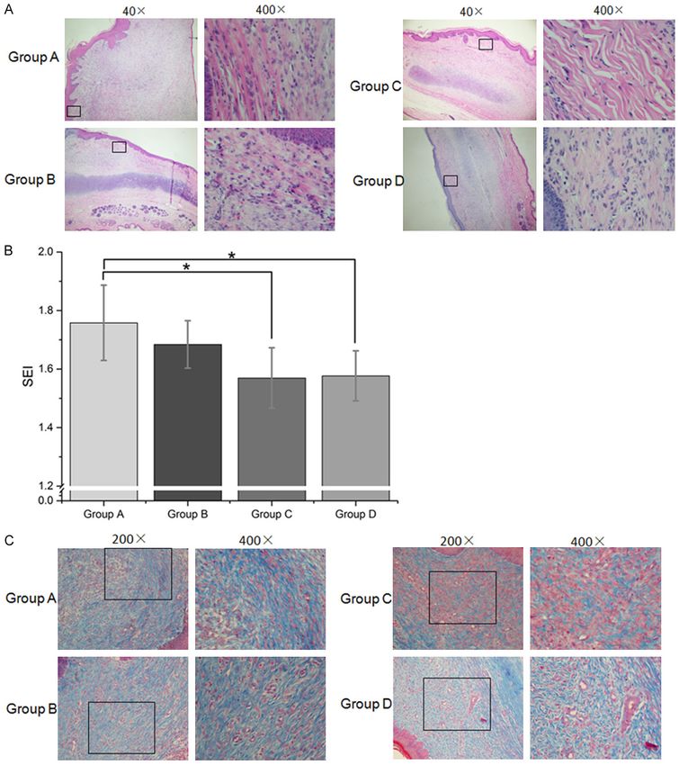
TF on hypertrophic scar

                                                                                ale SPF New Zealand white
                                                                                rabbits (weighing 1.8 to 2.6
                                                                                kg) were acquired from La-
                                                                                boratory Animal Center of
                                                                                Xinjiang Medical University
                                                                                (License No. SCXK 2003-001)
                                                                                and were single-housed under
                                                                                the same standard.

                                                                                Preparation of rabbit ear
                                                                                HS model, treatment and
                                                                                sampling

                                                                                On day 0, rabbits were anes-
                                                                                thetized by intramuscular in-
                                                                                jection with 0.15 mL/kg Zo-
                                                                                letile 50 (tiletamine hydro-
                                                                                chloride and zolazepam hyd-
                                                                                rochloride) and 0.1 mL/kg
                                                                                Ketamine, respectively. Und-
                                                                                er sterile conditions, six full-
                                                                                thickness 8-mm diameter ro-
                                                                                und excisional wounds were
                                                                                created down to the bare car-
                                                                                tilage on the ventral surface
                                                                                of each ear [17]. To bare the
                                                                                cartilage, the epidermis, der-
                                                                                mis, and perichondrium were
                                                                                removed under a surgical op-
                                                                                erating microscope [18]. Du-
                                                                                ring the excision, visible ves-
                                                                                sels were avoided and bleed-
                                                                                ing was treated by manually
                                                                                pressing with gauze. All of the
Figure 1. Gross images of samples representative scarring. A. On Day 15         wounds were then covered
post-wounding, the epithelialization was completed and the HS model was         with a sterile adhesive dress-
accomplished. B. After the second injection (Day 20), the HSs in Group C
and D were softer and had lower protuberant heights. C. At the end of the       ing. A total of 240 wounds
injection (Day 27), a much more significant effect in the TF injection groups   were created in 20 rabbits.
was observed. D. A special phenomenon was noticed, after the third injec-
tion (Day 24), the rabbit ear of Group A and Group B was slouched (left), and   On day 15, successfully estab-
the rabbit ear of the Group C and D became erect again (right). E. On day 27,   lished HS models were divid-
after the injection procedure, the HSs were harvest for further experiments.    ed into four groups along the
G.A: Group A; G.B: Group B; G.C: Group C; G.D: Group D.
                                                                                vertical axis of each ear from
                                                                                left to right in 19 rabbits (one
and its role in regulating the expression of key              rabbit died on day 2 probably due to the anes-
factors in TGF-β/Smads signaling pathway was                  thetization side effects) (Figure 1). Subsequent-
studied.                                                      ly, 219 HSs (9 scars were excluded because
                                                              of invagination) were treated by intralesional
Materials and methods                                         injection of either normal saline (Group A,
                                                              n=55) or different doses of TF solution (Group
Animals                                                       B, n=54, 2 mg/mL; Group C, n=56, 1 mg/mL;
                                                              Group D, n=54, 0.5 mg/mL) [17, 19]. The injec-
The Ethics Committee of the First Affiliated Hos-             tion was performed every three days and four
pital of Xinjiang Medical University approved                 times in total. In addition, since day 15 post-
all procedures of this experiment. Twenty fem-                wounding, photographs of ears and scarring

5629                                                                Int J Clin Exp Med 2018;11(6):5628-5637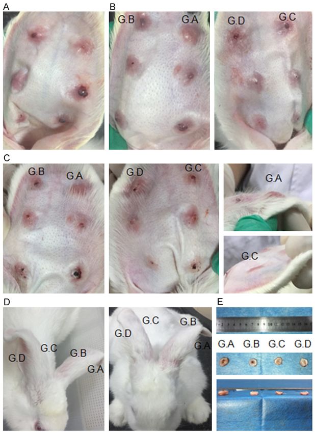
TF on hypertrophic scar

Table 1. Primer sequences for PCR amplification       night. Finally, sections were incubated with
Gene         Sequence (5’-3’)                         goat anti-rabbit secondary antibody (Boster,
                                                      China) or goat anti-mouse secondary antibody
Collagen III Forward: TGG GAA GCC AGG AGT TAA TG
                                                      (Boster, China) for 30 minutes at 37°C, followed
             Reverse: ATC CAG GGT TTC CGT CTC TT
                                                      by DAB (Boster, China) coloration for 5 minutes
TGF-β1       Forward: CCA GGA ATA CAG CAA CGA TTC C
                                                      and hematoxylin staining.
             Reverse: CCA CTG CCT CAC AAC TCC A
Smad 7       Forward: GTG GCA TAC TGG GAG GAG AA      All histological images were obtained with an
             Reverse: TTG TTG TCC GAA TTG AGC TG      optical microscope connected to cell Sens soft-
GAPDH        Forward: ATT TGA AGG GCG GAG CCA AA      ware (Olympus, Japan) and all of the measure-
             Reverse: TCA TGA GCC CCT CCA CAA TG      ments were performed with its measurement
                                                      function by two observers, blind to this experi-
                                                      ment, and then averaged. The cell number was
areas were taken before each time of injection        counted in five different microscopic regions
for qualitative changes in scar morphology [20].      (400×) for each section and then averaged.
On day 27, rabbits were sacrificed and 207 HSs
(a rabbit died after the second injection) were       Real-time PCR
harvested with a 3-mm unwounded margin
(Figure 1E). From each group, 45 HSs were ran-        Sixty (15 for each group) HS samples were
domly collected.                                      ground into powders in liquid nitrogen and then
                                                      total RNA was extracted using TRIzol® (Invit-
Histopathological analysis                            rogen) according to the manufacturer’s inst-
                                                      ructions. RNA was reverse-transcribed into
The 60 HSs (15 in each group) were fixed with         cDNA with a RevetAid First Strand cDNA Sy-
formaldehyde, embedded in paraffin, cut into          nthesis Kit (Thermo Scientific, USA). Real-time
4-μm sections, and stained for histopathologi-        PCR was performed using the Maxima SYBR
cal analysis.                                         Green qPCR Master Mix (Thermo Scientific,
                                                      USA) on the iQCycler thermocycler (Bio-Rad).
After hematoxylin and eosin (HE) staining, the        The primer sequences (synthesized by San-
protuberant degree was quantified by measur-          gon Biotech, China) are listed in Table 1. Real-
ing the scar elevation index (SEI) which repre-       time PCR was carried out as follows: initial
sents the ratio of total scar area to the estimat-    denaturation for 10 minutes at 95°C and 40
ed area below the protuberant portion that had        cycles of 15 seconds at 95°C and 60 seconds
new tissue with the same height as the sur-           at 60°C. Quantification was always normalized
rounding non-scarred dermis [17].                     to the internal control GAPDH and each tem-
                                                      plate was repeated three times under the same
To analyze collagen deposition and arrange-           condition.
ment, Masson’s trichrome staining was carried
out using a quick Masson’s trichrome staining         Western blot
kit (Njjcbio, China), following the manufactur-
er’s instructions.                                    Briefly, 60 HS tissue samples (15 each group)
                                                      were lysed with Pierce® RIPA buffer (Thermo
For immunohistochemistry, endogenous perox-           Scientific, USA) and PMSF (100:1). The extract-
idase was inactivated by 3% H2O2 incubation           ed proteins were quantified by Pierce® BCA
for 10 minutes at room temperature and then           Protein Assay Kit (Thermo Scientific, USA). Pro-
the sections were incubated with 1% citric acid       teins were separated on 12% SDS-PAGE and
for 10 minutes in microwave for antigen retriev-      transferred to polyvinylidene fluoride (PVDF)
al. After that, the sections were blocked with        membranes (Millipore, Bedford, USA). Then, the
normal goat serum (ZSGB-Bio, China) for 15            membranes were incubated with primary anti-
minutes at 37°C and then incubated with pri-          bodies of collagen I (Cat# ab90395; 1:500 dilu-
mary antibodies of collagen I (Cat# ab90395;          tion; Abcam), collagen III (Cat# ab7778; 1:1000
1:1000 dilution; Abcam), collagen III (Cat#           dilution; Abcam), TGF-β1 (Cat# sc-146; 1:200
ab7778; 1:5000 dilution; Abcam), TGF-β1 (Cat#         dilution; Abcam), Smad-7 (Cat# sc-365846;
sc-146; 1:500 dilution; Santa Cruz Biotech-           1:200 dilution; Abcam), and β-actin (Cat# sc-
nology), and Smad-7 (Cat# sc-365846; 1:500            47778; 1:500 dilution; Santa Cruz Biotech-
dilution; Santa Cruz Biotechnology) at 4°C over-      nology) overnight at 4°C. After washing, HRP-

5630                                                       Int J Clin Exp Med 2018;11(6):5628-5637
TF on hypertrophic scar
Figure 2. Morphology analysis of hyper-
trophic scars after TF injection. A. HE
staining showed different pro-tuberant
degrees among these groups. B. The SEI
was measured according to the observa-
tion of HE staining. The SEI significantly
decreased in Group C and D. *P < 0.05,
compared with the control group (saline
injection). C. Masson staining revealed
the collagen distribution and arrange-
ment in the HS tissues.
conjugated goat anti-rabbit or goat anti-mouse dent’s t-test. One-way ANOVA was used to com-
secondary antibody (Abbkine, USA) was added pare mean values of the four groups simultane-
and incubated at room temperature for 2 hours. ously, followed by Bonferroni’s post hoc test or
The protein bands were visualized with the ECL Tamhane’s method when the variance was not
Kit (Thermo Scientific, USA) and densities of homogenous. A P value < 0.05 was considered
the bands were compared with that of β-Actin. statistically significant.
Statistical analysis Results
All data were analyzed using SPSS version 22 Effect of TF on gross scar formation
(SPSS, Inc., Chicago, IL, USA) and are present-
ed as mean ± standard deviation (SD). Inter- To dynamically observe the scar formation and
group comparisons were analyzed by Stu- the effect of TF on scar formation since day
5631 Int J Clin Exp Med 2018;11(6):5628-5637TF on hypertrophic scar
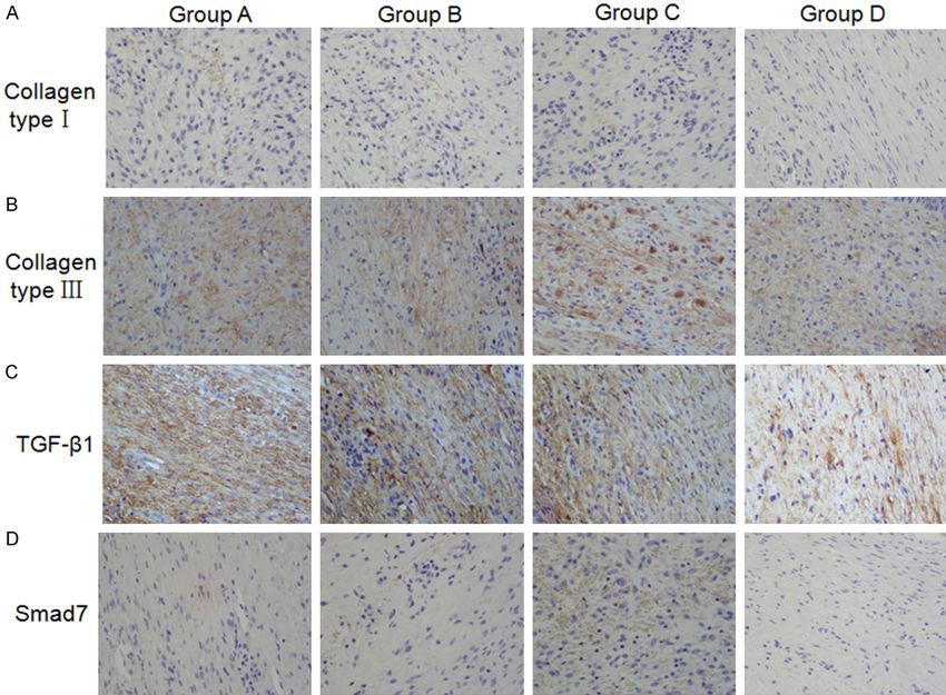
Figure 3. Representative immunohistochemical staining of Type I (A) and Type III collagens (B), TGF-β1 (C) and Smad
7 (D) in each group.
15 post-wounding (Figure 1A), scars were tive analysis of SEI presented a remarkable
observed every day and photographed be- decreasing in Groups C and D (Figure 2B).
fore each injection. The intralesional TF in- These results suggest that treatment with a
jection groups showed obvious softness after proper dose of TF may inhibit the proliferation
the second injection compared with the control of fibroblasts and less decrease the protuber-
group (Figure 1B). Some HSs in Groups C and ant degree of HSs.
D almost resembled normal skin, which had
lower protuberant heights (Figure 1C). Some The HS tissues were subjected to Masson
staining to examine the effect of TF on
vulnerable epidermis samples were observed
collagen arrangement. It showed that the
in Group B, in a form of bleeding or ulceration.
collagen was sparsely distributed and arrang-
In addition, a rabbit showed slouched ears
ed in a net-like shape similar to the adjacent
because of the scar contracture but the sides
unwounded tissues in the TF treated groups,
of rabbit ears in Groups C and D became erect-
especially in Groups C and D, while the collagen
ed again after the third injection (Figure 1D).
of the control group was densely arranged, par-
These results indicate that TF may soften the
allel to the epithelium and less mature with
scar and delay wound-healing. more fibroblasts (Figure 2C). Therefore, treat-
Effect of TF on SEI and collagen ment with a proper dose of TF may lead to col-
lagen arranging and depositing in a beneficial
To assess the effects of TF on cell morphology, direction.
HE staining was performed. The cells of the The effect of TF on the protein expressions of
control group were primarily spindle-shaped Type I and III collagens, TGF-β1 and Smad 7
while those in the TF injection groups were
round. The number of fibroblasts was lowest in In order to observe the expression of Type I and
Group C (Figure 2A). Furthermore, the quantita- III collagens, TGF-β1 and Smad 7 in the HS tis-
5632 Int J Clin Exp Med 2018;11(6):5628-5637TF on hypertrophic scar
Figure 4. The protein expression levels of Type I and Type III collagens, TGF-β1 and Smad 7. The protein levels
were analyzed by Western-blot. β-actin was used as an internal control. The representative and quantitative results
of Type I (A) and Type III collagens (B), TGF-β1 (C) and Smad 7 (D) were shown. *P < 0.05, compared to the control
group.
sues, immunohistochemical staining was per- and 4B, P < 0.05). TGF-β1 protein expression
formed. There was less Type I and III collagen was effectively suppressed in Group C and D,
deposition in cellular mesenchyme after TF tre- while a higher expression was detected in
atment compared with the control group (Figure Group B (Figure 4C, P < 0.05). The high expres-
3A and 3B). Also, expression of TGF-β1 was sion of TGF-β1 protein in Group B (TF, 2 mg/mL)
obviously reduced in Group C and D (Figure 3C) might be due to the damage of tissues and
while that of Smad 7 increased in the TF treat- cells caused by the high dose of TF [13]. By con-
ed groups, especially in Group C (Figure 3D). trast, Smad 7 protein expression significantly
increased after TF treatment, among which
To further verify the effects of TF treatment on Group D (TF, 0.5 mg/mL) showed the highest
the protein expression of Type I and Type III col- expression (Figure 4D, P < 0.05).
lagens, TGF-β1 and Smad 7, Western blot was
performed. The results showed that Type I Thus, it is assumed that TF might reduce colla-
and Type III collagen expressions remarkably gen deposition and TGF-β1 expression but pro-
reduced in the TF treated groups (Figure 4A mote Smad 7 expression.
5633 Int J Clin Exp Med 2018;11(6):5628-5637TF on hypertrophic scar
Figure 5. The effect of TF on the mRNA expression of Type I (A) collagen, Type III collagen (B), TGF-β1 (C) and Smad
7 (D). mRNA expression was detected with real-time PCR and normalized to that of GADPH. The horizontal bar indi-
cated the mean of the measurements in each group. *P < 0.05, compared with the control group.
The effect of TF on the mRNA expression of Discussion
Type I and III collagens, TGF-β1, and Smad 7
Scar formation is the normal consequence of
To detect the mRNA expression of Type I and III skin injury. It is thin and resembles normal skin
collagens, TGF-β1 and Smad 7, quantitative under correct regulation. However, prolong-
RT-PCR was carried out. The results showed ed inflammation and chronic stimulation may
that Type I collagen mRNA expression was cause excessive deposition of collagen and pr-
suppressed in all the TF treated groups (Figure oliferation of fibroblasts, eventually leading to
5A, P < 0.05), especially in Group C (TF, 1 mg/ HS formation [6, 7, 21]. So far, the treatment for
mL). The mRNA expression of Type III collagen HS remains unsatisfying [22]. TF, one effective
and TGF-β1 was inhibited in the TF treated ingredient in traditional Chinese medicine, is
groups, among which the effect in Group D (TF, found to have an anti-proliferative property to
certain cells [11, 23, 24]. Preliminary studies
0.5 mg/mL) was the most significant (Figure 5B
have shown that total flavonoids (TF) extracted
and 5C, P < 0.05). In contrast, mRNA expres-
from ASMq might prevent HS formation [11-14].
sion of Smad 7, which is considered as a
The present study found that TF could soften
unique inhibitor of the TGF-β/Smad signaling
HSs and decrease the SEI of the rabbit ear
pathway, in the TF treated groups was much model in vivo.
higher than that of the control group (Figure
5D, P < 0.05). These results indicate that Previous studies have suggested that wound-
mRNA levels of collagen Type I and III, and TGF- healing and scarring are closely related to TGF-
β1 were downregulated after a proper dose of β family members, which also regulate a wide
TF treatment, while Smad 7 was upregulated. spectrum of cellular functions such as prolifer-
5634 Int J Clin Exp Med 2018;11(6):5628-5637TF on hypertrophic scar
ation, apoptosis, differentiation, and migration ads), common-partner Smads (Co-Smads), and
[25, 26]. Increased amounts of TGF-β have inhibitory Smads (I-Smads). Smad 7, which acts
been found in HS and the scar-free healing in as the I-Smads of TGF-β family signaling, can
human fetal is considered as a result of TGF-β bind with TGF-β receptor and thus prevent the
deficient [27]. Furthermore, it has been report- recruitment and phosphorylation of effector
ed that TGF-β can mediate fibroblast prolifera- Smads and inhibiting TGF-β/Smads signaling
tion, angiogenesis, ECM synthesis, and re-epi- [7, 37, 41]. The TGF-β/Smad signaling system is
thelialization in the wound-healing process [28, an auto inhibitory loop to control the intensity
29]. Overexpression of TGF-β1 and β2 were and duration of TGF-β signaling response. TGF-
detected in HSs, whereas TGF-β3 was reported β stimulation could result in the export of
to have anti-fibrotic effects [7]. Particularly, Smad 7 from nucleus [36]. Therefore, the
TGF-β1 transcriptionally regulates various fibro- expression level of Smad 7 will influence TGF-β
sis-related proteins, including Type I and III col- transcriptional responsiveness. Cells with a
lagens [30, 31]. It can also promote the trans- higher level of Smad 7 are more inclined to
formation of fibroblasts to myofibroblasts, resist the pro-fibrotic actions of TGF-β. As this
which are the major cells contributing to HS study indicated, TF treatment blocked the
formation and characterized by an increased TGF-β/Smad signaling via increasing the
propensity to synthesize collagen and upregula- expression of Smad 7 and thus the stimula-
tion of cytokines [32, 33]. Therefore, it is tion effect of TGF-β1 on collagen deposition in
speculated that inhibition of TGF-β1 activity ECM was inhibited.
would have potential benefits in suppressing
HS formation. In conclusion, a rabbit ear HS model was estab-
lished. A proper dose of TF could negatively
In spite of the limited varieties of TGF-β recep- regulate expression of Type I and III collagens
tors and Smads, there may be a greater versa- and collagen deposition. TGF-β1 transcription
tility of the signaling possibilities than we used was down regulated which might be partially
to expect. Combinatorial interactions between due to Smad 7 upregulation. This study sug-
TGF-β receptors and Smads in oligomeric com- gests that TF may have potential to prevent HS
plexes allow substantial diversity and are com- formation.
plemented by various sequence-specific tran-
scription factors cooperating with Smads, Acknowledgements
resulting in context-dependent transcriptional
This research work was supported by the
regulation [34-36]. In addition, other signaling
National Natural Science Foundation of China
pathways may also help to define the respons-
(No. 81260291).
es to TGF-β and it is evidently shown that TGF-
β-related protein activation is not a linear sig- Disclosure of conflict of interest
naling transduction pathway. These pathways
that regulate Smad-dependent responses also None.
induce Smad-independent responses [37]. On
one hand, the TGF-β1-activated Smad-depen- Address correspondence to: Shaolin Ma, Depart-
dent pathways can cause cellular changes su- ment of Plastic Surgery, The First Affiliated Hospi-
ch as the synthesis and secretion of collagen, tal of Xinjiang Medical University, 137 South
leading to increased scar formation. However, Liyushan Road, Urumqi 830054, China. Tel: 0086-
Smad-independent signaling pathways may 13639934408; E-mail: mashaolin9@outlook.com
improve healing. Therefore, inhibition of TGF-β1
transcription may reduce scarring but delay References
wound healing [38-40]. In this study, it was
[1] Gurtner GC, Werner S, Barrandon Y and Lon-
observed that treatment with a proper dose of gaker MT. Wound repair and regeneration. Na-
TF could lessen scarring and delay healing. ture 2008; 453: 314-321.
Meanwhile, expression of Smad 7 was up- [2] Butzelaar L, Ulrich MM, Mink van der Molen
regulated and that of TGF-β1 was decreased. AB, Niessen FB and Beelen RH. Currently
known risk factors for hypertrophic skin scar-
The Smad family is divided into three distinct ring: a review. J Plast Reconstr Aesthet Surg
subfamilies: receptor-regulated Smads (R-Sm- 2016; 69: 163-169.
5635 Int J Clin Exp Med 2018;11(6):5628-5637TF on hypertrophic scar
[3] Rha EY, Kim YH, Kim TJ, Yoo G, Rhie JW, Kim HJ [15] Wang H, Gao W and Ma S. Preliminary study of
and Park IK. Topical application of a silicone the effect of abnormal savda munziq on
gel sheet with verapamil microparticles in a TGF-β1 and Smad 7 expression in hypertrophic
rabbit model of hypertrophic scar. Plast Recon- scar fibroblasts. Int J Clin Exp Med 2015; 8:
str Surg 2016; 137: 144-151. 519-525.
[4] Tredget EE, Levi B and Donelan MB. Biology [16] Wang HJ, Gao WC and Ma SL. Effect of abnor-
and principles of scar management and burn mal Savda Munziq on hypertrophic scar forma-
reconstruction. Surg Clin North Am 2014; 94: tion in a rabbit ear model. Chin J Integr Med
793-815. 2014; 21: 537-541.
[5] Finnerty CC, Jeschke MG, Branski LK, Barret [17] Morris DE, Wu L, Zhao LL, Bolton L, Roth SI,
JP, Dziewulski P and Herndon DN. Hypertro- Ladin DA and Mustoe TA. Acute and chronic
phic scarring: the greatest unmet challenge animal models for excessive dermal scarring:
after burn injury. Lancet 2016; 388: 1427- quantative studies. Plast Reconstr Surg 1997;
1436. 100: 674-81.
[6] Armour A, Scott PG and Tredget EE. Cellular [18] Yagmur C, Guneren E, Kefeli M and Ogawa R.
and molecular pathology of HTS: basis for The effect of surgical denervation on preven-
treatment. Wound Repair Regen 2007; 15: S6- tion of excessive dermal scarring: a study on
S17. rabbit ear hypertrophic scar model. J Plast Re-
[7] van der Veer WM, Bloemen MC, Ulrich MM, constr Aesthet Surg 2011; 64: 1359-1365.
Molema G, van Zuijlen PP, Middelkoop E and [19] Meuli M, Liu Y, Liggitt D, Kashani-Sabet M,
Niessen FB. Potential cellular and molecular Knauer S, Meuli-Simmen C, Harrison MR,
causes of hypertrophic scar formation. Burns Adzick NS, Heath TD and Debs RJ. Efficient
2009; 35: 15-29. gene expression in skin wound sites following
[8] Jin JH, Kim JS, Kang SS, Son KH, Chang HW, local plasmid injection. J Invest Dermatol
Kim HP. Anti-inflammatory and anti-arthritic 2001; 116: 131-5.
activity of total flavonoids of the roots of so- [20] Saulis AS, Mogford JH and Mustoe TA. Effect of
phora flavescens. J Ethnopharmacol 2010; mederma on hypertrophic scarring in the rab-
127: 589-595. bit ear model. Plast Reconstr Surg 2002; 110:
[9] Yuan LP, Chen FH, Ling L, Dou PF, Bo H, Zhong 177-83; discussion 184-6.
MM, Xia LJ. Protective effects of total flavo- [21] Berman B, Maderal A and Raphael B. Keloids
noids of Bidens pilosa L. (TFB) on animal liver and hypertrophic scars: pathophysiology, clas-
injury and liver fibrosis. J Ethnopharmacol
sification, and treatment. Dermatol Surg 2017;
2008; 116: 539-546.
43 Suppl 1: S3-S18.
[10] Chien ST, Shi MD, Lee YC, Te CC, Shih YW.
[22] Ogawa R, Akaishi S, Kuribayashi S and Miyas-
Galangin, a novel dietary flavonoid, attenuates
hita T. Keloids and hypertrophic scars can now
metastatic feature via PKC/ERK signaling
be cured completely: recent progress in our
pathway in TPA-treated liver cancer HepG2
understanding of the pathogenesis of keloids
cells. Cancer Cell Int 2015; 15: 15.
and hypertrophic scars and the most promis-
[11] Zhang Y, Shan S, Wang J, Cheng X, Yi B, Zhou J
ing current therapeutic strategy. J Nippon Med
and Li Q. Galangin inhibits hypertrophic scar
Sch 2016; 83: 46-53.
formation via ALK5/Smad2/3 signaling path-
[23] Santos EO, Kabeya LM, Figueiredo-Rinhel AS,
way. Mol Cell Biochem 2016; 413: 109-118.
Marchi LF, Andrade MF, Piatesi F, Paoliello-Pas-
[12] Middleton E Jr, Kandaswami C, Theoharides
choalato AB, Azzolini AE, Lucisano-Valim YM.
TC. The effects of plant flavonoids on mamma-
Flavonols modulate the effector functions of
lian cells: implications for inflammation, heart
disease, and cancer. Pharmacol Rev 2000; healthy individuals’ immune complex-stimulat-
52: 673-751. ed neutrophils: a therapeutic perspective for
[13] Li N, Kong M, Ma T, Gao W and Ma S. Uighur rheumatoid arthritis. Int Immunopharmacol
medicine abnormal savda munzip (ASMq) sup- 2014; 21: 102-111.
presses expression of collagen and TGF-β1 [24] Johnson JL, Gonzalez d ME. Interactions be-
with concomitant induce Smad 7 in human hy- tween dietary flavonoids apigenin or luteolin
pertrophic scar fibroblasts. Int J Clin Exp Med and chemotherapeutic drugs to potentiate an-
2015; 8: 8551-60. ti-proliferative effect on human pancreatic can-
[14] Wang H, Gao W, Kong M, Li N and Shaolin M. cer cells, in vitro. Food Chem Toxicol 2013; 60:
Effects of abnormal Savda Munzip on the pro- 83-91.
liferation activity and migration ability of fibro- [25] Tziotzios C, Profyris C and Sterling J. Cutane-
blasts derived from hypertrophic scar in vitro. ous scarring: pathophysiology, molecular
Evid Based Complement Alternat Med 2015; mechanisms, and scar reduction therapeutics
2015: 1-6. Part II. Strategies to reduce scar formation af-
5636 Int J Clin Exp Med 2018;11(6):5628-5637TF on hypertrophic scar
ter dermatologic procedures. J Am Acad Der- [34] Qi SH, Xie JL, Pan S, Xu YB, Li TZ, Tang JM, Liu
matol 2012; 66: 13-24. XS, Shu B and Liu P. Effects of asiaticoside on
[26] Hwangbo C, Tae N, Lee S, Kim O, Park OK, Kim the expression of Smad protein by normal skin
J, Kwon SH and Lee JH. Syntenin regulates fibroblasts and hypertrophic scar fibroblasts.
TGF-β1-induced Smad activation and the epi- Clin Exp Dermatol 2008; 33: 171-175.
thelial-to-mesenchymal transition by inhibiting [35] Itoh S, Itoh F, Goumans MJ and Ten Dijke P. Sig-
caveolin-mediated TGF-β type I receptor inter- naling of transforming growth factor-β family
nalization. Oncogene 2015; 35: 389-401. members through Smad proteins. Eur J Bio-
[27] Zheng Z, Zhang X, Dang C, Beanes S, Chang chem 2000; 267: 6954-6967.
GX, Chen Y, Li CS, Lee KS, Ting K and Soo C. [36] Itóh S, Landström M, Hermansson A, Itoh F,
Fibromodulin is essential for fetal-type scar- Heldin CH, Heldin NE and ten Dijke P. Trans-
less cutaneous wound healing. Am J Pathol forming growth factor beta1 induces nuclear
2016; 186: 2824-2832. export of inhibitory Smad 7. J Biol Chem 1998;
[28] Wick G, Grundtman C, Mayerl C, Wimpissinger 273: 29195-29201.
TF, Feichtinger J, Zelger B, Sgonc R and Wol- [37] Derynck R and Zhang YE. Smad-dependent
fram D. The immunology of fibrosis. Annu Rev and Smad-independent pathways in TGF-β
Immunol 2013; 31: 107-135. family signalling. Nature 2003; 425: 577-84.
[29] Muppala S, Xiao R, Krukovets I, Verbovetsky D, [38] Altan ZM and Fenteany G. c-Jun N-terminal ki-
Yendamuri R, Habib N, Raman P, Plow E and nase regulates lamellipodial protrusion and
Stenina-Adognravi O. Thrombospondin-4 medi- cell sheet1 migration during epithelial wound
ates TGF-beta-induced angiogenesis. Onco- closure by a gene expression-independent
gene 2017; 36: 5189-5198. mechanism. Biochem Biophys Res Commun
[30] Hsieh YP, Chen HM, Lin HY, Yang H and Chang 2004; 322: 56-67.
JZ. Epigallocatechin-3-gallate inhibits transfor- [39] Liu X, Li P, Liu P, Xiong R, Zhang E, Chen X, Gu
ming-growth-factor-β1-induced collagen syn- D, Zhao Y, Wang Z and Zhou Y. The essential
thesis by suppressing early growth response-1 role for c-Ski in mediating TGF-β1-induced bi-
in human buccal mucosal fibroblasts. J Formos directional effects on skin fibroblast prolifera-
Med Assoc 2017; 116: 107-113. tion through a feedback loop. Biochem J 2008;
[31] Ide M, Jinnin M, Tomizawa Y, Wang Z, Kajihara 409: 289-297.
I, Fukushima S, Hashizume Y, Asano Y and Ihn [40] Lu L, Saulis AS, Liu WR, Roy NK, Chao JD, Led-
H. Transforming growth factor beta-inhibitor better S and Mustoe TA. The temporal effects
Repsox downregulates collagen expression of of anti-TGF-β1, 2, and 3 monoclonal antibody
scleroderma dermal fibroblasts and prevents on wound healing and hypertrophic scar for-
bleomycin-induced mice skin fibrosis. Exp Der- mation. J Am Coll Surg 2005; 201: 391-397.
matol 2017; 26: 1139-1143. [41] Massagué J. How cells read TGF-β signals. Nat
[32] Klingberg F, Hinz B and White ES. The myofi- Rev Mol Cell Biol 2000; 1: 169-78.
broblast matrix: implications for tissue repair
and fibrosis. J Pathol 2013; 229: 298-309.
[33] Crider BJ, Risinger GM Jr, Haaksma CJ, Howard
EW and Tomasek JJ. Myocardin-related tran-
scription factors A and B are key regulators of
TGF-beta1-induced fibroblast to myofibroblast
differentiation. J Invest Dermatol 2011; 131:
2378-2385.
5637 Int J Clin Exp Med 2018;11(6):5628-5637You can also read