Aggravation of hepatic ischemia reperfusion injury with increased inflammatory cell infiltration is associated with the TGF β/Smad3 signaling pathway
←
→
Page content transcription
If your browser does not render page correctly, please read the page content below
MOLECULAR MEDICINE REPORTS 24: 580, 2021

             Aggravation of hepatic ischemia‑reperfusion injury with
              increased inflammatory cell infiltration is associated
                   with the TGF‑β/Smad3 signaling pathway
                HAIXIA LI1*, XIAOYUN SHEN2*, YIFAN TONG2, TONG JI2, YAN FENG3, YANPING TANG3,
                          RONGYUN MAI3, JIAXIANG YE1, TING QUE1 and XIAOLING LUO1

               1
                Department of Immunology, School of Basic Medical Sciences, Guangxi Medical University, Nanning,
                Guangxi 530021; 2Key Laboratory of Endoscopic Technology Research, Sir Run Run Shaw Hospital,
                   Zhejiang University School of Medicine, Hangzhou, Zhejiang 310016; 3Research Department,
                  Affiliated Tumor Hospital of Guangxi Medical University, Nanning, Guangxi 530021, P.R. China

                                        Received November 15, 2020; Accepted May 18, 2021

                                                     DOI: 10.3892/mmr.2021.12219

Abstract. Ischemia‑reperfusion (IR) injury is a major chal‑             Aggravated hepatic injury, increased apoptosis and enhanced
lenge influencing the outcomes of hepatic transplantation.              inflammatory cell infiltration induced by hepatic IR injury
Transforming growth factor‑β (TGF‑β) and its downstream                 were observed in the Smad3‑/‑ mice compared with in Smad3+/+
gene, SMAD family member 3 (Smad3), have been implicated                mice. Collectively, the current findings suggested that activation
in the pathogenesis of chronic hepatic injuries, such as hepatic        of the TGF‑β/Smad3 signaling pathway was present alongside
fibrosis. Thus, the present study aimed to investigate the role of      the hepatic injury induced by IR. However, the TGF‑β/Smad3
the TGF‑β/Smad3 signaling pathway on hepatic injury induced             signaling pathway may have an effect on protecting against liver
by IR in vivo. In total, 20 129S2/SvPasCrl wild‑type (WT) mice          tissue damage caused by IR injury in vivo.
were randomized into two groups; 10 mice underwent IR injury
surgery and 10 mice were sham‑operated. Histopathological               Introduction
changes in liver tissues and serum levels of alanine aminotrans‑
ferase (ALT) were examined to confirm hepatic injury caused             Hepatic ischemia‑reperfusion (IR) injury is a common compli‑
by IR surgery. The expression levels of TGF‑β1, Smad3 and               cation that occurs due to a variety of factors, such as liver
phosphorylated‑Smad3 (p‑Smad3) were detected via western                transplantation, shock and trauma (1). Temporary blood flow
blotting. Furthermore, a total of five Smad3‑/‑ 129S2/SvPasCrl          deprivation (ischemia) and restoration (reperfusion) of the organs
mice (Smad3‑/‑ mice) and 10 Smad3+/+ littermates received IR            are the primary pathological processes occurring in IR (2). Liver
surgery, while another five Smad3‑/‑ mice and 10 Smad3+/+ litter‑       parenchymal cell death is caused by ischemic injury, which
mates received the sham operation. Histopathological changes            involves metabolic disorders and oxidative stress. Moreover,
in liver tissues and serum levels of ALT were then compared             inflammatory mediators cause further damage during blood
between the groups. Furthermore, hepatic apoptosis and inflam‑          reperfusion (3). In addition, hepatic IR injury affects the quality
matory cell infiltration after IR were evaluated in the liver tissues   of donor livers and the prognosis of liver transplantation (4).
of Smad3‑/‑ mice and Smad3+/+ mice. The results demonstrated                The transforming growth factor‑β (TGF‑β) superfamily
that the expression levels of TGF‑β1, Smad3 and p‑Smad3                 exerts multiple biological functions via the secretion of inhibins,
were elevated in hepatic tissue from WT mice after IR injury.           activins and bone morphogenetic proteins (BMPs), which are
                                                                        involved in regulating a range of biological processes, for
                                                                        example BMPs can induce endochondral bone formation (5‑7).
                                                                        TGF‑β is an important activated mediator of myofibroblasts,
                                                                        which activates stellate cells to secrete collagen fibers, in turn
Correspondence to: Professor Xiaoling Luo, Department of
                                                                        leading to liver fibrosis (8,9). The abnormal expression of
Immunology, School of Basic Medical Sciences, Guangxi Medical
University, 22 Shuangyong Road, Nanning, Guangxi 530021,
                                                                        TGF‑β not only promotes the proliferation and migration of
P.R. China                                                              liver cancer cells, but it is also associated with viral hepatitis,
E‑mail: luoxiaoling67@126.com                                           hepatic failure and other chronic hepatic diseases (10‑14). As
                                                                        the downstream effector of TGF‑β, SMAD family member 3
*
    Contributed equally                                                 (Smad3) activation is induced by its phosphorylation to phos‑
                                                                        phorylated‑Smad3 (p‑Smad3) and the signal is transported
Key words: transforming growth factor‑β, SMAD family member 3,          to the nucleus, thus forming the classic TGF‑β/Smad3 signal
ischemia‑reperfusion, hepatic injury, Smad3 mutant mice                 transduction pathway (15). The Smad3 linker region can be
                                                                        phosphorylated by intracellular kinases and affects TGF‑β
                                                                        responses, such as tumor growth inhibition (16).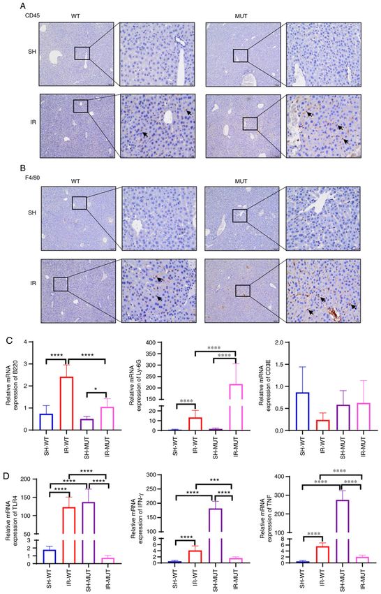
2                                 LI et al: ROLE OF TGF‑β/Smad3 IN ISCHEMIA‑REPERFUSION INJURY

    A previous study reported that the TGF‑β/Smad3 signaling          In brief, a vertical incision was made to each layer to expose
pathway was activated and promoted ventricular remodeling             the liver after the mouse was anesthetized (4% chloral hydrate
after IR injury in rats (17), while mediating the protection          sodium; 400 mg/kg) by intraperitoneal injection. The blood
of myocardial cells against IR injury via the stimulation of          vessels, except those of the caudate and right lobes, were
sphingosine‑1‑phosphate (S1P)/S1P receptor 1. TGF‑β1/Smad3            occluded with the vascular clamp for 30 min to block the blood
has also been reported to exert an important effect on cerebral       flow without injuring the remaining liver tissues. Subsequently,
ischemia. For example, Smad3 has been shown to exhibit neuro‑         the vascular clamp was gently removed and the blood flow and
protective effects on the brain following IR via the induction of     liver tissues were assessed without injuring before the wound
anti‑inflammatory and anti‑apoptotic pathways (18). Thus, it was      was sutured. In the sham group, surgery was performed by
hypothesized that the TGF‑β1/Smad3 signaling pathway may be           exposing the blood vessels only for 30 min without blocking.
able to protect liver cells from IR injury. Therefore, the present    Subsequently, recipient animals were intraperitoneally injected
study investigated the role of TGF‑β/Smad3 in achieving clinical      with anesthesia (4% chloral hydrate sodium; 400 mg/kg) at the
transformation in IR‑induced acute liver injury and provide a         end of the predetermined period (after 6 h of reperfusion), and
novel therapeutic target for the prognosis of liver surgery.          then the blood samples were collected from the orbital venous
                                                                      plexus to assess serum alanine aminotransferase (ALT) levels.
Materials and methods                                                 Mice were then sacrificed by cervical dislocation, and the
                                                                      death of mice was confirmed when the respiration and various
Animals. In total, 20 129S2/SvPasCrl wild‑type (WT) mice,             reflexes had ceased. A portion of the IR injury tissue was used
aged 6‑8 weeks and weighing 22‑25 g, were purchased                   for extraction and detection of total protein and RNA. Then,
from the Laboratory Animal Center of Zhejiang University              ~1.0x1.0x1.0‑cm liver tissues were promptly fixed overnight
School of Medicine (Hangzhou, China) and were random‑                 in 4% paraformaldehyde (cat. no. 6148; Sigma‑Aldrich;
ized into sham‑operated (SH‑WT) and IR injury (IR‑WT)                 Merck KGaA) at room temperature.
groups (n=10/group). A total of four Smad3+/‑ heterozygous
129S2/SvPasCrl mice were donated by The Second Military               Histopathological evaluation. The hepatic tissues were fixed
Medical University (Shanghai, China). Mating of Smad3+/‑              in 4% paraformaldehyde for 8 h at room temperature, dehy‑
mice was followed by sibling mating of offspring‑generated            drated, embedded in paraffin, sectioned to 3‑µm thickness,
Smad3+/‑ mice, to obtain Smad3+/+ mice and Smad3‑/‑ mice for          stained with hematoxylin for 3 min, rinsed under running
experiments. The mouse genotypes were identified at 3 weeks           water for 5 min, stained with eosin for 2 min and sealed at room
of age, and DNA was isolated from the toes of mice by heating         temperature. Stained tissues were viewed and imaged using a
to 100˚C with 50 mM NaOH for 10 min. Then, 2X Phanta Max              light microscope. The Suzuki histological grading scores for
Master Mix (cat. no. P515‑01; Vazyme Biotech Co., Ltd.) was           liver damage were determined as described previously (21).
used for PCR amplification. The amplification conditions were         A score of 0 indicated minimal or no evidence of damage,
set as follows: Initial denaturation at 95˚C for 3 min, followed by   1 indicated mild damage with cytoplasmic vacuolation and
35 cycles at 95˚C for 15 sec, 59˚C for 15 sec and 72˚C for 30 sec.    interstitial disorder, 2 indicated moderate to severe damage
Then, 3% Tris‑acetate‑EDTA buffer agarose gel electrophoresis         with extensive nuclear pyknosis and interstitial congestion,
was used to identify the Smad3 gene‑deficient mouse genotype,         and 3 indicated serious necrosis with disintegration of liver
and visualized under the ChemiDoc™ System (cat. no. 1708265;          cells, hemorrhage and inflammatory cell infiltration.
Bio‑Rad Laboratories, Inc.). The primers used for genotyping
are listed in Table I. The overall morphology of Smad3 mice           Detection of ALT levels. Blood samples were centrifuged
was assessed and homozygous Smad3‑/‑ mutant (MUT) mice                at 4˚C for 5 min at 1,500 x g after standing for 1 h at room
were found to be smaller than littermate Smad3+/‑ and Smad3+/+        temperature and the serum was separated. Serum levels of ALT
mice. Then, the mice were divided into four groups: Smad3+/+          were determined using an ALT Assay kit (cat. no. C009‑2‑1;
mice that underwent a sham operation (SH‑WT; n=10); Smad3+/+          Nanjing Jiancheng Biological Technology), according to the
mice with IR liver injury (IR‑WT; n=10); Smad3‑/‑ mice that           manufacturer's instructions.
underwent a sham operation (SH‑MUT; n=5); and Smad3‑/‑
mice with IR liver injury (IR‑MUT; n=5).                              Reverse transcription‑quantitative PCR (RT‑qPCR). The fresh
    All mice were maintained on a 12/12‑h light/dark cycle under      hepatic tissue was homogenized using an automatic sample quick
controlled humidity (50±10%) and temperature (25±0.5˚C) in            grinding machine (model JXFSTPRP‑24; Shanghai Jingxin
a specific pathogen‑free environment and allowed free access          Industrial Development Co., Ltd.). Total RNA was extracted using
to standard chow and water in the Experimental Animal                 TRIzol® reagent (cat. no. 10296‑028; Invitrogen; Thermo Fisher
Center of Sir Run Run Shaw Hospital, Zhejiang University              Scientific, Inc.) according to the manufacturer's instructions.
School of Medicine. All animal experiments included in                cDNA was synthesized from RNA using the Hifair® II 1st Strand
this protocol adhere to the Animal Research: Reporting                cDNA Synthesis SuperMix for RT (cat. no. 11121ES60; Shanghai
In Vivo Experiments guidelines (19), and were approved by             Yeasen Biotechnology Co., Ltd.). The temperature conditions
the Animal Testing Ethics Committee of Sir Run Run Shaw               reverse transcription were as follows: 42˚C for 15 min and 85˚C
Hospital, Zhejiang University School of Medicine (approval            for 2 min. qPCR was performed using the FastStart Universal
no. 20171120‑14).                                                     SYBR Green Master (ROX) mix (cat. no. 04913914001; Roche
                                                                      Diagnostics), according to the manufacturer's instructions. The
Model of IR liver injury. The present study used a non‑lethal         amplification conditions were set as: Initial denaturation at 95˚C
segmental (70%) liver IR model as previously described (20).          for 10 min, followed by 40 cycles at 95˚C for 15 sec, 60˚C for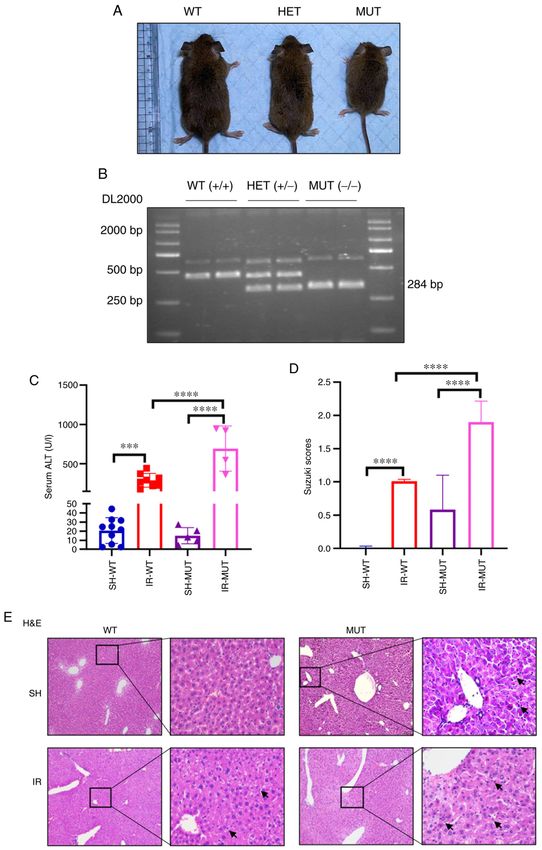
MOLECULAR MEDICINE REPORTS 24: 580, 2021                                                  3

Table I. Primers used for genotyping and reverse transcription‑quantitative PCR.

Target gene                              Forward primer (5'→3')                                    Reverse primer (5'→3')

Smad3 WT/MUT                     CCACTTCATTGCCATATGCCCTG
Smad3 WT                         CCCGAACAGTTGGATTCACACA
Smad3 MUT                        CCAGACTGCCTTGGGAAAAGC
β‑actin                          GGCTGTATTCCCCTCCATCG                                    CCAGTTGGTAACAATGCCATGT
TLR4                             ATGGCATGGCTTACACCACC                                    GAGGCCAATTTTGTCTCCACA
IFN‑γ                            AGGTCAACAACCCACAGGTC                                    ATCAGCAGCGACTCCTTTTC
TNF                              TAGCTCCCAGAAAAGCAAGC                                    TTTTCTGGAGGGAGATGTGG
B220                             GTTTTCGCTACATGACTGCACA                                  AGGTTGTCCAACTGACATCTTTC
CD3E                             ATGCGGTGGAACACTTTCTGG                                   GCACGTCAACTCTACACTGGT
Ly‑6G                            CGCCCCACTACTCTGGACAATAC                                 AAACCAGGCTGAACAGAAGCACCC

MUT, mutant; WT, wild‑type; Smad3, SMAD family member 3; TLR4, Toll‑like receptor 4; Ly‑6G, lymphocyte antigen 6 complex locus G6D.

1 min and 60˚C for 30 sec. TsingKe Biological Technology            the tissues were cut into 3‑µm sections and the antigen
synthesized the primers for PCR (Table I). The 2‑ΔΔCq method        retrieval process was performed in sodium citrate solution
was used to analyze the data (22).                                  (cat. no. C1010; Beijing Solarbio Science & Technology Co.,
                                                                    Ltd.) in a high‑pressure steam boiler for 10 min, followed by
Western blotting. RIPA lysis buffer (Beyotime Institute of          incubation with 3% H2O2 for 15 min at room temperature and
Biotechnology), supplemented with protease inhibitor cock‑          blocking with 10% goat serum (cat. no. G9023; Sigma‑Aldrich;
tail (cat. no. HY‑K0011; MedChemExpress) and phosphatase            Merck KGaA) in PBS for 1 h. Slides were incubated with
inhibitor cocktail (cat. no. HY‑K0022; MedChemExpress), was         primary antibodies against CD45 (1:250; cat. no. 14‑0454‑85;
used to extract total proteins. Liver tissues were homogenized      eBioscience; Thermo Fisher Scientific, Inc.), Ki67 (1:10,000;
in protein lysis buffer on ice and the protein concentrations       cat. no. ab15580; Abcam) or F4/80 (1:250; cat. no. 4339486;
were measured using a BCA protein assay kit (cat. no. CW0014;       Invitrogen; Thermo Fisher Scientific, Inc.) at 4˚C overnight.
CoWin Biosciences). The proteins (20 µg) were loaded                Next, the slides were restored to normal temperature and washed
onto each lane and separated via 15% SDS‑PAGE, and then             with PBS three times and then processed using the GTvision
transferred onto a PVDF membrane (cat. no. IPVH00010;               immunohistochemical kit (cat. no. GK600710; Shanghai, Gene
MilliporeSigma). Then, 5% skimmed milk (BD Biosciences)             Tech Company Ltd.) according to the manufacturer's instruc‑
was used to block the membrane for 1 h using a rocking shaker       tions. Slides were incubated with anti‑mouse/rabbit secondary
at room temperature; then, the membranes were incubated             antibodies, included in the aforementioned kit, at room tempera‑
for 8 h, at a minimum temperature of 4˚C, with primary anti‑        ture for 30 min from the working solution B of the kit, the DAB
bodies against Smad3 (1:1,000; cat. no. ab208182; Abcam),           solution was prepared according to the kit instructions and the
p‑Smad3 (1:500; cat. no. bs‑3425R; BIOSS), TGF‑β1 (1:1,000;         reaction time was controlled under the light microscope. After
cat. no. ab215715; Abcam), β‑actin (1:1,000; cat. no. 20536‑1‑AP;   counterstaining with hematoxylin (same procedure as afore‑
ProteinTech Group, Inc.) and cleaved caspase‑3 (1:2,000;            mentioned), it was sealed with neutral resin (cat. no. MB0722;
cat. no. ab214430; Abcam). The following day, the membrane          Dalian Meilun Biology Technology Co., Ltd.). Stained tissues
was thoroughly rinsed three times in western blot washing           were viewed and imaged under a light microscope.
buffer (cat. no. CW0043S; CoWin Biosciences), followed by
incubation with an appropriate HRP‑conjugated secondary             TUNEL staining. The hepatic tissues were fixed in 4% para‑
antibody (1:4,000; cat. no. FDR007; Fdbio Science) for 1 h at       formaldehyde for 8 h at room temperature, fixed tissue was
room temperature. Finally, ECL reagents (cat. no. FD8030;           processed using an autoprocessor machine, and then the
Fdbio Science) were used to assess the antigen‑antibody             tissues were cut into 3‑µm sections. Hepatocyte apoptosis in
complex on the membrane. The bands were detected using the          paraffin‑embedded sections was determined with the TUNEL kit
ChemiDoc™ Touch Imager (Bio‑Rad Laboratories, Inc.) and             (cat. no. KGA703; Nanjing KeyGen Biotech Co., Ltd.) according
analysis was performed using ImageLab software version 5.2          to the manufacturer's instructions, as previously described (20).
(Bio‑Rad Laboratories, Inc.). The ratio of phosphorylated           After staining the nuclei with hematoxylin (same procedure as
protein/total protein was evaluated by ImageJ version 1.0           aforementioned), it was sealed with neutral resin. Stained tissues
software (National Institutes of Health).                           were viewed and imaged using a light microscope (23).

Immunohistochemistry. The hepatic tissues were fixed in             Statistical analysis. GraphPad Prism 8.00 software (GraphPad
4% paraformaldehyde for 8 h at room temperature, fixed              Software, Inc.) was used for all statistical analyses. Data are
tissue was processed using an autoprocessor machine                 presented as the mean ± SEM of a minimum of three indepen‑
(cat. no. ASP200S; Leica Microsystems GmbH), and then               dent experiments. Differences among multiple groups were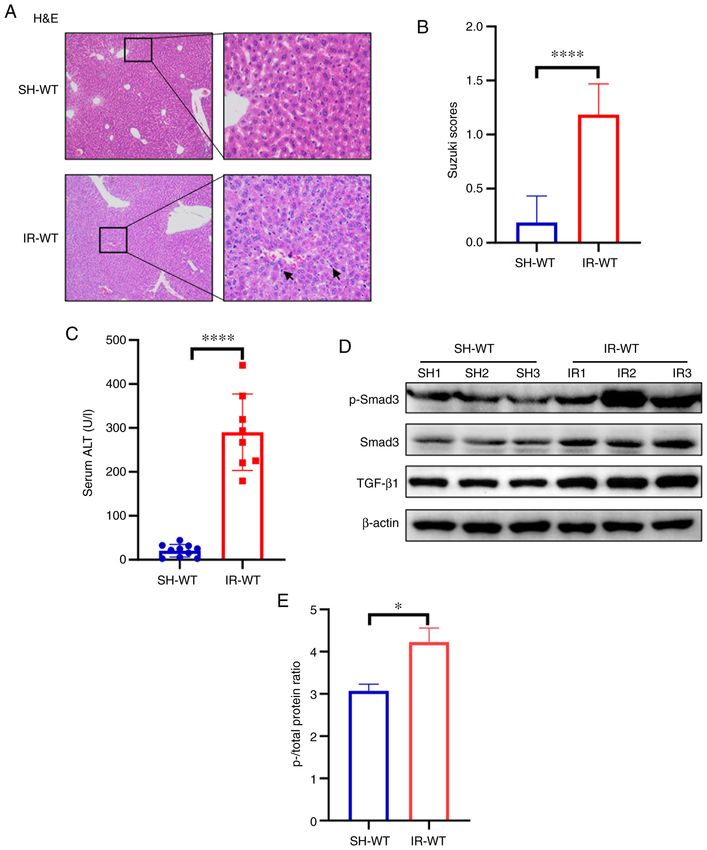
4                                       LI et al: ROLE OF TGF‑β/Smad3 IN ISCHEMIA‑REPERFUSION INJURY

Figure 1. TGF‑β1/Smad3 signaling is activated during hepatic IR injury in 129S2/SvPasCrl WT mice. (A) Liver pathological changes were analyzed via histo‑
pathological evaluation in hepatic IR injury tissue (magnification, x100 and x400). The black arrows indicate the positive inflammatory cells. (B) Suzuki injury
score from H&E staining. (C) Levels of the transaminase ALT were detected in serum samples from the IR‑WT and SH‑WT groups. (D) Protein expression
levels of TGF‑β1, Smad3 and p‑Smad3 were determined in hepatic tissues by western blotting. SH1, SH2 and SH3 indicate three randomly selected samples
from the SH‑WT group. IR1, IR2 and IR3 indicate three randomly selected samples from the IR‑WT group. (E) ImageJ software was used to perform grayscale
analysis, to evaluate the ratio of p‑ vs. total protein. β‑actin was used as the internal reference. *P
MOLECULAR MEDICINE REPORTS 24: 580, 2021                                                              5

Figure 2. Aggravated hepatic IR injury in Smad3‑/‑ mice. (A) Smad3‑/‑ MUT mice appeared smaller than littermate WT mice or HET mice. (B) Genotyping
was performed via agarose gel electrophoresis. The 431 bp band is the WT transcript and the 284 bp band is the MUT transcript. (C) Levels of serum ALT
were detected. (D) Suzuki injury score from H&E staining. (E) Liver pathological changes were analyzed via histopathological evaluation in hepatic tissue
(magnification, x100 and x400). The black arrows indicate the positive inflammatory cells. ***P
6 LI et al: ROLE OF TGF‑β/Smad3 IN ISCHEMIA‑REPERFUSION INJURY
To further identify which component served an important compared with SH‑WT groups; however, the opposite result
role in the mouse liver following IR injury, the current study was observed in Smad3‑/‑ mice, in which the expression levels
investigated whether TGF‑β/Smad3 possessed a protective of TLR4, IFN‑γ and TNF mRNA in the IR‑MUT group were
effect. The results indicated that the levels of serum ALT decreased compared with the SH‑MUT group (Fig. 4D). These
were increased in the IR‑MUT group compared with those results suggested that the TGF‑β/Smad3 signaling pathway
in the SH‑MUT, SH‑WT and IR‑WT groups (Fig. 2C). The may directly or indirectly regulate the TLR4 signaling
pathological changes after liver injury in the IR‑MUT group pathway. These findings demonstrated that inflammatory cell
were more notable than in the IR‑WT group, these changes infiltration was significantly aggravated in Smad3 gene‑defi‑
included swelling, mild vacuolation and hepatic sinus hyper‑ cient mice following liver IR injury.
emia (Fig. 2E). In addition, the Suzuki histological grading
score was significantly higher in the IR‑MUT group compared Discussion
with that in the IR‑WT group (Fig. 2D).
IR injury in the liver is one of the most severe side effects
Apoptosis is increased in Smad3 gene‑deficient mice following of liver surgery and transplantation, and is also the main
liver IR injury. To further examine the hepatic cell apoptosis factor affecting the quality of the transplanted liver (24).
and proliferation in Smad3‑/‑ mice following liver IR injury, Tissue IR injury can occur during organ harvesting and
TUNEL and Ki67 staining assays were conducted. The expres‑ peri‑transplantation. It has been shown that microcirculatory
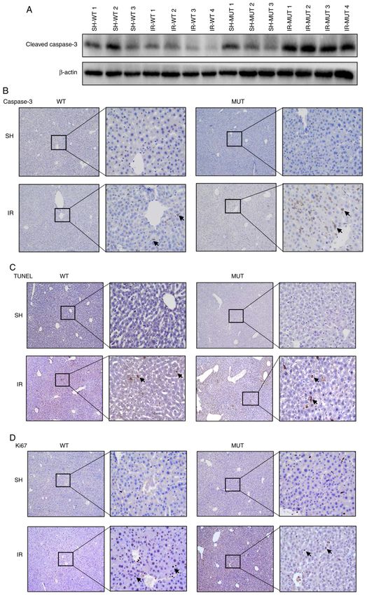
sion levels of cleaved caspase‑3 were detected via western dysfunction and immune adjustment are associated with the
blotting, and caspase‑3 expression levels were measured pathogenesis of liver IR damage (26,27). Previous studies have
using immunohistochemistry. Elevated expression of cleaved reported that neutrophil inflammatory cells were detected
caspase‑3 was detected in the IR‑MUT group compared with following IR injury in the liver (28‑30). In conditions such
IR‑WT group (Fig. 3A). Furthermore, increased staining as ischemia, hypoxia and IR liver injury, a large number of
of caspase‑3‑positive hepatocytes was found in the hepatic oxygen free radicals can be produced, which induce the oxida‑
lobule portal area of the IR‑MUT group compared with that tive stress response and liver damage (31). The present study
in the IR‑WT group (Fig. 3B). It was also identified that the demonstrated that the TGF‑β/Smad3 signaling pathway was
staining of TUNEL‑positive hepatocytes was increased in activated after liver IR injury in a mouse model of liver IR and
the IR‑MUT mice compared with that in the IR‑WT group revealed that TGF‑β/Smad3 signaling pathway activation may
(Fig. 3C). Although a high level of proliferation was observed serve a role in protection of hepatic cells, as the attenuation of
in both the IR groups in comparison with that in the sham‑oper‑ this pathway caused aggravated hepatic cell injury.
ated mice, no differences were observed between the IR‑WT TGF‑ β1 is one of three isoforms of the TGF‑ β super‑
and IR‑MUT groups based on Ki67 staining results (Fig. 3D). family (32). TGF‑β/Smad3 has been revealed to be associated
These findings demonstrated that Smad3 gene deficiency may with IR injury in several organs. For example, the interaction
aggravate liver IR damage by promoting hepatocyte apoptosis of Wnt/β ‑catenin and TGF‑β/Smad signaling pathways was
in mice. shown to exert neuroprotective effect in rats with cerebral
IR injury (33). Moreover, in a previous study, microRNA‑211
Inflammatory cell infiltration in Smad3 gene‑deficient mice suppressed apoptosis and relieved kidney injury following IR
following liver IR injury. IR not only causes damage to liver by targeting the TGF‑β/Smad3 signaling pathway (34). It has
parenchymal cells, but also causes the infiltration of inflam‑ also been shown that TGF‑β1 may contribute to isoflurane
matory cells and the secretion of inflammatory factors (25). post‑conditioning against cerebral IR injury by inhibiting
To further examine the possible mechanism via which Smad3 the JNK signaling pathway (35). In the present study, it was
knockout could aggravate liver injury, inflammatory cells, demonstrated that TGF‑ β1 was more highly expressed in
including inflammatory neutrophils [lymphocyte antigen 6 hepatic tissue derived from mice with liver IR injury compared
complex locus G6D (Ly‑6G)], leukocyte infiltration (CD3) and with that in the sham‑operated group.
macrophage cells (F4/80) were analyzed. The results revealed Considering that TGF‑ β is the strongest fibrotic factor
increased staining of CD45‑ (Fig. 4A) and F4/80‑positive and pro‑inflammatory factor, which can also aggravate
(Fig. 4B) cells in the hepatic tissues of the IR‑MUT group IR injury (36), the present study aimed to reduce the effect
compared with that in the IR‑WT group. of IR injury by blocking TGF‑β/Smad signaling. As homozy‑
To further verify the inflammatory cell infiltration in gous TGF‑β1 (37,38) and Smad2 mutations in mice were all
Smad3‑/‑ mice following liver IR injury, the mRNA expres‑ embryonic lethal (39,40), the current study selected Smad3
sion levels of B220, CD3E and Ly‑6G were assessed. The gene‑deficient mice (41,42) as a model for further examina‑
results indicated that the mRNA expression levels of Ly‑6G tion. This strain of Smad3 MUT mice was reported to have
were significantly increased, whereas B220 mRNA expression deficient TGF‑ β signaling, and studies using the TGF‑ β
levels were decreased in the hepatic tissue homogenates of the responsive reporter 3TP‑Lux failed to show any activation
IR‑MUT group compared with those in the IR‑WT group; and by the MUT construct (24,42), which indicated that TGF‑β
there was no significant difference in CD3E mRNA expression signaling was attenuated in these mice. However, the present
(Fig. 4C). Additionally, it was observed that Toll‑like receptor results indicated that IR injury was more severe in Smad3
(TLR)4, TNF and IFN‑ γ mRNA expression levels were gene‑deficient mice, with increased hepatocyte apoptosis and
significantly increased in the IR‑WT group compared with higher inflammatory cell infiltration. The results showed that
SH‑WT groups; and TLR4, TNF and IFN‑γ mRNA expres‑ endogenous deletion of the TGF‑β/Smad3 signaling pathway
sion levels were significantly increased in the SH‑MUT group could aggravate IR injury, which indirectly demonstratedMOLECULAR MEDICINE REPORTS 24: 580, 2021 7 Figure 3. Apoptosis is increased in Smad3‑/‑ mice following hepatic IR injury. (A) Protein expression levels of cleaved caspase‑3 were determined via western blot analysis. SH‑WT1, SH‑WT2 and SH‑WT3 indicate three randomly selected samples from the SH‑WT group. IR‑WT1, IR‑WT2, IR‑WT3 and IR‑WT4 indicate three randomly selected samples from the IR‑WT group. SH‑MUT1, SH‑MUT2 and SH‑MUT3 indicate three randomly selected samples from the SH‑WT group. IR‑MUT1, IR‑MUT2, IR‑MUT3 and IR‑MUT4 indicate three randomly selected samples from the IR‑WT group. (B) Caspase‑3 staining analysis in hepatic tissue from WT and Smad3 MUT mice. The positive cells are colored brown (black arrows; magnification, x100 and x400). (C) TUNEL staining indicated liver cell apoptosis in WT and Smad3 MUT mice. The positive cells are colored brown (black arrows; magnification, x100 and x400). (D) Ki67 staining analysis in hepatic tissue of WT and Smad3 MUT mice. The positive cells are colored brown (black arrows; magnification, x100 and x400). IR‑WT, IR injury in Smad3 WT mice (n=10); SH‑WT, sham‑operated in Smad3 WT mice (n=10); IR‑MUT, IR injury in Smad3 MUT mice (n=5); SH‑MUT, sham‑operated in Smad3 MUT mice (n=5). IR, ischemia‑reperfusion; MUT, mutant; Smad3, SMAD family member 3; WT, wild‑type.
8                                      LI et al: ROLE OF TGF‑β/Smad3 IN ISCHEMIA‑REPERFUSION INJURY

Figure 4. Inflammatory cell infiltration in Smad3‑/‑ mice following liver IR injury. (A) CD45 staining analysis in hepatic tissue from WT and Smad3 MUT mice.
The positive cells are colored brown (black arrows; magnification, x100 and x400). (B) F4/80 staining analysis in hepatic tissue from WT and Smad3
MUT mice. The positive cells are colored brown (black arrows; magnification, x100 and x400). (C) B220, Ly‑6G and CD3E mRNA expression levels were
assessed in hepatic tissue homogenates via reverse transcription‑quantitative PCR. The results of the relative mRNA expression levels of represent at least
triplicate determinations. (D) mRNA expression levels of TLR4, IFN‑γ and TNF were assessed in the liver tissues. The graph represents the relative mRNA
expression levels from triplicate determinations. *P
MOLECULAR MEDICINE REPORTS 24: 580, 2021                                                             9

    TLR4 is an intermediary agent of inflammation and tissue           Authors' contributions
damage in different IR damage models, such as hepatic (43),
renal (44) and pulmonary (45) models. The activated TLR4               HL and XS conceived and performed the experiments,
signaling pathway can promote an increase in the secretion of          analyzed the data, prepared the figures, authored or reviewed
TNF‑α, IFN‑β and other inflammatory cytokines, thus causing            drafts of the paper, and approved the final draft. YTo, TJ and
increased blood reperfusion injury (46). A previous study observed     XS conceived and directed the experiments, and reviewed and
increased TLR4 mRNA expression in Smad3‑/‑ mice, and found             approved the final draft. YTo, TJ and XS confirm the authen‑
that TLR4 was associated with lipopolysaccharide (LPS)                 ticity of all the raw data. XL designed and guided the project
hyperresponsiveness, leading to the increased expression of            research, and examined and approved the final manuscripts.
inflammatory cytokines (47). Therefore, the changes in the             YF and YTa conducted the experiments, reviewed and modi‑
expression of TLR4 and other inflammatory cytokines, such as           fied drafts of the paper, and approved the final draft. JY, TQ
TNF and IFN‑γ, in Smad3+/+ mice after hepatic IR injury in the         and RM performed the animal experiments, and approved the
present study supported the aforementioned conclusions. In addi‑       final draft. All authors read and approved the final manuscript.
tion, in the sham operated group, the expression levels of TLR4,
TNF and IFN‑γ mRNA were significantly increased in Smad3‑/‑            Ethics approval and consent to participate
mice compared with in the Smad3+/+. These results indicated that
the TGF‑β/Smad3 signaling pathway acts as an immunosuppres‑            All animal experiments included in this protocol adhere to the
sive factor that has a direct or indirect negative regulatory effect   Animal Research: Reporting In Vivo Experiments guidelines,
on the TLR4 signaling pathway, and loss of the TGF‑β/Smad3             and have been approved by the Animal Testing Ethics Committee
signaling pathway can promote TLR4‑mediated inflammatory               of Sir Run Run Shaw Hospital, Zhejiang University School of
injury. Significant increases in the expression of TLR4, TNF and       Medicine (approval no. 20171120‑14; Hangzhou, China).
IFN‑γ inflammatory cytokine genes were observed in Smad3‑/‑
mice, indicating that endogenous deletion of the Smad3 gene            Patient consent for publication
in mice can lead to hyperreactivity of LPS in vivo, which can
increase the secretion of inflammatory cytokines and hyperen‑          Not applicable.
dotoxemia. In addition, mRNA expression levels of TLR4, TNF
and IFN‑γ were significantly downregulated in Smad3‑/‑ mice            Competing interests
after IR injury. It was suggested that Smad3‑/‑ mice were resistant
to IR injury and the associated endotoxic shock, resulting in an       The authors declare that they have no competing interests.
immune non‑response, which may be associated with the loss
of the TGF‑β/Smad3 signaling pathway and persistently high             References
LPS responses. Although the underlying mechanism between
TGF‑β/Smad3 signaling and TLR4 signaling is unclear, it is clear        1. Takasu C, Vaziri ND, Li S, Robles L, Vo K, Takasu M, Pham C,
that the loss of the negative regulatory effects of TGF‑β/Smad3,           Farzaneh SH, Shimada M, Stamos MJ, et al: Treatment with
                                                                           dimethyl fumarate ameliorates liver ischemia/reperfusion injury.
through either environmental or endogenous stimuli, can trigger            World J Gastroenterol 23: 4508‑4516, 2017.
the activation of TLR4 and downstream elements, and lead to an          2. Peralta C, Jiménez‑Castro MB and Gracia‑Sancho J: Hepatic
imbalance in the number of inflammatory cells.                             ischemia and reperfusion injury: Effects on the liver sinusoidal
                                                                           milieu. J Hepatol 59: 1094‑1106, 2013.
    In conclusion, the present study demonstrated that the              3. Shen XD, Ke B, Zhai Y, Amersi F, Gao F, Anselmo DM,
TGF‑ β /Smad3 signaling pathway could protect against                      Busuttil RW and Kupiec‑Weglinski JW: CD154‑CD40 T‑cell
IR injury‑induced damage in liver tissue repair and immune                 costimulation pathway is required in the mechanism of hepatic
                                                                           ischemia/reperfusion injury, and its blockade facilitates and
response. However, whether exogenous intervention treatment                depends on heme oxygenase‑1 mediated cytoprotection.
can reduce IR injury in the early stages of disease still needs            Transplantation 74: 315‑319, 2002.
further clarification, which provides a novel research direction        4. Zabala V, Boylan JM, Thevenot P, Frank A, Senthoor D, Iyengar V,
                                                                           Kim H, Cohen A, Gruppuso PA and Sanders JA: Transcriptional
for the prevention of IR injury.                                           changes during hepatic ischemia‑reperfusion in the rat. PLoS
                                                                           One 14: e0227038, 2019.
Acknowledgements                                                        5. Suzuki E, Ochiai‑Shino H, Aoki H, Onodera S, Saito A, Saito A
                                                                           and Azuma T: Akt activation is required for TGF‑ β1‑induced
                                                                           osteoblast differentiation of MC3T3‑E1 pre‑osteoblasts. PLoS
Not applicable.                                                            One 9: e112566, 2014.
                                                                        6. Derynck R and Budi EH: Specificity, versatility, and control of
                                                                           TGF‑β family signaling. Sci Signal 12: eaav5183, 2019.
Funding                                                                 7. Brown KA, Pietenpol JA and Moses HL: A tale of two proteins:
                                                                           Differential roles and regulation of Smad2 and Smad3 in
This work was supported by the National Natural Science                    TGF‑beta signaling. J Cell Biochem 101: 9‑33, 2007.
                                                                        8. Zou GL, Zuo S, Lu S, Hu RH, Lu YY, Yang J, Deng KS, Wu YT,
Foundation of China (grant nos. 81702854 and 81700551),                    Mu M, Zhu JJ, et al: Bone morphogenetic protein‑7 represses hepatic
and the Natural Science Foundation of Guangxi Zhuang                       stellate cell activation and liver fibrosis via regulation of TGF‑β/Smad
Autonomous Region (grant no. 2018GXNBSFBA138033).                          signaling pathway. World J Gastroenterol 25: 4222‑4234, 2019.
                                                                        9. Inagaki Y and Okazaki I: Emerging insights into Transforming
                                                                           growth factor beta Smad signal in hepatic fibrogenesis. Gut 56:
Availability of data and materials                                         284‑292, 2007.
                                                                       10. Li T, Zhao S, Song B, Wei Z, Lu G, Zhou J and Huo T: Effects
                                                                           of transforming growth factor β‑1 infected human bone marrow
The datasets used and/or analyzed during the present study are             mesenchymal stem cells on high‑ and low‑metastatic potential
available from the corresponding author on reasonable request.             hepatocellular carcinoma. Eur J Med Res 20: 56, 2015.
10 LI et al: ROLE OF TGF‑β/Smad3 IN ISCHEMIA‑REPERFUSION INJURY
11. Porowski D, Wirkowska A, Hryniewiecka E, Wyzgał J, 31. Cutrn JC, Perrelli MG, Cavalieri B, Peralta C, Rosell Catafau J
Pacholczyk M and Pączek L: Liver Failure Impairs the and Poli G: Microvascular dysfunction induced by reperfusion
Intrahepatic Elimination of Interleukin‑6, Tumor Necrosis injury and protective effect of ischemic preconditioning. Free
Factor‑Alpha, Hepatocyte Growth Factor, and Transforming Radic Biol Med 33: 1200‑1208, 2002.
Growth Factor‑Beta. BioMed Res Int 2015: 934065, 2015. 32. Bielecka‑Dabrowa A, Gluba‑Brzózka A, Michalska‑Kasiczak M,
12. Wang RQ, Mi HM, Li H, Zhao SX, Jia YH and Nan YM: Modulation Misztal M, Rysz J and Banach M: The multi‑biomarker approach
of IKKβ/NF‑κ B and TGF‑β1/Smad via Fuzheng Huayu recipe for heart failure in patients with hypertension. Int J Mol Sci 16:
involves in prevention of nutritional steatohepatitis and fibrosis 10715‑10733, 2015.
in mice. Iran J Basic Med Sci 18: 404‑411, 2015. 33. Zhang G, Ge M, Han Z, Wang S, Yin J, Peng L, Xu F, Zhang Q,
13. Park SO, Kumar M and Gupta S: TGF‑β and iron differently alter Dai Z, Xie L, et al: Wnt/β‑catenin signaling pathway contributes
HBV replication in human hepatocytes through TGF‑ β/BMP to isoflurane postconditioning against cerebral ischemia‑reper‑
signaling and cellular microRNA expression. PLoS One 7: fusion injury and is possibly related to the transforming growth
e39276, 2012. factorβ1/Smad3 signaling pathway. Biomed Pharmacother 110:
14. Akhmetshina A, Palumbo K, Dees C, Bergmann C, Venalis P, 420‑430, 2019.
Zerr P, Horn A, Kireva T, Beyer C, Zwerina J, et al: Activation 34. Shang J, Sun S, Zhang L, Hao F and Zhang D: miR‑211 alle‑
of canonical Wnt signalling is required for TGF‑ β ‑mediated viates ischaemia/reperfusion‑induced kidney injury by targeting
fibrosis. Nat Commun 3: 735, 2012. TGFβR2/TGF‑β/SMAD3 pathway. Bioengineered 11: 547‑557,
15. Feng XH and Derynck R: Specificity and versatility in tgf‑beta 2020.
signaling through Smads. Annu Rev Cell Dev Biol 21: 659‑693, 35. Wang S, Yin J, Ge M, Dai Z, Li Y, Si J, Ma K, Li L and Yao S:
2005. Transforming growth‑beta 1 contributes to isoflurane post‑
16. Ooshima A, Park J and Kim SJ: Phosphorylation status at Smad3 conditioning against cerebral ischemia‑reperfusion injury by
linker region modulates transforming growth factor‑β‑induced regulating the c‑Jun N‑terminal kinase signaling pathway.
epithelial‑mesenchymal transition and cancer progression. Biomed Pharmacother 78: 280‑290, 2016.
Cancer Sci 110: 481‑488, 2019. 36. Yang T, Zhang X, Ma C and Chen Y: TGF‑β/Smad3 pathway
17. Liu ZY, Pan HW, Cao Y, Zheng J, Zhang Y, Tang Y, He J, Hu YJ, enhances the cardio‑protection of S1R/SIPR1 in in vitro
Wang CL, Zou QC, et al: Downregulated microRNA‑330 ischemia‑reperfusion myocardial cell model. Exp Ther Med 16:
suppresses left ventricular remodeling via the TGF‑β1/Smad3 178‑184, 2018.
signaling pathway by targeting SRY in mice with myocardial 37. Larsson J, Goumans MJ, Sjöstrand LJ, van Rooijen MA, Ward D,
ischemia‑reperfusion injury. J Cell Physiol 234: 11440‑11450, 2019. Levéen P, Xu X, ten Dijke P, Mummery CL and Karlsson S:
18. Liu FF, Liu CY, Li XP, Zheng SZ, Li QQ, Liu Q and Song L: Abnormal angiogenesis but intact hematopoietic potential in
Neuroprotective effects of SMADs in a rat model of cerebral TGF‑beta type I receptor‑deficient mice. EMBO J 20: 1663‑1673,
ischemia/reperfusion. Neural Regen Res 10: 438‑444, 2015. 2001.
19. Percie du Sert N, Ahluwalia A, Alam S, Avey MT, Baker M, 38. Vander Ark A, Cao J and Li X: TGF‑β receptors: In and beyond
Browne WJ, Clark A, Cuthill IC, Dirnagl U, Emerson M, et al: TGF‑β signaling. Cell Signal 52: 112‑120, 2018.
Reporting animal research: Explanation and elaboration for the 39. Schepers D, Tortora G, Morisaki H, MacCarrick G, Lindsay M,
ARRIVE guidelines 2.0. PLoS Biol 18: e3000411, 2020. Liang D, Mehta SG, Hague J, Verhagen J, van de Laar I, et al:
20. Liu Y, Lu T, Zhang C, Xu J, Xue Z, Busuttil RW, Xu N, Xia Q, A mutation update on the LDS‑associated genes TGFB2/3 and
Kupiec‑Weglinski JW and Ji H: Activation of YAP attenuates SMAD2/3. Hum Mutat 39: 621‑634, 2018.
hepatic damage and fibrosis in liver ischemia‑reperfusion injury. 40. Waldrip WR, Bikoff EK, Hoodless PA, Wrana JL and
J Hepatol 71: 719‑730, 2019. Robertson EJ: Smad2 signaling in extraembryonic tissues
21. Malý O, Zajak J, Hyšpler R, Turek Z, Astapenko D, Jun D, determines anterior‑posterior polarity of the early mouse embryo.
Váňová N, Kohout A, Radochová V, Kotek J, et al: Inhalation of Cell 92: 797‑808, 1998.
molecular hydrogen prevents ischemia‑reperfusion liver damage 41. Stewart AG, Thomas B and Koff J: TGF‑β: Master regulator of
during major liver resection. Ann Transl Med 7: 774, 2019. inflammation and fibrosis. Respirology 23: 1096‑1097, 2018.
22. Livak KJ and Schmittgen TD: Analysis of relative gene expression 42. Goumans MJ and Mummery C: Functional analysis of the
data using real‑time quantitative PCR and the 2(‑Delta Delta TGFbeta receptor/Smad pathway through gene ablation in mice.
C(T)) Method. Methods 25: 402‑408, 2001. Int J Dev Biol 44: 253‑265, 2000.
23. Han L, Wang JN, Cao XQ, Sun CX and Du X: An‑te‑xiao capsule 43. Taylor KR, Trowbridge JM, Rudisill JA, Termeer CC, Simon JC
inhibits tumor growth in non‑small cell lung cancer by targeting and Gallo RL: Hyaluronan fragments stimulate endothelial
angiogenesis. Biomed Pharmacother 108: 941‑951, 2018. recognition of injury through TLR4. J Biol Chem 279:
24. Yang X, Letterio JJ, Lechleider RJ, Chen L, Hayman R, Gu H, 17079‑17084, 2004.
Roberts AB and Deng C: Targeted disruption of SMAD3 results 44. Wu H, Chen G, Wyburn KR, Yin J, Bertolino P, Eris JM,
in impaired mucosal immunity and diminished T cell respon‑ Alexander SI, Sharland AF and Chadban SJ: TLR4 activation
siveness to TGF‑beta. EMBO J 18: 1280‑1291, 1999. mediates kidney ischemia/reperfusion injury. J Clin Invest 117:
25. Bravatà V, Cammarata FP, Minafra L, Pisciotta P, Scazzone C, 2847‑2859, 2007.
Manti L, Savoca G, Petringa G, Cirrone GAP, Cuttone G, et al: 45. Imai Y, Kuba K, Neely GG, Yaghubian‑Malhami R, Perkmann T,
Proton‑irradiated breast cells: Molecular points of view. J Radiat van Loo G, Ermolaeva M, Veldhuizen R, Leung YH,
Res (Tokyo) 60: 451‑465, 2019. Wang H, et al: Identification of oxidative stress and Toll‑like
26. Z ha i Y, Pet rowsk y H, Hong JC, Busut ti l RW a nd receptor 4 signaling as a key pathway of acute lung injury.
Kupiec‑Weglinski JW: Ischaemia‑reperfusion injury in liver Cell 133: 235‑249, 2008.
transplantation‑‑from bench to bedside. Nat Rev Gastroenterol 46. Liu Q and Zhang Y: PRDX1 enhances cerebral ischemia‑reper‑
Hepatol 10: 79‑89, 2013. fusion injury through activation of TLR4‑regulated inflammation
27. Jiménez‑Castro MB, Cornide‑Petronio ME, Gracia‑Sancho J and apoptosis. Biochem Biophys Res Commun 519: 453‑461,
and Peralta C: Inflammasome‑Mediated Inflammation in Liver 2019.
Ischemia‑Reperfusion Injury. Cells 8: E1131, 2019. 47. McCartney‑Francis N, Jin W and Wahl SM: Aberrant Toll receptor
28. Lee PY, Wang JX, Parisini E, Dascher CC and Nigrovic PA: Ly6 expression and endotoxin hypersensitivity in mice lacking
family proteins in neutrophil biology. J Leukoc Biol 94: 585‑594, a functional TGF‑beta 1 signaling pathway. J Immunol 172:
2013. 3814‑3821, 2004.
29. Xiang S, Chen K, Xu L, Wang T and Guo C: Bergenin
Exerts Hepatoprotective Effects by Inhibiting the Release of This work is licensed under a Creative Commons
Inflammatory Factors, Apoptosis and Autophagy via the PPAR‑γ Attribution-NonCommercial-NoDerivatives 4.0
Pathway. Drug Des Devel Ther 14: 129‑143, 2020.
International (CC BY-NC-ND 4.0) License.
30. Palomino‑Schätzlein M, Simó R, Hernández C, Ciudin A,
Mateos‑Gregorio P, Hernández‑Mijares A, Pineda‑Lucena A and
Herance JR: Metabolic fingerprint of insulin resistance in human
polymorphonuclear leucocytes. PLoS One 13: e0199351, 2018.You can also read