Fine-tuning the stimulation of MLL1 methyltransferase
←
→
Page content transcription
If your browser does not render page correctly, please read the page content below
The FASEB Journal • Research Communication
Fine-tuning the stimulation of MLL1 methyltransferase
activity by a histone H3-based peptide mimetic
Vanja Avdic,*,†,1 Pamela Zhang,*,†,1 Sylvain Lanouette,*,†, Anastassia Voronova,†
Ilona Skerjanc,† and Jean-Francois Couture*,†,2
*Ottawa Institute of Systems Biology and †Department of Biochemistry, Microbiology, and
Immunology, University of Ottawa, Ottawa, Ontario, Canada
ABSTRACT The SET1 family of methyltransferases while the heterozygotes display retarded growth and
carries out the bulk of histone H3 Lys-4 methylation in hematopoietic abnormalities (5, 6). Correlative to these
vivo. One of the common features of this family is the observations, MLL proteins regulate several differenti-
regulation of their methyltransferase activity by a tri- ation programs, including hematopoiesis (7), myogen-
partite complex composed of WDR5, RbBP5, and esis (8, 9), and neurogenesis (10).
Ash2L. To selectively probe the role of the SET1 family MLL1 and its yeast homologue SET1 (ySET1) harbor
of methyltransferases, we have developed a library of several evolutionarily conserved domains and are virtu-
histone H3 peptide mimetics and report herein the ally always found in multisubunit complexes. Interac-
characterization of an N␣ acetylated form of histone tions with other protein subunits regulate their cellular
H3 peptide (N␣H3). Binding and inhibition studies localization and enzymatic activity (11). The catalytic
reveal that the addition of an acetyl moiety to the N domain of MLL1, also referred to as the suppressor of
terminus of histone H3 significantly enhances its bind- variegation 3–9, enhancer of zeste and trithorax (SET)
ing to WDR5 and prevents the stimulation of MLL1 domain (12), is located on its C terminus and is pivotal
methyltransferase activity by the WDR5-RbBP5- in controlling skeletal development and HOX gene
Ash2L complex. The crystal structure of N␣H3 in expression (6). Similar to other members of the SET1
complex with WDR5 reveals that a high-affinity hy- family, MLL1 has minimal methyltransferase activity
drophobic pocket accommodates the binding of the toward its substrate; however, upon interaction with a
acetyl moiety. These results provide the structural 3-protein complex composed of WDR5, Ash2L, and
basis to control WDR5-RbBP5-Ash2L-MLL1 activity RbBP5, MLL1 enzymatic activity is enhanced (13–15).
and a tool to manipulate stem cell differentiation The biochemical determinants underlying the for-
programs.—Avdic, V., Zhang, P., Lanouette, S., mation of the MLL complex have recently been de-
Voronova, A., Skerjanc, I., Couture, J.-F. Fine-tuning tailed. Initial work carried out by Dou et al. (15) showed
the stimulation of MLL1 methyltransferase activity by that each subunit contributes in modulating the enzy-
a histone H3-based peptide mimetic. FASEB J. 25, matic activity of MLL1, and the level of histone H3
960 –967 (2011). www.fasebj.org Lys-4 methylation in vivo. Notably, the same researchers
also demonstrated the importance of WDR5 within the
Key Words: epigenetics 䡠 chromatin biology 䡠 nucleosomes core complex as its depletion impaired MLL1 methyl-
䡠 SET1 family of methyltransferases transferase activity. Similarly, Patel and coworkers (14,
16) found that WDR5 is central in binding a region on
MLL1 termed the WDR5 interacting motif (WIN) and
Histone methylation plays an important role in a bridging the methyltransferase to the other subunits of
large variety of biological processes. Depending on its the core complex. Binding of the MLL1WIN peptide is
position on a substrate and the number of methyl achieved through a peptidyl arginine binding pocket
groups transferred to its ε-amine, lysine methylation has located in one of the central clefts of the  propeller.
been associated with DNA damage response (1), DNA Site-directed mutagenesis studies of the MLL1WIN pep-
replication (2), and transcription (3). Of these posi- tide and the WDR5 peptidyl arginine binding cleft
tions, lysine 4 of histone H3 (H3K4) is a key methyl- revealed that loss of binding between MLL1 and WDR5
ation site and commonly found in the promotor re-
gions of actively transcribed genes.
1
Histone H3 K4 methylation is predominantly cata- These authors equally contributed to this work.
2
lyzed by the SET1 family of methyltransferases. Com- Correspondence: University of Ottawa, Ottawa Institute of
posed of 7 members, this family includes Ash1, Systems Biology, 451 Smyth Rd., Roger Guindon Hall, Ot-
tawa, ON K1H 8M5, Canada. E-mail: jean-francois.couture@
SET1A/B, and 5 myeloid lymphoma leukemia proteins uottawa.ca
(MLL1–5) (4). MLL1 is essential for proper develop- doi: 10.1096/fj.10-171959
ment during embryogenesis (5). Homozygous disrup- This article includes supplemental data. Please visit http://
tion of the mouse MLL1 gene is embryonically lethal, www.fasebj.org to obtain this information.
960 0892-6638/11/0025-0960 © FASEB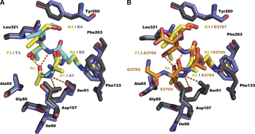
caused severe impairment in MLL1 methyltransferase 2H14.pdb) as a search model. After 2 rounds of refinement
activity, further underscoring the importance of the with Refmac5 (23), the structure and electron density map
role of WDR5-MLL1 interaction (15–17). were inspected using Coot (24). The structure has been
solved at 1.7 Å with final Rfactor/Rfree values of 15.4/20.5
The detrimental effects of misregulating histone H3 molecules in the asymmetric unit. Geometric parameters
Lys-4 methylation have been documented. The MLL1 were calculated using Molprobity (25) (Table 1).
gene is prone to chromosomal aberrations, such as
translocations and duplications, which are associated Isothermal titration calorimetry
with aggressive leukemic disorders (17). Alterations of
H3K4me3 levels are observed in peripheral blood Equilibrium dissociation constants were obtained as de-
mononuclear cells from systemic erythematosus pa- scribed previously (20). Briefly, binding of the N␣H3 peptide
tients (18, 19), while, in C. elegans, misregulation of was measured using a VP-ITC microcalorimeter from Micro-
H3K4 methyl mark levels leads to a reduction in life Cal (Piscataway, NJ, USA) with 0.5–1 mM of peptide and
30 –50 M of WDR5. Equilibrium dissociation constants were
span (18). The involvement of the SET1 family of calculated with the Origin software (OriginLab, Northamp-
methyltransferases in various diseases underlines the ton, MA, USA).
importance of gaining a better understanding in their
functions and modulating their enzymatic activity by Overexpression and purification of MLL1 SET domain,
the WDR5-RbBP5-Ash2L complex. Ash2L, and RbBP5
In this study, we report the characterization of a
histone H3-based peptide mimetic on MLL1 methyl- Fragments corresponding to full-length Ash2L and RbBP5
transferase activity. We found that the addition of an were PCR amplified using the OpenBiosystem clones 3921999
N␣-acetyl moiety to histone H3 N terminus (N␣H3) and 5266066, respectively (OpenBiosystem, Huntsville, AL,
USA). PCR fragments were cloned in a modified version of
increases its binding affinity for WDR5 by 84-fold when
pET3d (26), and the proteins were overexpressed with 0.1
compared to its unacetylated form. The crystal struc- mM IPTG in Rosetta cells (EMD, Gibbstown, NJ, USA) for
ture of the WDR5-N␣H3 complex reveals that N␣H3 16 h at 18°C. Cells were harvested in 50 mM sodium phos-
undergoes structural reorganization to fit in a shallow phate, 500 mM NaCl, and 5 mM -mercaptoethanol; lysed by
pocket that stabilizes the acetyl moiety using novel sonication; clarified by centrifugation; and purified by Talon
hydrophobic contacts and hydrogen bonds. We also Co2⫹ affinity chromatography (Clontech, Mountain View,
show that incubation of the N␣H3 peptide mimetic
with WDR5 impairs the stimulation of MLL1 methyl- TABLE 1. Data collection and refinement statistics
transferase activity. These results provide a basis for the for WDR5-N␣H3
use of histone H3 peptide mimetics in regulating MLL1
activity and present a new tool for the study of biolog- Statistic Value
ical processes regulated by MLL1 methyltransferase
activity. Data Collection
Space group P1
Cell dimensions
a, b, c (Å) 45.9, 48.6, 63.3
MATERIALS AND METHODS ␣, , ␥ (deg) 98.8, 90.9, 117.5
Resolution 40.0–1.7 (1.76–1.70)
Peptides Rmerge 6.5 (23.2)
I/I 20.6 (2.4)
Peptides were purchased either from Peptide2.0 (Chantilly, Completeness (%) 97.0 (93.8)
VA, USA) or New England Peptides (Gardner, MA, USA) with Redundancy 3.1 (2.4)
95–99% purity with an additional tyrosine on their C termi- Refinement
nus for UV quantification. Peptides were suspended in water Resolution (Å) 35.65–1.70
at 100 mM concentration and stored at ⫺20°C. Reflections 47,864
Rwork/Rfree 15.4/20.5
Atoms
WDR5 expression, purification, crystallization, and
Protein 4601
structure determination
Peptides 67
Water 351
A fragment of WDR5 corresponding to residues 22–334 was B factors (Å2)
overexpressed and purified as described previously (20). Protein 17.7
WDR5 and the peptides were mixed in equal molar ratio and Ligands 15.6
incubated on ice for 15 min. Crystals were grown at 22°C in a Water 31.7
buffer containing 50 mM NaAcetate, 100 mM NH4SO4, and Root mean square deviations
20% PEG4000, pH 4.6. Crystals were harvested and soaked in Bond lengths (Å) 0.007
a cryoprotecting solution composed of the mother liquor Bond angles (deg) 1.108
supplemented with 20% glycerol. Complete data sets were Molprobity scores
collected at the General Medicine and Cancer Institutes Ramachandran favored (%) 96.6
Collaborative Access Team (GM/CA-CAT) at the Advanced Ramachandran allowed (%) 3.4
Photon Source (Argonne National Laboratory, Argonne, IL,
USA). Reflections were integrated and scaled using HKL2000 Data were collected at GM/CA-CAT (Argonne National Labora-
(21). Using Molrep (22), a molecular replacement solution tory, Argonne, IL, USA). Values in parentheses are for highest-
has been found using the apo-WDR5 structure (RCSB code resolution shell.
INHIBITION OF MLL1 METHYLTRANSFERASE ACTIVITY 961CA, USA). TEV-cleaved Ash2L and RbBP5 were further 27–29). A comparison of the KD values revealed that
purified by metal affinity and by size exclusion chromatogra- WDR5 bound an N-␣-acetylated form of an histone H3
phy steps (Superdex 200; GE Healthcare, Piscataway, NJ, peptide (N␣H3) ⬃84- and ⬃13-fold more tightly than
USA).
A DNA fragment encoding the WIN and SET domains histone H3 and MLL1WIN peptides, respectively (16,
(3753–3969) of MLL1 has been synthesized (Eurofins; MWG 20) (Fig. 1A). We then sought to verify whether this
Operon, Huntsville, AL, USA) and cloned in fusion with a peptide could prevent the stimulation of MLL1 meth-
TEV-cleavable glutathione sulfotransferase. GST-MLL1 has yltransferase activity by the core complex proteins.
been overexpressed similarly to Ash2L. Cells were harvested Using a radiometric methyltransferase assay, we deter-
in PBS buffer, lysed by sonication, and centrifuged. The mined that the N␣H3 peptide inhibited MLL1 methyl-
supernatant was applied to glutathione Sepharose 4B for 1 h,
transferase activity with an IC50 of 4.1 M (Fig. 1C). To
and unbound proteins were washed with 50 column vol of
PBS. The slurry was resuspended in 25 mM Tris (pH 8.0), 100 assess the methylation state of histone H3 on Lys-4
mM NaCl, 5 mM -mercaptoethanol, and 250 g of TEV following inhibition of the complex with N␣H3, we
protease for 12 h. Cleaved MLL1 was further purified by performed Western blot with antibodies specific for the
size-exclusion chromatography (Superdex 75) preequili- mono- and dimethylated state of H3K4 (H3K4me and
brated with the slurry buffer. H3K4me2). Consistent with the role of the core com-
plex on MLL1 methyltransferase activity, a significant
In vitro methyltransferase assay loss of H3K4me and H3K4me2 was observed upon
incubation of the WDR5-RbBP5-Ash2L-MLL1 complex
Methyltransferase assays were conducted using 5 M of with N␣H3, confirming that blocking the WDR5 pepti-
recombinantly purified MLL1 SET domain with equimolar dyl arginine binding cleft provides a way to control the
amounts of Ash2L, RbBP5, and WDR5. Reactions were initi-
stimulation of MLL1 methyltransferase activity by its
ated by the addition of 1 Ci of radiolabeled S-adenosyl-l-
methionine and incubated during 2 h at 22°C. Methyltrans- core complex subunits. However, the persistence of
ferase assays were carried out in 50 mM Tris (pH 8.5), 200 H3K4me and H3K4me2 and incomplete inhibition of
mM NaCl, 3 mM DTT, 5 mM MgCl2, 5% glycerol, and 1 mM MLL1 methyltransferase activity suggest that other do-
histone H3 peptide (ARTKQTARKSTGGKAPRKQY), and mains of Ash2L or RbBP5 also play important roles in
stopped by spotting the reactions onto Whatman P-81 filter the stimulation of MLL1 methyltransferase activity. As
papers (Whatman, Piscataway, NJ, USA). Free AdoMet was expected, incubation of WDR5-RbBP5-Ash2L-MLL1
removed by washing the filter papers in 250 ml of 50 mM
NaHCO3 at pH 9.0. Activity was quantified by liquid scintilla-
did not alter the substrate specificity of MLL1 (Supple-
tion counts. Inhibition analyses were performed similarly, mental Fig. S1).
with the exception that the peptide inhibitor was added to the These initial results indicated that adding an acetyl
assay prior to adding AdoMet and the substrate peptide. moiety to the N terminus of histone H3 improves the
binding capabilities of a peptide mimetic to WDR5.
Western blot analysis However, an initial modeling of an N␣Ac moiety on
the available WDR5-H3 crystal structure (RCSB code
To evaluate the product specificity of the WDR5-RbBP5- 2H13.pdb; ref. 20) revealed numerous steric clashes
Ash2L-MLL1 complex upon inhibition with N␣H3, methyl- between the WDR5 side chains and the N␣Ac moiety
ation reactions were performed as described previously, with (data not shown), suggesting that the N␣Ac H3
the exception that the peptide substrate was replaced by peptide must undergo significant structural rear-
full-length histone H3. Methylated histone proteins were
separated on a 15% SDS-PAGE, transferred onto PVDF, and rangement to bind the peptidyl arginine binding
blotted with histone H3, H3-K4me1, or H3-K4me2 specific cleft.
antibodies (Abcam, Cambridge, MA, USA).
Crystal structure of WDR5 in complex with N␣H3
RESULTS To understand the structural basis underlying the recogni-
tion of N␣H3, we have solved its crystal structure in complex
Biochemical analysis of a histone H3-based peptide with WDR5. Overall, the WDR5 structure is similar to the
mimetic previously published apo-structure with root-mean-square
deviations of 0.42 Å for all protein atoms. The density of the
On the basis of initial studies showing that MLL1-WDR5 peptide is unambiguous (Fig. 2A) for the first three amino
interaction is essential for proper histone H3 Lys-4 acids including the N␣-acetyl moiety, while there is no
methylation (14), we surmised that the development of visible electron density for K4 ε-amine and Q5 resi-
a high-affinity peptide mimetic targeting WDR5 pepti- due (single letter will refer therein to WDR5-bound
dyl arginine binding cleft would prevent its interaction peptide residues).
with MLL1 and impair its methyltransferase activity. To The peptide is maintained in the central peptidyl
verify this hypothesis, we performed isothermal titra- arginine binding cleft of WDR5, in which it engages in
tion calorimetry (ITC) and determined the equilibrium several hydrogen bonds and hydrophobic contacts.
dissociation constants (KD) of a peptide library for Briefly, N␣H3 T3 makes van der Waals contacts with
WDR5 (data not shown). All peptides were designed to residues delineating a cleft composed of Tyr-260, Leu-
include an arginine residue in position 2, which is 321, AL-47, and Ala-65 side chains (Fig. 2B, D). As
considered to be essential for binding to WDR5 (20, observed for the WDR5-H3 and WDR5-MLL1WIN com-
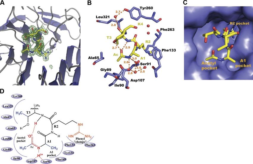
962 Vol. 25 March 2011 The FASEB Journal 䡠 www.fasebj.org AVDIC ET AL.carbonyl groups. Similar to T3, A1 is stabilized by
several hydrophobic and polar contacts. Notably, A1 is
maintained in a shallow depression, henceforth re-
ferred to as the A1 pocket (Fig. 2D), comprising
Phe-133, Phe-149, and Tyr-131 phenyl rings. In addi-
tion, the A1 amide group is stabilized by two hydrogen
bonds with the Asp-107 carboxylate and Ser-91 hydroxyl
groups, respectively. Finally, the A1 carbonyl group is
maintained by a 3.1-Å hydrogen bond with the K4
amide group.
Unambiguous electron density for this region of the
peptide allowed us to accurately model the N␣ acetyl
moiety. Initial analysis of the acetyl group reveals that it
has comparable geometry to an amide bond, as its
carbonyl group is planar with the nitrogen atom of A1.
This geometry allows the methyl group of the N␣ acetyl
moiety to snuggly fit into an hydrophobic cleft, hence-
forth referred as the N␣ acetyl pocket, composed of
Ala-65, Leu-88, Gly-89, and Ile-90 (Fig. 2D). The car-
bonyl group of the acetyl moiety is further stabilized by
two intramolecular hydrogen bonds of 2.7 and 2.9 Å
with T3 hydroxyl and amide groups, respectively. Over-
all, the hydrogen bonding pattern concurs with a
recent modeling analysis and molecular dynamics
study showing that intramolecular hydrogen bonds
are important to maintain high-affinity binding of
peptide mimetics in the WDR5 peptidyl arginine
binding cavity (30).
Peptide scanning
N␣H3 is maintained through several hydrogen bonds
and van der Waals contacts within the WDR5 peptidyl
arginine-binding cleft. To probe the role of these
interactions, we mutated A1, R2, and T3 residues and
measured their effects on WDR5 binding and inhibi-
tion of MLL1 methyltransferase activity. Mutation of A1
to a glycine or serine residue had variable effects on the
affinity for WDR5 and inhibition of histone H3 meth-
ylation. The alanine substitution weakened the interac-
tion between WDR5 and N␣H3 (Table 2) and resulted
in a loss of inhibition of histone H3 methylation by
MLL1 (Fig. 3). In contrast, the A1S mutation bound
WDR5 with similar affinity when compared to the
wild-type N␣H3 and inhibited histone H3 methylation
with comparable activity.
Figure 1. Binding profiles of N␣H3. A) Table illustrating the
As observed for the WDR5-H3 and WDR5-MLL1WIN
equilibrium dissociation constants for N␣H3, histone H3 complexes, R2 of N␣H3 is stabilized, in the central
peptide, and MLL1WIN. B) ITC titration experiment with cavity of the -propeller, by several hydrogen bonds
N␣H3 peptide and WDR5 (top panel) and the fitted binding and hydrophobic contacts. Given the high-affinity bind-
curve (bottom panel). C) Inhibition assays of MLL1 methyl- ing capability of N␣H3, we posited that the addition of
transferase activity with increasing concentration of N␣H3 an acetyl moiety to the N terminus of H3 would
peptide and 5 M of MLL1 and its core complex subunits. alleviate the requirement of R2. However, substitution
Inset: histone H3 K4 methylation state in the presence or
absence of N␣H3. of R2 to a leucine, glutamic acid, or alanine residue
abolished binding of the mutant peptides (Table 2)
and, correlatively, failed to inhibit MLL1 methyltrans-
plexes, R2 of N␣H3 is maintained in the central cavity ferase activity (Fig. 3). These results concur with previ-
of the -propeller by a pair of phenylalanine residues ous studies (20, 27–29, 31) and highlight the impor-
and several direct and water-mediated hydrogen bonds tance of R2 for high-affinity binding of a peptide
with Ser-91, Phe-133, Ser-175, and Cys-261 backbone mimetic to WDR5.
INHIBITION OF MLL1 METHYLTRANSFERASE ACTIVITY 963Figure 2. WDR5 binds N␣H3 through the peptidyl arginine binding cleft.
A) Zoom view of a simulated annealing fo-fc omit map (green) contoured
at 2. B) View of the N␣-acetylated histone H3 binding pocket, WDR5
carbon atoms are shown in blue. Hydrogen bonds are rendered as orange
dashed lines. C) Surface representation of the WDR5 peptidyl arginine
binding cleft showing the 3 pockets. D) Two-dimensional schematic
representation of important hydrogen bonds and resonance of the new
amide bond. To improve clarity of the figure, the hydrogen bond
between A1 carbonyl group and K4 amide group is not shown.
To probe the role of the hydrogen bond between the Comparison of N␣H3 binding mode
T3 hydroxyl group and the acetyl carbonyl moiety,
binding studies were performed with a T3V substituted Structural alignment and analysis of the WDR5-N␣H3
peptide. To our surprise, no loss of binding was ob- complex with the previously described WDR5-MLL1WIN
served, as identical binding constants were calculated and WDR5-H3 complexes allowed us to further under-
for the N␣H3 T3V peptide (Table 2). Consistent with
the binding studies, the T3V peptide inhibited histone H3
methylation to similar levels as the WT N␣H3 (Fig. 4).
These results suggest that the hydrogen bond between
the carbonyl group of the acetyl moiety and the T3
amide group is sufficient to maintain high-affinity bind-
ing capability of the N␣H3 peptide. Overall, the map-
ping and structural analyses suggest that for high-
affinity binding, a peptide mimetic requires an N␣-
acetyl moiety, a residue other than a glycine in position
1 and an arginine residue in position 2.
TABLE 2. Mutational analysis of the N␣-acetylated peptides
␣-A1R2T3K4Q5T6A7R8K9S10Y KD (M)
WT 0.13 ⫾ 0.02
A1S 0.09 ⫾ 0.01
A1G 1.30 ⫾ 0.18
R2L N.B.
R2A N.B. Figure 3. Mapping of the N␣H3 peptide. Methyltransferase
R2E ⬎500a assay of MLL1 SET domain performed in presence of the
T3V 0.10 ⫾ 0.01 RbBP5/WDR5/Ash2L complex. Enzymatic assays have been
A1G/R2S N.B. carried out either in the absence (NP) or presence of 4 M of
N␣H3 peptides outlined in Table 1. Inhibition is shown as a
N.B., no heat of binding could be detected; WT, wild type. relative percentage of activity compared to methylation reac-
a
Saturation could not be achieved. tions performed in absence of N␣H3 peptides.
964 Vol. 25 March 2011 The FASEB Journal 䡠 www.fasebj.org AVDIC ET AL.Figure 4. Difference in compaction of peptide in WDR5 peptidyl arginine binding cleft. A) Comparison of WDR5-H3 and WDR5-N␣H3 complexes. Carbon atoms are shown in yellow and cyan for N␣H3 and H3 complexes, respectively. B) Comparison of WDR5-N␣H3 and WDR5-MLL1WIN complexes. MLL1WIN carbon atoms are shown in orange. Hydrogen bonds are rendered as in Fig. 2. stand the structural basis underlying the differences in consequently leads to shorter hydrogen bonds. In the equilibrium dissociation constants between MLL1WIN, WDR5-MLL1WIN complex, residues preceding A3764 histone H3, and N␣H3 peptides. Overall, the structure stack onto Ala-65. This additional interaction likely of WDR5-N␣H3 is similar to the previously published forces the peptide to adopt a more relaxed conforma- structure of WDR5-MLL1WIN and WDR5-H3 with root- tion in comparison to N␣H3. Collectively, our observa- mean-square deviations of 0.33 and 0.34 Å for all atoms, tions point to a model in which the compaction, respectively. However, close inspection of the peptides achieved by a shift of A1 and an intramolecular hydro- revealed several conformational differences. The addi- gen bond between the carbonyl group of the acetyl tional van der Waals contacts of A1 side chain in its moiety and the T3 amide proton, plays an important aromatic pocket are permitted by a 180° rotation along role in high-affinity binding to WDR5. its C␣-C bond comparatively to H3 peptide (Fig. 4A). This rotation places the A1 amide group within hydro- gen bond distances with Ser-91 and Asp-107 hydroxyl DISCUSSION and carboxylate groups, respectively. Moreover, the addition of the N-terminal acetyl moiety also permits The MLL1 protein catalyzes methylation of histone H3 new van der Waals contacts in the WDR5 peptide on Lys-4, an enzymatic activity required for proper binding cleft. Furthermore, the carbonyl group of the control of skeletal development (6). As MLL1 methyl- acetyl moiety allows the formation of intramolecular transferase activity is dependent on its association with hydrogen bonds with T3, inducing a tighter packing of the core complex and relies on WDR5 peptide-binding the peptide. ability, a peptide mimetic with a high affinity for its The equilibrium dissociation constant calculated for peptidyl arginine cleft would be expected to impair the N␣H3 peptide is ⬃13-fold lower than the MLL1WIN MLL1 activity. As demonstrated for many other protein peptide. Although the number of contacts with WDR5 targets (32–34), an understanding of the structural is greater for the MLL1WIN peptide (16, 27), the determinants of peptide binding is key to the design of differences in binding affinities between these peptides such mimetics. To achieve this, we sought to character- can be rationalized by the peptide-binding mode em- ize the interaction of WDR5 with specifically designed ployed by N␣H3. Close inspection of the overlay of peptides. WDR5- MLL1WIN and WDR5-N␣H3 reveals a clear shift Our results show that the addition of an acetyl moiety of 1 Å inward the propeller cleft for N␣H3 A1 in to the N terminus of a histone H3 peptide increases its comparison to MLL A3764 (Fig. 4B). This shift places binding affinity to WDR5 by 84-fold. The same peptide the A1 amide group closer to Ser-91 and Asp-107 has a 13-fold higher affinity comparatively to the previ- hydroxyl and carboxylate groups, respectively, which ously characterized MLL1WIN peptide. These results are INHIBITION OF MLL1 METHYLTRANSFERASE ACTIVITY 965
supported by a recent study by Karatas et al. (30), who         in controlling the high-affinity binding of N␣H3 to
also observed a higher affinity for short N␣-acetylated         WDR5.
peptide mimetics of histone H3 and MLL1WIN pep-                    Analysis of apo-WDR5 reveals that, in the absence of
tides.                                                          peptides, WDR5’s acetyl pocket binds 3 water molecules
                                                                (data not shown). In light of these observations, one
Three binding pockets within WDR5 control its                   alternate possibility is that the addition of an acetyl
high-affinity binding to the peptide mimetic                    group in the N terminus of histone H3 decreases the
                                                                energy of desolvation comparatively to a free N-termi-
                                                                nal group. Another explanation of how peptide acety-
Comparison of the WDR5-peptide complexes and the
                                                                lation may increase binding affinity by 2 orders of
probing analyses provided several insights on the struc-
                                                                magnitude is simply by virtue that acetylation blocks a
tural determinants required for the high-affinity bind-
                                                                repulsive positive charge at the N terminus. This idea is
ing of N␣H3 to WDR5. First, the arginine in position 2
                                                                further supported by the 10-fold increase in binding
is an obligatory element for binding WDR5, as its
                                                                affinity for the MLL1WIN peptide in comparison to a
substitution to leucine, glutamic acid, and glycine resi-
                                                                histone H3 peptide (Fig. 1A).
dues completely abrogated binding to WDR5. These
observations are supported by a comparison of WDR5-
peptide structures, which reveals that an identical net-        N␣H3 as a lead molecule?
work of direct and water-mediated hydrogen bonds, as
well as hydrophobic interactions maintain the arginine          The combination of in vitro methyltransferase and
side chain in the central aperture of WDR5 (20, 29, 31,         binding assays, as well as structural studies reveal that
35). Second, the addition of an acetyl group to the             N␣H3 potently inhibits the stimulation of MLL1 meth-
histone H3 N terminus allows two intramolecular hy-             yltransferase activity by the WDR5-RbBP5-Ash2L com-
drogen bonds in the peptide structure. However, our             plex. The crystal structure of N␣H3 bound to WDR5
mutational analysis suggests that the hydrogen bond             gives us a rationale to design new peptide mimetics with
between the carbonyl group of the N␣-acetyl moiety              high binding capabilities. The proximity of the T3 side
and T3 hydroxyl group is dispensable for high-affinity          chain and the acetyl moiety suggests that cyclization of
binding to WDR5. These observations may be attrib-              N␣H3, through the formation of oxaloacetate or ma-
uted to the change in polarity of the peptidyl arginine         lonate bridges, would increase the binding of the
binding pocket as substitution of T3 to a valine residue        peptide to WDR5 by inducing a tighter packing in the
decreases the polarity of the microenvironment,                 binding cleft. Notwithstanding that N␣H3 does not
whereby reinforcing the hydrogen bond between T3                penetrate cells efficiently (data not shown), it is likely
amide proton and N␣Ac carbonyl group (36). Alterna-             that inducing a planar geometry to the peptide would
tively, these results may simply imply that only two            likely increase its ability to penetrate cells.
intramolecular hydrogen bonds are important for high-              In summary, our data offer the first structural basis
affinity binding to WDR5 peptidyl arginine binding              for controlling the assembly of WDR5-mediated protein
cleft, an hypothesis that is further supported by previ-        scaffolding with an unnatural peptide mimetic and
ous studies (30). Collectively, our study suggests that         hold promise for future studies of biological processes
modulating intramolecular contacts will influence the           related to the enzymatic activity of the SET1 family of
packing of the peptide in the WDR5 peptidyl arginine            methyltransferases.
binding cleft, allowing stronger hydrogen bonds be-
tween A1 amide group and WDR5 residues. These                      The authors thank Dr. Alexandre Blais for reviewing this
bonds are likely to be further strengthened by the              manuscript. This work was supported by a Canadian Institutes
delocalization of A1 nitrogen atom electrons along the          of Health Research grant to J.-F.C. J.-F.C. holds a Canadian
amide bond created by the addition of the acetyl                Research Chair in Structural Biology and Epigenetics. The
moiety. This conformation is further stabilized by a            coordinates for the WDR5-NaH3 complex have been depos-
                                                                ited in the Research Collaboratory for Structural Bioinformat-
delocalization of the charge through T3 amide proton,           ics (RCSB) Protein Data Bank under accession number
carbonyl oxygen of N␣Ac and Asp-107 carboxylate                 3PSL.pdb.
group (Fig. 2D). The network of water-mediated hydro-
gen bonds with the Ser-91 hydroxyl group likely stabi-
lizes the free electron doublet of Asp-107 carboxylate
group. The formation of an amide bond orients the A1            REFERENCES
side chain, T3 side chain, and the methyl group of the
N␣ acetyl moiety toward three new hydrophobic pock-              1. Yang, H., and Mizzen, C. A. (2009) The multiple facets of
ets where they engage in additional van der Waals                   histone H4-lysine 20 methylation. Biochem. Cell Biol. 87, 151–161
                                                                 2. Probst, A. V., Dunleavy, E., and Almouzni, G. (2009) Epigenetic
contacts. The importance of these pockets is exempli-               inheritance during the cell cycle. Nat. Rev. Mol. Cell. Biol. 10,
fied by the 10-fold decrease in affinity after mutating A1          192–206
to a glycine residue, thus impairing the hydrophobic             3. Martin, C., and Zhang, Y. (2005) The diverse functions of
contacts within A1 pocket. Finally, the additional inter-           histone lysine methylation. Nat. Rev. Mol. Cell. Biol. 6, 838 – 849
                                                                 4. Cosgrove, M. S., and Patel, A. (2010) Mixed lineage leukemia: a
actions made by N␣H3 with Ala-65, Leu-88, Gly-89, and               structure-function perspective of the MLL1 protein. FEBS J. 277,
Ile-90 side chains are unique and are likely key factors            1832–1842

966   Vol. 25   March 2011                   The FASEB Journal 䡠 www.fasebj.org                                        AVDIC ET AL.
5. Yu, B. D., Hess, J. L., Horning, S. E., Brown, G. A., and 21. Otwinowski, Z., and Minor, W. (1997) Processing of x-ray
Korsmeyer, S. J. (1995) Altered Hox expression and segmental diffraction data collected in oscillation mode. Methods Enzymol.
identity in Mll-mutant mice. Nature 378, 505–508 Macromol. Crystallogr. A 276, 307–326
6. Terranova, R., Agherbi, H., Boned, A., Meresse, S., and Djabali, 22. Vagin, A., and Teplyakov, A. (2000) An approach to multi-copy
M. (2006) Histone and DNA methylation defects at Hox genes search in molecular replacement. Acta Crystallogr. D Biol. Crystal-
in mice expressing a SET domain-truncated form of Mll. Proc. logr. 56, 1622–1624
Natl. Acad. Sci. U. S. A. 103, 6629 – 6634 23. Vagin, A. A., Steiner, R. A., Lebedev, A. A., Potterton, L.,
7. Argiropoulos, B., and Humphries, R. K. (2007) Hox genes in McNicholas, S., Long, F., and Murshudov, G. N. (2004) REF-
hematopoiesis and leukemogenesis. Oncogene 26, 6766 – 6776 MAC5 dictionary: organization of prior chemical knowledge
8. Rampalli, S., Li, L., Mak, E., Ge, K., Brand, M., Tapscott, S. J., and guidelines for its use. Acta Crystallogr. D Biol. Crystallogr. 60,
and Dilworth, F. J. (2007) p38 MAPK signaling regulates recruit- 2184 –2195
ment of Ash2L-containing methyltransferase complexes to 24. Emsley, P., and Cowtan, K. (2004) Coot: model-building tools
specific genes during differentiation. Nat. Struct. Mol. Biol. 14, for molecular graphics. Acta Crystallogr. D Biol. Crystallogr. 60,
1150 –1156 2126 –2132
9. McKinnell, I. W., Ishibashi, J., Le Grand, F., Punch, V. G., 25. Chen, V. B., Arendall, W. B., 3rd, Headd, J. J., Keedy, D. A.,
Addicks, G. C., Greenblatt, J. F., Dilworth, F. J., and Rudnicki, Immormino, R. M., Kapral, G. J., Murray, L. W., Richardson,
M. A. (2008) Pax7 activates myogenic genes by recruitment of a J. S., and Richardson, D. C. (2010) MolProbity: all-atom struc-
histone methyltransferase complex. Nat. Cell Biol. 10, 77– 84 ture validation for macromolecular crystallography. Acta Crystal-
10. Lim, D. A., Huang, Y. C., Swigut, T., Mirick, A. L., Garcia- logr. D Biol. Crystallogr. 66, 12–21
Verdugo, J. M., Wysocka, J., Ernst, P., and Alvarez-Buylla, A. 26. Couture, J. F., Collazo, E., Ortiz-Tello, P. A., Brunzelle, J. S., and
(2009) Chromatin remodelling factor Mll1 is essential for Trievel, R. C. (2007) Specificity and mechanism of JMJD2A, a
neurogenesis from postnatal neural stem cells. Nature 458, trimethyllysine-specific histone demethylase. Nat. Struct. Mol.
529 –533 Biol. 14, 689 – 695
11. Popovic, R., and Zeleznik-Le, N. J. (2005) MLL: how complex 27. Song, J. J., and Kingston, R. E. (2008) WDR5 interacts with
does it get? J. Cell. Biochem. 95, 234 –242 mixed lineage leukemia (MLL) protein via the histone H3-
12. Couture, J. F., and Trievel, R. C. (2006) Histone-modifying binding pocket. J. Biol. Chem. 283, 35258 –35264
enzymes: encrypting an enigmatic epigenetic code. Curr. Opin. 28. Patel, A., Dharmarajan, V., and Cosgrove, M. S. (2008) Structure
Struct. Biol. 16, 753–760 of WDR5 bound to mixed lineage leukemia protein-1 peptide.
13. Southall, S. M., Wong, P. S., Odho, Z., Roe, S. M., and Wilson, J. Biol. Chem. 283, 32158 –32161
J. R. (2009) Structural basis for the requirement of additional 29. Ruthenburg, A. J., Wang, W., Graybosch, D. M., Li, H., Allis,
factors for MLL1 SET domain activity and recognition of C. D., Patel, D. J., and Verdine, G. L. (2006) Histone H3
epigenetic marks. Mol. Cell 33, 181–191 recognition and presentation by the WDR5 module of the
14. Patel, A., Dharmarajan, V., Vought, V. E., and Cosgrove, M. S. MLL1 complex. Nat. Struct. Mol. Biol. 13, 704 –712
(2009) On the mechanism of multiple lysine methylation by the 30. Karatas, H., Townsend, E. C., Bernard, D., Dou, Y., and Wang, S.
human mixed lineage leukemia protein-1 (MLL1) core com- (2010) Analysis of the binding of mixed lineage leukemia 1
plex. J. Biol. Chem. 284, 24242–24256 (MLL1) and histone 3 peptides to WD repeat domain 5 (WDR5)
for the design of inhibitors of the MLL1-WDR5 interaction. J.
15. Dou, Y., Milne, T. A., Ruthenburg, A. J., Lee, S., Lee, J. W.,
Med. Chem. 53, 5179 –5185
Verdine, G. L., Allis, C. D., and Roeder, R. G. (2006) Regulation
31. Han, Z., Guo, L., Wang, H., Shen, Y., Deng, X. W., and Chai, J.
of MLL1 H3K4 methyltransferase activity by its core compo-
(2006) Structural basis for the specific recognition of methyl-
nents. Nat. Struct. Mol. Biol. 13, 713–719
ated histone H3 lysine 4 by the WD-40 protein WDR5. Mol. Cell
16. Patel, A., Vought, V. E., Dharmarajan, V., and Cosgrove, M. S.
22, 137–144
(2008) A conserved arginine-containing motif crucial for the
32. Mendez, A. J. (2010) The promise of apolipoprotein A-I mimet-
assembly and enzymatic activity of the mixed lineage leukemia
ics. Curr. Opin. Endocrinol. Diabetes Obes. 17, 171–176
protein-1 core complex. J. Biol. Chem. 283, 32162–32175
33. Chonghaile, T. N., and Letai, A. (2008) Mimicking the BH3
17. Martens, J. H., and Stunnenberg, H. G. (2010) The molecular
domain to kill cancer cells. Oncogene 27(Suppl. 1), S149 –S157
signature of oncofusion proteins in acute myeloid leukemia.
34. Cragg, M. S., Harris, C., Strasser, A., and Scott, C. L. (2009)
FEBS Lett. 584, 2662–2669
Unleashing the power of inhibitors of oncogenic kinases
18. Greer, E. L., Maures, T. J., Hauswirth, A. G., Green, E. M., through BH3 mimetics. Nat. Rev. Cancer 9, 321–326
Leeman, D. S., Maro, G. S., Han, S., Banko, M. R., Gozani, O., 35. Schuetz, A., Allali-Hassani, A., Martin, F., Loppnau, P., Vedadi,
and Brunet, A. (2010) Members of the H3K4 trimethylation M., Bochkarev, A., Plotnikov, A. N., Arrowsmith, C. H., and Min,
complex regulate lifespan in a germline-dependent manner in J. (2006) Structural basis for molecular recognition and presen-
C. elegans. Nature 466, 383–387 tation of histone H3 by WDR5. EMBO J. 25, 4245– 4252
19. Dai, Y., Zhang, L., Hu, C., and Zhang, Y. (2010) Genome-wide 36. Gao, J., Bosco, D. A., Powers, E. T., and Kelly, J. W. (2009)
analysis of histone H3 lysine 4 trimethylation by ChIP-chip in Localized thermodynamic coupling between hydrogen bonding
peripheral blood mononuclear cells of systemic lupus erythem- and microenvironment polarity substantially stabilizes proteins.
atosus patients. Clin. Exp. Rheumatol. 28, 158 –168 Nat. Struct. Mol. Biol. 16, 684 – 690
20. Couture, J. F., Collazo, E., and Trievel, R. C. (2006) Molecular
recognition of histone H3 by the WD40 protein WDR5. Nat. Received for publication August 31, 2010.
Struct. Mol. Biol. 13, 698 –703 Accepted for publication September 18, 2010.
INHIBITION OF MLL1 METHYLTRANSFERASE ACTIVITY 967You can also read