Comparison of clinical findings in 293 dogs with suspect acute pancreatitis: Different clinical presentation with left lobe, right lobe or diffuse ...
←
→
Page content transcription
If your browser does not render page correctly, please read the page content below
Journal of the South African Veterinary Association
ISSN: (Online) 2224-9435, (Print) 1019-9128
                                                          Page 1 of 10      Original Research

Comparison of clinical findings in 293 dogs with suspect
  acute pancreatitis: Different clinical presentation
   with left lobe, right lobe or diffuse involvement
                     of the pancreas

 Authors:                              Pancreatitis is a common clinical condition seen in companion animals. The correlation of the
 Chad F. Berman1, 2
                                       region of the pancreas affected to the presentation of clinical signs has not been previously
 Remo G. Lobetti1
 Eric Lindquist3                       described. A retrospective study on the clinical findings in 293 client-owned dogs diagnosed
                                       with suspect pancreatitis based on history, clinical signs, laboratory testing and abdominal
 Affiliations:                         ultrasonography was performed. Based on ultrasonography, dogs were divided into three
 1
  Bryanston Veterinary
                                       groups: group 1: 41 dogs with ultrasonographic changes consistent with pancreatitis within
 Hospital, Johannesburg,
 South Africa                          the left lobe of the pancreas; group 2: 105 dogs with ultrasonographic changes compatible with
                                       pancreatitis within the right lobe of the pancreas; and group 3: 147 dogs with ultrasonographic
 2
  Department of Companion              evidence of diffuse pancreatitis. No significant differences regarding age, breed and sex were
 Animal and Clinical Studies,          evident. Furthermore, statistical significance was demonstrated with the presence of pain in
 Onderstepoort, South Africa
                                       group 3; poor appetite in groups 2 and 3; and vomiting and diarrhoea in group 3. Pain is
 Sonopath, New Jersey,
 3                                     expected to occur with a higher frequency in diffuse pancreatitis, but it is not a common clinical
 United States                         sign. This may represent a more severe form of the disease when the pancreas is diffusely
                                       affected. Vomiting was more common than diarrhoea with both clinical signs more prevalent
 Corresponding author:                 in dogs with diffuse pancreatitis, and this could be ascribed to gastric and intestinal tract
 Chad Berman,
 c_berman@hotmail.com
                                       involvement. Poor appetite occurred more frequently in dogs with diffuse and right lobe
                                       pancreatitis. A possible explanation can be attributed to the fact that the duodenum has many
 Dates:                                receptors and is referred to as the ‘organ of nausea’.
 Received: 20 Sept. 2019
 Accepted: 12 Feb. 2020                Keywords: gastroenterology; pancreatitis; internal medicine; companion animals; organ of
 Published: 21 Apr. 2020               nausea.
 How to cite this article:
 Berman, C.F., Lobetti, R.G. &
 Lindquist, E., 2020,                 Introduction
 ‘Comparison of clinical
 findings in 293 dogs with            Pancreatitis is the most common condition of the exocrine pancreas in the dog and is defined as
 suspect acute pancreatitis:          inflammation of the exocrine pancreas. This term includes diseases of the pancreas characterised
 Different clinical presentation      by necrosis as well as irreversible structural changes such as fibrosis (Xenoulis 2015). Pancreatitis
 with left lobe, right lobe or
                                      is generally divided into acute and chronic forms based on the absence or presence of certain
 diffuse involvement of the
 pancreas’, Journal of the            histopathological features such as fibrosis and/or atrophy (Xenoulis, Suchodolski & Steiner 2008).
 South African Veterinary             The presence of permanent histopathological changes (namely fibrosis and acinar atrophy) is
 Association 91(0), a2022.            considered suggestive of chronic pancreatitis, whereas the absence of such changes together with
 https://doi.org/10.4102/             an inflammatory reaction is more indicative of acute pancreatitis (Bostrom et al. 2013; Newman et
 jsava.v91i0.2022
                                      al. 2004; Watson et al. 2007). Cellular infiltrates can also be used to differentiate pancreatitis into
 Copyright:                           acute and chronic forms with suppurative inflammation or lymphocytic infiltration compatible
 © 2020. The Authors.                 with acute disease and chronic disease, respectively (Ferreri et al. 2003; Hill & Van Winkle 1993).
 Licensee: AOSIS. This work           However, histopathological differentiation is not always clear as many animals can have
 is licensed under the
                                      histopathological evidence of both acute and chronic pancreatitis (Xenoulis 2015).
 Creative Commons
 Attribution License.
                                      The majority of dogs with pancreatitis are middle aged to old (usually > 5 years of age), but can
                                      vary from a few months to older than 15 years (Akol et al. 1993; Cook et al. 1993; Hess et al. 1998).
                                      The miniature schnauzer and terrier breeds are at an increased risk (De Cock et al. 2007; Ferreri
                                      et al. 2003). Most cases of pancreatitis are considered idiopathic although pathological conditions
     Read online:                     such as hypertriglyceridaemia, endocrine disease, adverse drug reactions, anti-convulsant
                    Scan this QR      therapy, surgery, and infectious and dietary factors have all been implicated (Xenoulis 2015).
                    code with your
                    smart phone or    Although there are no pathognomonic clinical signs for pancreatitis in dogs, the typical presenting
                    mobile device     sign in dogs with acute severe pancreatitis is the acute onset of abdominal pain (Hess et al. 1998;
                    to read online.
                                      Weatherton & Streeter 2009). Depending on disease severity, clinical presentation can vary

                                                http://www.jsava.co.za      Open Access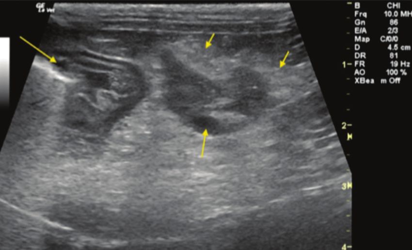
Page 2 of 10 Original Research
markedly and may consist of non-specific findings such as in a small number of cases (n = 7) (Shanaman et al. 2013).
poor appetite, vomiting, lethargy, diarrhoea, abdominal pain Moreover, a pilot study of CT angiography in 10 dogs
and weight loss. These are, however, non-specific clinical allowed a more complete evaluation of the entire pancreas
signs that can be seen with other conditions (Hess et al. 1998; than did ultrasound. In addition to this, identification of a
Steiner 2003). The differential diagnostic list includes a heterogeneous contrast enhancement of the pancreas may
plethora of primary diseases of the gastrointestinal, be a negative prognostic indicator in dogs with acute
hepatobiliary and urogenital tract, intra-abdominal tumours, necrotising pancreatitis (Adrian et al. 2015). Furthermore, in
splenic torsion and hypoadrenocorticism. Dogs may also humans, contrast CT is frequently used for the diagnosis of
display a combination of different clinical signs, including pancreatitis (Arvanitakis et al. 2007; Cappell 2008; Kim &
dehydration, icterus, fever, hypothermia, bleeding diathesis Pickhardt 2007; Scaglione et al. 2008; Sheu et al. 2012). To the
or ascites (Hess et al. 1998), as well as present with severe authors’ knowledge, the use of magnetic resonance imaging
systemic complications (Weatherton & Streeter 2009). These (MRI) to investigate the canine pancreas has not been
systemic complications include those already listed as well reported in the literature.
as tachycardia, arrhythmias, hypovolemic shock, acute
respiratory distress syndrome and death. Ultrasonography is an alternative technique for imaging the
pancreas and is considered the imaging method of choice for
The diagnosis of acute pancreatitis can be difficult because the diagnosis of pancreatitis in dogs (Xenoulis 2015), with a
of the anatomic inaccessibility of the pancreas and vague reported sensitivity of approximately 68% in dogs with
clinical signs and findings on clinical examination. Despite acute severe pancreatitis; however, this is operator
numerous improvements in various diagnostic tests, the dependant (Hess et al. 1998; Ferreri et al. 2003; Saunders
diagnosis of pancreatitis is still challenging. The only et al. 2002; Swift et al. 2000). The specificity of this modality
definitive diagnosis of pancreatitis is histopathology, which has, to date, not been reported as no histopathology
is highly invasive with localised disease possibly being confirmation has been performed to establish this (Mansfield
missed with a single biopsy (Newman et al. 2004). Although 2012). Contrast-enhanced ultrasound has been used in dogs
routine clinical pathology is non-specific, it may help in with pancreatitis and can demonstrate pancreatic perfusion
estimating the severity of the pancreatitis (Xenoulis 2015). changes. It has also been shown to aid in the diagnosis of
Animals with pancreatitis can show varying haematological pancreatitis, pancreatic necrosis as well as disease monitoring
abnormalities (Akol et al. 1993; Ferreri et al. 2003; Hess et al. in dogs following therapy (Lim et al. 2015; Rademacher et al.
1998; Hill & Van Winkle 1993 ). Serum-specific lipase (cPL) 2016). Thus, the clinical diagnosis of pancreatitis is generally
has been shown to be both a sensitive and specific serum based on a combination of clinicopathologic and imaging
marker for pancreatitis in dogs (McCord et al. 2012; Steiner findings.
et al. 2008; Trivedi et al. 2011; Watson et al. 2010), with a
sensitivity ranging between 72% and 78% (McCord et al. The aims of this study were to compare the clinical signs with
2012) and a specificity of between 81% and 100% (Mansfield the ultrasonographic findings in dogs with acute pancreatitis
& Jones 2000; McCord et al. 2012; Neilson-Carley et al. 2011; and to account for possible differences in the clinical
presentation depending on the region of the pancreas affected
Strombeck, Farver & Kaneko 1981; Trivedi et al. 2011). SNAP
as determined by ultrasonography.
canine pancreatic lipase (cPL) has a sensitivity between 91%
and 94% and a specificity between 71% and 78% for
pancreatitis (McCord et al. 2012). Other diagnostic tests Materials and methods
available include trypsin-like immunoreactivity (TLI), Multi-institutional cross-sectional retrospective study was
serum lipase and amylase activity, triolein and performed from first opinion and referral practices in
1,2-o-dilauryl-rac-glycero-3-glutaric acid-6-methylresorufin, Canada, North America and South Africa. Medical records
which is a lipase-based test (DGGR lipase assay, precision were searched in which a final diagnosis of pancreatitis had
pancreatic specific lipase [PSL]). Recent studies have shown been made based on typical history and clinical signs,
a high agreement between the Spec cPL and the DGGR laboratory testing and abdominal ultrasonography over a
lipase assay. However, this same article did demonstrate a 24-month period (2013–2014). A total of 293 cases met the
fair agreement between pancreatic ultrasonography results search criteria.
and serum lipase results (Kook et al. 2014). Thus, lipase
results are more accurate, and so ultrasonography should Inclusion criteria into the study included supportive clinical
therefore be interpreted carefully. In the dog radiography is signs and/or supportive clinical examination findings,
an insensitive diagnostic modality for pancreatitis because supportive ultrasonographic findings as well as an abnormal
of non-specific findings and often no findings at all. It does, SNAP cPL test. Animals had to have both appropriate
however, aid in ruling out other possible differentials (Akol ultrasonographic findings and an abnormal SNAP cPL to be
et al. 1993; Hess et al. 1998; Hill & Van Winkle 1993). included as well as ≥ 1 clinical sign and/or clinical
Contrast-enhanced computed tomography (CT) was examination finding. Indicative presenting clinical signs were
reported to be promising in the diagnosis of two cases of the acute (< 2 weeks) onset of poor appetite, and/or vomiting
canine pancreatitis (Jaeger et al. 2003); however, another or diarrhoea. Suggestive clinical examination findings
study reported a low sensitivity for diagnosing pancreatitis were abdominal discomfort and/or pain on palpation.
http://www.jsava.co.za Open Access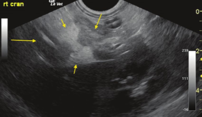
Page 3 of 10             Original Research

Full abdominal cavity ultrasound was performed by using an                                       appropriate organs, reducing variability amongst the
ultrasound machine (General Electric Logic E Ultrasound                                          different sonographers. All ultrasound images and videos
machine, GE Healthcare Biosciences, Shenzhen-China) with                                         were subsequently reviewed in a blind manner by one author
an 8-MHz probe, and the same type of machine and probe                                           (E.L.) with extensive experience in clinical sonography
were used across multiple institutions. Findings consistent                                      (Diplomate of the American Board of Veterinary Practitioners
with pancreatitis are an abnormal echogenic appearance of                                        [DABVP], certified International Veterinary Ultrasound
the pancreas, which included a mixed hypoechoic pancreatic                                       Society [Cert.IVUSS]). Exclusion criterion was the presence of
parenchyma and irregular capsule, and the presence of ill-                                       other primary diseases (such as gastrointestinal, hepato-
defined hyperechoic surrounding peri-pancreatic fat and                                          biliary, urinary tract diseases) and incomplete visualisation of
mesentery (Figures 1–4). In addition to this, assessment for                                     the pancreas on ultrasound. Animals with no ultrasonographic
the dilatation of the pancreatic or biliary ducts as well as the                                 changes to the pancreas were also excluded from this study.
                                                                                                 Based on ultrasonographic findings, dogs were divided into
presence of any ascites was performed (Hecht & Henry 2007).
                                                                                                 three groups: group 1 – 41 dogs with changes within the left
Moreover, ultrasonographic changes (as described above)
                                                                                                 lobe of the pancreas exclusively; group 2 – 105 dogs with
had to be restricted to the left or right lobe of the pancreas
                                                                                                 changes within the right lobe of the pancreas exclusively; and
exclusively for patients to be classified as left- or right-sided
                                                                                                 group 3 – 147 dogs with diffuse pancreatic involvement.
pancreatitis. In addition, any involvement of the body of the
pancreas would be ascribed as diffuse pancreatitis, and any
                                                                                                 As this was a retrospective study, abdominal pain was either
dilatation of the pancreatic or biliary duct would be assigned
                                                                                                 present or absent and not graded according to a pain scale.
to the right lobe pancreatitis group. In all cases the left, right                               Each animal had their entire abdomen palpated from cranial
and body of the pancreatic lobes were available for review.                                      to caudal and dorsal to ventral. If any discomfort or
Unobstructed images of the left, right and body of the                                           vocalisation was demonstrated, pain was regarded as
pancreas as well as a full video sweep of all areas of the                                       present. In addition, if no discomfort and/or vocalisation
pancreas were required to be included into the study. Multiple                                   was exhibited, pain was interpreted as absent. Poor appetite,
sonographers with training in small animal ultrasound                                            vomiting and diarrhoea were recorded as either present or
performed the initial ultrasound scans. All ultrasonographers                                    absent. Any dog with either complete loss or reduced
were either board certified internal medicine specialists or                                     appetite up to and including 2 weeks prior to presentation as
clinical sonographers and had to follow a standardised                                           described by the owner would be assigned the clinical sign of
ultrasound procedure taking images and videos of all                                             poor appetite.

Source: Photo courtesy of Doug Casey, DMV, DABVP English Bay Ultrasound, Vancouver, BC, Canada
FIGURE 1: Example of pancreatitis affecting the left limb of the pancreas (group 1): Focal mixed hypoechoic lesion (middle arrow) in the near field at the base of the left
pancreatic limb. Low-grade ill-defined inflamed fat (small arrows) is noted bordering the hypoechoic pancreatic parenchyma typical of pancreatitis. The location is caudal
to the gastric fundus (long arrow) free from influence upon the duodenum or pyloric outflow.

                                                           http://www.jsava.co.za                Open Access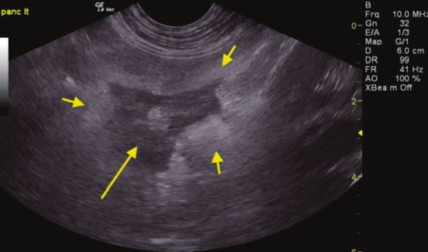
Page 4 of 10 Original Research
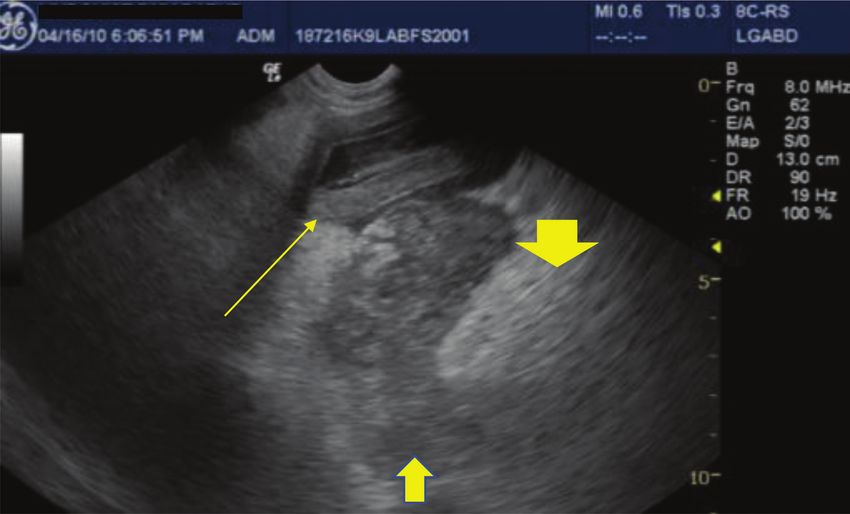
Source: Photo courtesy of Andi Parkinson, Intrapet Imaging, Baltimore, MD, United States
FIGURE 2: Example of pancreatitis affecting the left limb of the pancreas (group 1): Left limb of the pancreas demonstrating coarse mixed hypoechoic (long arrow)
pancreatic parenchyma with ill-defined hyperechoic surrounding fat (small arrows) typical of acute pancreatitis.
FIGURE 3: Example of pancreatitis affecting the right limb of the pancreas (group 2): Hypoechoic oedematous pancreatic parenchyma (middle arrow) and hyperechoic
ill-defined surrounding fat (small thick arrow) consistent with saponification and inflamed mesentery. The upper descending duodenum in the near field is mildly
oedematous and adjacent to the pancreatic inflammation (long thin arrow).
http://www.jsava.co.za Open AccessPage 5 of 10 Original Research
Source: Photo courtesy of Andi Parkinson, Intrapet Imaging, Baltimore, MD, United States
FIGURE 4: Example of pancreatitis affecting the right limb of the pancreas (group 2): Right limb pancreatitis with mixed hypoechoic oedematous parenchyma (middle
arrow), ill-defined inflamed surrounding fat (short arrows), all of which surround promote inflammation of the upper descending duodenum (long arrow). Right kidney is
present in far field.
All data were tabulated in an Excel spreadsheet programme Shih Tzu (18), Maltese/Maltese cross (18), Labrador/
(Excel®, Microsoft Corporation, Washington, United States.) Labrador mix (17), Bischon Frise (16), Miniature Schnauzer
and statistical analysis was performed by using a statistical (11), Dachshund (7), Terrier/Terrier mix (4), Boxer (3), Golden
software package (NCSS®, Kaysville, UT, United States). Retriever/Golden Retriever mix (3), Miniature Poodle (3),
Descriptive statistics were used to describe the data. For two of each American Eskimo mix, Bassett Hound, Beagle,
single parameters (abdominal pain, poor appetite, vomiting Border Collie/Border Collie cross, Chihuahua/Chihuahua
and diarrhoea), differences between the groups were tested mix, Husky mix, Jack Russell terrier/Jack Russel terrier mix,
using one-way analysis of variance with Bonferroni and Pomeranian, Shetland Sheepdog and Weimaraner; and one
Tukey–Kramer comparisons. Fischer’s exact test was used of each Akita, Boston Terrier, Cocker Spaniel, Collie mix,
to determine the association between ultrasonographic English Setter, Fox terrier, Lhasa Apso mix, Miniature
findings and clinical signs. The data were normally Pincher, Pekingese, Pitbull mix, Pyrenees Mountain dog, Rat
distributed, and the level of significance was set at p < 0.05. terrier, Vizsla and West Highland White terrier. Sex
distribution in group 1 was 18 males and 23 females; in
Ethical considerations group 2, it was 52 males and 53 females; and in group 3,
it was 68 males and 79 females, with no statistical difference
This article followed all ethical standards for carrying out a
between the groups.
research without direct contact with human or animal
subjects. No ethical approval was required as this was a
In group 1, pain was noted in 4/41 dogs (10%), poor appetite
retrospective non-invasive clinical study.
in 13/41 dogs (32%), vomiting in 27/41 dogs (66%) and
diarrhoea in 17/41 dogs (41%). In group 2, pain was present
Results in 11/105 dogs (10%), poor appetite in 44/105 dogs (42%),
A total of 293 client-owned dogs were used with a median vomiting in 44/105 dogs (42%) and diarrhoea in 20/105 dogs
age of the groups 1–3 were 8.8, 9.8 and 10.1 years, respectively, (19%). In group 3, pain was noted in 29/147 dogs (20%), poor
with no statistical difference between the groups. Various appetite in 46/147 dogs (31%), vomiting in 76/147 dogs
breeds were present with no one breed or sex appearing to be (52%) and diarrhoea in 35/147 dogs (24%) (Table 1). Statistical
over-represented in any one group. The following breeds significance was presence of pain in group 3; poor appetite in
were represented in the order of decreasing frequency: mixed groups 2 and 3; and vomiting and diarrhoea in group 3
breeds (128), Yorkshire terrier/Yorkshire terrier cross (31), (Figure 5).
http://www.jsava.co.za Open AccessPage 6 of 10 Original Research
TABLE 1: Percentage of dogs displaying clinical signs according to the region of and followed caudally, either in transverse or longitudinal
pancreas affected.
Group Clinical signs (% dogs affected)
plane (Larson 2016). The pancreatic body lies immediately
Pain Anorexia Vomiting Diarrhoea
caudal and dorsal to the pyloroduodenal junction, ventral to
1 (left limb) 11 32 66 41 the portal vein and medial to the proximal duodenum. The
2 (right limb) 10 42 42 19 left lobe in the dog lies dorsal and caudal to the body of the
3 (diffuse) 20 31 52 24 stomach, cranial to the transverse colon. It follows the course
of the splenic vein as it travels from the splenic hilus medially
to the portal vein at the level of the pancreatic body. The
canine left lobe is often much smaller than the right lobe and
Group 1 Group 2 Group 3
is more difficult to consistently detect because of shadowing
80 76
and reverberation artefact from gas in the stomach and
70
transverse colon (Larson 2016). The anatomic location of each
60 lobe is important as it can explain the findings of this study.
50 46
44 44
40 35
Abdominal ultrasound is considered the imaging method of
29 27 choice for the diagnosis of pancreatitis in dogs with the
30
20 added advantage of ruling out differential diagnoses that
20 17
11 13 share similar clinical signs (Xenoulis 2015). The performance
10 4 of abdominal ultrasonography is, however, dependent on
0 the expertise of the user. Ultrasonographic findings in dogs
Pain Anorexia Voming Diarrhoea
with acute pancreatitis include hypoechoic areas within the
*, statistical significance between groups ( p < 0.05). pancreas, increased echogenicity of the mesentery and
FIGURE 5: Clinical signs between the three groups with number of dogs enlargement or irregularity of the pancreas as well as
exhibiting the signs. Group 1 – pancreatitis of the left lobe, group 2 – pancreatitis dilatation of the pancreatic or biliary duct, and ascites
within the right lobe and group 3 – diffuse pancreatitis.
(Ferreri et al. 2003; Hecht & Henry 2007; Hess et al. 1998;
Saunders et al. 2002; Swift et al. 2000). Dilatation of the
Discussion pancreatic duct is considered pathognomonic for pancreatitis
in human patients, and possibly also in dogs (Simpson &
This study focused on relating clinical signs of pancreatitis
Lamb 1995; Watson 2004). In pancreatitis, the surrounding
to the area of the pancreas affected, and it documented
mesentery can be hyperechoic indicating peri-pancreatic
different presenting clinical signs depending on which
steatitis and fat necrosis (Hecht & Henry 2007). In the dog,
lobe of the pancreas was involved. No obvious breed
the right lobe seems to be the most commonly affected
predisposition was identified in this study, which agrees
portion of the pancreas in acute disease, which may be
with previous studies that reported that dogs of any age,
related to the fact that the right limb is more easily identified
breed or sex can develop pancreatitis. Most dogs that are
on ultrasound (Larson 2016). These ultrasonographic
presented with pancreatitis are usually greater than 5 years findings were present in this current study and were used to
of age (Cook et al. 1993; Ferreri et al. 2003; Hess et al. 1998; aid the researchers in the diagnosis of acute pancreatitis.
Watson et al. 2010). Various studies have highlighted Previous research has described normal ultrasonographic
differences in breed predilections because of different findings in healthy dogs. They showed that the right lobe of
geographic regions. In the United States, miniature the pancreas was most consistently visible and measured in
schnauzers and terrier breeds (particularly Yorkshire 98% of dogs versus the left lobe at 88% of all dogs and the
terriers) are at increased risk (Cook et al. 1993; Hess et al. body at 64%, with the pancreatic duct detected in only 8% of
1998; Lem et al. 2008). In the United Kingdom, Cocker all the dogs (Murtaugh et al. 1985). In an experimental study
spaniels, Cavalier King Charles spaniels, Border collies and of induced pancreatitis in six healthy dogs, an ill-defined
Boxers have been reported to be at increased risk for chronic mass with a heterogeneous echogenicity accompanied by an
pancreatitis (Watson et al. 2007). overall decrease in echogenicity was reported in both the left
and right pancreatic lobes. Right pancreatic lobe involvement
The differences between this current study and those was detected in 5/6 dogs, and the left pancreatic lobe was
previously reported can be ascribed to the difference in involved in 4/6 dogs (Murtaugh et al. 1985). In a study that
population size, where sample size in the current study was assessed the ultrasonographic findings in three dogs with
significantly higher than that in previous studies. experimentally induced pancreatitis, right pancreatic lobe
pathology was detected more often than the left lobe
In the dog, the pancreas is divided into the right and left pathology (Nyland et al. 1983). These findings are also in
lobes, joined at the body with the right lobe easiest to identify agreement with another study (Lamb & Simpson 1995).
on ultrasound, located dorsomedial to the descending Although these researches are experimental and were
duodenum, ventral to the right kidney and lateral to the conducted on a small number of dogs, these results are,
ascending colon (Larson 2016). The duodenum and right however, like the findings in our study where lesions
pancreatic lobe can be located in the dorsal aspect of the isolated to the right lobe of the pancreas was identified in
10th–12th intercostal space (can vary with dog conformation) 36% of cases in comparison with lesions isolated to the left
http://www.jsava.co.za Open AccessPage 7 of 10 Original Research
lobe of the pancreas in 14% of cases. Diffuse pancreatic In addition to this, pain, vomiting and diarrhoea were more
disease was detected in 50% of patients. Thus, based on our commonly identified in diffuse pancreatitis. Poor appetite
study, diffuse pancreatic pathology seems to be the most was more prevalent in right-sided and diffuse pancreatitis.
common ultrasonographic finding in patients with acute These differences between the groups can possibly be
pancreatitis. attributed to gastric and/or intestinal tract involvement
when various lobes of the pancreas are affected. In humans,
Ultrasonographic findings consistent with acute pancreatitis numerous studies have demonstrated the relationship of
were noted in 68% of cases in a previous study (Hess et al. nausea with the onset of gastric dysrhythmias in individuals
1998). Ultrasonography has advantages over other imaging with motion sickness, pregnant women and gastroparesis
methods in being relatively low in cost and non- (Hasler et al. 1995; Koch 2014; Xu et al. 1993). Previous
invasive. However, the low negative predictive value of studies have suggested a relationship between gastric
normal ultrasonographic findings prevents the use of dysrhythmias and nausea (Xu et al. 1993). In addition to this,
ultrasonography as an exclusion diagnostic procedure for medications that decrease dysrhythmias, decrease nausea
acute pancreatitis as false-negative results are possible and stimuli that increase dysrhythmias may promote the
(Ruaux 2003). Not all lesions can be correlated with the sensation of nausea (Koch 2014). It can thus be speculated
clinical status of the patient (Mix & Jones 2006). As non- that diffuse pancreatitis, because of its close association with
significant pancreatic lesions can be detected on ultrasound the stomach, may result in gastric dysrhythmias that would
examination, the significance of pancreatic abnormalities cause nausea and vomiting. Possibilities for the increased
identified via ultrasonography must be interpreted in the frequency of diarrhoea in diffuse pancreatitis would be
light of a patient’s clinical signs (Mix & Jones 2006). A thickening of the gastric and colonic wall and/or gastric
previous study showed that the ultrasound pattern of dumping syndrome. A recent study in humans with acute
pancreatitis, by using MRI to record abnormalities in the
corrugated and thickened bowel wall was associated with
gastrointestinal tract, showed a thickened stomach and
pancreatitis. In that study 18 dogs had a corrugated bowel;
transverse colon in 20% and 15% of patients, respectively
however, a final diagnosis of acute pancreatitis was made in
(Ji et al. 2017). In humans following gastrectomy a dumping
only 50% of cases (Moon, Biller & Armbrust 2003). Therefore,
syndrome can develop. This entails rapid gastric emptying
a diagnosis of pancreatitis should not be based exclusively
of hyperosmolar contents into the proximal intestine (Davis
on ultrasonographic findings, and correlating appropriate
& Ripley 2017; Machella 1950). Because of the proximity of
history, clinical examination findings and biochemical blood
the left lobe of the pancreas with the stomach, it can be
tests are still crucial.
speculated that gastric motility may be affected because of
the localised inflammation, resulting in dumping of gastric
No clinical sign is pathognomonic for pancreatitis in dogs;
contents into the proximal intestine causing diarrhoea. In
however, dogs with severe acute pancreatitis are typically
humans, an acute necrotising pancreatitis may result in
presented with acute onset abdominal pain (Hess et al.
colonic complications. A previous study demonstrated that
1998; Weatherton & Streeter 2009). One study reported
in human patients with surgical management of acute
abdominal pain in 58% of dogs with acute pancreatitis
necrotising pancreatitis, 6.1% had colonic infarction
(Hess et al. 1998), which is much higher than the findings in secondary to the inflammatory process as a complication
this current study where abdominal pain was only present (Adams, Davis & Anderson 1994). These findings are similar
in 15% of all cases. Poor appetite has been reported to occur to a previous study which showed in 22 patients with acute
in 91% of cases (Hess et al. 1998), which is different from the pancreatitis, nine had colonic involvement. The transverse
current study where a poor appetite was present in only colon was affected in three of the nine patients (Aldridge et
35% of all dogs in the study. Acute vomiting has been al. 1989). In the dog the left lobe of the pancreas lies cranially
reported in 90% (Hess et al. 1998) of cases, whereas in this to the transverse colon. Albeit not as severe as the
study it occurred in only 50% of all patients. Diarrhoea has complications that develop in people, we hypothesise that
been reported in 33% (Hess et al. 1998) of dogs, whereas in the localised inflammatory process affecting the left lobe of
this study it occurred only in 25% of all animals. The the pancreas might result in a colitis of the transverse colon
differences between the current study and Hess et al.(1998) and may not necessarily always cause vomiting; however,
can be ascribed to the different focus of the two studies as this may change overtime as the condition progresses.
well as improvement with abdominal ultrasonography Although mild clinical signs associated with left lobe
technology and operator expertise. Furthermore, the pancreatitis have not been described in the veterinary
sample size in the current study was significantly higher literature, a new publication assessing CT angiography and
than that of Hess et al.’s study. Moreover, differences ultrasonography in dogs with acute pancreatitis has
amongst these results could also be attributed to the demonstrated some interesting findings. This study showed
dissimilar patient populations. Hess et al.’s (1998) study that dogs with heterogeneous contrast enhancement of the
only contained patients that died; thus, those patients may pancreas had significantly longer duration of hospitalisation
have had more severe clinical signs. In another study on including the likelihood to be hospitalised for more than
20 dogs with pancreatitis, only one dog (5%) did not have 5 days, had increased number of relapses and were
gastrointestinal signs (Kis et al. 2013). Once again this was a significantly more likely to have portal vein thrombosis
very small sample size. (French et al. 2019). A recent study highlighted that there was
http://www.jsava.co.za Open AccessPage 8 of 10 Original Research
a weak but significant linear correlation between thickness of that this influenced the outcome of our results as presence
right lobe of the pancreas with that of the mural thickness of and not severity of clinical signs were the aims of our study.
the duodenum and the duodenal diameter (Wickramasekara However, future studies assessing prognostic significance of
Rajapakshage et al. 2016). This demonstrates a dimensional these findings should include a grading scale for abdominal
relationship between the right pancreatic limb and adjacent pain and poor appetite. Additional limitations were that the
duodenum and can be used to assess pancreatic size in dogs true prevalence of pancreatitis confined to the limb of the
(Wickramasekara Rajapakshage et al. 2016). In addition, pancreas may have been under-represented in this study as
recent studies have demonstrated that the intestine is thought interference from gastric content may have resulted in
to contribute to or exacerbate pancreatic inflammation animals with left limb pancreatitis being unnecessarily
directly because of intestinal ischaemia (Flint & Windsor excluded, and no odds ratio was performed to determine any
2003). The right pancreatic lobe lies dorsomedial to the breed predilection. Dogs had to have ultrasonographic
descending duodenum. Poor appetite was a statistical findings consistent with pancreatitis to be included into the
finding in this study, and it did appear more prevalent in study. Thus, patients with no ultrasonographic findings may
dogs with right-sided and diffuse pancreatitis. A possible have been excluded from the study, which may have biased
explanation can be ascribed to the fact that the duodenum the results. However, it is unlikely that these patients would
has a large number of receptors and is referred to as the have had the other inclusion criteria. Moreover, the
‘organ of nausea’ (Schoor 2011). Stimulation of these concentration of serum canine pancreatic lipase was not
receptors within the duodenum could have resulted in the assessed. The definitive Spec cPLI concentration test has
poor appetite because of nausea and not necessarily a grey zone when the result is between 200 mg/L and
vomiting. Furthermore, pain seen in canine patients with 400 mg/L and values > 400 mg/L indicate pancreatitis. The
acute pancreatitis was noted with a significantly higher SNAP cPL results can only be differentiated as normal or
frequency in diffuse pancreatic disease compared with abnormal. Thus, some of the patients may have had a false-
disease restricted to the left or right lobe of the pancreas. This positive test result for the SNAP cPL.
may be because dogs with diffuse pancreatitis have a more
severe form of the disease. Despite overlap between groups, Despite the limitations of the study, the findings nevertheless
this study indicates that pain response is expected to occur present pertinent questions, including how dogs with
with a higher frequency in diffuse pancreatitis but overall is pancreatitis may display a variety of different clinical signs
not a very common clinical sign. This is important as lack of depending on the region of the pancreas affected. Although
abdominal pain does not exclude pancreatitis especially if these results have not been previously described, these
either of the two lobes is affected. findings are important, as they may aid in the understanding
of how these vague clinical signs occur in the dog and may
The results of our study are in contrast to a previous study help practitioners with their treatment regimens. Moreover,
that showed no significant differences between clinical further studies are required to validate these results and to
signs and ultrasonographic changes in the pancreas assess whether the area of the pancreas affected has
(Myung-Jin et al. 2017). However, that study had a small prognostic significance in the dog.
sample size of 40 client-owned dogs (Myung-Jin et al.
2017). In addition to this, the aims of that study focussed Based on the results obtained in this study, it can be concluded
on comparing abnormal serum canine pancreas-specific that animals with left, right and diffuse pancreatitis have
lipase results and pancreatic ultrasonographic changes in different clinical presentations. In addition, pain occurs with
dogs with pancreatitis, which are different from the aims of a higher frequency in diffuse pancreatitis, although vomiting
our study. and diarrhoea are more prevalent in dogs with diffuse
pancreatitis. Furthermore, reduced appetite appears more
Important limitations of this study were as follows: it was a
commonly in dogs with diffuse and right lobe pancreatitis.
retrospective study with dogs assessed only at one single
time point; only a single ultrasound examination was
performed with no follow-up scans; and no pancreatic Acknowledgements
histopathology was conducted. Furthermore, multiple Competing interests
individuals performed the ultrasound, and thus there could
The authors have declared that no competing interest exists.
have been interobserver variability as the skills of the operator
as well as the sensitivity of the ultrasound scanner are
extremely important with ultrasonography. However, all Authors’ contributions
persons performing the ultrasonography had training in All the authors made substantial contributions to the study
sonography and had to follow a standard ultrasonographic design, interpretation of data, and drafting and revising of
procedure using the same ultrasound scanner. Moreover, all the article.
images and videos were assessed by one reviewer with
extensive experience in small animal ultrasound, which
reduced the impact of these limitations. Although grading for Funding information
lesions (clinical and ultrasonographic findings) observed in This research received no specific grant from any funding
dogs with pancreatitis was not discussed, we do not believe agency in the public, commercial or not-for-profit sectors.
http://www.jsava.co.za Open AccessPage 9 of 10 Original Research
Data availability statement Kim, D.H. & Pickhardt, P.J., 2007, ‘Radiologic assessment of acute and chronic
pancreatitis’, Surgical Clinics of North America 87(6), 1341–1358, viii. https://doi.
org/10.1016/j.suc.2007.08.005
Data sharing is not applicable to this article as no new data
Kis, I., Brkljacic, M., Mayer, I., Selanec, J., Jovic, I., Kucer, N. et al., 2013, ‘Diagnostics of
were created or analysed in this study. acute canine pancreatitis and comparison of clinical, laboratory and ultrasound
findings’, in 5th International Congress ‘Veterinary Science and Profession’, Zagreb,
Croatia, October 3–4, 2013. Book of abstracts, 38.
Disclaimer Koch, K.L., 2014, ‘Gastric dysrhythmias: A potential objective measure of nausea’,
Experimental Brain Research 232(8), 2553–2561. https://doi.org/10.1007/
s00221-014-4007-9
The views expressed in this article are those of the authors
Kook, P.H., Kohler, N., Hartnack, S., Riond, B. & Reusch, C.E., 2014, ‘Agreement of
and not the views of an official position of an institution or serum Spec cPL with the 1,2-o-dilauryl-rac-glycero glutaric acid-(6’-
funder. methylresorufin) ester (DGGR) lipase assay and with pancreatic ultrasonography
in dogs with suspected pancreatitis’, Journal of Veterinary Internal Medicine 28(3),
863–870. https://doi.org/10.1111/jvim.12334
References Lamb, C.R. & Simpson, K.W., 1995, ‘Ultrasonographic findings in cholecystokinin-
induced pancreatitis in dogs’, Veterinary Radiology & Ultrasound 36, 139–145.
https://doi.org/10.1111/j.1740-8261.1995.tb00234.x
Adams, D.B., Davis, B.R. & Anderson, M.C., 1994, ‘Colonic complications of
pancreatitis’, The American Surgeon 60(1), 44–49. Larson, M.M., 2016, ‘Ultrasound imaging of the hepatobiliary system and pancreas’,
Veterinary Clinics of North America: Small Animal Practice 46(3), 453–480.
Adrian, A.M., Twedt, D.C., Kraft, S.L. & Marolf, A.J., 2015, ‘Computed tomographic https://doi.org/10.1016/j.cvsm.2015.12.004
angiography under sedation in the diagnosis of suspected canine pancreatitis: A
pilot study’, Journal of Veterinary Internal Medicine 29(1), 97–103. https://doi. Lem, K.Y., Fosgate, G.T., Norby, B. & Steiner, J.M., 2008, ‘Associations between dietary
org/10.1111/jvim.12467 factors and pancreatitis in dogs’, Journal of the American Veterinary Medical
Association 233(9), 1425–1431. https://doi.org/10.2460/javma.233.9.1425
Akol, K.G., Washabau, R.J., Saunders, H.M. & Hendrick, M.J., 1993, ‘Acute pancreatitis
in cats with hepatic lipidosis’, Journal of Veterinary Internal Medicine 7(4), Lim, S.Y., Nakamura, K., Morishita, K., Sasaki, N., Murakami, M., Osuga, T. et al., 2015,
205–209. https://doi.org/10.1111/j.1939-1676.1993.tb01008.x ‘Quantitative contrast-enhanced ultrasonographic assessment of naturally
occurring pancreatitis in dogs’, Journal of Veterinary Internal Medicine 29(1), 71–
Aldridge, M.C., Francis, N.D., Glazer, G. & Dudley, H.A., 1989, ‘Colonic complications of 78. https://doi.org/10.1111/jvim.12470
severe acute pancreatitis’, British Journal of Surgery 76(4), 362–367. https://doi.
org/10.1002/bjs.1800760416 Machella, T.E., 1950, ‘Mechanism of the post-gastrectomy dumping syndrome’,
Gastroenterology 14(2), 237–255. https://doi.org/10.1016/S0016-5085(50)80037-9
Arvanitakis, M., Koustiani, G., Gantzarou, A., Grollios, G., Tsitouridis, I., Haritandi-
Kouridou, A. et al., 2007, ‘Staging of severity and prognosis of acute pancreatitis Mansfield, C., 2012, ‘Acute pancreatitis in dogs: Advances in understanding,
by computed tomography and magnetic resonance imaging-a comparative study’, diagnostics, and treatment’, Topics in Companion Animal Medicine 27(3), 123–
Digestive and Liver Disease 39(5), 473–482. https://doi.org/10.1016/j.dld. 132. https://doi.org/10.1053/j.tcam.2012.04.003
2007.01.015
Mansfield, C.S. & Jones, B.R., 2000, ‘Plasma and urinary trypsinogen activation peptide in
Bostrom, B.M., Xenoulis, P.G., Newman, S.J., Pool, R.R., Fosgate, G.T. & Steiner, J.M., healthy dogs, dogs with pancreatitis and dogs with other systemic diseases’, Australian
2013, ‘Chronic pancreatitis in dogs: A retrospective study of clinical, Veterinary Journal 78(6), 416–422. https://doi.org/10.1111/j.1751-0813.2000.tb
clinicopathological, and histopathological findings in 61 cases’, The Veterinary 11833.x
Journal 195(1), 73–79. https://doi.org/10.1016/j.tvjl.2012.06.034
Mccord, K., Morley, P.S., Armstrong, J., Simpson, K., Rishniw, M., Forman, M.A. et al.,
Cappell, M.S., 2008, ‘Acute pancreatitis: Etiology, clinical presentation, diagnosis, and 2012, ‘A multi-institutional study evaluating the diagnostic utility of the spec cPL
therapy’, Medical Clinics of North America 92(4), 889–923, ix–x. https://doi. and SNAP(R) cPL in clinical acute pancreatitis in 84 dogs’, Journal of Veterinary
org/10.1016/j.mcna.2008.04.013 Internal Medicine 26(4), 888–896. https://doi.org/10.1111/j.1939-1676.2012.
Cook, A.K., Breitschwerdt, E.B., Levine, J.F., Bunch, S.E. & Linn, L.O., 1993, ‘Risk factors 00951.x
associated with acute pancreatitis in dogs: 101 cases (1985–1990)’, Journal of the Mix, K. & Jones, C., 2006, ‘Diagnosing acute pancreatitis in dogs’ Compendium on
American Veterinary Medical Association 203(5), 673–679. Continuing Education for the Practicing Veterinarian 28:226–234.
Davis, J.L. & Ripley, R.T., 2017, ‘Postgastrectomy syndromes and nutritional Moon, M.L., Biller, D.S. & Armbrust, L.J., 2003, ‘Ultrasonographic appearance and
considerations following gastric surgery’, Surgical Clinics of North America 97(2), etiology of corrugated small intestine’, Veterinary Radiology & Ultrasound 44(2),
277–293. https://doi.org/10.1016/j.suc.2016.11.005 199–203. https://doi.org/10.1111/j.1740-8261.2003.tb01271.x
De Cock, H.E., Forman, M.A., Farver, T.B. & Marks, S.L., 2007, ‘Prevalence and Murtaugh, R.J., Herring, D.S., Jacobs, R.M. & Dehoff, W.D., 1985, ‘Pancreatic
histopathologic characteristics of pancreatitis in cats’, Veterinary Pathology 44(1), ultrasonography in dogs with experimentally induced acute pancreatitis’, Veterinary
39–49. https://doi.org/10.1354/vp.44-1-39 Radiology 26(1), 27–32. https://doi.org/10.1111/j.1740-8261.1985.tb01111.x
Ferreri, J.A., Hardam, E., Kimmel, S.E., Saunders, H.M., Van Winkle, T.J., Drobatz, K.J. et Myung-Jin, K., Joong-Hyun, S., Tae-Sung, H., Hee-Chun, L. & Dong-In, J., 2017,
al., 2003, ‘Clinical differentiation of acute necrotizing from chronic nonsuppurative ‘Comparison between SNAP canine pancreas-specific Lipase (cPL) test results and
pancreatitis in cats: 63 cases (1996–2001)’, Journal of the American Veterinary pancreatic ultrasonographic findings in dogs with pancreatitis’, Journal of
Medical Association 223(4), 469–474. https://doi.org/10.2460/javma.2003. Veterinary Clinics 34(4), 229–233. https://doi.org/10.17555/jvc.2017.08.34.4.229
223.469
Neilson-Carley, S.C., Robertson, J.E., Newman, S.J., Kutchmarick, D., Relford, R.,
Flint, R.S. & Windsor, J.A., 2003, ‘The role of the intestine in the pathophysiology and Woosley, K. et al., 2011, ‘Specificity of a canine pancreas-specific lipase assay for
management of severe acute pancreatitis’, HPB: The Official Journal of the diagnosing pancreatitis in dogs without clinical or histologic evidence of the
International Hepato Pancreato Biliary Association 5(2), 69–85. https://doi.
org/10.1080/13651820310001108 disease’, American Journal of Veterinary Research 72(3), 302–307. https://doi.
org/10.2460/ajvr.72.3.302
French, J.M., Twedt, D.C., Rao, S. & Marolf, A.J., 2019, ‘Computed tomographic
angiography and ultrasonography in the diagnosis and evaluation of acute Newman, S., Steiner, J., Woosley, K., Barton, L., Ruaux, C. & Williams, D., 2004,
pancreatitis in dogs’, Journal of Veterinary Internal Medicine 33(1), 79–88. https:// ‘Localization of pancreatic inflammation and necrosis in dogs’, Journal of Veterinary
doi.org/10.1111/jvim.15364 Internal Medicine 18(4), 488–493. https://doi.org/10.1111/j.1939-1676.2004.
tb02572.x
Hasler, W.L., Soudah, H.C., Dulai, G. & Owyang, C., 1995, ‘Mediation of hyperglycemia-
evoked gastric slow-wave dysrhythmias by endogenous prostaglandins’, Nyland, T.G., Mulvany, M.H. & Strombeck, D.R., 1983, ‘Ultrasonic features of
Gastroenterology 108(3), 727–736. https://doi.org/10.1016/0016-5085(95)90445-X experimentally induced, acute pancreatitis in the dog’, Veterinary Radiology
24(6), 260–266. https://doi.org/10.1111/j.1740-8261.1983.tb00729.x
Hecht, S. & Henry, G., 2007, ‘Sonographic evaluation of the normal and abnormal
pancreas’, Clinical Techniques in Small Animal Practice 22(3), 115–121. https:// Rademacher, N., Schur, D., Gaschen, F., Kearney, M. & Gaschen, L., 2016, ‘Contrast-
doi.org/10.1053/j.ctsap.2007.05.005 enhanced ultrasonography of the pancreas in healthy dogs and in dogs with acute
pancreatitis’, Veterinary Radiology & Ultrasound 57(1), 58–64. https://doi.
Hess, R.S., Saunders, H.M., Van Winkle, T.J., Shofer, F.S. & Washabau, R.J., 1998, org/10.1111/vru.12285
‘Clinical, clinicopathologic, radiographic, and ultrasonographic abnormalities in
dogs with fatal acute pancreatitis: 70 cases (1986–1995)’, Journal of the American Ruaux, C.G., 2003, ‘Diagnostic approaches to acute pancreatitis’, Clinical Techniques in
Veterinary Medical Association 213(5), 665–670. Small Animal Practice 18(4), 245–249. https://doi.org/10.1016/S1096-2867(03)
00072-0
Hill, R.C. & Van Winkle, T.J., 1993, ‘Acute necrotizing pancreatitis and acute suppurative
pancreatitis in the cat. A retrospective study of 40 cases (1976–1989)’, Journal of Saunders, H.M., Vanwinkle, T.J., Drobatz, K., Kimmel, S.E. & Washabau, R.J., 2002,
Veterinary Internal Medicine7(1), 25–33.https://doi.org/10.1111/j.1939-1676.1993. ‘Ultrasonographic findings in cats with clinical, gross pathologic, and histologic
tb03165.x evidence of acute pancreatic necrosis: 20 cases (1994–2001)’, Journal of the
American Veterinary Medical Association 221(12), 1724–1730. https://doi.
Jaeger, J.Q., Mattoon, J.S., Bateman, S.W. & Morandi, F., 2003, ‘Combined use of org/10.2460/javma.2002.221.1724
ultrasonography and contrast enhanced computed tomography to evaluate acute
necrotizing pancreatitis in two dogs’, Veterinary Radiology & Ultrasound 44(1), Scaglione, M., Casciani, E., Pinto, A., Andreoli, C., De Vargas, M. & Gualdi, G.F., 2008,
72–79. https://doi.org/10.1111/j.1740-8261.2003.tb01453.x ‘Imaging assessment of acute pancreatitis: A review’, Seminars in Ultrasound, CT
and MR 29(5), 322–340. https://doi.org/10.1053/j.sult.2008.06.009
Ji, Y.-F., Zhang, X.-M., Mitchell, D.G., Li, X.-H., Chen, T.-W., Li, Y. et al., 2017,
‘Gastrointestinal tract involvement in acute pancreatitis: Initial findings and Schoor, M.V., 2011, ‘Approach to the vomitting dog’, in Proceedings of the World
follow-up by magnetic resonance imaging’, Quantitative Imaging in Medicine and Veterinary Association Congress, Cape Town, South Africa, October 10–14, 2011,
Surgery 7(6), 641–653. https://doi.org/10.21037/qims.2017.12.03 pp. 1–5.
http://www.jsava.co.za Open AccessPage 10 of 10 Original Research
Shanaman, M.M., Schwarz, T., Gal, A. & O’brien, R.T., 2013, ‘Comparison between Watson, P., 2004, ‘Pancreatitis in the dog: Dealing with a spectrum of disease’
survey radiography, B-mode ultrasonography, contrast-enhanced ultrasonography In Practice 26.
and contrast-enhanced multi-detector computed tomography findings in dogs
with acute abdominal signs’, Veterinary Radiology & Ultrasound 54(6), 591–604. Watson, P.J., Archer, J., Roulois, A.J., Scase, T.J. & Herrtage, M.E., 2010, ‘Observational
https://doi.org/10.1111/vru.12079 study of 14 cases of chronic pancreatitis in dogs’, Veterinary Record 167(25),
968–976. https://doi.org/10.1136/vr.c4912
Sheu, Y., Furlan, A., Almusa, O., Papachristou, G. & Bae, K.T., 2012, ‘The revised Atlanta
classification for acute pancreatitis: A CT imaging guide for radiologists’, Emergency Watson, P.J., Roulois, A.J., Scase, T., Johnston, P.E., Thompson, H. & Herrtage, M.E.,
Radiology 19(3), 237–243. https://doi.org/10.1007/s10140-011-1001-4 2007, ‘Prevalence and breed distribution of chronic pancreatitis at post-mortem
examination in first-opinion dogs’, Journal of Small Animal Practice 48(11),
Simpson, K. & Lamb, C., 1995, ‘Acute pancreatitis in the dog’, In Practice 17, 328–337. 609–618. https://doi.org/10.1111/j.1748-5827.2007.00448.x
https://doi.org/10.1136/inpract.17.7.328
Weatherton, L.K. & Streeter, E.M., 2009, ‘Evaluation of fresh frozen plasma
Steiner, J.M., 2003, ‘Diagnosis of pancreatitis’, Veterinary Clinics of North America: administration in dogs with pancreatitis: 77 cases (1995–2005)’, Journal of
Small Animal Practice 33, 1181–1195. https://doi.org/10.1016/S0195- Veterinary Emergency and Critical Care (San Antonio) 19(6), 617–622. https://doi.
5616(03)00061-5 org/10.1111/j.1476-4431.2009.00483.x
Steiner, J.M., Newman, S., Xenoulis, P., Woosley, K., Suchodolski, J., Williams, D. et al., Wickramasekara Rajapakshage, B.K., Ellearaewe Garuhamilage, J.P., De Silva, D.D. &
2008, ‘Sensitivity of serum markers for pancreatitis in dogs with macroscopic Dangolla, A., 2016, ‘Dimensional ultrasonographic relationship of the right lobe
evidence of pancreatitis’, Veterinary Therapeutics 9(4), 263–273.
of pancreas with associated anatomic landmarks in clinically normal dogs’,
Strombeck, D.R., Farver, T. & Kaneko, J.J., 1981, ‘Serum amylase and lipase activities in Journal of Veterinary Medical Science 78(1), 1–5. https://doi.org/10.1292/
the diagnosis of pancreatitis in dogs’, American Journal of Veterinary Research jvms.15-0209
42(11), 1966–1970.
Xenoulis, P.G., 2015, ‘Diagnosis of pancreatitis in dogs and cats’, Journal of Small
Swift, N.C., Marks, S.L., Maclachlan, N.J. & Norris, C.R., 2000, ‘Evaluation of serum Animal Practice 56(1), 13–26. https://doi.org/10.1111/jsap.12274
feline trypsin-like immunoreactivity for the diagnosis of pancreatitis in cats’,
Journal of the American Veterinary Medical Association 217(1), 37–42. https:// Xenoulis, P.G., Suchodolski, J.S. & Steiner, J.M., 2008, ‘Chronic pancreatitis in dogs and
doi.org/10.2460/javma.2000.217.37 cats’, Compendium: Continuing Education for Veterinarians 30(3), 166–180; quiz
180–181.
Trivedi, S., Marks, S.L., Kass, P.H., Luff, J.A., Keller, S.M., Johnson, E.G. et al., 2011,
‘Sensitivity and specificity of canine pancreas-specific lipase (cPL) and other Xu, L.H., Koch, K.L., Summy-Long, J., Stern, R.M., Seaton, J.F., Harrison, T.S. et al., 1993,
markers for pancreatitis in 70 dogs with and without histopathologic evidence of ‘Hypothalamic and gastric myoelectrical responses during vection-induced
pancreatitis’, Journal of Veterinary Internal Medicine 25(6), 1241–1247. https:// nausea in healthy Chinese subjects’, American Journal of Physiology 265(4 Pt 1),
doi.org/10.1111/j.1939-1676.2011.00793.x E578–E584. https://doi.org/10.1152/ajpendo.1993.265.4.E578
http://www.jsava.co.za Open AccessYou can also read