Vaccine Potential of a Recombinant Bivalent Fusion Protein LcrV-HSP70 Against Plague and Yersiniosis - Frontiers
←
→
Page content transcription
If your browser does not render page correctly, please read the page content below
ORIGINAL RESEARCH
                                                                                                                                              published: 12 June 2020
                                                                                                                                      doi: 10.3389/fimmu.2020.00988

                                            Vaccine Potential of a Recombinant
                                            Bivalent Fusion Protein LcrV-HSP70
                                            Against Plague and Yersiniosis
                                            Ankit Gupta, Bineet Narayan, Subodh Kumar and Shailendra Kumar Verma*

                                            Microbiology Division, Defence Research and Development Establishment, Gwalior, India

                                            To counteract the deadly pathogens, i.e., Y. pestis, Y. enetrocolitica, and
                                            Y. pseudotuberculosis, we prepared a recombinant DNA construct lcrV-hsp70
                                            encoding the bivalent fusion protein LcrV-HSP70. The lcrV gene of Y. pestis and
                                            hsp70 domain II DNA fragment of M. tuberculosis were amplified by PCR. The lcrV
                                            amplicon was first ligated in the pET vector using NcoI and BamHI restriction sites. Just
                                            downstream to the lcrV gene, the hsp70 domain II was ligated using BamHI and Hind
                                            III restriction sites. The in-frame and the orientation of cloned lcrV-hsp70 were checked
                                            by restriction analysis and nucleotide sequencing. The recombinant bivalent fusion
                            Edited by:      protein LcrV-HSP70 was expressed in E. coli and purified by affinity chromatography.
                      Fabiano Oliveira,     The vaccine potential of LcrV-HSP70 fusion protein was evaluated in formulation with
    National Institutes of Health (NIH),
                          United States
                                            alum. BALB/c mice were vaccinated, and the humoral and cellular immune responses
                        Reviewed by:
                                            were studied. The fusion protein LcrV-HSP70 induced a strong and significant humoral
             Rodrigo Javier Gonzalez,       immune response in comparison to control animals. We also observed a significant
             Harvard Medical School,
                                            difference in the expression levels of IFN-γ and TNF-α in LcrV–HSP70-immunized mice
                         United States
                        Axel T. Lehrer,     in comparison to control, HSP70, and LcrV groups. To test the protective efficacy of the
        University of Hawaii at Manoa,      LcrV–HSP70 fusion protein against plague and Yersiniosis, the vaccinated mice were
                         United States
                                            challenged with Y. pestis, Y. enterocolitica, and Y. pseudotuberculosis separately. The
                  *Correspondence:
             Shailendra Kumar Verma
                                            bivalent fusion protein LcrV–HSP70 imparted 100% protection against the plague. In
              vermask@drde.drdo.in;         the case of Yersiniosis, on day 2 post challenge, there was a significant reduction in
                skv0005@gmail.com
                                            the number of CFU of Y. enterocolitica and Y. pseudotuberculosis in the blood (CFU/ml)
                   Specialty section:
                                            and the spleen (CFU/g) of vaccinated animals in comparison to the LcrV, HSP70, and
         This article was submitted to      control group animals.
Vaccines and Molecular Therapeutics,
               a section of the journal     Keywords: cellular immune response, humoral immune response, LcrV-HSP70, plague, yersiniosis, Yersinia
              Frontiers in Immunology       enterocolitica, Yersinia pestis, Yersinia pseudotuberculosis

       Received: 12 November 2019
           Accepted: 27 April 2020
          Published: 12 June 2020
                                            INTRODUCTION
                             Citation:      The Yersinia genus contains three pathogenic species, i.e., Y. pestis, Y. pseudotuberculosis, and Y.
    Gupta A, Narayan B, Kumar S and
                                            enterocolitica, which cause fatal infections to human beings (1). Of these, Y. enterocolitica and Y.
 Verma SK (2020) Vaccine Potential of
a Recombinant Bivalent Fusion Protein
                                            pseudotuberculosis are responsible for Yersiniosis, a self-limiting infection. The bacilli responsible
      LcrV-HSP70 Against Plague and         for Yersiniosis are passed on through oral or fecal routes mainly from water, soil, and food
   Yersiniosis. Front. Immunol. 11:988.     (1). The symptoms of Yersiniosis are typically mesenteric lymphadenitis, mild diarrhea, acute
      doi: 10.3389/fimmu.2020.00988         gastroenteritis, and reactive arthritis (1, 2).

Frontiers in Immunology | www.frontiersin.org                                    1                                                  June 2020 | Volume 11 | Article 988
Gupta et al.                                                                                                 Vaccine Against Plague and Yersiniosis

    Plague is a highly lethal and rapid disease caused by Y. pestis.        was carried out in strict accordance with recommendations
This pathogen is responsible for millions of deaths throughout              from the Care and Use of Laboratory Animals committee for the
the world. According to the World Health Organization (WHO),                purpose of control and supervision of experiments on animals
the plague is a re-emerging disease, and it remains an important            (CPCSEA), Govt. of India.
public health issue (3, 4). The Center for Disease Control and
Prevention (CDC) has classified Y. pestis as a group-3 risk                 Bacterial Strains, Plasmids, and Reagents
pathogen. Plague is a zoonotic infection, and infected wild rats            Escherichia coli bacterial strains, i.e., DH5α and BL21 (DE3),
exist as reservoirs in endemic areas throughout the world. The              were procured from Invitrogen, USA. The plasmid pET28a
human population is highly susceptible to Y. pestis infection,              was purchased from Novagen, USA. The bacterial strains, i.e.,
and the manifestation of the infection is mainly dependent on               Y. pestis (S1 strain), Y. pseudotuberculosis (A87 strain), and
the route of transmission and infection source. Consequentially,            Y. enterocolitica (O:8 serotype) were collected from DRDE
the plague develops in one of the three main clinical forms                 repository. All the challenge experiments using Y. pestis were
bubonic, septicaemic, and pneumonic (5). Mostly in nature, Y.               conducted in a biosafety level-3 facility at DRDE, Gwalior.
pestis transmitted to humans accidentally after the bite of an
infected flea. However, it can also be transmitted via inhalation           Cloning of lcrV–hsp70 Construct in pET
of aerosolized plague bacilli (6, 7). Transmission of bacilli after         Vector
bite of a flea develops into a bubonic form of the disease, which           Y. pestis (S1 strain) was grown on a Brain Heart Infusion
is typically characterized by the rapid dissemination of bacilli            (BHI) agar plate at 28◦ C for 48 h. In order to isolate the
into the lymph nodes, and their replication is responsible for the          genomic DNA, one colony from the BHI agar plate was picked
development of swollen buboes, an identifying characteristic of             up, inoculated in BHI broth (5 ml), and grown at 28◦ C for
the disease. The bubonic form can develop into septicemic or                48 h. The culture was pelleted at 10,000 × g for 1 min. The
secondary pneumonic plague if the disease is not treated in time            genomic DNA was isolated using commercially available kit
(8, 9).                                                                     (Qiagen, Germany). To clone the lcrV-hsp70 construct, the lcrV
    In this modern world, the intentional use of aerosolized Y.             gene of Y. pestis was amplified by PCR using the forward 5′ -
pestis is a serious threat because of its high fatality rate and its        ATACCATGGGCATGATTAGAGCCTACGAACAAAAC-3′ and
rapid individual-to-individual transmission competence. For the             reverse 5′ -TAGGATCCTTTACCAGACGTGTCATCTAGCA-3′
treatment of plague, antibiotics are available. The effectiveness           primers. The PCR amplicon was cloned in the pET28a vector
of these antibiotics has been confirmed in humans as well as                using the NcoI and BamHI restriction sites (underlined
in animal models (10, 11). However, according to some reports,              nucleotide sequence). Similarly, the hsp70 domain (II)
multidrug-resistant strains of Y. pestis have been isolated (12, 13).       of M. tuberculosis was also amplified using forward 5′ -
By genetic engineering, antibiotic-resistant strains of virulent Y.         TAGGATCCGAGAAGGAGCAGCGAATCCTG-3′ and reverse
pestis may be engineered by manipulating the plasmids harboring             5′ -TAAAGCTTCGGGGTAACATCAAGCAGCAG-3′ primers.
the antibiotic-resistant genes (13, 14). In these circumstances,            This amplified DNA product encoding HSP70 (domain II) was
the development of a new generation drugs or vaccines is of the             ligated to just downstream to the cloned lcrV gene using the
utmost importance to control the disease.                                   BamHI and Hind III restriction sites (underlined nucleotide
    F1 and LcrV are the major vaccine antigens that have been               sequence) in the same plasmid. The pET plasmid carrying the
targeted by various scientists to develop a potential vaccine.              in-frame of the lcrV-hsp70 DNA construct was transformed into
However, there is no approved vaccine yet. In Y. pestis, LcrV is            DH5α cells. The positive transformants were selected on LB-agar
one virulence factor and an essential part of the type III secretion        plates containing kanamycin (50 µg/ml).
system (T3SS) (15). In continuation of our efforts to develop an
anti-plague vaccine, here, we designed a recombinant construct              Preparations of Recombinant Proteins
lcrV–hsp70 encoding a fusion protein LcrV-HSP70 of 60 kDa.                  Expression
This recombinant protein was successfully expressed in E. coli              One of the positive clones encoding the bivalent fusion protein
and purified by immobilized metal affinity chromatography. In               LcrV-HSP70 was inoculated in 5 ml of LB broth containing
order to evaluate the vaccine potential of bivalent fusion protein          50 µg/ml of kanamycin. As reported earlier, LcrV of Yersinia
LcrV-HSP70, Balb/C mice were immunized. The humoral                         pestis and HSP70 domain II of M. tuberculosis were also
and cellular immune responses were studied, and, ultimately,                expressed individually (16). In brief, one full loop from stock
the protective efficacy against challenges with Y. pestis, Y.               cultures corresponding to LcrV and HSP70 (stored at −80◦ C)
pseudotuberculosis, and Y. enterocolitica were evaluated.                   was inoculated individually into 5 ml of LB broth containing
                                                                            kanamycin. All three cultures were grown individually at 37◦ C
MATERIALS AND METHODS                                                       at 200 rpm overnight. The next day, 5 ml LB broth was
                                                                            inoculated with 1% (v/v) individually to each overnight grown
Ethics Statement                                                            culture, and all cultures were grown at 37◦ C. The cultures were
All the protocols for conducting the experiments (MB-                       induced with IPTG and grown further for 4 h. One milliliter of
44/57/SKV) using BALB/c mice were approved by the                           uninduced culture was collected individually from each culture
Institutional Animal Ethics Committee (IAEC) of Defense                     prior to adding IPTG. The induced and uninduced cultures were
Research and Development Establishment (DRDE). This study                   centrifuged at 10,000 × g for 1 min. For SDS-PAGE analysis,

Frontiers in Immunology | www.frontiersin.org                           2                                       June 2020 | Volume 11 | Article 988
Gupta et al.                                                                                                   Vaccine Against Plague and Yersiniosis

the cultures were lysed in 1X sample buffer (0.313 m Tris-HCL;              Animal Immunization
pH 6.8; 50% glycerol; 10% SDS and 0.05% bromophenol blue;                   To test the protective efficacy and immune responses of the
100 mM DDT), and they were electrophoresed on SDS-PAGE.                     bivalent fusion protein LcrV-HSP70, 5-week-old female BALB/c
                                                                            mice were taken from DRDE animal facility. Mice were divided
                                                                            into four batches, and each batch was divided into four groups
Purification                                                                (10 mice/group): the control group, HSP70 group, LcrV group,
The recombinant proteins LcrV-HSP70, LcrV, and HSP70                        and the LcrV-HSP70 group (Figure 3A). All the groups, except
fused with histidine tags were purified under native conditions             for the control group, were vaccinated subcutaneously with 15
using Ni-NTA columns (Qiagen, Germany). One full loop                       µg/mouse of each vaccine antigen in formulation with alum
of cultures corresponding to LcrV-HSP70, LcrV, and HSP70                    adjuvant. The animals of batch I, II, and III were used for the
were inoculated individually into 5 ml of LB broth containing               evaluation of humoral immune response and protection studies
50 µg/ml kanamycin, grown for overnight at 37◦ C at 200 rpm.                against Y. pestis, Y. pseudotuberculosis, and Y. enterocolitica
The next day, 500 ml of LB broth was inoculated individually with           challenges, respectively. The animals of batch IV were used
1% overnight grown culture for each protein and grown at 37◦ C              for the study of the cellular immune response. The animals of
at 200 rpm. Cultures were induced with IPTG and grown further               the control group in each batch received sterile PBS only. The
for 4 h. All the cultures were pelleted individually at 8,000 × g for       animals were immunized on day 0 followed by two boosters on
10 min at 4◦ C. Each pellet was suspended individually in 20 ml of          day 14 and 21. In order to study the IgG response, blood was
lysis buffer (50 mM NaH2 PO4 , 250 mM NaCl; 10 mm imidazole;                collected on day 0, 21, and 28 (Figure 3A).
pH 8.0). The suspended pellets were sonicated, centrifuged at
20,000 × g for 30 min at 4◦ C, and supernatants were separated              Anti-LcrV-HSP70 IgG Response
individually. Three Ni-NTA columns were prepared for each                   ELISA plates (Nunc, USA) were coated individually with
protein. Each column was equilibrated with lysis buffer (∼10 ml).           LcrV, HSP70, and LcrV–HSP70 proteins using 1.0 µg/well and
The supernatant of individual protein was poured on to the Ni-              incubated for overnight at 4◦ C. Plates were washed with 0.05%
NTA column and allowed to pass. Each column was washed                      Tween 20 in PBS (PBS-T), blocked with 3% bovine serum
with 50 ml of wash buffer containing 50 mM NaH2 PO4 , pH-                   albumin (BSA), and incubated for 2 h at 37◦ C. After washing
8.0; 250 mM NaCl; 30 mM imidazole. The proteins, LcrV-HSP70,                the plates thrice with PBS-T, test sera were added in 2-fold serial
LcrV, and HSP70, were eluted individually by applying 15 ml                 dilutions in triplicate wells (100 µl/well) and incubated for 1 h
of elution buffer (50 mM NaH2 PO4, 250 mM NaCl; 200 mM                      at 37◦ C. After three washes, the wells were probed with rabbit
imidazole; pH 8.0). The collected fractions for each protein were           anti-mouse IgG-HRP (Sigma, USA) at 1:15000 dilutions in PBS
analyzed by SDS-PAGE. The fractions containing each purified                and incubated for 1 h at 37◦ C. The ELISA plates were washed,
protein were pooled individually and subjected to dialysis by               and the reaction was developed with OPD substrate. 2N H2 SO4
gradually lowering the salt concentration against dialysis buffer           was added to terminate the reaction, and the absorbance was read
(50 mM NaH2 PO4 ; 50 mm NaCl; pH 8.0). The concentration                    at 490 nm by a multimode reader (Biotek, USA). The IgG titers
of each purified protein was estimated using a BCA kit (Sigma               were represented as log10 titers of the highest serial dilution with
Aldrich, USA). The endotoxin contents were measured in                      a mean of OD490 value >2-fold OD490 value of control serum at
each purified recombinant protein using Limulus Amoebocyte                  the same dilution.
Lysates (LAL) QCL-1000 kit (Cambrex Biosciences, USA) as per
the instructions.                                                           Cytokines
                                                                            The animals of batch IV were sacrificed 1 month after the second
                                                                            booster to measure the cytokine levels. The spleens were removed
Western Blot                                                                aseptically and homogenized to prepare single-cell suspension.
All three prepared recombinant proteins, i.e., LcrV, HSP70,                 The splenocytes were counted, and 1 × 10⁶ cells/well were seeded
and LcrV-HSP70, were analyzed by SDS-PAGE for their                         in triplicate in a 96-well-plate. The cultures were stimulated with
purity, as shown in Figure 2Da. In order to confirm the                     the same vaccine antigen the animal groups were vaccinated
presence of proteins of interest, an immunoblot experiment                  with, i.e., HSP70, LcrV, LcrV–HSP70, or ConA (5 µg/ml each).
was performed. The recombinant proteins were separated by                   Concanavalin A (Sigma, USA) was used as a positive control.
SDS-PAGE and transferred onto the nitrocellulose membrane                   After 48 h, the supernatants of the cultured splenocytes were
electrophoretically. The nitrocellulose membrane was blocked                harvested and stored at −80◦ C for further use. The expression
overnight at 4◦ C in 5% skim milk. The membrane was probed                  levels of IFN-γ and TNF-α in the supernatants were measured
with Penta-His HRP conjugate anti-histidine antibody (Cat                   using ELISA kit (BD Biosciences, USA).
No./ID: 34460, Qiagen) at a dilution of 1:1,000 in 5% skimmed
milk for 1 h at 37◦ C. The membrane was washed thrice with PBS-             Bacterial Challenge
containing 0.05% Tween 20 (PBS-T). The reaction was developed               To determine the protective efficacy, on day 60 after priming, all
in 20 ml of PBS containing 8.8 mM H2 O2 and 10 mg of 3,3′ -                 the animal groups of batch I were challenged with 100 LD50 (1
diaminobenzidine/ml. Development was carried out for 2–3 min                × 105 CFU/mouse) of Y. pestis (S1 strain) via i.p. route (16, 17).
until bands of the desired intensity appeared, and thereafter the           The infected animals were observed for 20 days for their survival
membrane was washed.                                                        (Figure 3A). The protection experiments were repeated thrice.

Frontiers in Immunology | www.frontiersin.org                           3                                         June 2020 | Volume 11 | Article 988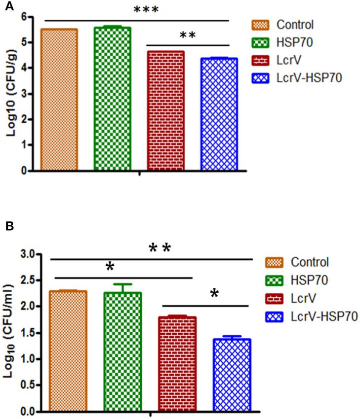
Gupta et al.                                                                                                                    Vaccine Against Plague and Yersiniosis

 FIGURE 1 | PCR amplification and agarose gel electrophoresis of lcrV gene and hsp70 (domain II) of M. tuberculosis and ligation of lcrV-hsp70 in pET vector: LcrV
 gene (A) Lane-1, 1Kb DNA ladder; Lane-2, lcrV gene product of 981 bp. (B) Lane-1, 1 Kb DNA ladder; Lane-2, hsp70 gene product of 630 bp. (C) The restriction
 map of cloned lcrV-hsp70 construct in pET vector. (D) Screening of positive clones of lcrV-hsp70 construct after ligation in pET28 vector, Lane-M, 1 Kb DNA ladder;
 Lane 1–3, released inserts of lcrV-hsp70 (1,611 bp) after restriction digestion using NcoI and Hind III restriction enzymes.

    Similarly, the animals of batch II and III were challenged (1                     PCR (Figures 1A,B). The amplicon of the lcrV gene was first
× 108 CFU/mouse) with Y. enterocolitica (O:8 serotype) and                            ligated in pET28a plasmid using NcoI and BamHI restriction
(1 × 10⁹ CFU/mouse) and with Y. pseudotuberculosis (A87                               sites. Just downstream to lcrV gene, a 630 bp fragment of hsp70
strain), respectively, via i.p. route (Figure 3A). To count the                       domain II was ligated using BamHI and HindIII restriction sites,
bacterial load in the blood and spleen, on days 1, 2, 3, 4, and                       as shown in Figure 1C. The pET vector harboring the in-frame
5 post-challenge, two mice from each group of batch II and                            of lcrV-hsp70 DNA construct (pET-lcrV-hsp70) was transformed
III were sacrificed. Their spleens were removed aseptically and                       into competent DH5α cells. The cells were grown on LB agar
weighed. Each spleen was homogenized, and serial dilutions were                       plates containing kanamycin (50 µg/ml). Positive clones were
made in PBS. One hundred microliter suspensions from each                             selected by restriction analysis using NcoI and HindIII restriction
countable dilution were spread on BHI agar plates in duplicate.                       enzymes. The digested products were analyzed by agarose gel
Y. pseudotuberculosis culture plates were incubated at 28◦ C for                      electrophoresis. A DNA insert of 1,611 bp was observed, as shown
48 h, and the bacterial colonies were counted. Y. enterocolitica                      in Figure 1D. The in-frame and the orientation of the ligated
culture plates were incubated at 37◦ C for 24 h, and the bacterial                    construct were further confirmed by nucleotide sequencing
colonies were counted. Prior to sacrifice the remaining animals,                      (Chromous Biotech, Bangalore, India).
the blood was collected. Each blood sample was serially diluted
in PBS. One hundred microliter from each countable dilution
was spread on BHI agar plates in duplicate. The bacterial load                        Expression and Purification of
(CFU/ml) and (CFU/g) was determined in the blood and spleen,                          Recombinant Proteins
respectively. The protection experiments were repeated thrice,                        LcrV-HSP70
and the results were expressed as the mean log CFU ± SD per                           To express and purify the bivalent fusion protein LcrV-
group of three experiments.                                                           HSP70 (60 kDa), one of the positive clones of lcrV-hsp70 was
                                                                                      transformed into E. coli expression host strain BL-21 (DE3). The
Statistical Analysis                                                                  colonies were selected on LB-agar plates containing kanamycin.
Data were analyzed using One Way Analysis of Variance                                 One colony was inoculated in 5 ml LB broth, and the culture was
(ANOVA) to compare IgG titers, CFU count, and cytokines                               induced with 1 mM IPTG. The IPTG-induced and uninduced
response. Log-rank (Mantel-Cox) test was used to generate                             E. coli cell lysates were analyzed by SDS-PAGE, as shown in
survival curves using GraphPad Prism 5.0. Statistically significant                   Figure 2Aa. For purification of LcrV-HSP70 protein, the culture
p-values are represented as ∗∗∗ p < 0.0001; ∗∗ p < 0.01; ∗ p < 0.05.                  was inoculated in 500 ml LB broth. The cells were pelleted by
                                                                                      centrifugation, and the lysis under native conditions revealed
RESULTS                                                                               the association of LcrV-HSP70 protein in the soluble fraction.
                                                                                      The recombinant construct of lcrV-hsp70 was engineered to
Cloning of lcrV–hsp70 in pET Vector                                                   carry a histidine tag at the carboxyl terminus of the LcrV-
For the expression of a bivalent fusion protein LcrV-HSP70, a 981                     HSP70 protein. The purification was performed under native
bp gene of Y. pestis encoding LcrV and a 630 bp DNA fragment                          conditions by affinity chromatography using Ni-NTA resin. All
encoding HSP70 domain II of M. tuberculosis were amplified by                         the eluted fractions were analyzed by SDS-PAGE, as shown in

Frontiers in Immunology | www.frontiersin.org                                     4                                                 June 2020 | Volume 11 | Article 988
Gupta et al.                                                                                                                     Vaccine Against Plague and Yersiniosis

 FIGURE 2 | (A) Recombinant bivalent fusion protein LcrV-HSP70 expression profile and purification: (a) SDS–PAGE profile of LcrV-HSP70 protein expression;
 Lane-M, Pre-stained protein marker; Lane-U, Uninduced cell lysate; Lane-I, Induced cell lysate. The arrow at the right side of the SDS-PAGE profile indicates the
 position of LcrV-HSP70 protein of 60 kDa. (b) Purification of LcrV-HSP70 protein. Lane-M, Protein marker; Lane-1, Cell lysate; Lane-2, Flow through; Lane-3, Wash;
                                                                                                                                                            (Continued)

Frontiers in Immunology | www.frontiersin.org                                      5                                                 June 2020 | Volume 11 | Article 988
Gupta et al.                                                                                                                        Vaccine Against Plague and Yersiniosis

 FIGURE 2 | Lane 4–9, eluted fractions of LcrV-HSP70 protein. The arrow at the right side of the SDS-PAGE profile indicates the position of LcrV-HSP70 protein of 60
 kDa. (B) Recombinant LcrV protein expression profile and purification: (a) SDS–PAGE profile of LcrV protein expression; Lane-M, Pre-stained protein marker; Lane-U,
 Uninduced cell lysate; Lane-I, Induced cell lysate. The arrow at the right side of the SDS-PAGE profile indicates the position of LcrV protein of 37 kDa. (b) Purification
 of LcrV protein. Lane-M, Protein marker; Lane-1, Cell lysate; Lane-2, Flow through; Lane-3, Wash; Lane 4–9, eluted fractions of LcrV protein. The arrow at the right
 side of the SDS-PAGE profile indicates the position of LcrV protein of 37 kDa. (C) Recombinant HSP70 protein expression and purification: (a) SDS–PAGE profile of
 HSP70 protein expression; Lane-M, Pre-stained protein marker; Lane-U, Uninduced cell lysate; Lane-I, Induced cell lysate. The arrow at the right side of the
 SDS-PAGE profile indicates the position of HSP70 protein of 23 kDa. (b) Purification of HSP70 protein. Lane-M, Protein marker; Lane-1, Cell lysate; Lane-2, Flow
 through; Lane-3, Wash; Lane 4–8, eluted fractions of HSP70 protein. The arrow at the right side of the SDS-PAGE profile indicates the position of HSP70 protein of 23
 kDa. (D) Western blot analysis of purified recombinant proteins LcrV, HSP70, and LcrV-HSP70: (a) SDS–PAGE profile of purified proteins, Lane-M, Pre-stained protein
 marker; Lane-1, LcrV, Lane-2, HSP70 and Lane-3, LcrV-HSP70 protein. (b) Western blot analysis of LcrV, HSP70, and LcrV-HSP70 proteins showing reaction with
 anti-HIS antibody: Lane-M, Pre-stained protein marker; Lane-1, LcrV, Lane-2, HSP70 and Lane-3, LcrV-HSP70 protein.

Figure 2Ab. The eluted fractions were pooled, dialyzed, and the                          Cellular Immune Response
protein concentration was estimated. The obtained yield of the                           The expression levels of IFN-γ and TNF-α in the collected
purified LcrV-HSP70 was 30 mg/L of shake flask culture. The                              supernatants of splenocytes were measured. We observed a
endotoxin level was observed
Gupta et al.                                                                                                    Vaccine Against Plague and Yersiniosis

                                                                      FIGURE 3 | (A) Representation of animal groups and schedule of vaccination
                                                                      activities: Animal groups (10 mice/group) for vaccination studies; Vaccine
                                                                      formulations of LcrV, HSP70 and LcrV–HSP70 vaccine antigens with alum.
                                                                      Schematic representation of vaccination schedule, blood collection for
                                                                      humoral and cell-mediated studies, and challenge experiments. (B) The ELISA
                                                                      plates were coated by the same antigens, which were used as immunizing
                                                                      antigens. IgG titers were determined by ELISA and represented as log10 titers.
                                                                      Serum samples were collected after first and second boosters from
                                                                      vaccinated animal groups, i.e., LcrV, HSP70, and LcrV–HSP70. Analysis was
                                                                      done by one-way ANOVA, ***p < 0.0001; **p < 0.01; *p < 0.05. (C) Cytokine
                                                                      profile of IFN-γ and (D) TNF-α; the induced levels of cytokines in vaccinated
                                                                      mice were measured in pg/ml, as represented in graphs. Splenocytes were
                                                                      prepared from all the vaccinated and control groups. The cells were induced
                                                                      with the same vaccine antigens as the immunizing antigens, i.e., LcrV, HSP70,
                                                                      and LcrV-HSP70 (5 µg/ml each) and grown for 48 h. All the statistical
                                                                      comparisons were done by one-way ANOVA, ***p < 0.0001; **p < 0.01; *p <
                                                                      0.05. (E) Recombinant bivalent fusion protein LcrV–HSP70 imparts protection
                                                                      against challenge with Y. pestis. The immunized and control group mice were
                                                                      challenged against 100 LD50 of Y. pestis. All the animals were observed for 20
                                                                      days after the challenge for their survival. Log-rank (Mantel-Cox) test was used
                                                                      to compare the survival against plague infection amongst different vaccinated
                                                                      groups (***p < 0.0001).

                                                                     vaccine formulations based on the live attenuated and killed
 FIGURE 3 | Continued                                                whole-cell of Y. pestis have been developed. Despite having
                                                                     ethical issues and safety concerns, i.e., high fever, headache,
                                                                     and inflammation at the site of injection, these formulations
DISCUSSION                                                           are in use in some countries (18). The main limitations of
                                                                     these vaccine formulations are partial or incomplete protective
Despite continuous and dedicated efforts by the scientific           efficacy, poor memory, and repeated boosters (9, 19, 20). In the
community, an ideal plague vaccine is yet to come. Many plague       recent past, many reports have been published on F1/LcrV based

Frontiers in Immunology | www.frontiersin.org                    7                                                 June 2020 | Volume 11 | Article 988
Gupta et al. Vaccine Against Plague and Yersiniosis
FIGURE 4 | (A) CFU count in the spleens of Y. enterocolitica-challenged mice.
FIGURE 5 | (A) CFU count in the spleens of Y. pseudotuberculosis-challenged
Culture plates were incubated at 37◦ C for 24 h, and the bacterial colonies
mice. Culture plates were incubated at 28◦ C for 48 h, and the bacterial
were counted. The number of bacteria in the spleens is represented as the
colonies were counted. The number of bacteria in the spleens is represented
mean log CFU ± SD per group. Protection was found statistically significant in
as the mean log CFU ± SD per group. Protection was found statistically
LcrV-HSP70 vaccinated group when compared to LcrV (**p < 0.01), HSP70
significant in LcrV-HSP70 vaccinated group when compared to LcrV (**p <
and control group (***p < 0.0001). (B) CFU count in the blood of Y.
0.01), HSP70 and control group (***p < 0.0001). (B) CFU count in the blood of
enterocolitica challenged mice: Y. enterocolitica culture plates were incubated
Y. pseudotuberculosis challenged mice: Y. pseudotuberculosis culture plates
at 37◦ C for 24 h and the bacterial colonies were counted. The number of
were incubated at 28◦ C for 48 h, and the bacterial colonies were counted. The
bacteria in the blood is represented as the mean log CFU ± SD per group.
number of bacteria in the blood is represented as the mean log CFU ± SD
per group (*p = 0.05).
vaccine formulations. The F1/LcrV vaccine antigens adjuvanted
with alum induce robust humoral immune responses and protective potential of YopE (35); the SA-4-1BBL adjuvant,
impart 100% protection in a mouse model (16, 21–24) with a strong inducer of the Th1 response, improved the vaccine
no side effects in humans (25). F1/LcrV antigen-based vaccine potential of F1-LcrV (30); and the HSP70 domain II of
formulations protect cynomolgus macaques against aerosolized M. tuberculosis augments the humoral and cellular immune
Y. pestis but failed to protect African Green monkeys (26, response of F1/LcrV vaccine (16, 24). HSP70 proteins have
27). The immune mechanisms responsible for this poor and been characterized to immunomodulate the immune response of
inconsistent protection in the African Green monkey model vaccine antigens (36–42). The domain II of heat shock protein
yet to be analyzed. To improve the efficacy of these vaccine HSP70(II), when formulated with vaccine antigen/s, evoked the
formulations by inducing stronger cellular immunity might be T-cell responses. A bivalent fusion protein ovalbumin-HSP70(II)
the best solution. A number of strategies are in development to induces CD8 cytotoxic T lymphocytes specific to ovalbumin (36).
advance the efficacy of F1/LcrV antigens, e.g., use of novel and HSP70(II) of M. tuberculosis modulated the immune responses
competent adjuvants (28–30), vaccine antigen delivery systems of vaccine antigen p24 of HIV-1 (36). The amino acids (359–
(31, 32), and genetically modified antigens (33). To counteract 610) of the carboxy-terminus of HSP70 evoke the expression
the pathogen effectively, addition of one or more vaccine of IFN-γ, IL-2, and TNF-α and a high titer of IgG2a and IgG3
antigens might be considered which can significantly augment antibodies (43).
the immune response. Earlier, we have also characterized the potential role of
In the recent years, the efficacy of F1/LcrV based vaccines HSP70(II) of M. tuberculosis to augment the vaccine efficacy
have been improved by adding molecular adjuvants, i.e., F1/V of F1/LcrV in a mouse model (16, 24). Our findings showed
adjuvanted with flagellin (Flagellin/F1/V) for phase I safety that HSP70(II) improved the cellular immune response (IL-2,
and immunogenicity trial in healthy adult volunteer (34); the IFN-γ, and TNF-α) of F1+LcrV+HSP70(II)-vaccinated mice.
recombinant flagellin of Salmonella typhi also improved the It also enhanced the IFN-γ secreting CD4+ and CD8+ T cells.
Frontiers in Immunology | www.frontiersin.org 8 June 2020 | Volume 11 | Article 988Gupta et al. Vaccine Against Plague and Yersiniosis TABLE 1 | Protection on day 2 post-challenge in LcrV-HSP70 vaccinated mice against Y. enterocolitica. Groups Log10 CFU Y. enterocolitica (Spleen) Unit of protection Log10 CFU Y. enterocolitica (Blood) Unit of protection Control 5.365 ± 0.01343 – 2.0695 ± 0.013435 – HSP70 5.344 ± 0.01060 0.0215 2.2170 ± 0.235467 – LcrV 4.378 ± 0.05091 0.9875 1.6015 ± 0.063851 0.468 LcrV-HSP70 4.175 ± 0.01060 1.1905 1.1230 ± 0.053460 0.9465 The number of bacteria in the spleens (CFU/spleen) is represented as the mean log CFU ± SD per group. The unit of protection was determined by subtracting the mean log CFU of the immunized group from the mean log CFU of the control group. TABLE 2 | Protection on day 2 post-challenge in LcrV-HSP70 vaccinated mice against Y. pseudotuberculosis. Groups Log10 CFU Y. pseudotuberculosis (Spleen) Unit of protection Log10 CFU Y. pseudotuberculosis (Blood) Unit of protection Control 5.511 ± 0.0 – 2.2895 ± 0.016263 – HSP70 5.574 ± 0.06929 – 2.2625 ± 0.235467 0.027 LcrV 4.625 ± 0.02121 0.886 1.7995 ± 0.030406 0.49 LcrV-HSP70 4.527 ± 0.03676 0.984 1.371 ± 0.097581 0.9185 The number of bacteria in the spleens (CFU/spleen) is represented as the mean log CFU ± SD per group. The unit of protection was determined by subtracting the mean log CFU of the immunized group from the mean log CFU of the control group. Earlier, we developed a recombinant trivalent fusion protein F1- role by suppressing the expression of proinflammatory cytokines LcrV-HSP70(II) and evaluated as a vaccine candidate against (47, 48). As the LcrV is a common virulence factor of Y. pestis, plague in a mouse model (24). We experienced difficulties in enterocolitica, and pseudotuberculosis, we took advantage of this the expression and purification of this trivalent fusion protein to test the protective efficacy of LcrV-HSP70 against these human in E. coli. The expression was not found to be optimal, making pathogens. However, our primary focus is to develop the subunit inclusion bodies during expression. Hence the protein can only vaccine against plague. be purified under denaturing conditions. Moreover, this trivalent Clinical symptoms of yersiniosis by Y. enterocolitica first fusion protein is highly degradable at room temperature. In appear after an incubation period of about 5 days (range 1–11 this study, we prepared a recombinant bivalent fusion protein days) and include diarrhea, fever, vomiting, tenesmus, and LcrV-HSP70, and we evaluated its vaccine potential in BALB/c abdominal pain. The incubation period of Y. pseudotuberculosis mice. The expression of the bivalent fusion protein is up to is 5–10 days; however, durations of 2–20 days have been reported the mark in E. coli, and this protein does not make inclusion in occasional outbreaks, with the average time being 4 days during the expression. The bivalent protein can be easily purified after exposure to the bacterium when symptoms are present under native conditions to the optimum yield. All the three (https://www.cdc.gov/yersinia/healthcare.html). Therefore, antigens, i.e., LcrV-HSP70, LcrV, and HSP70, were adjuvanted in our studies, we determined the presence of both of the individually with alum, and the animals were vaccinated. There pathogens in the spleen and in the blood to know whether our was a significantly higher anti-LcrV IgG titer in the sera of LcrV- vaccine formulation is effective or not. We challenged all the HSP70 vaccinated mice in comparison to HSP70 and control vaccinated and control mice on day 60, and we determined the group. No significant difference was observed in the anti-LcrV CFUs in the blood and spleen on day 61–65. On day 2 post- IgG titer between LcrV alone and fusion protein LcrV-HSP70 challenge, we observed a significant reduction in the number vaccinated sera. However, there was a significant difference in of CFUs in the spleen and blood of Y. enterocolitica- and Y. the IgG titers in the sera after first and second booster. We also pseudotuberculosis-challenged mice that were immunized with observed a significant difference in the expression of IFN-γ and LcrV-HSP70 in comparison to LcrV and HSP70 alone. Since the TNF-α in LcrV-HSP70 vaccinated mice in comparison to groups oral route is the natural route of infection for Y. enterocolitica vaccinated with LcrV and HSP70 alone. or Y. pseudotuberculosis, it would be relevant in the future to Yersinia species use Type III secretion system (T3SS) to determine the protective efficacy of the above vaccine candidate translocate effector proteins into the host target cells to breach by a mucosal route. In the case of plague, the bivalent fusion host immune barriers (44). LcrV is an essential virulent factor protein LcrV-HSP70 imparts 100% protection against plague, that makes the tip of T3SS. Before contact with the host cells, whereas LcrV alone provided 70% protection only. There was LcrV protein is expressed on the cell membrane of Y. pestis, no protection in control and HSP70-vaccinated mice. Taken enterocolitica, and pseudotuberculosis (45). LcrV essentially helps together, the recombinant bivalent fusion protein LcrV-HSP70 in the regulation and translocation of other virulence factors has the scope for further evaluation of its mucosal immune into the host cell (46). It also helps Yersiniae to defeat the response in animal models, thus increasing its potential to host immune response via IL-10 mediated immunomodulatory become a vaccine against plague and Yersiniosis. Frontiers in Immunology | www.frontiersin.org 9 June 2020 | Volume 11 | Article 988
Gupta et al.                                                                                                                           Vaccine Against Plague and Yersiniosis

DATA AVAILABILITY STATEMENT                                                               FUNDING
All datasets generated for this study are included in the article/                        The necessary funding and facilities to complete these
Supplementary Material.                                                                   studies were provided by Defense Research and Development
                                                                                          Establishment (DRDE), Ministry of Defense, Govt. of India.

ETHICS STATEMENT                                                                          ACKNOWLEDGMENTS
The animal study was reviewed and approved by Institutional
                                                                                          The authors are thankful to the Director of the Defense
Animal Ethics Committee (IAEC) of Defence Research and
                                                                                          Research and Development Establishment (DRDE), Ministry of
Development Establishment.
                                                                                          Defense, Govt. of India, for providing the necessary facilities to
                                                                                          complete these studies. The article has been assigned an accession
AUTHOR CONTRIBUTIONS                                                                      No. DRDE/MB/37/2019.

SV initiated this project and was responsible for the overall design                      SUPPLEMENTARY MATERIAL
of the study, conducting challenge experiments, interpretation of
data and writing of this manuscript. AG and BN conducted the                              The Supplementary Material for this article can be found
laboratory experiments. SK contributed in the interpretation of                           online at: https://www.frontiersin.org/articles/10.3389/fimmu.
results and reviewing of this manuscript before submission.                               2020.00988/full#supplementary-material

REFERENCES                                                                                      Yersinia pestis. J Bacteriol. (1995) 177:2530–42. doi: 10.1128/JB.177.9.2530-
                                                                                                2542.1995
 1. Brubaker RR. Factors promoting acute and chronic diseases caused by                   16.   Batra L, Verma SK, Nagar DP, Saxena N, Pathak PP, Pant SC, et al. HSP70
    yersiniae. Clin Microbiol Rev. (1991) 4:309–24. doi: 10.1128/CMR.4.3.309                    domain II of Mycobacterium tuberculosis modulates immune response and
 2. Bottone EJ. Yersinia enterocolitica: the charisma continues. Clin Microbiol                 protective potential of F1 and LcrV antigens of Yersinia pestis in a mouse
    Rev. (1997) 10:257–76. doi: 10.1128/CMR.10.2.257                                            model. PLoS Neg Trop Dis. (2014) 8:e3322. doi: 10.1371/journal.pntd.0003322
 3. WHO. Plague. Geneva: World Health Organization (2005). p. 267.                        17.   Verma SK, Batra L, Athmaram TN, Pathak P, Katram N, Agarwal GS,
 4. Sun W, Singh AK. Plague vaccine: recent progress and prospects. NPJ                         et al. Characterization of immune responses to Yersinia pestis (Indian
    Vaccines. (2019) 4:11. doi: 10.1038/s41541-019-0105-9                                       isolate) infection in mouse model. J Clin Cell Immunol. (2013) 4:151.
 5. Williamson        ED.       Plague.      Vaccine.      (2009)      27:D56–60.               doi: 10.4172/2155-9899.1000151
    doi: 10.1016/j.vaccine.2009.07.068                                                    18.   Hart MK, Saviolakis GA, Welkos SL, House RV. Advanced development of the
 6. Dennis DT, Chow CC. Plague. Pediatr Infect Dis J. (2004) 23:69–71.                          rF1V and rBV A/B vaccines: progress and challenges. Adv Prev Med. (2012)
    doi: 10.1097/01.inf.0000106918.18570.dd                                                     2012:731604. doi: 10.1155/2012/731604
 7. Pechous RD, Sivaraman V, Stasulli NM, Goldman WE. Pneumonic plague:                   19.   Wang X, Zhang X, Zhou D, Yang R. Live-attenuated Yersinia pestis vaccines.
    the darker side of Yersinia pestis. Trends Microbiol. (2016) 24:190–7.                      Expert Rev Vaccines. (2013) 12:677–86. doi: 10.1586/erv.13.42
    doi: 10.1016/j.tim.2015.11.008                                                        20.   Chu K, Hu J, Meng F, Li J, Luo L, Xu J, et al. Immunogenicity and safety of
 8. Prentice MB, Rahalison L. Plague. Lancet. (2007) 369:1196–207.                              subunit plague vaccine: a randomized phase. 2 a clinical trial. Hum Vaccine
    doi: 10.1016/S0140-6736(07)60566-2                                                          Immunother. (2016) 12:2334–40. doi: 10.1080/21645515.2016.1175261
 9. Verma SK, Tuteja U. Plague vaccine development: current research and future           21.   Williamson ED, Eley SM, Stagg AJ, Green M, Russell P, Titball RW. A sub-
    trends. Front Immunol. (2016) 7:602. doi: 10.3389/fimmu.2016.00602                          unit vaccine elicits IgG in serum, spleen cell cultures and bronchial washings
10. Inglesby TV, Dennis DT, Henderson DA, Bartlett JG, Ascher MS, Eitzen                        and protects immunized animals against pneumonic plague. Vaccine. (1997)
    E, et al. Plague as a biological weapon: medical and public health                          15:1079–84. doi: 10.1016/S0264-410X(96)00303-9
    management. working group on civilian biodefense. JAMA. (2000) 283:2281–              22.   Lin JS, Park S, Adamovicz JJ, Hill J, Bliska JB, Cote CK, et al. TNF-
    90. doi: 10.1001/jama.283.17.2281                                                           α and IFN-γ contribute to F1/LcrV-targeted immune defense in mouse
11. Levy Y, Flashner Y, Tidhar A, Zauberman A, Aftalion M, Lazar S, et al. T cells              models of fully virulent pneumonic plague. Vaccine. (2010) 29:357–62.
    play an essential role in anti-F1 mediated rapid protection against bubonic                 doi: 10.1016/j.vaccine.2010.08.099
    plague. Vaccine. (2011) 29:6866–73. doi: 10.1016/j.vaccine.2011.07.059                23.   Levy Y, Vagima Y, Tidhar A, Aftalion M, Gur D, Nili U, et al. Targeting of
12. Galimand M, Guiyoule A, Gerbaud G, Rasoamanana B, Chanteau                                  the Yersinia pestis F1 capsular antigen by innate like B1b cells mediates a
    S, Carniel E, et al. Multi drug resistance in Yersinia pestis mediated                      rapid protective response against bubonic plague. NPJ Vaccines. (2018) 3:52.
    by a transferable plasmid. N Engl J Med. (1997) 337:677–80.                                 doi: 10.1038/s41541-018-0087-z
    doi: 10.1056/NEJM199709043371004                                                      24.   Verma SK, Batra L, Tuteja U. A recombinant trivalent fusion protein F1-
13. Guiyoule A, Gerbaud G, Buchrieser C, Galimand M, Rahalison L, Chanteau                      LcrV-HSP70(II) augments humoral and cellular immune responses and
    S, et al. Transferable plasmid-mediated resistance to streptomycin in                       imparts full protection against Yersinia pestis. Front Microbiol. (2016) 7:1053.
    a clinical isolate of Yersinia pestis. Emerg Infect Dis. (2001) 7:43–8.                     doi: 10.3389/fmicb.2016.01053
    doi: 10.3201/eid0701.010106                                                           25.   Price JL, Manetz TS, Shearer JD, House RV. Preclinical safety assessment
14. Chain PSG, Carniel E, Larimer FW, Lamerdin J, Stoutland PO, Regala WM,                      of a recombinant plague vaccine (rF1V). Int J Toxicol. (2013) 32:327–335.
    et al. Insights into the evolution of Yersinia pestis through whole-genome                  doi: 10.1177/1091581813497405
    comparison with Yersinia pseudotuberculosis. Proc Natl Acad Sci USA. (2004)           26.   Smiley ST. Current challenges in the development of vaccines for pneumonic
    101:13826–31. doi: 10.1073/pnas.0404012101                                                  plague. Expert Rev Vaccines. (2008) 7:209–21. doi: 10.1586/14760584.7.2.209
15. Skrzypek E, Straley SC. Differential effects of deletions in lcrV on secretion        27.   Williamson ED, Packer PJ, Waters EL, Simpson AJ, Dyer D,
    of V-antigen, regulation of the low-Ca++ response and virulence of                          Hartings J, et al. Recombinant (F1+V) vaccine protects cynomolgus

Frontiers in Immunology | www.frontiersin.org                                        10                                                    June 2020 | Volume 11 | Article 988
Gupta et al. Vaccine Against Plague and Yersiniosis
macaques against pneumonic plague. Vaccine. (2011) 29:4771–7. 39. Srivastava PK, Amato RJ. Heat shock proteins: the ‘Swiss army knife’
doi: 10.1016/j.vaccine.2011.04.084 vaccines against cancers and infectious agents. Vaccine. (2001) 19:2590–7.
28. Amemiya K, Meyers JL, Rogers TE, Fast RL, Bassett AD, Worsham PL, et doi: 10.1016/S0264-410X(00)00492-8
al. CpG oligodeoxynucleotides augment the murine immune response to the 40. Pockley AG. Heat shock proteins as regulators of the immune
Yersinia pestis F1-V vaccine in bubonic and pneumonic models of plague. response. Lancet. (2003) 362:469–76. doi: 10.1016/S0140-6736(03)
Vaccine. (2009) 27:2220–9. doi: 10.1016/j.vaccine.2009.02.016 14075-5
29. Honko AN, Sriranganathan N, Lees CJ, Mizel SB. Flagellin is 41. Robert J. Evolution of heat shock protein and immunity. Dev Comp Immunol.
an effective adjuvant for immunization against lethal respiratory (2003) 27:449–64. doi: 10.1016/S0145-305X(02)00160-X
challenge with Yersinia pestis. Infect Immun. (2006) 74:1113–20. 42. Hauser H, Chen SY. Augmentation of DNA vaccine potency through
doi: 10.1128/IAI.74.2.1113-1120.2006 secretory heat shock protein-mediated antigen targeting. Methods. (2003)
30. Bowen W, Batra L, Pulsifer AR, Yolcu ES, Lawrenz MB, Shirwan H. Robust 31:225–31. doi: 10.1016/S1046-2023(03)00136-1
Th1 cellular and humoral responses generated by the Yersinia pestis rF1- 43. Wang Y, Kelly CG, Singh M, McGowan EG, Carrara AS, Bergmeier
V subunit vaccine formulated to contain an agonist of the CD137 pathway do LA, et al. Stimulation of Th1-polarizing cytokines, C-C chemokines,
not translate into increased protection against pneumonic plague. Vaccine. maturation of dendritic cells, and adjuvant function by the peptide
(2019) 37:5708–16. doi: 10.1016/j.vaccine.2019.07.103 binding fragment of heat shock protein70. J Immunol. (2002) 169:2422–9.
31. Chiuchiolo MJ, Boyer JL, Krause A, Senina S, Hackett NR, Crystal RG. doi: 10.4049/jimmunol.169.5.2422
Protective immunity against respiratory tract challenge with Yersinia pestis 44. Cornelis GR. Yersinia type III secretion send in the effectors. J Cell Biol. (2002)
in mice immunized with an adenovirus-based vaccine vector expressing V 158:401–8. doi: 10.1083/jcb.200205077
antigen. J Infect Dis. (2006) 194:1249–57. doi: 10.1086/507644 45. Pettersson J, Holmström A, Hill J, Leary S, Frithz-Lindsten E, von Euler-
32. Huang SS, Li IH, Hong PD, Yeh MK. Development of Yersinia pestis F1 Matell A, et al. The V-antigen of Yersinia is surface exposed before target cell
antigen-loaded microspheres vaccine against plague. Int J Nanomed. (2014) contact and involved in virulence protein translocation. Mol Microbiol. (1999)
9:813–22. doi: 10.2147/IJN.S56260 32:961–76. doi: 10.1046/j.1365-2958.1999.01408.x
33. DeBord KL, Anderson DM, Marketon MM, Overheim KA, DePaolo 46. Mueller CA, Broz P, Müller SA, Ringler P, Erne-Brand F, Sorg I, et al. The V-
RW, Ciletti NA, et al. Immunogenicity protective immunity against antigen of Yersinia forms a distinct structure at the tip of injectisome needles.
bubonic plague pneumonic plague by immunization of mice with the Science. (2005) 310:674–6. doi: 10.1126/science.1118476
recombinant V10 antigen, a variant of LcrV. Infect Immun. (2006) 74:4910–4. 47. Sing A, Reithmeier-Rost D, Granfors K, Hill J, Roggenkamp A, Heesemann
doi: 10.1128/IAI.01860-05 J. A hypervariable N-terminal region of Yersinia LcrV determines Toll-like
34. Frey SE, Lottenbach K, Graham I, Anderson E, Bajwa K, May RC, et receptor 2-mediated IL-10 induction and mouse virulence. Proc Natl Acad Sci
al. A phase I safety and immunogenicity dose escalation trial of plague USA. (2005) 102:16049–54. doi: 10.1073/pnas.0504728102
vaccine, Flagellin/F1/V, in healthy adult volunteers (DMID 08-0066). Vaccine. 48. Overheim KA, Depaolo RW, Debord KL, Morrin EM, Anderson DM,
(2017) 35:6759–65. doi: 10.1016/j.vaccine.2017.09.070 Green NM, et al. LcrV plague vaccine with altered immunomodulatory
35. Verma SK, Batra L, Tuteja U. Escherichia coli expressed flagellin C (FliC) properties. Infect. Immun. (2005) 73:5152–9. doi: 10.1128/IAI.73.8.5152-51
of Salmonella Typhi improved the protective efficacy of YopE against plague 59.2005
infection. Vaccine. (2019) 37:19–24. doi: 10.1016/j.vaccine.2018.11.057
36. Suzue K, Young RA. Adjuvant-free HSP70(II) fusion protein system Conflict of Interest: The authors declare that the research was conducted in the
elicits humoral and cellular responses to HIV-1 p24. J Immunol. (1996) absence of any commercial or financial relationships that could be construed as a
156:873–9. potential conflict of interest.
37. Suzue K, Zhou X, Eisen HN, Young RA. Heat shock protein fusion proteins
as vehicles for antigen delivery into the major histocompatibility complex Copyright © 2020 Gupta, Narayan, Kumar and Verma. This is an open-access article
class I presentation pathway. Proc Natl Acad Sci USA. (1997) 94:13146–51. distributed under the terms of the Creative Commons Attribution License (CC BY).
doi: 10.1073/pnas.94.24.13146 The use, distribution or reproduction in other forums is permitted, provided the
38. Huang Q, Richmond JF, Suzue K, Eisen HN, Young RA. In vivo cytotoxic T original author(s) and the copyright owner(s) are credited and that the original
lymphocyte elicitation by mycobacterial heat shock protein 70 fusion proteins publication in this journal is cited, in accordance with accepted academic practice.
maps to a discrete domain and is CD4+ T cell independent. J Exp Med. (2000) No use, distribution or reproduction is permitted which does not comply with these
191:403–8. doi: 10.1084/jem.191.2.403 terms.
Frontiers in Immunology | www.frontiersin.org 11 June 2020 | Volume 11 | Article 988You can also read