Collagen I Induces Preeclampsia- Like Symptoms by Suppressing Proliferation and Invasion of Trophoblasts
←
→
Page content transcription
If your browser does not render page correctly, please read the page content below
ORIGINAL RESEARCH
                                                                                                                                           published: 06 August 2021
                                                                                                                                     doi: 10.3389/fendo.2021.664766

                                           Collagen I Induces Preeclampsia-
                                           Like Symptoms by Suppressing
                                           Proliferation and Invasion of
                                           Trophoblasts
                                           Yinglin Feng 1†, Xia Chen 2†, Huiqiao Wang 3, Xueping Chen 1, Zixin Lan 3, Pan Li 4,
                                           Yingshi Cao 1, Mian Liu 1, Jin Lv 5, Yun Chen 1, Yu Wang 1, Chao Sheng 1, Yingying Huang 1,
                            Edited by:     Mei Zhong 1, Zhijian Wang 1, Xiaojing Yue 1* and Liping Huang 1*
                     Ganesh Acharya,
                                           1 Department of Obstetrics and Gynecology, Nanfang Hospital, Southern Medical University, Guangzhou, China, 2 Department of
          Karolinska Institute, Sweden
                                           Obstetrics and Gynecology, Foshan First People’s Hospital, Foshan, China, 3 Zhujiang Hospital, Southern Medical University,
                        Reviewed by:
                                           Guangzhou, China, 4 Microbiome Research Center, University of New South Wales, Sydney, NSW, Australia, 5 Department of
                            Rahul Pal,
                                           Pathology, Foshan First People’s Hospital, Foshan, China
National Institute of Immunology (NII),
                                 India
                              Wei Wu,      Preeclampsia is a common obstetric disorder affecting 2-8% of pregnancy worldwide.
    Nanjing Medical University, China
                        Mona Nystad,
                                           Fibrosis is an important histological change occurring in preeclamptic placenta, and might
 University Hospital of North Norway,      depend on the excess deposition of collagen I. However, the role of fibrotic placenta and
                              Norway
                                           collagen I in the pathogenesis of preeclampsia remains unclear. Therefore, we analyzed
                  *Correspondence:
                                           the collagen deposition and the expression of Collagen I in human placenta by Masson
                        Liping Huang
              lphuang2006@126.com          staining, Sirius red staining and western blotting. Further, the role of collagen I in
                         Xiaojing Yue      preeclampsia pathogenesis was studied in C57BL/6 mice. HTR-8/SVneo cells were
                 yuexj1986@163.com
     †
                                           used to investigate the mechanisms underlying the effects of collagen I in trophoblasts by
      These authors have contributed
                 equally to this work
                                           transcriptome sequencing and pharmacological agonists. Human preeclamptic placenta
                                           exhibited a significantly higher degree of fibrosis in stem villi and terminal villi than normal
                   Specialty section:      placenta, and was characterized by collagen I deposition. In vivo, a single injection of
         This article was submitted to
                         Reproduction,
                                           collagen I on gestational day 0.5 led to an increase in systolic pressure of pregnant mice
               a section of the journal    from gestational days 4.5–17.5, to a decrease in weight and number of embryos, and to
            Frontiers in Endocrinology     enhanced placental collagen I expression and degree of fibrosis compared with control
          Received: 09 March 2021          mice. In vitro, collagen I attenuated the proliferation and invasion of HTR-8SV/neo cells.
           Accepted: 14 June 2021
         Published: 06 August 2021         This effect could be reversed by treatment with agonists of ERK and b-catenin. Moreover,
                              Citation:    transcriptome sequencing demonstrated that signaling pathways related to cell
            Feng Y, Chen X, Wang H,        proliferation and invasion were significantly downregulated in HTR-8SV/neo cells. Thus,
          Chen Y, Lan Z, Li P, Cao Y,
        Liu M, Lv J, Chen X, Wang Y,
                                           we propose that collagen I induced preeclampsia-like symptoms by suppressing the
       Sheng C, Huang Y, Zhong M,          proliferation and invasion of trophoblasts through inhibition of the ERK phosphorylation
  Wang Z, Yue X and Huang L (2021)         and WNT/b-catenin signaling pathways. Our findings could pave the way to the discovery
Collagen I Induces Preeclampsia-Like
Symptoms by Suppressing Proliferation      of small-molecule inhibitors for preeclampsia treatment and future studies with larger
         and Invasion of Trophoblasts.     sample size are required.
         Front. Endocrinol. 12:664766.
     doi: 10.3389/fendo.2021.664766        Keywords: fibrosis, collagen I, preeclampsia, placenta, pathogenesis

Frontiers in Endocrinology | www.frontiersin.org                                  1                                       August 2021 | Volume 12 | Article 664766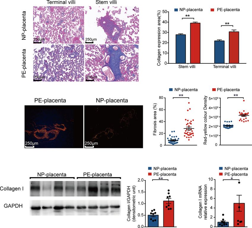
Feng et al.                                                                                                              Collagen I in Preeclampsia

INTRODUCTION                                                               and in vitro experiments to clarify the relationship between
                                                                           placental fibrosis and preeclampsia.
Preeclampsia is a common, pregnancy-specific disorder that
threatens 2–8% of pregnancies globally and constitutes one of the
leading causes of maternal and perinatal mortality worldwide (1). It       MATERIALS AND METHODS
is characterized by the occurrence of new-onset hypertension,
accompanied by proteinuria or other end-organ dysfunction after            Collection of Human Placenta
20 weeks of gestation in previously normotensive women (2). To             Women with PE and normotensive pregnant (NP) women
date, the pathogenesis of this morbidity is poorly understood. The         without previous treatments were recruited in the third
placenta is generally perceived as the primary organ during disease        trimester from March 2018 to 2019 in the Department of
development because the removal of placenta is necessary for               Obstetrics of the Nanfang Hospital, Southern Medical
symptoms to regress (3). Previous research has shown that                  University, China. According to the current American College
histological changes exist in pre-eclampsia (PE) placenta include          of Obstetricians and Gynecologists criteria (1), preeclampsia is
chronic inflammation, vascular lesions, villous coagulation, and            diagnosed as maternal systolic blood pressure blood pressure
villous fibrosis. Among the changes occurring in these placentae,           ≥140mmHg and/or diastolic blood pressure ≥90mmHg on at
fibrosis is an important feature (4). In particular, microscopic            least two occasions, four hours apart, with proteinuria, after 20
comparison with control placenta has revealed that villous                 weeks of gestation in previously normotensive women. In the
fibrosis is more frequent in preeclamptic placentas (4, 5). Notably,        absence of proteinuria, patients with new-onset of hypertension
fibrosis in preeclamptic placenta is reported to be related to the          with the new onset of any of the following: thrombocytopenia,
activation of stromal fibroblasts (6). Whether placental fibrosis is         renal insufficiency, impaired liver function, or pulmonary edema
involved in the pathogenesis of preeclampsia is still unclear.             can be induced. The clinical characteristics of placenta donors are
    Fibrosis is a well-known pathogenic process resulting in the           summarized in Supplementary Table S1.
progressive loss of organ structure and function. It is defined by              Fresh placentae were obtained immediately after delivery. The
overproduction of the extracellular matrix (ECM) in connective             tissue was collected from the area near the umbilical cord (1 cm
tissues and plays a significant role in the impairment of organ             from the cord insert), from both the maternal and fetal side of
function, such as during liver cirrhosis and cardiac fibrosis (7, 8).       the placenta.
It is noteworthy that diverse diseases in different organs are                 A sample size of 10 placentae in each group was used for the
associated with common pathogenic pathways, as excessive ECM               identification of collagen and quantification of collagen I by
deposition causes physical organ deformation, which impairs                Masson staining, Sirius red staining, and Western blotting. The
organ function and ultimately induces organ failure (9).                   experimental procedures were detailed in the Supplementary
    Collagens are the predominant components of the ECM (8,                Material. Following staining, the placentae were assessed for the
10). Consequently, increases in collagen expression and                    degree of fibrosis in a blinded fashion by a trained pathologist. For
deposition are directly associated with fibrosis (11). The family           Masson trichrome straining, collagen fiber was stained blue, while
of collagens comprises twenty-eight members and regulates cell             cytoplasm and red blood cells were stained red and nucleus blue
proliferation and migration, cell-matrix interactions, and cell            and brown. Fibrosis area% was calculated in µm digitally using the
signaling (12). Furthermore, emerging studies have indicated               software NDP.view2 (Hamamatsu Photonics, Hamamatsu,
that collagen exerts contrasting effects on the physiology of              Japan). Sirius Red staining is a method used for collagen
different organs. For example, collagen could stimulate                    identification. Collagen I appears red-yellow when observed
angiogenesis in a myocardial infarction model (13). Moreover,              under a polarizing microscope. Each analyzed field was chosen
collagens have been demonstrated to reduce melanoma cell                   randomly and the positive red-stained areas and red-yellow
proliferation and invasion by inhibiting the FAK/PI3K/AKT                  density were quantified using computerized image analysis
pathway (14).                                                              software (NIH, MD, USA).
    Collagen I is well known as one of the main member of the                  The collection of placentae was approved by the Ethics
collagen family, constituting over 90% of the collagen of the body         Committee of Nanfang Hospital(NFEC-2017-055). All
(15, 16). Higher production of Collagen I was once detected in             participants gave written consent prior to donating their placenta.
preeclamptic placenta (17). Takako and his colleagues indicated
that the fibroblasts isolated from preeclamptic placenta showed a           Collagen I Preparation
higher expression of collagen I (6). Although the similar studies          Collagen I (C7774, Sigma-Aldrich, St. Louis, MO, USA) was
were limited, that collagen I may be the characteristic collagen           dissolved in 0.1 mM acetic acid and emulsified in an equal
deposited in preeclamptic placenta, and may influences the                  volume incomplete Freund’s adjuvant (IFA) (F5506, Sigma-
placental function, warrants further investigation.                        Aldrich, St. Louis, MO, USA) to obtain a 1000 mg/ml solution
    To date, detailed investigation of the collagen I in                   as the stock solution.
preeclamptic placenta is lacking. Whether placental fibrosis is
involved in the pathogenesis of preeclampsia is still unclear. In          Animals and Experimental Protocol
this study, we hypothesize that exacerbation of fibrosis leads to           This project was performed in accordance with animal protocol
impairment of placental function and is involved in the                    procedures approved by the Department of Laboratory Animal
pathogenesis of preeclampsia. Therefore, we performed in vivo              Sciences, Southern Medical University (L-2019216), and the

Frontiers in Endocrinology | www.frontiersin.org                       2                                  August 2021 | Volume 12 | Article 664766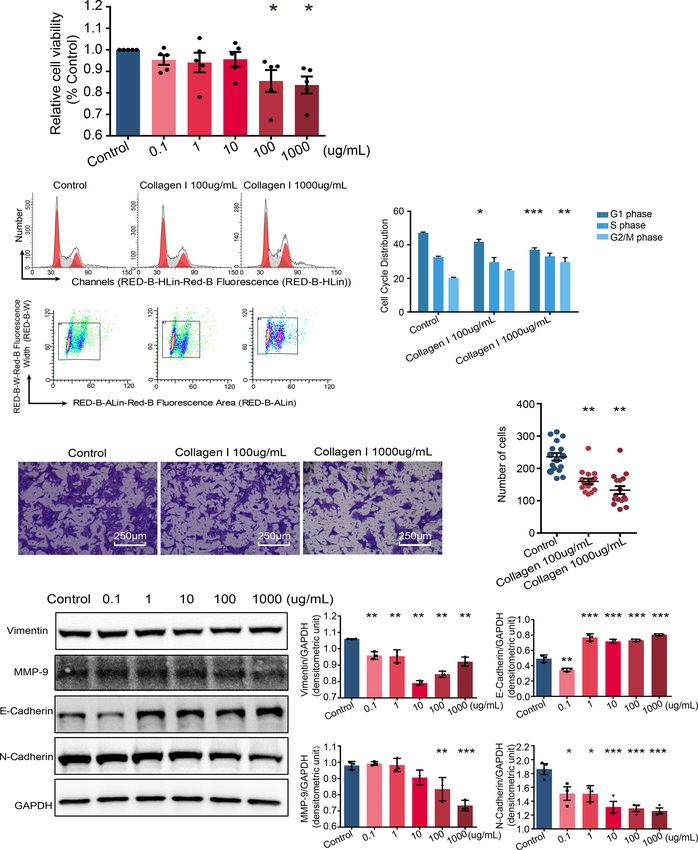
Feng et al.                                                                                                                     Collagen I in Preeclampsia

animals were handled according to the guiding principles                      was removed from the plate surface; plates were then dried
published in the National Institutes of Health Guide for the                  overnight at room temperature and washed with phosphate
Care of Animals.                                                              buffered saline to remove the unattached protein.
    Female C57/BL6 mice strain (8 weeks, weight 18–20 g,
purchased from the Animal Laboratory Center of Southern                       Cell Stimulation
Medical University) were used in the experiments. They were                   HTR-8SV/neo cells were seeded in six-well plate which were
maintained on a 12 hour/12hour light/dark schedule with free                  incubated with different concentration of collagen I for 48h.
access to food and water. All female mice were housed with male               CCK-8 (ab228554, Abcam, MA, USA) assay was conducted to
mice at a 2:1 ratio overnight, and pregnancy was confirmed by                  detect cell viability and cell cycle analysis cell cycle stage was
the presence of vaginal spermatozoa. The presence of                          analyzed using flow cytometry. Western blotting was used to
spermatozoa was indicative of gestational day 0.5 (E0.5d).                    measure the relative protein level. The experimental procedures
    Pregnant female mice were randomly divided into five groups                were detailed in the Supplementary Material.
on E0.5d(six mice per group): pregnant control group, 0.5 mg/kg                  Honokiol, which enhance the phosphorylation of ERK
collagen I-treated group, 5 mg/kg collagen I-treated group,                   phosphorylation, (HY-N0003, MedChemExpress, USA) dissolved
IFA-treated group, and NG-nitro-L-arginine methyl ester                       at a concentration of 10mM. In the co-treatment group, HTR-8/
(L-NAME)-treated group. In the collagen I-treated groups, 100 mL              SVneo cells were treated with Honokiol and collagen I 48h. SKL-
of collagen I (0.5 mg/kg and 5 mg/kg) was injected intradermally              2001 (HY-101085, MedChemExpress, USA), an agonist of b-
at the base of the tail of each mouse on E0.5d. The mice in IFA-              catenin which was used at a concentration of 5mM. Co-treament
treated group were injected 100mL intradermally at the base of                with collagen I 24h. Cell viability, cell cycle and relative protein level
the tail on E0.5.                                                             were analyzed by CCK-8, cycle cell and western blotting.
    The mice in the L-NAME group were treated with continuous
administration of 125 mg/kg/d of L-NAME, a common
                                                                              Protein Expression
vasoconstrictor, starting at 10.5 days of gestation via
subcutaneous injection on the nucha (18). Control mice were
                                                                              and Biochemical Analysis
                                                                              Protein was extracted from cells and placental tissue with
not injected with any solution. Systolic blood pressure (SBP) was
                                                                              commercial lysis buffer RIPA (89900, Thermo Scientific, USA).
recorded every four days. All mice were sacrificed, and placental
                                                                              Western blotting was performed as described in Supplementary
tissues were harvested on gestational day 17.5 (E17.5d). The
                                                                              Material. Primary antibodies against collagen I (ab260043, Abcam,
numbers of fetus were counted and recorded. We cut open the
                                                                              Cambridge, UK), E-cadherin (#14472, Cell Signaling, USA), N-
uterus along the uterine membrane to remove the fetus. Then, we
                                                                              cadherin (#13116, Cell Signaling, USA), MMP-9(ab38898, Abcam,
washed the fetus with PBS and placed into a clean dish. The
                                                                              Cambridge, UK), Vimentin (ab92547, Abcam, Cambridge, UK),
excess liquid was removed by filter paper before weighting.
                                                                              GAPDH (ab8245, Abcam, Cambridge, UK), ERK (#4695, Cell
                                                                              Signaling, USA), p-ERK(#4370, Cell Signaling, USA), b-Catenin
Measurements of Blood Pressure in Mice
                                                                              (ab32572, Abcam, Cambridge, UK).
Monitor of blood pressure were to evaluate the preeclampsia
symptom of animal. We determined the blood pressure in
conscious mice by tail cuff plethysmography using the Softron                 Transwell Assay
BP 2010 (Softron Biotechnology, Beijing, China). All the mice                 Transwell assays were performed to test the invasion ability of
were habituated to the measurement procedure 10 times a day                   HTR-8/SVneo Matrigel (356234, BD, USA) and Transwell (3422,
before caging with male mice. At least 9 consecutive                          Costa, USA) were purchased and used as per manufacturer’s
measurements were recorded, but only when the condition of                    instructions (19). We precoated the upper chamber of each
the mice was stable. Six time point (pre-pregnancy, E0.5, E4.5,               Transwell (Costa, USA) insert with different doses of collagen I
E8.5, E12.5, and E16.5) were selected for assessing systolic blood            (100 or 1000 mg/mL). Cells were grown in the upper chamber in
pressure level throughout the pregnancy. Serial blood pressure                medium containing 3% FBS and assessed for their invasion
measures analyzed by two-way repeated measures ANOVA.                         ability through collagen I toward a chemoattractant (10% FBS)
                                                                              in the lower chamber for 48 h.
Cell Culture
HTR-8SV/neo cells (purchased from ATCC, Rockville, USA)                       Transcriptome Sequencing
were cultured in RPMI-1640 medium containing 10% fetal                        and Data Analysis
bovine serum (FBS) and 1% Penicillin-Streptomycin. Cells                      Differential gene expression analysis of 30,945 expressed genes
were maintained at 37°C, with 5% CO2. Liguid were changed                     was performed using DESeq2 (19, 20), and yielded 1237
every other day until cells reached 90% confluence.                            differentially expressed genes (an adjusted p value < 0.05). We
                                                                              collected PE-related genes from six meta-analyses (21–26), two
Precoating of Cell Culture Plates                                             microarray-based studies (27, 28), one review paper (29), and
With Collagen I                                                               one research paper based on RNA-seq (30) published since
The surfaces of six-well cell culture plates were coated with different       2009. A total of 3553 PE-associated genes were obtained. We
doses of collagen I, which was diluted in a minimal volume (500 mL            found 2347 of these PE-related genes to be expressed in our
per well). After 4 hours of incubation with collagen I, the solution          dataset, 227 of which were differentially expressed. KEGG (Kyoto

Frontiers in Endocrinology | www.frontiersin.org                          3                                     August 2021 | Volume 12 | Article 664766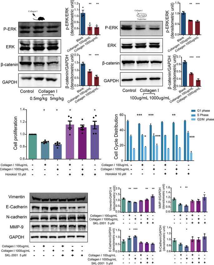
Feng et al.                                                                                                                Collagen I in Preeclampsia

Encyclopedia of Genes and Genomes) pathway enrichment                          At E4.5 d, collagen I-injected mice exhibited significantly higher
analysis for 227 genes was performed with the clusterProfiler               SBP than did control and IFA mice (p < 0.05). Moreover, the SBP of
package (Supplementary Table S2) (31). A cutoff of adjusted                the collagen I- and L-NAME-treated groups was elevated from the
P value < 0.05 was chosen to select the most significantly                  second trimester to the end of gestation (p < 0.01 Figure 2B).
enriched pathways. Finally, pathview (32) was used to visualize            Placental tissue was harvested at E17.5d to examine the weight of
the differentially expressed genes belonging to significantly               foetuses and the structure of the placenta. Fetal weight was decreased
enriched pathways.                                                         in both the collagen I- and L-NAME-treated groups (p < 0.01
                                                                           Figure 2C). However, the number of fetus only decreased in the
Statistics                                                                 group treated with 5 mg/kg of collagen I (p < 0.01 Figure 2C).
Statistical analyses were performed using Prism 7.0 software               Nevertheless, histomorphometric analysis revealed structural
(GraphPad Software, Inc. San Diego, CA, USA). After testing for            changes in the placenta of mice in the collagen I- and L-NAME-
normal distribution, differences among groups were analyzed by two-        treated groups; particularly, infarction was observed in the labyrinth
tailed, unpaired Student’s t-tests or one-way analysis of variance         layer (Figure 2D). Moreover, in both the labyrinth and junctional
(ANOVA) with Tukey’s post-hoc test for multiple comparisons. All           zones, a higher degree of fibrosis was observed in the collagen I- and
data are presented as mean ± standard error of mean (SEM).                 L-NAME-treated groups than in the control group (Figure 2E).
    Enrichment analyses were performed on the R platform, and              However, the relative protein expression of collagen I was higher only
a two-tailed Fisher’s exact test was used. Error bars were used to         in the collagen I-treated groups (p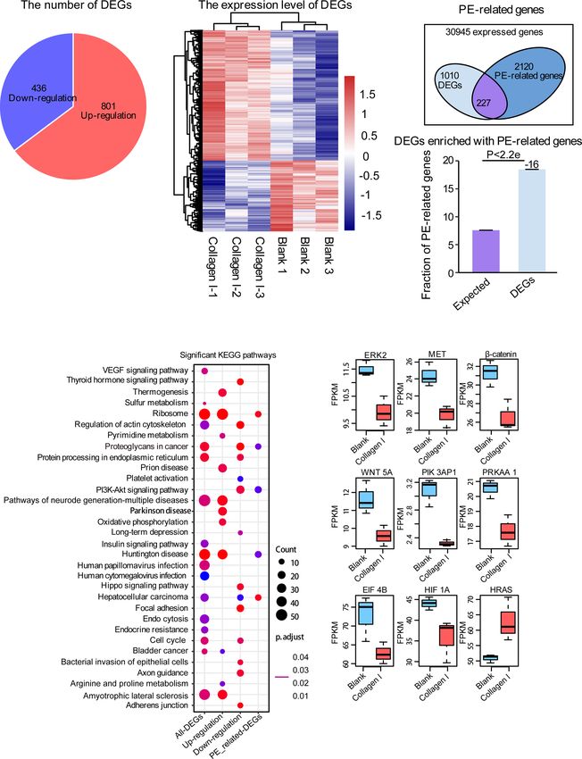
Feng et al.                                                                                                                                     Collagen I in Preeclampsia

                          A

                          B

                          C                                                                                          D

 FIGURE 1 | Identification and quantification of collagen types in human preeclamptic (PE) placentas. (A) Masson’s trichrome staining of placentas in the stem and
 terminal villus region from normotensive pregnant women (NP placenta) and from PE patients (PE placenta; n = 6 per group, scale bar = 250 nm). Collagen
 expression area of PE and NP placentas. Collagen fibers were stained blue, while cytoplasm and red blood cells were stained red, and the nucleus blue and brown.
 (B) Representative image of Sirius red staining of placenta (magnification, 200×). Measured area and red-yellow color intensity of Sirius Red staining in PE and NP
 placenta (three visual fields for each placental sample, n = 6 per group). (C) Representative image of western blotting and quantitative analysis of collagen I in PE and
 NP placenta (bar chart, n = 8 per group). (D) Relative gene expression of collagen I (bar chart, n = 8 per group). Data are presented as mean ± standard error of
 mean (SEM). *p < 0.05, **p < 0.01 by two-tailed unpaired t-test analysis.

total RNA samples from 100mg/mL collagen I-treated and                                  and WNT5A, genes related to cell proliferation and invasion in
untreated HTR-8SV/neo cells by RNA-seq. We found that the                               trophoblasts (Figure 4E).
expression of 801 genes increased while that of 436 genes
decreased under collagen I treatment compared with gene
expression in the control group (Figure 4A). The relative                               Collagen I Suppress Trophoblast
expression of upregulated and downregulated genes was                                   Proliferation and Invasion
illustrated in a heat map (Figure 4B). Additionally, we found                           Together with transcriptome sequencing analysis, western blotting
2347 PE-related genes to be expressed in our dataset, among                             was used to detect p-ERK/ERK and b-catenin expression in collagen
which 227 were differentially expressed (Figure 4C). With the                           I-treated and untreated mouse placenta and HTR-8SV/neo cells. In
latter gene set, we carried out KEGG pathway enrichment                                 both types of samples, we observed decreased expression of p-ERK
analysis; the significantly enriched pathways are summarized in                          and b-catenin after collagen I treatment (Figures 5A, B). ERK is a
Figure 4D. Interestingly, genes involved in proteoglycans in                            member of the MAPK signaling pathway. Alterations in ERK
cancer, cell cycle, and the PI3K-AKT signaling pathway (which                           phosphorylation can lead to changes in cell proliferation (39). The
is related to cell proliferation and invasion), were significantly                       present results demonstrated that the ERK phosphorylation level was
downregulated in our dataset. Consistently, collagen I treatment                        decreased following treatment with collagen I. Furthermore, the
downregulated the expression of ERK2, MET, PI3K, b-catenin,                             combination of the ERK activator, honokiol, with collagen I to treat

Frontiers in Endocrinology | www.frontiersin.org                                    5                                         August 2021 | Volume 12 | Article 664766
Feng et al. Collagen I in Preeclampsia
A B C
D
E
F
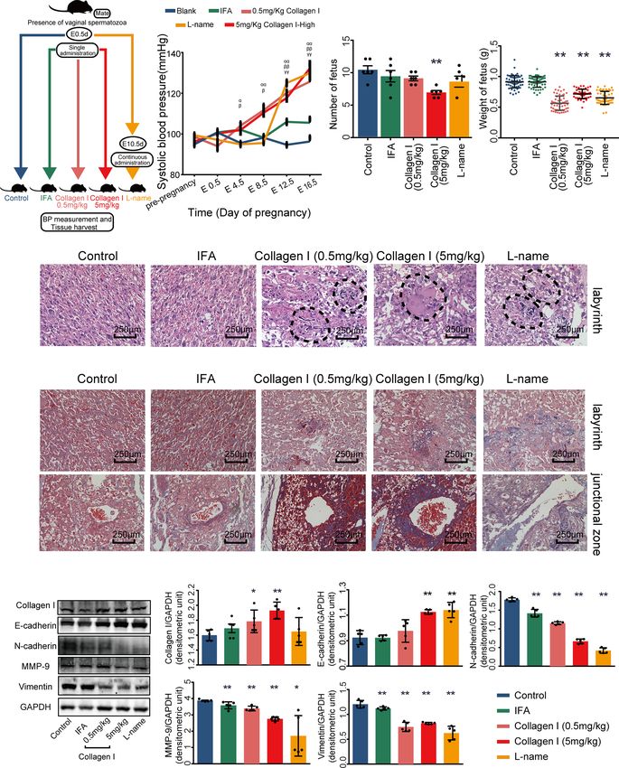
FIGURE 2 | Collagen I induces preeclampsia-like symptoms in mice. Effects of collagen I on the reproductive outcomes of pregnant mice. (A) Schematic representation of
the experimental groups. (B) The systolic blood pressure of members of the five groups during pregnancy (n = 6 per group, a indicates mice treated with 5 mg/kg of collagen
I vs. control, b indicates mice treated with 0.5 mg/kg of collagen I vs. control, g indicates L-NAME-treated mice vs. control, with a,b,gp < 0.05 and aa,bb,ggp < 0.01, according
to two-way repeated measures ANOVA). (C) The number of offspring significantly decreased in the group treated with 5 mg/kg of collagen I (n = 6 per group) compared with
the control at 17.5 days of gestation. Fetal weights in the groups treated with 0.5 mg/kg of collagen I (n = 54), 5 mg/kg of collagen I (n = 46), and L-NAME (n = 49) were
significantly decreased compared with those of the control (n = 59) and IFA-treated groups (n = 56). (D) Representative image of the H&E-stained labyrinth zone of mouse
placenta. Typical lesions are marked with dotted lines (original magnification, ×200; scale bar = 250 mm, n = 4). (E) Representative image of Masson’s trichrome staining in
the labyrinth and junctional zone of the mouse placenta. (original magnification, ×200; scale bar = 250 mm, n = 4). (F) Western blot analysis of the protein level of collagen I
and the proteins related to PE pathogenesis including MMP-9, E-cadherin, N-cadherin, and vimentin (n = 5, asterisks indicate differences between groups vs. control, with
*p < 0.05, **p < 0.01, according to one-way ANOVA followed by Dunnett post-hoc test).
HTR-8/SVneo cells, reversed the decline in proliferation and the plays a central role in canonical Wnt/b-catenin signaling. b-
G2/M arrest induced by collagen I (Figures 5C, D). catenin levels decreased in both collagen I-treated mouse
Wnt/b-catenin signaling is an essential pathway promoting placenta and HTR-8/SVneo cells. Therefore, SKL-2001, an
blastocyst activation and implantation. In particular, b-catenin agonist of the Wnt/b-catenin pathway that can stabilize
Frontiers in Endocrinology | www.frontiersin.org 6 August 2021 | Volume 12 | Article 664766Feng et al. Collagen I in Preeclampsia
A
B
C
D
FIGURE 3 | Collagen I induce trophoblast dysfunction in HTR-8/SVneo cells. Effect of collagen I on proliferation and invasion abilities of HTR-8/SVneo cells.
(A) HTR-8/SVneo cells were cultured at different concentrations of collagen I (0 mg/mL, 0.1 mg/mL, 1 mg/mL, 10 mg/mL, 100 mg/mL, and 1000 mg/mL). Cell
proliferation of HTR-8SV/neo cells was analyzed by CCK-8 assay after 48 hours of incubation (n = 5 per group). (B) Effect of collagen I on the cell cycle profile of
HTR-8SV/neo cells. After cultured at 100 mg/mL and 1000 mg/mL of collagen I for 48 hours, cells were labeled with propidium iodide and the stain was detected by
laser scanning cytometry(LSC). Bar plot representing the proportion of cells at each phase of the cell cycle (G1, S, and G2/M phases) in three different cell groups
(cells grown in presence of 100 mg/mL or 1000 mg/mL of collagen I and control cells; n = 5 per group). (C) Migration of HTR-8/SVneo cells. The total number of
invading cells was counted in four representative fields under ×200 magnification (n = 4 per group). (D) Representative western blot image and quantitative analysis
of vimentin, MMP-9, E-cadherin, and N-cadherin expression in the three cell groups (n = 3 per group). Asterisks indicate differences between groups vs. control, with
*p < 0.05, **p < 0.01, and ***p < 0.001, according to one-way ANOVA followed by Dunnett post-hoc test.
intracellular b-catenin levels, was used to clarify the role of Overall, our results showed that activators of ERK and b-catenin
b-catenin in collagen I-dependent remodulation of trophoblast (honokiol and SKL-2001) reversed the decline in cell proliferation
properties. The relative protein levels of MMP-9, vimentin, and invasion induced by collagen I, demonstrating that collagen I
N-cadherin, and E-cadherin were also recovered compared suppressed proliferation and invasion through inhibition of ERK
with those of collagen I-treated cells (Figure 5E). phosphorylation, and the Wnt/b-catenin signal pathway.
Frontiers in Endocrinology | www.frontiersin.org 7 August 2021 | Volume 12 | Article 664766Feng et al. Collagen I in Preeclampsia
A B C
D E
FIGURE 4 | Collagen I induce transcriptional changes in HTR-8SV/neo cells. Overview of differentially expressed genes upon collagen I treatment of HTR-8SV/
neo cells. (A) Pie chart showing the number of differentially expressed genes between collagen I-treated cells and the control group. (B) Heatmap showing the
expression levels of differentially expressed genes. (C) Venn diagram showing the PE-related genes expressed in our dataset. Differentially expressed genes
were enriched in PE-related genes (p < 0.05). (D) Significantly enriched KEGG pathways within total differentially expressed genes, including upregulated genes,
downregulated genes, and differentially expressed PE-related genes. (E) Boxplots showing the relative expression levels of ERK2 (MAPK1), MET, b-catenin,
WNT5A, PIK3AP1, PRKAA1, EIF4B, HIF1A, and EIF4B. Enrichment analyses were performed on the R platform, and two-tailed Fisher’s exact test was used.
Error bars represent the standard error of the fraction, estimated using a bootstrapping method with 100 re-samplings. The data of the experimental validations
are presented as mean ± standard error of mean (SEM) of two independent experiments. Comparisons between two independent groups were conducted using
Student’s t-test.
Frontiers in Endocrinology | www.frontiersin.org 8 August 2021 | Volume 12 | Article 664766Feng et al. Collagen I in Preeclampsia
A B
C D
E
FIGURE 5 | Collagen I suppressed proliferation and invasion of trophoblasts through inhibition of ERK phosphorylation and the WNT/b-catenin signaling pathway.
Honokiol and SKL-2001 reversed the decrease in proliferation and invasion ability of trophoblasts induced by collagen I. (A) Representative western blot image and
quantitative analysis of p-ERK, ERK, and b-catenin levels in collagen I-treated mouse placentas (n = 3 per group). (B) Representative western blot image and
quantitative analysis of p-ERK, ERK, and b-catenin levels in collagen I-treated HTR-8/SVneo cells (n = 3 per group). (C) After incubation with honokiol and collagen I
for 48 hours, HTR-8/SVneo cell proliferation was measured by CCK-8 assay (n = 5 per group). (D) After incubation with honokiol and collagen I for 48 hours, cells
were labelled with propidium iodide (PI) and the stain was detected by LSC. Bar plots representing the proportion of cells at each phase of the cell cycle (G1, S, and
G2/M phases) in three different cell groups (n = 5 per group). (E) Representative western blot image and quantitative analysis of MMP-9, vimentin, E-cadherin, and
N-cadherin levels in collagen I- and/or SKL-2001-treated HTR-8/SVneo cells (n = 3 per group). Asterisks indicate differences between groups vs. control, with
*p < 0.05, **p < 0.01, and ***p < 0.001, according to one-way ANOVA followed by Dunnett’s post-hoc test.
DISCUSSION associated with preeclampsia-like symptoms in pregnant mice.
Indeed, a single injection of collagen I in early pregnancy led to
Herein, we proved the characteristic role of Collagen I in increased blood pressure on E4.5d, which continued to stay
preeclamptic placenta is consistent with previous reports (17). elevated until term. Furthermore, the weight and number of
Moreover, we determined that excess collagen I accumulation is offspring decreased when the tissue was harvested at term.
Frontiers in Endocrinology | www.frontiersin.org 9 August 2021 | Volume 12 | Article 664766Feng et al. Collagen I in Preeclampsia
This phenomenon might depend on the decreased proliferation It has also been indicated that early pregnancy placentation is
and invasion ability of trophoblasts in the presence of excess closely related to trophoblast function and behavior (42). In our
collagen I. In summary, we propose that excessive collagen I in vitro study, excessive collagen I induced trophoblast
deposition in the placenta plays a crucial role in preeclampsia dysfunction by direct stimulation. It decreased the proliferation
pathogenesis (summarized in Figure 6). and invasion of HTR-8/SVneo cells. Surprisingly, we found that
The pathogenesis of preeclampsia is still unclear. A possible preeclampsia-like symptoms emerged in later pregnancy and
reason for this is the difficulty in performing high-quality research infarction and a high degree of fibrosis were observed in the
on the etiology of placental pathologies in humans because of ethical placenta after a single injection of collagen I on E0.5 d in vivo.
challenges associated with the examination of pregnant women in Moreover, changes in MMP-9, vimentin, E-cadherin and
the first trimester. Thus, animal models of preeclampsia are the N-cadherin expression both in placental and HTR-8SV/neo
most important tool for longitudinal investigation of adverse cells were consistent with the phenotypes of preeclampsia
pregnancy outcomes (40). For instance, treating third-trimester (36–38) after treatment with collagen I. Thus, we suggest that
pregnant mice with L-NAME, a common vasoconstrictor, to induce the single injection of collagen I in early pregnancy induced
PE symptoms is a common practice, as once drug treatment is preeclampsia development by interfering with placentation in
stopped, blood pressure returns to normal (41). In our study, a early pregnancy due to impaired trophoblast invasion. Collagen I
single injection of collagen I at early pregnancy led to changes in is the most abundant structural protein in most tissues (43).
placental structure at term. In addition to a higher degree offibrosis, However, excessive accumulation of collagen I may lead to
collagen I-treated placentas showed a significantly higher collagen I fibrosis, which in turn impairs normal organ function.
protein level than the control group. Moreover, preeclampsia-like Trigonelline hydrocholoride, targeting collagen I fibrillation,
symptoms were induced in later trimesters of pregnancy. In could ameliorate fibrosis of the myocardium (44). Therefore,
comparison with L-NAME treatment, the symptoms induced by collagen I may also be a novel target for therapeutic interventions
collagen I did not depend on continuous medication, but occurred in patients with preeclampsia.
in the second trimester and continued through the third. These The ERK/MAPK pathway is an important signaling cascades
disease dynamics were close to those observed in clinical practice. that is involved in cell proliferation and embryo development
It is widely accepted that primary defective trophoblast (45). We found that collagen I downregulated ERK
invasion, leading to inadequate transformation of maternal phosphorylation in HTR-8/SVneo cells and mouse placenta
uterine vasculature, probably drives preeclampsia pathogenesis. compared with the control. Honokiol which enhances the
FIGURE 6 | The abstract graphic. Collagen I deposited in placenta participate in the pathogenesis of preeclampsia: Collagen I is the characteristic collagen
deposited in preeclamptic placenta. Collagen I administration is associated with suppressed proliferation and invasion of trophoblasts, and with preeclampsia-like
symptoms in pregnant mice.
Frontiers in Endocrinology | www.frontiersin.org 10 August 2021 | Volume 12 | Article 664766Feng et al. Collagen I in Preeclampsia
phosphorylation of ERK phosphorylation, could reverse DATA AVAILABILITY STATEMENT
the weakened proliferation induced by collagen I. Further, the
canonical “Wnt/b-catenin” pathway regulates cell invasion, and The datasets presented in this study can be found in online
abnormal Wnt/b-catenin expression contributes to preeclampsia repositories. The names of the repository/repositories
development (46). b-catenin and GSK-3b were proteins levels and accession number(s) can be found below: https://data.
were reportedly significantly decreased in severe preeclamptic mendeley.com/datasets/hcd2c3n9tc/1, The Mendeley dataset DOI:
placenta (47). In our study, b-catenin expression in collagen 10.17632/hcd2c3n9tc.1. https://github.com/YingLin-Feng/Collagen-
I-treated placentas was less than that in the pregnant control I-and-preeclampsia.
Transcriptome analysis suggested b-catenin and GSK-3b
expression decreased in HTR-8/SVneo cells incubated with
100µg/mL collagen I. To verify these findings, SKL-2001, an ETHICS STATEMENT
agonist of that b-catenin was used. In vitro, SKL-2001 could
The collection of placentae was approved by the Ethics Committee
reverse the downregulation of MMP-9, N-cadherin and vimentin
of Nanfang Hospital (NFEC-2017-055). All participants gave
induced by collagen I. Thus, the results suggested that collagen I
written consent prior to donating their placenta. The patients/
suppresses trophoblast proliferation and invasion through
participants provided their written informed consent to participate
inhibition of ERK phosphorylation, and the WNT/b-catenin
in this study. This project was performed in accordance with animal
signaling pathway.
protocol procedures approved by the Department of Laboratory
There were some limitations of this study. First, our
Animal Sciences, Southern Medical University (L-2019216).
experiments demonstrated that the dysfunction of HTR-8/
SVneo cells induced by collagen I can be reversed by ERK and
b-catenin agonists. Therefore, the possible role of upstream AUTHOR CONTRIBUTIONS
regulators of the ERK/MAPK and WNT/b-catenin pathways in
preeclampsia pathogenesis should be investigated. Second, YF, XiC, and LH designed the study. Data collection was
placental Masson’s and Sirius red staining showed that the performed by HW, YiC, ML, ZL, XuC, YuC, YW, CS, and YH.
degree of fibrosis in PE patients was more serious than that in Data analysis was done by PL, JL, MZ, ZW, and XY. YF wrote the
normal patients after delivery. However, whether the extent of manuscript and designed the figures, whilst all other authors
placental fibrosis and its alteration can be determined by revised the manuscript. All authors contributed to the article and
ultrasound or other diagnostic methods during pregnancy, approved the submitted version.
calls for further clinical study and discussion. Third, small-
molecule inhibitors against placental collagen I deposition are
still lacking and should be explored as a future research avenue. FUNDING
Finally, the sample is relatively small, larger sample size is
This work was funded by the National Natural Science
required in our coming study.
Foundation of China (82071669), Foshan Dengfeng project
Overall, in this study we confirmed that the preeclamptic
(2020B002), the Natural Science Foundation of Guangdong
placenta shows significant histological signs of fibrosis and is
Province (2019A1515010637) and Science and Technology
characterized by collagen I deposition. Furthermore, an excess of
Planning Project of Guangdong Province (2017A010105025).
collagen I caused preeclampsia-like features in pregnant mice by
suppressing proliferation and invasion of trophoblasts. In vitro,
we detected altered expression of genes related to proliferation SUPPLEMENTARY MATERIAL
and invasion upon collagen I stimulation. In conclusion, our
study points at the relevant role of collagen I in the development The Supplementary Material for this article can be found online at:
of preeclamptic placentas, providing new insights into the https://www.frontiersin.org/articles/10.3389/fendo.2021.664766/
pathogenesis of preeclampsia. full#supplementary-material
REFERENCES Possible Maladaptations of the Fetal Component of the Placenta. Eur J
Obstet Gynecol Reprod Biol (2011) 156(1):29–34. doi: 10.1016/
1. Gestational Hypertension and Preeclampsia: ACOG Practice Bulletin, j.ejogrb.2010.12.038
Number 222. Obstet Gynecol (2020) 135(6):e237–60. doi: 10.1097/ 5. Devisme L, Merlot B, Ego A, Houfflin-Debarge V, Deruelle P, Subtil D.
AOG.0000000000003891 A Case-Control Study of Placental Lesions Associated With Pre-
2. Hypertension in Pregnancy. Report of the American College of Eclampsia. Int J Gynaecol Obstet (2013) 120(2):165–8. doi: 10.1016/
Obstetricians and Gynecologists’ Task Force on Hypertension in j.ijgo.2012.08.023
Pregnancy. Obstet Gynecol (2013) 122(5):1122–31. doi: 10.1097/01.AOG. 6. Ohmaru-Nakanishi T, Asanoma K, Fujikawa M, Fujita Y, Yagi H, Onoyama
0000437382.03963.88 I, et al. Fibrosis in Preeclamptic Placentas Is Associated With Stromal
3. Roberts JM, Taylor RN, Musci TJ, Rodgers GM, Hubel CA, McLaughlin MK. Fibroblasts Activated by the Transforming Growth Factor-Beta1 Signaling
Preeclampsia: An Endothelial Cell Disorder. Am J Obstet Gynecol (1989) 161 Pathway. Am J Pathol (2018) 188(3):683–95. doi: 10.1016/j.ajpath.
(5):1200–4. doi: 10.1016/0002-9378(89)90665-0 2017.11.008
4. Ducray JF, Naicker T, Moodley J. Pilot Study of Comparative Placental 7. Zeisberg M, Kalluri R. Cellular Mechanisms of Tissue Fibrosis. 1.
Morphometry in Pre-Eclamptic and Normotensive Pregnancies Suggests Common and Organ-Specific Mechanisms Associated With Tissue
Frontiers in Endocrinology | www.frontiersin.org 11 August 2021 | Volume 12 | Article 664766Feng et al. Collagen I in Preeclampsia
Fibrosis. Am J Physiol Cell Physiol (2013) 304(3):C216–25. doi: 10.1152/ Cancer Molecular Pathways in Placenta Leading to Preeclampsia. Sci Rep
ajpcell.00328.2012 (2013) 3:2407. doi: 10.1038/srep02407
8. Rockey DC, Bell PD, Hill JA. Fibrosis–A Common Pathway to Organ Injury 27. Song Y, Liu J, Huang S, Zhang L. Analysis of Differentially Expressed Genes in
and Failure. N Engl J Med (2015) 372(12):1138–49. doi: 10.1056/ Placental Tissues of Preeclampsia Patients Using Microarray Combined With
NEJMra1300575 the Connectivity Map Database. Placenta (2013) 34(12):1190–5. doi: 10.1016/
9. Weiskirchen R, Weiskirchen S, Tacke F. Organ and Tissue Fibrosis: Molecular j.placenta.2013.09.013
Signals, Cellular Mechanisms and Translational Implications. Mol Aspects 28. Sitras V, Paulssen RH, Gronaas H, Leirvik J, Hanssen TA, Vartun A, et al.
Med (2019) 65:2–15. doi: 10.1016/j.mam.2018.06.003 Differential Placental Gene Expression in Severe Preeclampsia. Placenta
10. Wahyudi H, Reynolds AA, Li Y, Owen SC, Yu SM. Targeting Collagen for (2009) 30(5):424–33. doi: 10.1016/j.placenta.2009.01.012
Diagnostic Imaging and Therapeutic Delivery. J Control Release (2016) 29. Kobayashi H. Characterization of the Down-Regulated Genes Identified in
240:323–31. doi: 10.1016/j.jconrel.2016.01.007 Preeclampsia Placenta. Hypertens Pregnancy (2016) 35(1):15–21. doi: 10.3109/
11. Zhang Y, Stefanovic B. LARP6 Meets Collagen mRNA: Specific Regulation of 10641955.2015.1116555
Type I Collagen Expression. Int J Mol Sci (2016) 17(3):419. doi: 10.3390/ 30. Kaartokallio T, Cervera A, Kyllonen A, Laivuori K, Kere J, Laivuori H. Gene
ijms17030419 Expression Profiling of Pre-Eclamptic Placentae by RNA Sequencing. Sci Rep
12. Ricard-Blum S, Baffet G, Theret N. Molecular and Tissue Alterations of (2015) 5:14107. doi: 10.1038/srep14107
Collagens in Fibrosis. Matrix Biol (2018) 68-69:122–49. doi: 10.1016/ 31. Yu G, Wang LG, Han Y, He QY. Clusterprofiler: An R Package for Comparing
j.matbio.2018.02.004 Biological Themes Among Gene Clusters. Omics (2012) 16(5):284–7.
13. Lindsey ML, Iyer RP, Zamilpa R, Yabluchanskiy A, DeLeon-Pennell KY, doi: 10.1089/omi.2011.0118
Hall ME, et al. A Novel Collagen Matricryptin Reduces Left Ventricular 32. Luo W, Brouwer C. Pathview: An R/Bioconductor Package for Pathway-Based
Dilation Post-Myocardial Infarction by Promoting Scar Formation and Data Integration and Visualization. Bioinformatics (2013) 29(14):1830–1.
Angiogenesis. J Am Coll Cardiol (2015) 66(12):1364–74. doi: 10.1016/ doi: 10.1093/bioinformatics/btt285
j.jacc.2015.07.035 33. Winters J, von Braunmuhl ME, Zeemering S, Gilbers M, Brink TT, Scaf B,
14. Lambert E, Fuselier E, Ramont L, Brassart B, Dukic S, Oudart JB, et al. et al. JavaCyte, A Novel Open-Source Tool for Automated Quantification of
Conformation-Dependent Binding of a Tetrastatin Peptide to Alphavbeta3 Key Hallmarks Of Cardiac Structural Remodeling. Sci Rep (2020) 10(1):20074.
Integrin Decreases Melanoma Progression Through FAK/PI3K/Akt doi: 10.1038/s41598-020-76932-3
Pathway Inhibition. Sci Rep (2018) 8(1):9837. doi: 10.1038/s41598-018- 34. Liang X, Yang LX, Guo R, Shi Y, Hou X, Yang Z, et al. Atorvastatin Attenuates
28003-x Plaque Vulnerability by Downregulation of EMMPRIN Expression via COX-
15. Stefanovic B, Manojlovic Z, Vied C, Badger CD, Stefanovic L. Discovery and 2/PGE2 Pathway. Exp Ther Med (2017) 13(3):835–44. doi: 10.3892/
Evaluation of Inhibitor of LARP6 as Specific Antifibrotic Compound. Sci Rep etm.2017.4062
(2019) 9(1):326. doi: 10.1038/s41598-018-36841-y 35. Cui Y, Wang W, Dong N, Lou J, Srinivasan DK, Cheng W, et al. Role of Corin
16. Sadeghi-Avalshahr AR, Nokhasteh S, Molavi AM, Mohammad-Pour N, in Trophoblast Invasion and Uterine Spiral Artery Remodelling in Pregnancy.
Sadeghi M. Tailored PCL Scaffolds as Skin Substitutes Using Sacrificial PVP Nature (2012) 484(7393):246–50. doi: 10.1038/nature10897
Fibers and Collagen/Chitosan Blend. Int J Mol Sci (2020) 21(7):2311. 36. Espino YSS, Flores-Pliego A, Espejel-Nunez A, Medina-Bastidas D, Vadillo-
doi: 10.3390/ijms21072311 Ortega F, Zaga-Clavellina V, et al. New Insights Into the Role of Matrix
17. Xu XH, Jia Y, Zhou X, Xie D, Huang X, Jia L, et al. Downregulation of Lysyl Metalloproteinases in Preeclampsia. Int J Mol Sci (2017) 18(7):1488.
Oxidase and Lysyl Oxidase-Like Protein 2 Suppressed the Migration and doi: 10.3390/ijms18071448
Invasion of Trophoblasts by Activating the TGF-Beta/Collagen Pathway in 37. Vicovac L, Aplin JD. Epithelial-Mesenchymal Transition During Trophoblast
Preeclampsia. Exp Mol Med (2019) 51(2):1–12. doi: 10.1038/s12276-019- Differentiation. Acta Anat (Basel) (1996) 156(3):202–16. doi: 10.1159/
0211-9 000147847
18. Shu W, Li H, Gong H, Zhang M, Niu X, Ma Y, et al. Evaluation of Blood 38. Yue D, Li H, Che J, Zhang Y, Tseng HH, Jin JQ, et al. Hedgehog/Gli Promotes
Vessel Injury, Oxidative Stress and Circulating Inflammatory Factors in an L- Epithelial-Mesenchymal Transition in Lung Squamous Cell Carcinomas.
NAME-Induced Preeclampsia-Like Rat Model. Exp Ther Med (2018) 16 J Exp Clin Cancer Res (2014) 33:34. doi: 10.1186/1756-9966-33-34
(2):585–94. doi: 10.3892/etm.2018.6217 39. Liu F, Feng XX, Zhu SL, Huang HY, Chen YD, Pan YF, et al. Sonic Hedgehog
19. Marshall J. Transwell((R)) Invasion Assays. Methods Mol Biol (2011) 769:97– Signaling Pathway Mediates Proliferation and Migration of Fibroblast-Like
110. doi: 10.1007/978-1-61779-207-6_8 Synoviocytes in Rheumatoid Arthritis via MAPK/ERK Signaling Pathway.
20. Love MI, Huber W, Anders S. Moderated Estimation of Fold Change and Front Immunol (2018) 9:2847. doi: 10.3389/fimmu.2018.02847
Dispersion for RNA-Seq Data With Deseq2. Genome Biol (2014) 15(12):550. 40. Sones JL, Davisson RL. Preeclampsia, of Mice and Women. Physiol Genomics
doi: 10.1186/s13059-014-0550-8 (2016) 48(8):565–72. doi: 10.1152/physiolgenomics.00125.2015
21. Kleinrouweler CE, van Uitert M, Moerland PD, Ris-Stalpers C, van der Post 41. Kanashiro CA, Cockrell KL, Alexander BT, Granger JP, Khalil RA. Pregnancy-
JA, Afink GB. Differentially Expressed Genes in the Pre-Eclamptic Placenta: A Associated Reduction in Vascular Protein Kinase C Activity Rebounds During
Systematic Review and Meta-Analysis. PloS One (2013) 8(7):e68991. Inhibition of NO Synthesis. Am J Physiol Regul Integr Comp Physiol (2000)
doi: 10.1371/journal.pone.0068991 278(2):R295–303. doi: 10.1152/ajpregu.2000.278.2.R295
22. Vaiman D, Calicchio R, Miralles F. Landscape of Transcriptional 42. Ridder A, Giorgione V, Khalil A, Thilaganathan B. Preeclampsia: The
Deregulations in the Preeclamptic Placenta. PloS One (2013) 8(6):e65498. Relationship Between Uterine Artery Blood Flow and Trophoblast
doi: 10.1371/journal.pone.0065498 Function. Int J Mol Sci (2019) 20(13):2363. doi: 10.3390/ijms20133263
23. Brew O, Sullivan MH, Woodman A. Comparison of Normal and Pre- 43. Yamauchi M, Sricholpech M, Terajima M, Tomer KB, Perdivara I.
Eclamptic Placental Gene Expression: A Systematic Review With Meta- Glycosylation of Type I Collage. Methods Mol Biol (2019) 1934:127–44.
Analysis. PloS One (2016) 11(8):e161504. doi: 10.1371/journal.pone.0161504 doi: 10.1007/978-1-4939-9055-9_9
24. Vaiman D, Miralles F. An Integrative Analysis of Preeclampsia Based on the 44. Rasheeda K, Fathima NN. Trigonelline Hydrochloride: A Promising Inhibitor
Construction of an Extended Composite Network Featuring Protein-Protein for Type I Collagen Fibrillation. Colloids Surf B Biointerfaces (2018) 170:273–9.
Physical Interactions and Transcriptional Relationship. PloS One (2016) 11 doi: 10.1016/j.colsurfb.2018.06.030
(11):e165849. doi: 10.1371/journal.pone.0165849 45. Xing CY, Zhang DX, Gui SQ, Tao MF. Kidney-Replenishing Herb Induces
25. Tejera E, Cruz-Monteagudo M, Burgos G, Sanchez ME, Sanchez-Rodriguez A, SOCS-3 Expression via ERK/MAPK Pathway and Improves Growth of the
Perez-Castillo Y, et al. Consensus Strategy in Genes Prioritization and First-Trimester Human Trophoblast Cell. Evid Based Complement Alternat
Combined Bioinformatics Analysis for Preeclampsia Pathogenesis. BMC Med (2017) 2017:2473431. doi: 10.1155/2017/2473431
Med Genomics (2017) 10(1):50. doi: 10.1186/s12920-017-0286-x 46. Zhang Z, Wang X, Zhang L, Shi Y, Wang J, Yan H. Wnt/beta-Catenin
26. Moslehi R, Mills JL, Signore C, Kumar A, Ambroggio X, Dzutsev A. Signaling Pathway in Trophoblasts and Abnormal Activation in Preeclampsia
Integrative Transcriptome Analysis Reveals Dysregulation of Canonical (Review). Mol Med Rep (2017) 16(2):1007–13. doi: 10.3892/mmr.2017.6718
Frontiers in Endocrinology | www.frontiersin.org 12 August 2021 | Volume 12 | Article 664766Feng et al. Collagen I in Preeclampsia
47. Wang X, Zhang Z, Zeng X, Wang J, Zhang L, Song W, et al. Wnt/beta-Catenin Copyright © 2021 Feng, Chen, Wang, Chen, Lan, Li, Cao, Liu, Lv, Chen, Wang,
Signaling Pathway in Severe Preeclampsia. J Mol Histol (2018) 49(3):317–27. Sheng, Huang, Zhong, Wang, Yue and Huang. This is an open-access article
doi: 10.1007/s10735-018-9770-7 distributed under the terms of the Creative Commons Attribution License
(CC BY). The use, distribution or reproduction in other forums is permitted,
provided the original author(s) and the copyright owner(s) are credited and that
Conflict of Interest: The authors declare that the research was conducted in the the original publication in this journal is cited, in accordance with accepted
absence of any commercial or financial relationships that could be construed as a academic practice. No use, distribution or reproduction is permitted which does
potential conflict of interest. not comply with these terms.
Frontiers in Endocrinology | www.frontiersin.org 13 August 2021 | Volume 12 | Article 664766You can also read