Pseudomonas aeruginosa biofilm killing beyond the spacer by antibiotic-loaded calcium sulfate beads: JBJI
←
→
Page content transcription
If your browser does not render page correctly, please read the page content below
JBJI

                                                                                                                                                   Original full-length article
J. Bone Joint Infect., 6, 119–129, 2021

                                                                                                                                     Open Access
https://doi.org/10.5194/jbji-6-119-2021
© Author(s) 2021. This work is distributed under                                                                 Journal of Bone
                                                                                                               and Joint Infection
the Creative Commons Attribution 4.0 License.

           Pseudomonas aeruginosa biofilm killing beyond the
           spacer by antibiotic-loaded calcium sulfate beads:
                            an in vitro study
             Jacob R. Brooks1, , Devendra H. Dusane1, , Kelly Moore1 , Tripti Gupta1 , Craig Delury2 ,
          Sean S. Aiken2 , Phillip A. Laycock2 , Anne C. Sullivan3 , Jeffrey F. Granger3 , Matthew V. Dipane4 ,
                                     Edward J. McPherson4 , and Paul Stoodley1,3,5
                       1 Department  of Microbial Infection and Immunity, The Ohio State University,
                                       Wexner Medical Center, Columbus, Ohio, USA
                        2 Biocomposites Ltd., Keele Science Park, Keele, Staffordshire, ST5 5NL, UK
          3 Department of Orthopaedics, The Ohio State University, Wexner Medical Center, Columbus, Ohio, USA
        4 Department of Orthopaedic Surgery, David Geffen School of Medicine at UCLA, Santa Monica, California,

                                                             USA
           5 National Centre for Advanced Tribology at Southampton (nCATS), National Biofilm Innovation Centre

               (NBIC), Department of Mechanical Engineering, University of Southampton, Southampton, UK
                                       These authors contributed equally to this work.

                               Correspondence: Paul Stoodley (paul.stoodley@osumc.edu)

             Received: 29 October 2020 – Revised: 1 March 2021 – Accepted: 4 March 2021 – Published: 23 March 2021

      Abstract. Introduction: Bacterial biofilms are an important virulence factor in chronic periprosthetic joint
      infection (PJI) and other orthopedic infection since they are highly tolerant to antibiotics and host immunity.
      Antibiotics are mixed into carriers such as bone cement and calcium sulfate bone void fillers to achieve sustained
      high concentrations of antibiotics required to more effectively manage biofilm infections through local release.
      The effect of antibiotic diffusion from antibiotic-loaded calcium sulfate beads (ALCS-B) in combination with
      PMMA bone cement spacers on the spread and killing of Pseudomonas aeruginosa Xen41 (PA-Xen41) biofilm
      was investigated using a “large agar plate” model scaled for clinical relevance. Methods: Bioluminescent PA-
      Xen41 biofilms grown on discs of various orthopedic materials were placed within a large agar plate containing
      a PMMA full-size mock “spacer” unloaded or loaded with vancomycin and tobramycin, with or without ALCS-
      B. The amount of biofilm spread and log reduction on discs at varying distances from the spacer was assessed
      by bioluminescent imaging and viable cell counts. Results: For the unloaded spacer control, PA-Xen41 spread
      from the biofilm to cover the entire plate. The loaded spacer generated a 3 cm zone of inhibition and significantly
      reduced biofilm bacteria on the discs immediately adjacent to the spacer but low or zero reductions on those
      further away. The combination of ALCS-B and a loaded PMMA spacer greatly reduced bacterial spread and
      resulted in significantly greater biofilm reductions on discs at all distances from the spacer. Discussion: The
      addition of ALCS-B to an antibiotic-loaded spacer mimic increased the area of antibiotic coverage and efficacy
      against biofilm, suggesting that a combination of these depots may provide greater physical antibiotic coverage
      and more effective dead space management, particularly in zones where the spread of antibiotic is limited by
      diffusion (zones with little or no fluid motion).

Published by Copernicus Publications on behalf of EBJIS and MSIS.
120                                                                             J. R. Brooks et al.: P. aeruginosa biofilm killing

1   Introduction                                                   Further, we have previously shown in a similar model that
                                                                   the antibiotic loading concentration in ALCS-B did not sig-
Periprosthetic joint infection (PJI) is a complex problem in       nificantly change the effective area of antibiotic activity over
total joint arthroplasty (TJA) and occurs in 2 %–2.4 % of          the observed time frame, from which we concluded that the
all total hip and knee replacement procedures (Kurtz et al.,       area of antibiotic potency was limited primarily by diffusion,
2012; Rasouli et al., 2014). While infrequent, PJI has a dra-      not the loading potency (Dusane et al., 2017). While we ex-
matic effect on the patient’s health, often resulting in joint     pect that increasing the area of spread of antibiotic depots
dysfunction, morbidity, and mortality (Vrgoc et al., 2014;         will result in increased spatial coverage of antibiotic activity,
Boddapati et al., 2018; Zmistowski et al., 2013). The finan-       we wish to determine the spatial effect of adding ALCS-B
cial burden placed on patients and the healthcare system is        to an antibiotic-loaded spacer mimic on the spread of bac-
staggering (Kurtz et al., 2012; Kamath et al., 2015). A ma-        teria from biofilm-colonized materials, as well as the killing
jor complicating factor in treating PJI is microbiota-produced     efficacy of biofilm bacteria on those materials in a scaled-up
biofilms (Gbejuade et al., 2015; McConoughey et al., 2014).        in vitro model. In the present study, we evaluate the effect of
Biofilms are pathogenic communities that adhere to living          ALCS-B and antibiotic-loaded bone cement spacers (ALBC-
and nonliving surfaces and exhibit greatly increased antibi-       S) on containing both the spread and killing of P. aeruginosa
otic tolerance and resistance against host immunity. The es-       biofilms using a “large agar plate” model. We used P. aerugi-
tablishment of biofilms is assisted by the presence of foreign     nosa Xen41 in part because of its strong bioluminescent sig-
materials of orthopedic implant components, such as various        nal, allowing for more sensitive and illustrative monitoring of
metals and polymers (Zimmerli, 2014; Moley et al., 2019).          the effect of antibiotic diffusing from the PMMA or ALSC-B
   Pseudomonas aeruginosa is a Gram-negative opportunis-           by an in vitro imaging system (IVIS).
tic nosocomial pathogen and is cultured up to 20 % of the
time in chronic Gram-negative PJI (McConoughey et al.,             2     Materials and methods
2014; Zmistowski et al., 2011; Rodríguez-Pardo et al., 2014).
In previous in vitro studies, P. aeruginosa has shown the abil-    2.1    Bacterial growth
ity to display tolerance and resistance to antibiotics com-
monly used to treat PJIs (Dusane et al., 2019). Although           A bioluminescent derivative strain of Pseudomonas aerug-
the Gram-positive Staphylococci are the most commonly iso-         inosa PAO1 (PA-Xen41; Xenogen Corp., USA) was used.
lated pathogens from PJI, the infecting organism (or organ-        Stock culture was streaked onto 1.5 % tryptic soy agar (TSA;
isms) may not be cultured as much as up to 25 % of the             Becton, Dickinson & Company, MD, USA) and incubated
time (Kapadia et al., 2016; Pulido et al., 2008; Choi et al.,      for 24 h. A single colony was transferred aseptically to 15 mL
2013) or treatment is started before culture data are available.   of tryptic soy broth (TSB; Becton, Dickinson & Company,
Thus, in treating PJI, combinations of antibiotics are com-        MD, USA) and incubated overnight at 37 ◦ C, 5 % CO2 on a
monly used to provide broad-spectrum coverage (Ciofu et            rotary shaker at 200 rpm. We used PA-Xen41 for our stud-
al., 2017). Vancomycin and/or aminoglycosides (most com-           ies because it gives off a very strong signal, allowing for our
monly gentamicin or tobramycin) can be mixed into poly-            long-term non-destructive monitoring of the spread of antibi-
methyl methacrylate (PMMA) bone cement and mineral-                otics on activity and killing of the lawn biofilms, and as men-
based absorbable bone fillers such as calcium sulfate dihy-        tioned previously, P. aeruginosa is a relevant Gram-negative
drate (CaSO4 ·2 H2 O) to be administered as spacers (Hansen        PJI pathogen.
et al., 2014) and beads (McPherson et al., 2013) in the joint
space and medullary canals. These depot forms have been            2.2    Formation of biofilms on circular discs
shown to release high concentrations of antibiotic required
                                                                   Overnight cultures were diluted to 0.1 % and used to inoc-
to more effectively prevent or manage biofilms that cannot be
                                                                   ulate sterile, circular discs (BioSurface Technologies, MT,
achieved by systemic administration (Mandell et al., 2019).
                                                                   USA) of hydroxyapatite (HA), ultra-high molecular weight
There is promising yet inconclusive clinical data suggesting
                                                                   polyethylene (UHMWPE), 316L stainless steel (SS-316),
that ALCS-B used as an adjuvant in revision arthroplasty re-
                                                                   and titanium (Ti). The “as received” roughnesses measured
sult in more favorable outcomes (Abosala and Ali, 2020).
                                                                   by contact profilometry were 976, 3867, 224, and 300 nm,
However, it is not known how ALCS-B used in combina-
                                                                   respectively. The discs had a diameter of 9.5 mm and a thick-
tion with antibiotic-loaded PMMA may impact the area of
                                                                   ness of 2 mm. Four milliliters of the diluted culture was
antimicrobial potency against biofilms, particularly in those
                                                                   added to four different wells of a six-well plate and three
areas such as dead zones where there is little fluid flow, and
                                                                   discs of each material were aseptically submerged in the in-
the spread of the antibiotic is limited by diffusion.
                                                                   oculum. The plate was incubated at 37 ◦ C, 5 % CO2 for 72 h
   Previous studies using in vitro biofilms of the biolumines-
                                                                   to establish 3 d biofilms.
cent P. aeruginosa Xen41 (PA-Xen41) showed that the num-
ber and spacing of ALCS-B were important in the rate and
extent of killing of an agar lawn biofilm (Dusane et al., 2019).

J. Bone Joint Infect., 6, 119–129, 2021                                                 https://doi.org/10.5194/jbji-6-119-2021
J. R. Brooks et al.: P. aeruginosa biofilm killing 121
2.3 Preparation of antibiotic-loaded calcium sulfate might be applied clinically, where they might not be spread
beads (ALCS-B) evenly. Also, small diameter antibiotic depots such as ALCS-
B can fill smaller void spaces than the larger PMMA beads
ALCS-B were prepared using Stimulan® Rapid Cure (Bio-
and thus can be distributed more thoroughly.
composites, Ltd., Keele, UK) 10-cc mixing kits. Twenty
After adding the beads, another 100 mL of cooled
grams of CaSO4 powder was mixed with 1000 mg of van-
(∼ 50 ◦ C) liquid agar was poured to cover the discs and
comycin hydrochloride powder (VAN; Tokyo Chemical In-
beads and the plate covered with plastic film wrap for incuba-
dustry, Tokyo, Japan) and 240 mg of tobramycin sulfate pow-
tion and imaging. The antibiotic-loaded spacer and biofilm-
der (TOB; VWR International, PA). Once blended, 6 mL of
colonized discs were embedded within layers of agar to
sterile liquid included in the kit was added and the mixture
allow for the diffusion of antibiotic and bacteria from all
stirred into a uniform paste for 30 s. The paste was pressed
surfaces. These agar “layers” merged to form one cohe-
into a flexible mold (Biocomposites, Ltd., Keele, UK) with
sive block (Fig. 1). The following conditions were tested:
bead sizes of 4.8 mm in diameter and allowed to set for
(1) unloaded PMMA spacer only (US), (2) a VAN + TOB
10–15 min at 20 ◦ C. Although Xen41 is resistant to van-
antibiotic-loaded spacer only (LS), and (3) a VAN + TOB-
comycin (Dusane et al., 2017), we used a combination of
loaded spacer with a 10-cc pack of ALCS-B (LS + ALCS-
vancomycin and tobramycin since the combination of van-
B). The plates were incubated at 37 ◦ C, 5 % CO2 for 5 d and
comycin with an aminoglycoside (tobramycin or gentamicin)
imaged daily. Previously we demonstrated that the spreads
has been mixed in PMMA and ALCS-B clinically to provide
of tobramycin as a function of time (t) over a 4 d period to
broad-spectrum coverage (Anagnostakos, 2017; Hanssen and
achieve killing of the Xen41 agar lawn biofilms from PMMA
Spangehl, 2004). Additionally, we wanted to be consistent
and ALCS beads were 4.2 t0.5 mm and 3.6 t0.5 mm respec-
with previous experiments using ALCS-B (McConoughey et
tively, with the loading concentration making little differ-
al., 2015, 2014; Dusane et al., 2017; Howlin et al., 2017)
ence, suggesting the transport through the agar was limited
and avoid changing the release characteristics and set time
by diffusion (Dusane et al., 2017).
by changing the loading formulation.
2.6 Bioluminescent imaging (BLI)
2.4 Preparation of antibiotic-loaded (LS) and unloaded
(US) PMMA bone cement spacer mimics BLI was executed using an in vitro imaging system (IVIS
100, Xenogen, MA) that semi-quantitatively measures the
PMMA bone cement spacer mimics were prepared using
relative amount of metabolically active biofilm. Each quad-
Simplex™ P SpeedSet™ Radiopaque Bone Cement con-
rant of the large plate model was individually imaged and
struction kits (Stryker, Kalamazoo, MI), which are com-
then stitched together (Photoshop, Adobe, CA) to show the
monly used clinically. Antibiotic-loaded spacers were fab-
whole plate. Red represented the highest metabolic activity
ricated using 2 g VAN powder, 2 g TOB powder, and 40 g
and blue or black low or no metabolic activity. White-light
PMMA powder. Unloaded spacers were prepared with only
(plain) images of each plate were captured with a cellphone.
PMMA bone cement powder. Twenty milliliters of sterile
methyl methacrylate monomer (liquid) was added to the
powder mixture, stirred into a dough-like mass, formed into 2.7 Viable cell counting
a “hockey puck-like” disc using a 7 cm diameter circular Colony-forming unit (CFU) counts were performed on mim-
mold (Silikomart Professional Silicone Baking Mold, Cylin- icked sets of discs, one set on the 3 d biofilms and one set af-
der 6 Cavities, Amazon, WA) and allowed to set for 30 min ter incubation in the large plate model. CFUs were performed
at 20 ◦ C. as previously described (Moley et al., 2018). These discs
were rinsed and then vortexed with 10 mL of phosphate-
2.5 Large plate model buffered saline (PBS; Dulbecco’s, Gibco, Thermo Fisher Sci-
entific, MA) in 15 mL Falcon tubes (Thermo Fisher Scien-
Fifty milliliters of 1.5 % TSA was added to a 21 cm diame-
tific, MA); 10 µL of each dilution of a 10-fold dilution series
ter glass pie baking dish sterilized with 70 % EtOH and al-
was spotted onto TSA. The plates were incubated at 37 ◦ C,
lowed to solidify, followed by central placement of a PMMA
5 % CO2 for 24 h and colonies enumerated to determine CFU
spacer followed by another 100 mL of molten agar. Once set,
per area of disc (CFU/cm2 ). CFU counts after the incubation
three discs of each material (12 in total, each colonized with
were done by first extracting the embedded discs as an “agar
pre-grown 3 d biofilms) were placed on top of the agar layer
plug” in which a 1.35 cm diameter circular glass tube was
radiating outwards (Fig. 1). For the experiments with ALCS-
used to punch out the coupons.
B, a 10-cc pack of beads was sprinkled somewhat randomly
around the spacer with greater bead density closer to the
spacer mimic. While sprinkling the ALCS-B without care-
ful placement in a specified pattern may cause difficulty with
interpretation, we wished to more closely mimic how they
https://doi.org/10.5194/jbji-6-119-2021 J. Bone Joint Infect., 6, 119–129, 2021122 J. R. Brooks et al.: P. aeruginosa biofilm killing
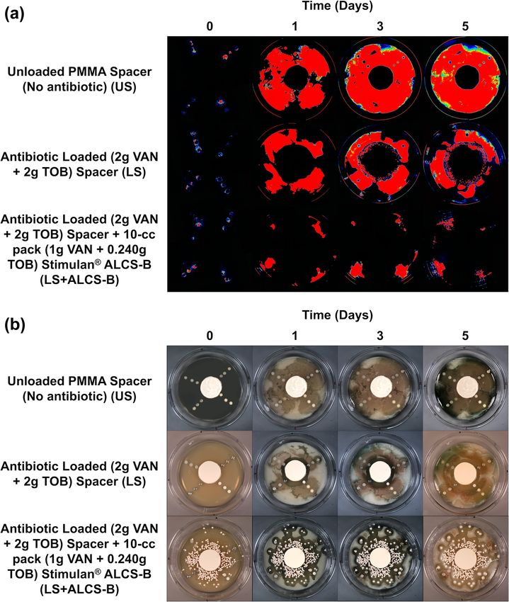
Figure 1. Side-view schematic showing the construction of the large plate agar model. This model allows for the spread of antibiotic released
from ALCS-B and an antibiotic-loaded spacer (ALBC-S) as it diffuses through the agar. Three discs of each material containing 3 d biofilms
were placed at distances of 1, 3, and 5 cm radiating linearly from the edge of the PMMA spacer mimic. ALCS-B were only included in the
LS + ALCS-B antibiotic condition.
2.8 Statistical analysis
All experiments were performed in triplicate. The effect of
the different conditions on the log reduction of biofilm at
different proximities from the spacer was compared by first
performing a log10 transformation and the geometric means
used to calculate log reductions. Our CFU detection limit
was 3.5 log10 CFU/cm2 . Discs that displayed no CFU growth
are shown as equal to or less than this limit. The log re-
ductions of the LS and LS + ALCS-B antibiotic conditions
were compared by a Student’s t test assuming unequal vari-
ances, where p < 0.05 was considered statistically signifi-
cant. Data were analyzed and graphed using Excel software
(version 2102, Microsoft 365).
3 Results
3.1 Prevention of biofilm spread by LS and ALCS-B
The bioluminescence of P. aeruginosa Xen41 allowed
biofilm on the discs and the spread of bacteria from these
discs to be easily visualized while remaining in situ in the
large plate model over the 5 d (Fig. 2a). The unloaded spacer
(US) condition of the large plate, containing no antibiotic,
showed substantial bacterial spread from the discs and was
confirmed by white-light imaging, showing P. aeruginosa Figure 2. IVIS (a) and white-light (b) images tracking the suppres-
coverage throughout the large plate (Fig. 2b). sion of P. aeruginosa Xen41 biofilm spread from biofilm-colonized
The antibiotic-loaded spacer (LS) alone exhibited anti- discs of four different orthopedic materials for three different an-
bacterial activity generating a zone of inhibition (ZOI) of ap- tibiotic conditions. Plates were imaged every 24 h for 5 d. At the
periphery of all plates, there is a loss of bioluminescence over time,
proximately 2.4–3.0 cm from the edge of the spacer (Fig. 2a).
even in the non-antibiotic control. This is likely due to nutrient de-
After 3 d a small number of colonies were observed grow-
pletion and a loss of metabolic activity in this region.
ing a few millimeters within the edge ZOI (Fig. 2a). These
were possibly antibiotic tolerant phenotypes and have been
observed previously (Dusane et al., 2019).
The LS + ALCS-B condition, which contained the addi- 3.2 Region of biofilm killing by viable cell counts
tion of VAN + TOB ALCS-B, prevented the formation of
antibiotic-tolerant phenotypes and suppressed the spreading After 3 d of inoculation, the amount of PA-Xen41 biofilm
of PA-Xen41 to sparse areas near the peripheral edge of the grown on various discs was quantified by cell counting
plate, where the bead density was lowest, or in areas where (CFU). This analysis was completed to confirm biofilm
beads were absent (Fig. 2). growth on each type of disc before implementation into
the large plate model. All four disc materials displayed
sizeable biofilm growth of ∼ 1010 CFU/cm2 , although there
J. Bone Joint Infect., 6, 119–129, 2021 https://doi.org/10.5194/jbji-6-119-2021J. R. Brooks et al.: P. aeruginosa biofilm killing 123
Figure 3. Quantification of viable bacteria (by CFU) on discs containing P. aeruginosa Xen41 3 d biofilm. Counts are reported in terms of
log10 CFUs per surface area of the disc (in cm2 ).
was no statistically significant difference in growth be- such implant surfaces, particularly in those locations where
tween any of the four materials (p > 0.05). UHMWPE discs the spread of released antibiotics is limited by diffusion, as
garnered the largest number of bacteria with a count of would be expected in the periprosthetic tissue, areas of a joint
10.6 log10 CFU/cm2 (Fig. 3), followed by HA, SS-316, and space, or the medullary canal. In a well-mixed system, the
Ti, indicating a weak trend with increasing roughness. specific distribution of antibiotic depots will be less impor-
The LS alone reduced biofilm concentrations on the clos- tant since the antibiotics will be well distributed by the mix-
est discs (1 cm from the spacer’s edge), with bacterial reduc- ing, but when the rate of spread of antibiotics is limited by
tion ranging from 1.5 to 5.5 log10 CFU/cm2 depending on diffusion, then the spatial distribution becomes significant.
the material type (Fig. 4a–d). For the more distant discs (3 The observed spread of bacteria from the discs over the
and 5 cm from the spacer), reduction of bacteria drastically surrounding agar appears similar to how biofilm is thought
decreased to a range of no reduction to 3.8 log10 CFU/cm2 . to spread throughout a joint space infection (Zimmerli and
The addition of loaded beads (LS + ALCS-B) promoted the Sendi, 2017; Jenkins et al., 2010). Antibiotics loaded into
log reduction of the more distant discs to levels of 2.5 to PMMA cement in the form of both beads and spacers have
6.6 log10 CFU/cm2 , depending on the material. Furthermore, been successfully used to treat PJI for many years (Hanssen
the addition of ALCS-B eradicated biofilm to below detec- and Spangehl, 2004). However, there are limitations to the
tion limits (3.5 log10 CFU/cm2 ) on SS-316 and Ti discs 5 cm use of PMMA alone with respect to the release of local an-
from the spacer (Fig. 4a, b), a statistically significant reduc- tibiotics at a surgical site. Once an antibiotic has eluted to
tion compared to the LS alone. In UHMWPE (Fig. 4d), the below MIC levels, the PMMA itself becomes a surface for
LS + ALCS-B condition produced significantly more (p < bacterial biofilm formation (Ma et al., 2018; Stoodley et al.,
0.05) biofilm reduction in discs at every distance. The Ti 2008). Further, even though retrieved PMMA spacers are still
discs 3 and 5 cm from the edge of the spacer (Fig. 4b), the HA shown to elute antibiotics and produce a ZOI in laboratory
disc 3 cm from the spacer (Fig. 4c), and the SS-316 disc 5 cm testing, the infection can persist, suggesting that the zone
from the spacer (Fig. 4a) also showed statistically significant of antimicrobial potency may be limited (Swearingen et al.,
biofilm killing in the LS + ALCS-B condition compared to 2016).
the LS alone (p < 0.05). Antibiotic-loaded PMMA beads have been used to in-
crease the effective area of antibiotic activity, but they have
4 Discussion
limited penetration into small spaces and require surgical re-
moval. Additionally, their relatively large size (∼ 1 cm di-
Here we show in our diffusion-based model that ALCS- ameter) limits the number that can be packed into a given
B significantly reduced (p < 0.05) P. aeruginosa Xen41 volume. In contrast, ALCS-B allow for the rapid, full release
biofilm growth on materials common to orthopedic implants of antibiotics (Dusane et al., 2017), and our visual demon-
in vitro while also increasing the zone of antimicrobial po- stration of ZOI produced by loaded PMMA spacers alone,
tency beyond that achieved by an antibiotic-loaded PMMA compared to spacers with ALCS-B, indicates how ALCS-B
spacer alone. Furthermore, our work demonstrates how such can aid in releasing antibiotics to areas beyond the spacer.
beads sprinkled around the spacer may more effectively man- We also note that since the beads in our study were de-
age dead space and control the spread of bacteria from liberately not positioned carefully but rather scattered semi-
https://doi.org/10.5194/jbji-6-119-2021 J. Bone Joint Infect., 6, 119–129, 2021124 J. R. Brooks et al.: P. aeruginosa biofilm killing Figure 4. Log reduction of P. aeruginosa Xen41 biofilms, after being grown for 3 d in TSB media and then introduced into the large plate model against one of three antibiotic conditions for 5 d. CFU counts of SS-316 (a), Ti (b), HA (c), and UHMWPE (d) from the biofilm- colonized discs, varying in distance from the edge of the PMMA spacer, were compared to those on an unloaded spacer (US) control. ≥ denotes discs eradicated of biofilm bacteria to below the CFU detection limit (3.54 log10 CFU/cm2 ) and indicates that the reduction could be greater than or equal to the value listed in the graph. * represents statistically significant (p < 0.05) differences in log reductions between LS and LS + ALCS-B antibiotic conditions. randomly, ALCS-B either ended up relatively close to one or may be less antibiotic-tolerant and less clinically relevant more of the distant (3 and 5 cm) discs or fell farther away. than more mature biofilms. Also, our in vitro model is sim- This resulted in variability of killing efficacy. If beads fell plistic compared to the complex mechanical and chemical close to a biofilm-colonized disc, then there was a significant environment of surrounding bones, particularly that of an ar- log reduction or eradication to below-detection limits, while ticulating human joint space. By design our model lacks any discs further away from beads exhibited less or no reduc- fluid flow, which would be expected to enhance the spread tion. This spatial variation is evidenced by some of the large of antibiotic from the spacer mimic and the ALCS-B while error bars (Fig. 4a–d) and illustrates the importance of ade- also removing antibiotic from the system (i.e., there is no quate spatial coverage within a periprosthetic joint or other infinite sink where antibiotic can be removed from the sys- infected surgical sites. Nevertheless, our in vitro data as well tem). A pharmacokinetic study by Hayes et al. (2014) found as those found clinically (Morgenstern et al., 2018; Ciofu et the intra-articular concentration of gentamicin released from al., 2017), and data from other in vitro studies (Howlin et al., a sponge in a dog dropped rapidly from approximately 2400 2015, 2017; Laycock et al., 2018; Knecht et al., 2018) sup- to 4 µg/mL after 22.4 h. They concluded that this drop was port the use of ALCS-B as an effective means of releasing due to vascular exchange and inflammation. However, con- antibiotic to prevent biofilm spread and kill pre-grown bacte- ventional in vitro studies of elution kinetics from antibiotic- rial biofilms. loaded depots are also oversimplified. In these studies, the We recognize limitations in our study. Our study was con- depot is placed in a reservoir of saline and the concentration ducted with only one bacterial strain. While we have shown measured periodically. In some cases there is periodic com- similar efficacy with other Gram-negative and -positive plete or partial exchange of the saline to create an infinite species with respect to prevention and killing efficacy by sink, where the flux of the released drug is not limited by its antibiotics released from beads alone (Howlin et al., 2017, build-up in the reservoir (Fu and Kao, 2010). While these es- 2015), to add rigor to our conclusions, we would need to in- tablished kinetic studies are useful for directly comparing the clude more pathogenic species and clinical strains. In addi- release kinetics of different materials and indeed have been tion, we used a relatively young biofilm grown for 3 d which used to demonstrate faster release from ALCS-B than from J. Bone Joint Infect., 6, 119–129, 2021 https://doi.org/10.5194/jbji-6-119-2021
J. R. Brooks et al.: P. aeruginosa biofilm killing                                                                            125

PMMA beads (McConoughey et al., 2015), these are com-                play between antibiotic release kinetics from various admin-
pletely mixed systems and there are no gradients other than          istration methods, including addition of antibiotic powder.
in the mass boundary layer at the depot surface. Thus, they
provide no information about how the spatial arrangement
of such depots may influence the local distribution of antibi-       Ethical statement. No human subjects or human subject data or
otics. Our diffusion model represents the opposite extreme           animals were used in this study.
of a completely mixed system, yet we argue it has value for
illustrating how antibiotics may spread from depots when
transport is limited by diffusion and how the size, number,
and spatial arrangement of such depots may influence such
spread. In a healthy knee, the flow of synovial fluid caused
by flexion of the joint during cyclic loading cycles is thought
to provide enough convective transport to deliver nutrients
to periarticular tissues, which can be up to 1 cm away from
blood vessels (Levick, 1995). Such a mechanism is also ex-
pected to distribute antibiotics eluted from local carriers or
delivered systemically from the vasculature. However, it is
not well understood how well this transport mechanism func-
tions in a reconstructed, revised, and potentially immobilized
joint. Radical debridement of the PJI joint has been shown to
compromise local blood flow to regions of the affected joint,
especially bone. While such debridement may be necessary
to rid the infected area of biofilm to the best extent possible,
when vasculature is compromised, the spread of antibiotics
through bone and joint tissues may be expected to be largely
limited by diffusion (Thabit et al., 2019). Other complicat-
ing factors such as diabetes, kidney disease, or osteonecrosis
secondary to osteomyelitis may also limit the flow of sys-
temic fluids. Diffusion is a much slower process, particularly
over longer distances, making ALCS-B a potentially useful
adjunct to antibiotic-loaded bone cement spacers (or other
forms) for distributing the necessary amount of antibiotics in
a timely and effective manner. It is also possible that the lack
of flow may have resulted in pH changes or high calcium lev-
els in the vicinity of the beads. In previous studies we have
demonstrated that unloaded beads alone do not have antibac-
terial activity (McConoughey et al., 2015), though we did not
investigate antibiotic–local chemistry interactions.
   The results from our in vitro study may support the adju-
vant use of ALCS-B in the management of PJI, particularly
for the release of antibiotics to the interstices of the peripros-
thetic joint space in light of limited PMMA diffusion with
compromised vasculature. However, further work is required
in order to determine how our in vitro findings relate to the
spread of antibiotics in the human joint or may relate to clin-
ical outcomes. Such studies are not trivial since measuring
antibiotic concentrations at different locations in tissues and
fluids in a joint space requires multiple samplings at different
times, since sampling of joint fluid may not represent condi-
tions everywhere bacteria or biofilm may be present. We are
currently developing a continuous flow reactor system which
can accommodate clinically relevant antibiotic-loaded spacer
mimics and beads, where we match the flow rate with re-
ported clinical drainage rates following TKA revision. Such
a system may provide further insight into the complex inter-

https://doi.org/10.5194/jbji-6-119-2021                                                    J. Bone Joint Infect., 6, 119–129, 2021
126                                                                       J. R. Brooks et al.: P. aeruginosa biofilm killing

Appendix A: Abbreviations

 PJI              Periprosthetic joint infection
 TJA              Total joint arthroplasty
 PMMA             Polymethyl methacrylate
 ALBC-S           Antibiotic-loaded bone cement spacer
 ALCS-B           Antibiotic-loaded calcium sulfate beads
 PA-Xen41         Pseudomonas aeruginosa Xen41
 TSA              Tryptic soy agar
 TSB              Tryptic soy broth
 HA               Hydroxyapatite
 UHMWPE           Ultra-high molecular weight polyethylene
 SS-316           316L stainless steel
 Ti               Titanium
 VAN              Vancomycin
 TOB              Tobramycin
 US               Unloaded spacer
 LS               Loaded spacer
 LS + ALCS-B      Loaded spacer and antibiotic-loaded calcium sulfate beads
 BLI              Bioluminescent imaging
 IVIS             In vitro imaging system
 CFUs             Colony-forming units
 ZOI              Zone of inhibition

J. Bone Joint Infect., 6, 119–129, 2021                                           https://doi.org/10.5194/jbji-6-119-2021
J. R. Brooks et al.: P. aeruginosa biofilm killing                                                                                           127

Data availability. Raw data used to generate the graphs in Figs. 3        Ciofu, O., Rojo-Molinero, E., Macià, M. D., and Oliver, A.: An-
and 4 and showing the statistical analysis available by request              tibiotic treatment of biofilm infections, APMIS, 125, 304–319,
or download from DOI https://doi.org/10.5061/dryad.hqbzkh1fq                 https://doi.org/10.1111/apm.12673, 2017.
(Stoodley and Brooks, 2021).                                              Dusane, D. H., Diamond, S. M., Knecht, C. S., Farrar, N. R., Peters,
                                                                             C. W., Howlin, R. P., Swearingen, M. C., Calhoun, J. H., Plaut, R.
                                                                             D., Nocera, T. M., Granger, J. F., and Stoodley, P.: Effects of load-
Author contributions. PS, DHD, JFG, ACS, EJM, SSA, PAL,                      ing concentration, blood and synovial fluid on antibiotic release
and CD conceived and designed the work. DHD oversaw labora-                  and anti-biofilm activity of bone cement beads, J. Control. Re-
tory experiments. JFG, ACS, and EJM oversaw preparation of the               lease, 248, 24–32, https://doi.org/10.1016/j.jconrel.2017.01.005,
PMMA spacers. TG measured the roughness of the coupons and                   2017.
interpreted data. JRB and KM performed the lab experiments and            Dusane, D. H., Brooks, J. R., Sindeldecker, D., Peters, C. W., Li, A.,
collected data. JRB, DHD, and PS performed data analysis and in-             Farrar, N. R., Diamond, S. M., Knecht, C. S., Plaut, R. D., Delury,
terpretation. JRB, DHD, and PS drafted the article. JFG, ACS, EJM,           C., Aiken, S. S., Laycock, P. A., Sullivan, A., Granger, J. F., and
and MVP provided clinical perspective to data and draft narrative.           Stoodley, P.: Complete killing of agar lawn biofilms by system-
PS, DHD, JFG, ACS, EJM, MVP, SSA, PAL, CD, and JRB per-                      atic spacing of antibiotic-loaded calcium sulfate beads, Materials
formed critical revision of the article. All the authors gave final ap-      (Basel), 12, 4052, https://doi.org/10.3390/MA12244052, 2019.
proval of the version to be submitted.                                    Fu, Y. and Kao, W. J.: Drug release kinetics and trans-
                                                                             port mechanisms of non-degradable and degradable poly-
                                                                             meric delivery systems, Expert Opin. Drug Deliv., 7, 429–444,
Competing interests. Paul Stoodley receives research funding                 https://doi.org/10.1517/17425241003602259, 2010.
from Biocomposites Ltd. and consults for Biocomposites Ltd. and           Gbejuade, H. O., Lovering, A. M., and Webb, J. C.: The role of mi-
Zimmer Biomet. Edward J. McPherson consults for Austin Medical               crobial biofilms in prosthetic joint infections, Acta Orthop., 86,
Ventures Inc., BoneSupport AB, and Zimmer Biomet.                            147–158, https://doi.org/10.3109/17453674.2014.966290, 2015.
                                                                          Hansen, E. N., Adeli, B., Kenyon, R., and Parvizi, J.: Rou-
                                                                             tine use of antibiotic laden bone cement for primary to-
                                                                             tal knee arthroplasty: Impact on infecting microbial pat-
Acknowledgements. We are grateful for the support of Maurice
                                                                             terns and resistance profiles, J. Arthroplasty, 29, 1123–1127,
Manring in the process of editing and submitting the manuscript.
                                                                             https://doi.org/10.1016/j.arth.2013.12.004, 2014.
                                                                          Hanssen, A. D. and Spangehl, M. J.: Practical applications
                                                                             of antibiotic-loaded bone cement for treatment of infected
Financial support. This research has been supported by Biocom-               joint replacements, Clin. Orthop. Relat. Res., 427, 79–85,
posites Ltd. Biocomposites Ltd. provided Stimulan bone void filler           https://doi.org/10.1097/01.blo.0000143806.72379.7d, 2004.
used in the study.                                                        Hayes, G. M., Gibson, T. W. G., Moens, N. M. M., Mon-
                                                                             teiro, B., and Johnson, R. J.: Intra-Articular Pharmacokinet-
                                                                             ics of a Gentamicin Impregnated Collagen Sponge in the Ca-
Review statement. This paper was edited by Martin Clauss and                 nine Stifle: An Experimental Study, Vet. Surg., 43, 166–173,
reviewed by four anonymous referees.                                         https://doi.org/10.1111/j.1532-950X.2014.12115.x, 2014.
                                                                          Howlin, R. P., Brayford, M. J., Webb, J. S., Cooper, J. J., Aiken, S.
                                                                             S., and Stoodley, P.: Antibiotic-loaded synthetic calcium sulfate
                                                                             beads for prevention of bacterial colonization and biofilm forma-
                                                                             tion in periprosthetic infections, Antimicrob. Agents Chemother.,
References                                                                   59, 111–120, https://doi.org/10.1128/AAC.03676-14, 2015.
                                                                          Howlin, R. P., Winnard, C., Frapwell, C. J., Webb, J. S.,
Abosala, A. and Ali, M.: The Use of Calcium Sulphate beads in                Cooper, J. J., Aiken, S. S., and Stoodley, P.: Biofilm preven-
  Periprosthetic Joint Infection, a systematic review, J. Bone Joint         tion of gram-negative bacterial pathogens involved in peripros-
  Infect., 5, 43–49, https://doi.org/10.7150/jbji.41743, 2020.               thetic infection by antibiotic-loaded calcium sulfate beads in
Anagnostakos, K.: Therapeutic Use of Antibiotic-loaded Bone Ce-              vitro, Biomed. Mater., 12, 015002, https://doi.org/10.1088/1748-
  ment in the Treatment of Hip and Knee Joint Infections, J. Bone            605X/12/1/015002, 2017.
  Joint Infect., 2, 29–37, https://doi.org/10.7150/jbji.16067, 2017.      Jenkins, P. J., Phillips, S. A., Gaston, P., Dave, J., and Breusch, S.
Boddapati, V., Fu, M. C., Mayman, D. J., Su, E. P., Sculco,                  J.: Be vigilant for secondary periprosthetic joint infection, Prac-
  P. K., and McLawhorn, A. S.: Revision Total Knee Arthro-                   titioner, 254, 28–32, 2010.
  plasty for Periprosthetic Joint Infection Is Associated With            Kamath, A. F., Ong, K. L., Lau, E., Chan, V., Vail, T.
  Increased Postoperative Morbidity and Mortality Relative                   P., Rubash, H. E., Berry, D. J., and Bozic, K. J.: Quan-
  to Noninfectious Revisions, J. Arthroplasty, 33, 521–526,                  tifying the Burden of Revision Total Joint Arthroplasty
  https://doi.org/10.1016/j.arth.2017.09.021, 2018.                          for Periprosthetic Infection, J. Arthroplasty, 30, 1492–1497,
Choi, H. R., Kwon, Y. M., Freiberg, A. A., Nelson, S. B., and                https://doi.org/10.1016/j.arth.2015.03.035, 2015.
  Malchau, H.: Periprosthetic joint infection with negative culture       Kapadia, B. H., Berg, R. A., Daley, J. A., Fritz, J., Bhave, A., and
  results: Clinical characteristics and treatment outcome, J. Arthro-        Mont, M. A.: Periprosthetic joint infection, Lancet, 387, 386–
  plasty, 28, 899–903, https://doi.org/10.1016/j.arth.2012.10.022,           394, https://doi.org/10.1016/S0140-6736(14)61798-0, 2016.
  2013.

https://doi.org/10.5194/jbji-6-119-2021                                                          J. Bone Joint Infect., 6, 119–129, 2021
128 J. R. Brooks et al.: P. aeruginosa biofilm killing Knecht, C. S., Moley, J. P., McGrath, M. S., Granger, J. F., Rasouli, M. R., Restrepo, C., Maltenfort, M. G., Purtill, J. J., and Stoodley, P., and Dusane, D. H.: Antibiotic loaded calcium Parvizi, J.: Risk factors for surgical site infection following to- sulfate bead and pulse lavage eradicates biofilms on metal tal joint arthroplasty, J. Bone Joint Surg. Am., 96, 1570–1575, implant materials in vitro, J. Orthop. Res., 36, 2349–2354, https://doi.org/10.2106/JBJS.M.01363, 2014. https://doi.org/10.1002/jor.23903, 2018. Rodríguez-Pardo, D., Pigrau, C., Lora-Tamayo, J., Soriano, A., Kurtz, S. M., Lau, E., Watson, H., Schmier, J. K., and del Toro, M. D., Cobo, J., Palomino, J., Euba, G., Riera, M., Parvizi, J.: Economic burden of periprosthetic joint in- Sánchez-Somolinos, M., Benito, N., Fernández-Sampedro, M., fection in the united states, J. Arthroplasty, 27, 61–65, Sorli, L., Guio, L., Iribarren, J. A., Baraia-Etxaburu, J. M., https://doi.org/10.1016/j.arth.2012.02.022, 2012. Ramos, A., Bahamonde, A., Flores-Sánchez, X., Corona, P. Laycock, P. A., Cooper, J. J., Howlin, R. P., Delury, C., Aiken, S., S., Ariza, J., Amat, C., Larrosa, M. N., Puig, M., Murillo, and Stoodley, P.: In vitro efficacy of antibiotics released from cal- O., Cabo, X., Goenaga, M. Á., Elola, M., De la Herrán, G., cium sulfate bone void filler beads, Materials (Basel), 11, 2265, Garcia-Arenzana, J. M., García-Ramiro, S., Martínez-Pastor, J. https://doi.org/10.3390/ma11112265, 2018. C., Tornero, E., García-Lechuz, J. M., Marín, M., Villanueva, M., Levick, J. R.: Microvascular Architecture and Exchange López, I., Cisterna, R., Santamaría, J. M., Gómez, M. J., Puente, in Synovial Joints, Microcirculation, 2, 217–233, A., Cano, P., Horcajada, J. P., González-Mínguez, P., Portillo, https://doi.org/10.3109/10739689509146768, 1995. E., Puig, L., Franco, M., Jordán, M., Coll, P., Amador-Mellado, Ma, D., Shanks, R. M. Q., Davis, C. M., Craft, D. W., Wood, J., Fuster-Foz, C., García-Paíno, L., Nieto, I., Muniain, M. Á., T. K., Hamlin, B. R., and Urish, K. L.: Viable bacteria per- Suárez, A. I., Praena, J., Gómez, M. J., Puente, A., Maseguer, M. sist on antibiotic spacers following two-stage revision for A., Garagorri, E., Pintado, V., Marinescu, C., Ramírez, A., Mon- periprosthetic joint infection, J. Orthop. Res., 36, 452–458, taner, F., Múñez, E., Álvarez, T., García, R., Puente, E., Salas, https://doi.org/10.1002/jor.23611, 2018. C., Fariñas, M. C., Pérez, J. M., Achabal, B. V., and Montejo Mandell, J. B., Orr, S., Koch, J., Nourie, B., Ma, D., Bonar, D. D., Baranda, J. M.: Gram-negative prosthetic joint infection: Out- Shah, N., and Urish, K. L.: Large variations in clinical antibi- come of a debridement, antibiotics and implant retention ap- otic activity against Staphylococcus aureus biofilms of peripros- proach. A large multicentre study, Clin. Microbiol. Infect., 20, thetic joint infection isolates, J. Orthop. Res., 37, 1604–1609, 911–919, https://doi.org/10.1111/1469-0691.12649, 2014. https://doi.org/10.1002/jor.24291, 2019. Stoodley, P. and Brooks, J.: Pseudomonas aeruginosa biofilm killing McConoughey, S. J., Howlin, R., Granger, J. F., Manring, M. beyond the spacer by antibiotic-loaded calcium sulfate beads: an M., Calhoun, J. H., Shirtliff, M., Kathju, S., and Stoodley, P.: in vitro study Excel spread sheet with raw data, Dryad, Dataset, Biofilms in periprosthetic orthopedic infections, Future Micro- https://doi.org/10.5061/dryad.hqbzkh1fq, 2021. biol., 9, 987–1007, https://doi.org/10.2217/fmb.14.64, 2014. Stoodley, P., Nistico, L., Johnson, S., Lasko, L. A., Baratz, M., McConoughey, S. J., Howlin, R. P., Wiseman, J., Stoodley, P., and Gahlot, V., Ehrlich, G. D., and Kathju, S.: Direct demonstration Calhoun, J. H.: Comparing PMMA and calcium sulfate as carri- of viable Staphylococcus aureus biofilms in an infected total joint ers for the local delivery of antibiotics to infected surgical sites, arthroplasty: A case report, J. Bone Joint Surg. Am., 90, 1751– J. Biomed. Mater. Res.-Part B Appl. Biomater., 103, 870–877, 1758, https://doi.org/10.2106/JBJS.G.00838, 2008. https://doi.org/10.1002/jbm.b.33247, 2015. Swearingen, M. C., Granger, J. F., Sullivan, A., and Stoodley, P.: McPherson, E., Dipane, M., and Sherif, S.: Dissolvable Antibiotic Elution of antibiotics from poly(methyl methacrylate) bone ce- Beads in Treatment of Periprosthetic Joint Infection and Revi- ment after extended implantation does not necessarily clear the sion Arthroplasty – The Use of Synthetic Pure Calcium Sulfate infection despite susceptibility of the clinical isolates, Pathog. (Stimulan® ) Impregnated with Vancomycin & Tobramycin, Re- Dis., 74, 1–4, https://doi.org/10.1093/femspd/ftv103, 2016. constr. Rev., 3, 32–43, https://doi.org/10.15438/rr.v3i1.27, 2013. Thabit, A. K., Fatani, D. F., Bamakhrama, M. S., Barnawi, O. A., Moley, J. P., McGrath, M. S., Granger, J. F., Stoodley, P., and Du- Basudan, L. O., and Alhejaili, S. F.: Antibiotic penetration into sane, D. H.: Reduction in Pseudomonas aeruginosa and Staphy- bone and joints: An updated review, Int. J. Infect. Dis., 81, 128– lococcus aureus biofilms from implant materials in a diffu- 136, https://doi.org/10.1016/j.ijid.2019.02.005, 2019. sion dominated environment, J. Orthop. Res., 36, 3081–3085, Vrgoc, G., Japjec, M., Gulan, G., Ravlić-Gulan, J., Marinović, M., https://doi.org/10.1002/jor.24074, 2018. and Bandalović, A.: Periprosthetic infections after total hip and Moley, J. P., McGrath, M. S., Granger, J. F., Sullivan, knee arthroplasty – a review, Coll. Antropol., 38, 1259–1264, A. C., Stoodley, P., and Dusane, D. H.: Mapping bac- 2014. terial biofilms on recovered orthopaedic implants by a Zimmerli, W.: Clinical presentation and treatment of orthopaedic novel agar candle dip method, APMIS, 127, 123–130, implant-associated infection, J. Intern. Med., 276, 111–119, https://doi.org/10.1111/apm.12923, 2019. https://doi.org/10.1111/joim.12233, 2014. Morgenstern, M., Vallejo, A., McNally, M. A., Moriarty, T. F., Zimmerli, W. and Sendi, P.: Orthopaedic biofilm infections, AP- Ferguson, J. Y., Nijs, S., and Metsemakers, W.: The effect MIS, 125, 353–364, https://doi.org/10.1111/apm.12687, 2017. of local antibiotic prophylaxis when treating open limb frac- Zmistowski, B., Fedorka, C. J., Sheehan, E., Deirmengian, G., tures, Bone Joint Res., 7, 447–456, https://doi.org/10.1302/2046- Austin, M. S., and Parvizi, J.: Prosthetic joint infection caused 3758.77.bjr-2018-0043.r1, 2018. by gram-negative organisms, J. Arthroplasty, 26, 104–108, Pulido, L., Ghanem, E., Joshi, A., Purtill, J. J., and Parvizi, J.: https://doi.org/10.1016/j.arth.2011.03.044, 2011. Periprosthetic joint infection: The incidence, timing, and pre- Zmistowski, B., Karam, J. A., Durinka, J. B., Casper, D. S., and disposing factors, Clin. Orthop. Relat. Res., 466, 1710–1715, Parvizi, J.: Periprosthetic joint infection increases the risk of https://doi.org/10.1007/s11999-008-0209-4, 2008. J. Bone Joint Infect., 6, 119–129, 2021 https://doi.org/10.5194/jbji-6-119-2021
J. R. Brooks et al.: P. aeruginosa biofilm killing                                                 129

  one-year mortality, J. Bone Joint Surg. Am., 95, 2177–2184,
  https://doi.org/10.2106/JBJS.L.00789, 2013.

https://doi.org/10.5194/jbji-6-119-2021                         J. Bone Joint Infect., 6, 119–129, 2021
You can also read