Effect of Multispecies Microbial Consortia on Microbially Influenced Corrosion of Carbon Steel - MDPI
←
→
Page content transcription
If your browser does not render page correctly, please read the page content below
corrosion and
               materials degradation

Article
Effect of Multispecies Microbial Consortia on Microbially
Influenced Corrosion of Carbon Steel
Hoang C. Phan 1, * , Linda L. Blackall 2                  and Scott A. Wade 1, *

                                          1   Faculty of Science, Engineering and Technology, Swinburne University of Technology,
                                              Hawthorn, VIC 3122, Australia
                                          2   School of BioSciences, University of Melbourne, Melbourne, VIC 3052, Australia;
                                              linda.blackall@unimelb.edu.au
                                          *   Correspondence: hoang.phan@virbac.vn (H.C.P.); swade@swin.edu.au (S.A.W.);
                                              Tel.: + 84-9-3396-8650 (H.C.P.); Tel.: +61-3-9214-4339 (S.A.W.)

                                          Abstract: Microbially influenced corrosion (MIC) is responsible for significant damage to major
                                          marine infrastructure worldwide. While the microbes responsible for MIC typically exist in the
                                          environment in a synergistic combination of different species, the vast majority of laboratory-based
                                          MIC experiments are performed with single microbial pure cultures. In this work, marine grade
                                          steel was exposed to a single sulfate reducing bacterium (SRB, Desulfovibrio desulfuricans) and various
                                          combinations of bacteria (both pure cultures and mixed communities), and the steel corrosion studied.
                                          Differences in the microbial biofilm composition and succession, steel weight loss and pitting attack
                                          were observed for the various test configurations studied. The sulfate reduction phenotype was
                                          successfully shown in half-strength marine broth for both single and mixed communities. The highest
                                          corrosion according to steel weight loss and pitting, was recorded in the tests with D. desulfuricans

                                   alone when incubated in a nominally aerobic environment. The multispecies microbial consortia
                                          yielded lower general corrosion rates compared to D. desulfuricans or for the uninoculated control.
Citation: Phan, H.C.; Blackall, L.L.;
Wade, S.A. Effect of Multispecies
Microbial Consortia on Microbially
                                          Keywords: corrosion; metabarcoding; MIC; multispecies; SRB
Influenced Corrosion of Carbon Steel.
Corros. Mater. Degrad. 2021, 2,
133–149. https://doi.org/10.3390/
cmd2020008                                1. Introduction
                                                The corrosion of metals immersed in the marine environment is a well-known issue,
Academic Editor: Philippe Refait          which can also be accelerated by the activities of a diverse range of microbes in a process
                                          known as microbially influenced corrosion (MIC) [1,2]. An example of MIC is the rapid
Received: 1 February 2021
                                          failure of metal structures, typically in the form of localised corrosion around low tide
Accepted: 18 March 2021
                                          level, which is referred to as accelerated low water corrosion (ALWC) [3,4]. ALWC has
Published: 25 March 2021
                                          been reported on steel structures (e.g., sheet piling) in many ports and harbors worldwide.
                                          The reduction in thickness of steel pilings, which can be up to several mm/yr, is one of
Publisher’s Note: MDPI stays neutral
                                          indications used to diagnose ALWC [5]. In the UK alone, the short-term cost of ALWC was
with regard to jurisdictional claims in
                                          estimated to be ~£250 million [3]. Methods to minimise ALWC include the application of
published maps and institutional affil-
                                          cathodic protection, coatings and the use of special grade steel. Further work, however,
iations.
                                          is required in order to obtain a better understanding of the complex roles and types of
                                          microbes involved in ALWC.
                                                Biofilm formation is a biological process that takes place in humid/aquatic environ-
                                          ments, and involves a series of developmental steps. In the natural environment these
Copyright: © 2021 by the authors.
                                          intricate microbial structures typically contain a diverse range of microbes with complex
Licensee MDPI, Basel, Switzerland.
                                          interactions [6,7]. In relation to ALWC, different microbes are likely to play specific roles in
This article is an open access article
                                          the overall corrosion process, including the development of a biofilm on the steel structure,
distributed under the terms and
                                          nutrient provision/cycling, as well as producing and maintaining an anaerobic environ-
conditions of the Creative Commons
Attribution (CC BY) license (https://
                                          ment. Specific microbes are responsible for the corrosion process [5,8,9]. Sulfate reducing
creativecommons.org/licenses/by/
                                          bacteria (SRB), e.g., Desulfovibrio desulfuricans, are probably the best known bacterial group
4.0/).
                                          in relation to microbial corrosion and ALWC [10,11]. The SRB corrosion process is typically

Corros. Mater. Degrad. 2021, 2, 133–149. https://doi.org/10.3390/cmd2020008                                    https://www.mdpi.com/journal/cmd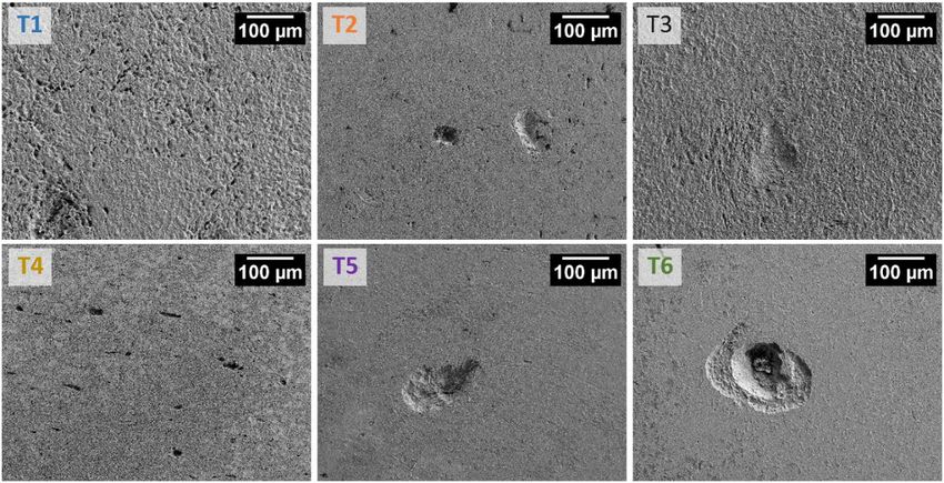
Corros. Mater. Degrad. 2021, 2                                                                                               134

                                 associated with a black biofilm on the surface and localised corrosion underneath [12].
                                 In addition to SRB, sulfur oxidising bacteria (SOB) have been implicated in ALWC, and
                                 the presence of both microbial groups will contribute to the sulfur cycle and subsequent
                                 corrosion [5,8,13–17].
                                       While there have been a large number of studies of MIC in the laboratory, most of
                                 these have failed to replicate the accelerated corrosion rates observed in the field. One
                                 of the possible reasons for this is that most tests use single microbial strains in the ex-
                                 periments (most commonly SRB), which is very different to the mixed microbial species
                                 found in biofilms associated with accelerated corrosion in the real environment [9,18–21].
                                 Another possible reason is that the testing arrangement used (e.g., test media, oxygen
                                 levels and test duration) may influence the processes (abiotic and biotic) required for the
                                 accelerated corrosion.
                                       Various laboratory MIC experiments have employed undefined combinations of mi-
                                 crobes (e.g., [5,22–25]). While this might be more similar to the field situation, the largely
                                 unknown combinations of microbes make replication impossible and the roles of any indi-
                                 vidual species cannot be determined. Several MIC studies using known microbial strains in
                                 various combinations have been undertaken, and have showed how the strains collectively
                                 affect corrosion (see Table S1). In some of the latter, the multispecies combinations have led
                                 to increased corrosion and in others decreased corrosion.
                                       The mechanisms responsible for MIC are hypothesised to include transfer of electrons
                                 from the metal of interest to the SRB and/or biochemicals produced as part of metabolic
                                 processes [26,27]. Both require the presence of a suitable electron donor and electron
                                 acceptor. The artificial supply of electron donors could facilitate increased corrosion
                                 compared to real world scenarios since seawater typically has relatively low levels of
                                 organic carbon sources [28]. Several researchers have shown that different test media used
                                 in MIC studies can affect the level of corrosion [29–31]. Additionally, specific strains of SRB
                                 (e.g., Desulfopila corrodens and Desulfovibrio sp. strain IS7) have been linked to the direct
                                 electron uptake from an iron/steel surface [26] and have also been detected in field studies
                                 of ALWC [9,17,23].
                                       This research adds to the understanding of the effect of multispecies microbial consor-
                                 tia in relation to microbial corrosion such as ALWC. The work involved exposing marine
                                 grade steel to an ALWC biofilm from a marine environment, pure culture isolates from
                                 an ALWC biofilm or adjacent locations, D. desulfuricans or to an uninoculated media (i.e.,
                                 negative control). The hypothesis of this work was that the combinations of microbes
                                 would produce increased rates of corrosion compared to tests with single isolates or an
                                 uninoculated control.

                                 2. Materials and Methods
                                 2.1. Steel Test Coupons
                                      Marine grade DH36 carbon steel [32] was chosen for this research due to its widespread use
                                 in shipbuilding and offshore structures. The metal coupon dimensions were 25 mm × 25 mm
                                 with a thickness of 6 mm. A 2 mm diameter hole was drilled in each coupon near the
                                 middle of one of the edges (3–4 mm from the edge) to allow coupons to be hung vertically
                                 in the aqueous corrosion tests. The top and bottom surfaces of the coupons were prepared
                                 using an automatic grinding machine (Tegramin-25, Struers, Cleveland, OH, USA) with
                                 a series of silicon carbide papers i.e., 320, 500 and 1200 grit. The edges were manually
                                 ground with 320 grit silicon carbide paper. After grinding, the coupons were cleaned by
                                 immersing in acetone in an ultrasonic bath for 8 min, rinsed thoroughly with distilled
                                 water and then absolute ethanol, and finally dried under warm air. Coupon weight was
                                 measured using a high accuracy analytical balance (MS205DU, readability 0.01 mg, Mettler
                                 Toledo, Mississauga, Canada) shortly before the corrosion tests.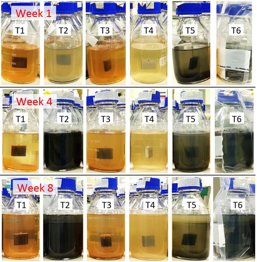
Corros. Mater. Degrad. 2021, 2                                                                                                135

                                 2.2. Bacteria and Test Media Details
                                       The four marine isolates were Halomonas korlensis, Bacillus aquimaris, Prolixibacter
                                 bellariivorans and Sulfitobacter pontiacus. Their presumed phenotypes cover a range of
                                 potential functions relevant to oxygen tolerance, biofilm formation and MIC. H. korlensis is
                                 a denitrifier/nitrate–nitrite reducing facultative aerobe [33] that was isolated from marine
                                 sediment [21]. B. aquimaris is a nitrate/metal ion utilising aerobe found in marine environ-
                                 ments/biofilms/corrosion steel surfaces [34,35] that was isolated from an orange ALWC
                                 tubercle [36]. P. bellariivorans is an iron-relating/sugar-fermenting anaerobe [37,38] that
                                 was isolated from an orange ALWC tubercle [36]. Finally, S. pontiacus is an obligate aerobe
                                 which belongs to the Rhodobacteraceae group which are noted for surface association [6]
                                 and can reduce thiosulfate and sulfite to sulfate [39]. It was isolated from a microbial test
                                 kit (used to determine ALWC microbial functions) that was inoculated with an ALWC
                                 tubercle sample.
                                       SRB are frequently associated with microbial corrosion (including ALWC), and a
                                 culture collection strain of an SRB, D. desulfuricans (ATCC 27774), was used. In addition
                                 to the five pure cultures, a consortium of microbes found in an orange tubercle from a
                                 suspected case of ALWC on steel sheet piling was also used in one of the test conditions [36].
                                       Pure cultures of the marine isolates (stored at –80 ◦ C in glycerol), were freshly grown
                                 on marine agar (MA, BD Difco, Franklin Lakes, NJ, USA) plates in an aerobic environment.
                                 These cultures were then used to inoculate 45 mL volumes of autoclaved (121 ◦ C, 1 atm
                                 for 16 min) half-strength marine broth (MB 12 , BD Difco, Franklin Lakes, NJ, USA), which
                                 were incubated aerobically at 25 ◦ C for 72 h. Since MB has relatively high nutrient levels, it
                                 was diluted to better mimic seawater. Plate counts on MA were performed for all culture
                                 broths using serial ten-fold dilution and triplicate plating.
                                       D. desulfuricans was grown in tryptic soy broth (Merck, Darmstadt, Germany), sup-
                                 plemented with sodium lactate (4 mL/L) and magnesium sulfate (2 g/L). The medium
                                 was adjusted to pH 7.5 prior to autoclaving (121 ◦ C, 1 atm for 16 min), after which 0.5 g/L
                                 of 0.22 µm filter-sterilised ferrous ammonium sulfate was added. D. desulfuricans was
                                 incubated in an anaerobic jar at 25 ◦ C for 72 h, with a gas generator sachet (AnaeroGen,
                                 Oxoid, Basingstoke, Hampshire, UK) and indicator (Resazurin, Oxoid, Basingstoke, Hamp-
                                 shire, UK). A black culture broth was obtained after incubation, which is an indication of
                                 ferrous sulfide production due to sulfate reduction. Plate counts of bacteria were performed
                                 anaerobically on modified tryptic soy agar (TSA+, prepared as for the modified tryptic soy
                                 broth but with 1.5% agar included prior to autoclaving).
                                       An orange tubercle from an ALWC setting had been sampled previously and was
                                 stored frozen at −80 ◦ C. The ALWC consortia inoculum for this work was prepared by
                                 adding 2.5 mL of thawed inner and 2.5 mL of outer tubercle layers to 45 mL of sterile MB1/2
                                 broth (10% inoculation), and incubated aerobically at 25 ◦ C for 72 h. All liquid cultures
                                 were gently shaken (~10 s) once a day to help stimulate microbial growth.
                                       Corrosion tests were performed in 500 mL sterilised glass bottles containing 350 mL
                                 of sterile MB 12 with each containing a sterile test coupon suspended by Nylon string. The
                                 coupons and Nylon string were sterilised by immersion in absolute ethanol (Merck) for
                                 30 min, then drying in a sterile dry bottle. Subsequently coupons were placed close to
                                 a Bunsen flame (10–15 cm) for 1–2 min to remove any excess ethanol prior to starting
                                 the corrosion tests. For the tests with pure cultures or orange tubercle material, 2 mL
                                 volumes of their 72 h culture broths were added and mixed thoroughly. A total of six test
                                 configurations were carried out (Table 1). All of the tests were nominally aerobic except
                                 for the test with D. desulfuricans (T6) for which anaerobic conditions were obtained by
                                 placing the test bottles in individual anaerobic bags together with AnaeroGen sachets and
                                 resazurin indicators (Oxoid). All the treatments were conducted statically with 6 individual
                                 replicates. All bottles were incubated in a Thermoline Scientific, Australia incubator at 25 ◦ C.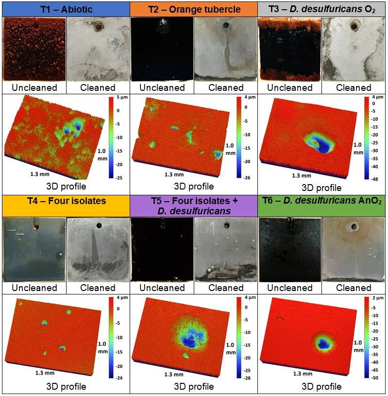
Corros. Mater. Degrad. 2021, 2                                                                                                     136

      Table 1. Relative abundance of microbes inoculated in laboratory corrosion tests and incubated at 25 ◦ C in aerobic or
      anaerobic conditions.

  Test (Incubation Atmosphere)               Brief Description                               Microbes (cfu/mL)
             T1 (aerobic)                       No inoculum                                            None
                                                                            Diverse consortium from an orange tubercle on steel
             T2 (aerobic)                     Orange tubercle
                                                                                               sheet piling (≥105 )
             T3 (aerobic)                      D. desulfuricans                            D. desulfuricans (2 × 10⁶ )
                                                                                 B. aquimaris (9 × 10⁵ ), H. korlensis (3 × 10⁵ ),
             T4 (aerobic)                   Four marine isolates
                                                                               S. pontiacus (3 × 10⁶ ), P. bellariivorans (7 × 10⁴ )
                                                                              D. desulfuricans (2 × 10⁶ ), B. aquimaris (9 × 10⁵ ),
                                           Four marine isolates +
             T5 (aerobic)                                                        H. korlensis (3 × 10⁵ ), S. pontiacus (3 × 10⁶ ),
                                              D. desulfuricans
                                                                                           P. bellariivorans (7 × 10⁴ )
            T6 (anaerobic)                     D. desulfuricans                            D. desulfuricans (2 × 10⁶ )

                                 2.3. Corrosion Tests
                                       The duration of the corrosion tests was 8 weeks. Testing six replicates allowed a range
                                 of different studies to be performed using separate coupons. Periodic photos of test bottles
                                 were taken to record any changes in media or test coupons.
                                       A total of 40 mL of fresh sterile MB 12 was added into each test bottle fortnightly (i.e.,
                                 fed-batch culture) to help maintain microbial viability during the study period. Sampling
                                 of the corrosion tests for bacterial counts was undertaken at weeks 2, 4 and 8 of incubation.
                                 This involved pipetting 10 mL of the culture (5 mL from near the test medium surface
                                 and 5 mL near the bottom of the medium) before nutrient amendment. Bacterial counts
                                 were obtained using MA plates for aerobic (T1–T5, Table 1) and TSA+ plates for anaerobic
                                 (T6, Table 1) corrosion test using serial ten-fold dilutions and duplicate plating. pH was
                                 measured at the beginning of incubation and then after 4 and 8 weeks of incubation.
                                 Aliquots of liquid cultures were stored in −20 ◦ C for use in subsequent microbial analyses.
                                       After 8 weeks of incubation, all coupons were removed from the test bottles. Shortly
                                 after removal, a gentle stream of sterile MilliQ water was run over the coupons to remove
                                 any planktonic cells from the biofilms. Pictures of the coupons were then taken. Certain
                                 coupon biofilms/corrosion products were examined by scanning electron microscopy
                                 (SEM), X-ray powder diffractometery (XRD) or energy-dispersive X-ray spectroscopy
                                 (EDS). For these, sterile MilliQ water was used to gently wash the coupons three times,
                                 after which they were air-dried in a physical containment level 2 cabinet for 2 h and
                                 then stored in a desiccator. For SEM of coupon biofilms, one coupon per test condition
                                 underwent fixation with glutaraldehyde solution (2.5 vol %) then dehydration via an
                                 ethanol series (25%, 50%, 75%, 90% and 100%) [40]. Coupons for steel surface analyses
                                 (SEM, 3D optical profilometery, weight loss), underwent a series of cleaning steps to remove
                                 biofilms/corrosion products. Individual coupons were sonicated in sterile MilliQ water for
                                 2 min (the fluid from this was stored at −20 ◦ C for subsequent microbial analyses), then in
                                 Clark’s solution [41] for 2 min, followed by rinsing with water and ethanol, drying under
                                 warm airflow and then storing in a desiccator.

                                 2.4. Metal Coupon and Microbial Analysis
                                       SEM (Zeiss SUPRA 40VP-25-38, Oberkochen, Germany) images were taken of the
                                 biofilms and the steel surfaces of separate fixed and cleaned coupons for each of the
                                 test conditions using a range of magnifications (150× to 12,000×). One coupon from
                                 each of the six treatments was used for EDS (Zeiss SUPRA 40VP-25-38, Oberkochen,
                                 Germany and INCA software, version 4.13) to provide semi-quantitative analyses of the
                                 biofilms/corrosion products formed on the coupons. Intensity with weight percent (wt.%)
                                 of chemical elements was averaged from two locations of the biofilm on each coupon
                                 surface. One uncleaned coupon from each of the six treatments was used for XRD (Bruker
                                 D8 Advance, Germany). Compositions of surface products were scanned with a copper line
                                 focus X-ray tube producing Kα radiation from a generator operating at 40 kV and 30 mA.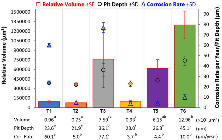
Corros. Mater. Degrad. 2021, 2                                                                                           137

                                      Three cleaned coupons from each of the six treatments were used to obtain average
                                 corrosion rates via weight loss using the method described in reference [41]. These coupons
                                 were also used for 3D optical profiling (Bruker Contour GT-K1) to obtain quantitative
                                 information about corrosion morphology. Each metal coupon was scanned over the entire
                                 surface (using 5× objective), and any areas with localised pitting were subjected to more
                                 detailed, higher resolution analysis. Pit depth (µm) and relative volume (µm3 ) for pits
                                 greater than 10 µm depth were recorded and analysed using VISION 64™ software (Bruker),
                                 and then pit density was calculated by number of pit/mm2 .
                                      For corrosion morphology analysis, the pitting depth, relative pit volume and cor-
                                 rosion rates were statistically compared for significant differences among the treatments
                                 (p < 0.05) using one-way ANOVA and Tukey’s post-hoc test (IBM® SPSS™ Statistics ver-
                                 sion 25).
                                      Microbial analysis was based on agar plate counts and 16S rRNA gene metabarcoding.
                                 Relative abundance was determined by a sum of the aerobic and anaerobic counts from the
                                 liquid in each treatment. For metabarcoding, liquid samples of T2, T4 and T5 experiments
                                 (Table 1) obtained at week 2 and week 8, together with biofilm suspensions obtained
                                 following sonication from week 8 samples were sent to a commercial test facility (AGRF,
                                 Adelaide, Australia) for DNA extraction and Illumina MiSeq sequencing. The primer pairs
                                 used were 341F (CCTAYGGGRBGCASCAG) and 806R (GGACTACNNGGGTATCTAAT)
                                 for amplification of the V3–V4 region of 16S rRNA genes. Data were processed using
                                 Quantitative Insights Into Microbial Ecology (QIIME 1.9) followed by METAGENassist [36].

                                 3. Results
                                 3.1. Surfaces of Coupons Before Cleaning
                                        Photos of coupon surfaces were taken directly after removal from the test solution
                                 at the end of the 8-week immersion period (see Figure 1). The T1 and T3 test condition
                                 coupons had orange and black corrosion products on the surfaces, the T4 coupons were rel-
                                 atively free of corrosion products and black films were present on the remaining treatments
                                 (i.e., T2, T5 and T6).
                                        The surfaces of individual test replicate coupons set aside for biofilm/corrosion
                                 product examination were observed using the SEM (Figure 2), and analysed by EDS
                                 (Figure 3). The coupon from the uninoculated test (T1) was covered in a general corrosion
                                 product, which appears to be a form of iron oxide. The images of the coupon exposed
                                 to the orange tubercle biofilm inoculum (T2) showed a biofilm present, with phosphorus
                                 and a relatively high sulfur content detected by the EDS (Figure 3). The coupon from the
                                 D. desulfuricans inoculum under aerobic incubation (T3) visually had a thicker biofilm on
                                 the surface compared to the coupon from the D. desulfuricans inoculum under anaerobic
                                 incubation (T6) (Figure 2). The biofilm of the T3 coupon also had much more sulfur present
                                 (relative to iron) compared to the T6 coupon, according to EDS (Figure 3). For the defined
                                 mixed microbial species tests (T4 and T5), the consortia including D. desulfuricans (T5)
                                 visually had a much denser biofilm compared to the sparse biofilm of T4 (Figure 2). No
                                 sulfur was detected in the mixed consortia without D. desulfuricans (T4) (Figure 3), while a
                                 small amount of sulfur in the biofilm was detected in the defined microbial consortia when
                                 the D. desulfuricans were present (T5) (Figure 3). The iron:sulfur ratios shown in Figure 3
                                 provide an indication of the relative amount of sulfur detected in surface product analysis.
Corros. Mater. Degrad. 2021, 2 138
Corros. Mater. Degrad. 2021, 2, FOR PEER REVIEW 6
Figure 1.
Figure 1. Photos
Photos of
of uncleaned
uncleanedand
andcleaned
cleanedcoupons
couponsafter
after8-weeks
8-weeksimmersion
immersionininhalf-strength
half-strength ma-
marine
Corros. Mater. Degrad. 2021, 2, FOR PEER
rineREVIEW
broth for the six different microbial testing conditions, together with 3D surface profiles of7
broth for the six different microbial testing conditions, together with 3D surface profiles of cleaned
cleaned coupons showing surface and pit morphology.
coupons showing surface and pit morphology.
The surfaces of individual test replicate coupons set aside for biofilm/corrosion prod-
uct examination were observed using the SEM (Figure 2), and analysed by EDS (Figure
3). The coupon from the uninoculated test (T1) was covered in a general corrosion prod-
uct, which appears to be a form of iron oxide. The images of the coupon exposed to the
orange tubercle biofilm inoculum (T2) showed a biofilm present, with phosphorus and a
relatively high sulfur content detected by the EDS (Figure 3). The coupon from the D.
desulfuricans inoculum under aerobic incubation (T3) visually had a thicker biofilm on the
surface compared to the coupon from the D. desulfuricans inoculum under anaerobic in-
cubation (T6) (Figure 2). The biofilm of the T3 coupon also had much more sulfur present
(relative to iron) compared to the T6 coupon, according to EDS (Figure 3). For the defined
mixed microbial species tests (T4 and T5), the consortia including D. desulfuricans (T5)
visually had a much denser biofilm compared to the sparse biofilm of T4 (Figure 2). No
sulfur was detected in the mixed consortia without D. desulfuricans (T4) (Figure 3), while
a small amount of sulfur in the biofilm was detected in the defined microbial consortia
when the D. desulfuricans were present (T5) (Figure 3). The iron:sulfur ratios shown in
Figure 3 provide an indication of the relative amount of sulfur detected in surface product
Figure 2. SEM
SEM images
images of analysis.
of biofilms/corrosion
biofilms/corrosion products
Figure 2. products on
on surfaces
surfaces of
of coupons
coupons tested
tested after
after 8-weeks
8-weeks incubation
incubation in
in the six
the six
different test conditions. All of these coupons underwent fixation (dehydration and 2% glutaraldehyde immersion) prior
different test conditions. All of these coupons underwent fixation (dehydration and 2% glutaraldehyde immersion) prior
to imaging.
to imaging.Corros.Figure
Mater. Degrad.
2. SEM2021, 2
images of biofilms/corrosion products on surfaces of coupons tested after 8-weeks incubation in the six 139
different test conditions. All of these coupons underwent fixation (dehydration and 2% glutaraldehyde immersion) prior
to imaging.
Figure 3. EDS
Figure 3. EDS spectra
spectra of
of the
the biofilms/corrosion
biofilms/corrosion products
products formed
formed on
on DH36
DH36 coupons
coupons tested
tested in
in the
the six
six different
different treatments
treatments
after 8-week incubation.
after 8-week incubation.
The XRD spectra peaks of individual phases were identified by comparing the diffrac-
tion pattern to a known standard from the diffraction powder database of the International
Centre for Diffraction Data (Diffrac Eva software, version 4.1). Unfortunately, the XRD
spectra obtained (Figure S1) provided ambiguous results for corrosion products/biofilms,
with the three peaks observed in each scan matching that of iron. It is possible that the
intensity of iron dominated the scanning spectra or that the corrosion products formed
were amorphous in nature.
3.2. Corrosion Evaluation
Example photos of the surfaces of test coupons after removing any biofilms and
corrosion products are shown in Figure 1. The key difference observed was that while
the uninoculated samples (T1) showed signs of general corrosion and occasional pitting,
most of the coupons tested in different biotic conditions appeared to suffer from sparse
localised pitting corrosion. SEM scans of the cleaned surfaces (Figure 4) confirmed that the
uninoculated control (T1) coupon had a combination of general and localised corrosion
attack, as did the coupon tested in the D. desulfuricans aerobic test configuration (T3). The
coupons from the other tests (T2, T4, T5 and T6) contained localised corrosion but were
relatively free from general corrosion. Finally, the diameters of pits observed for coupons
tested with D. desulfuricans (T3, T5 and T6) appeared to be generally larger than those for
the other test configurations.ised pitting corrosion. SEM scans of the cleaned surfaces (Figure 4) confirmed that the
uninoculated control (T1) coupon had a combination of general and localised corrosion
attack, as did the coupon tested in the D. desulfuricans aerobic test configuration (T3). The
coupons from the other tests (T2, T4, T5 and T6) contained localised corrosion but were
Corros. Mater. Degrad. 2021, 2 relatively free from general corrosion. Finally, the diameters of pits observed for coupons 140
tested with D. desulfuricans (T3, T5 and T6) appeared to be generally larger than those for
the other test configurations.
Figure
Figure 4. 4.
SEMSEM imagesofofsurfaces
images surfacesofofcleaned
cleanedtest
test coupons
coupons after
after 8-week
8-weekimmersion
immersionininthe
thesix
sixdifferent test
different conditions.
test conditions.
Tofurther
To furthercharacterise
characterisethethemorphology
morphologyofoflocalised
localisedcorrosion,
corrosion,3D 3Doptical
opticalprofiling
profilingwas
performed on one side of one of the cleaned coupons for each test condition (see(see
was performed on one side of one of the cleaned coupons for each test condition ex-
examples
amples in Figure 1). The pit density, which was calculated for all treatments
in Figure 1). The pit density, which was calculated for all treatments based on the number based on the
ofnumber of pits
µm)(>10
perµm)
mmper mm2~2 , was ~2 (pit/mm
2 , was 2 ) for2) for T1, (uninoculated treatment), and
pits (>10 (pit/mm T1, (uninoculated treatment), and T2,
T4 and T5 (mixed consortia inoculum), and ~1 (pit/mm2))for
T2, T4 and T5 (mixed consortia inoculum), and ~1 (pit/mm forT3
T3and
andT6T6(D.(D.
desulfuricans
2
desulfuricans
alone). The coupons with the largest pits were from tests containing
alone). The coupons with the largest pits were from tests containing D. desulfuricans D. desulfuricans (T3,(T3,
T5 and T6, see details in Figure 5). Apart from the coupon from T4
T5 and T6, see details in Figure 5). Apart from the coupon from T4 (four isolates) which(four isolates) which
had one pit deeper than 30 µm, only the coupons from tests with D. desulfuricans (T3 and
had one pit deeper than 30 µm, only the coupons from tests with D. desulfuricans (T3 and
T6) had any pits greater than 30 µm depth (five and six pits, respectively). The deepest pit
T6) had any pits greater than 30 µm depth (five and six pits, respectively). The deepest pit
found was 50.2 µm for the coupon tested in D. desulfuricans under anaerobic conditions
found was 50.2 µm for the coupon tested in D. desulfuricans under anaerobic conditions
(T6), compared to 27 µm for the deepest pit for the uninoculated control (T1). The average
(T6), compared to 27 µm for the deepest pit for the uninoculated control (T1). The average
pitREVIEW
Corros. Mater. Degrad. 2021, 2, FOR PEER
volumes (of five deepest pits) were nearly an order of magnitude greater for the tests
pit volumes (of five deepest pits) were nearly an order of magnitude greater for the9tests
containing D. desulfuricans (T3, T5 and T6) compared to the tests lacking D. desulfuricans.
containing D. desulfuricans (T3, T5 and T6) compared to the tests lacking D. desulfuricans.
Figure 5.
Figure 5. Summary
Summary of of
thethe
corrosion attack
corrosion on steel
attack onsamples from thefrom
steel samples six different
the sixtreatments. Av-
different treatments.
erages of 5 highest values of relative volume and pit depth as well as average corrosion rate are
Averages of 5 highest values of relative volume and pit depth as well as average corrosion rate are
shown with statistical analyses. Significant differences (p < 0.05) were shown by different series of
shown withletters
superscript statistical analyses.
on each Significant differences (p < 0.05) were shown by different series of
parameter.
superscript letters on each parameter.
The average corrosion rates of the DH36 coupons after 8 weeks of incubation in the
different test scenarios were determined via mass loss (Figure 5). The highest corrosion
rates among the six treatments obtained (p < 0.05) were for the T3 (D. desulfuricans tested
aerobically) and T1 (uninoculated) test conditions. For all the other test conditions veryCorros. Mater. Degrad. 2021, 2 141
The average corrosion rates of the DH36 coupons after 8 weeks of incubation in the
different test scenarios were determined via mass loss (Figure 5). The highest corrosion
rates among the six treatments obtained (p < 0.05) were for the T3 (D. desulfuricans tested
aerobically) and T1 (uninoculated) test conditions. For all the other test conditions very low
average corrosion rates were measured; always much less than the uninoculated control,
indicating a form of corrosion inhibition. Given that the localised attack of the uninoculated
control (T1) was relatively minor, the relatively high weight loss for this test condition will
have been dominated by general/uniform corrosion.
3.3. Test Solutions
Over the 8-week duration of the corrosion tests, the colours of the test solutions
changed (see Figure 6), which is a potential indication of the metabolic and corrosion
processes that took place. Of particular interest was the general blackening of solutions
for tests T2 (orange tubercle inoculum), T5 (four isolates + D. desulfuricans aerobic) and T6
(D. desulfuricans anaerobic), and the black precipitation layer observed at the bottom of test
Corros. Mater. Degrad. 2021, 2, FOR PEER REVIEW
condition T3 (D. desulfuricans aerobic). This blackening might be an indication of sulfate10
reduction and FeS production.
Figure6.6.Changes
Figure Changesininappearance
appearanceofoftest
testbottles
bottlesofofsix
sixtreatments
treatmentsduring
during8 8weeks
weeksofofimmersion.
immersion.
InIngeneral,
general,thethepHpHvalues
valuesofofthe
thesolutions
solutions(Table
(Table2)2)were
werereasonably
reasonablyconstant
constantforforall
all
ofofthe
thetest
testconditions
conditionsoveroverthe
the8-week
8-weekincubation
incubationperiod,
period,with
withpH
pHvalues
valuesstaying
stayingwithin
within
±±0.5
0.5 of
of the
the initial
initial value
value ofof7.54.
7.54.The
Thegreatest
greatestchanges
changesobserved
observedwere
werefor forT4,
T4,which
whichwent
went
slightly
slightlymore
morebasic
basictoto7.9,
7.9,and
andT6T6which
whichwent
wentslightly
slightlymore
moreacidic
acidictotojust
justbelow
below7.0.
7.0.
Table 2. Bacterial counts and pH during 8-week incubation test of six treatments (Table 1). Initial pH of sterile media
before inoculation was ~7.54. Total plate count in each treatment was the sum of both aerobic and anaerobic counts on
marine agar (MA) and tryptic soy agar (TSA+) media, respectively.
Total Bacterial Plate Count (cfu/mL) pH MeasurementCorros. Mater. Degrad. 2021, 2 142
Table 2. Bacterial counts and pH during 8-week incubation test of six treatments (Table 1). Initial pH of sterile media before
inoculation was ~7.54. Total plate count in each treatment was the sum of both aerobic and anaerobic counts on marine agar
(MA) and tryptic soy agar (TSA+ ) media, respectively.
Total Bacterial Plate Count (cfu/mL) pH Measurement
Test (T1–T5: O2 ; T6: AnO2 )
Initial Week 2 Week 4 Week 8 Week 4 Week 8
T1 (uninoculated) – – – – 7.58 ± 0.02 7.55 ± 0.02
T2 (orange tubercle) ≥105 107 –108 1 × 10⁸ 108 –109 7.47 ± 0.03 7.32 ± 0.06
T3 (D. desulfuricans) 2 × 10⁶ 7 × 10⁵ 2 × 10⁶ 103 –104 7.56 ± 0.04 7.73 ± 0.10
T4 (Four isolates) 4 × 10⁶ 4 × 10⁷ 6 × 10⁷ 9 × 10⁷ 7.74 ± 0.05 7.90 ± 0.06
T5 (Four isolates +
6 × 10⁶ 3 × 10⁷ 1 × 10⁸ 106 –107 7.32 ± 0.04 7.42 ± 0.06
D. desulfuricans)
T6 (D. desulfuricans) 2× 10⁶ 5× 10⁷ 5× 10⁷ 104 –105 6.98 ± 0.10 6.98 ± 0.09
3.4. Analysis of Microbial Populations
Total plate count results are provided in Table 2. While a reduction in counts for the
D. desulfuricans inoculum tests (T3 and T6) was seen between the start and the end of the
tests, in general the microbial populations were reasonably constant throughout incubation
times. It is also worth noting that no colonies were observed in the aerobic plate tests for
D. desulfuricans alone treatments (T3 and T6), suggesting that these solutions were free of
any aerobic microbial contamination.
Figure 7 summarises the metabarcoding analysis of the mixed microbial consortium
treatments (i.e., T2, T4 and T5) at week 2 (planktonic) and week 8 (planktonic and biofilm).
The solution inoculated with the orange tubercle (T2) has, as expected, the most diverse
community. While there were few bacterial taxa that reduced in relative abundance in the
T2 solution during the test (e.g., Exiguobacterium spp. dropped from 13% to 3% in planktonic
phase from 2 to 8 weeks), many bacteria maintained their presence in both planktonic
and biofilm phases (e.g., Clostridiales and Rhizobiales). However, several bacteria were
only detected after 8 weeks including Desulfarculus baarsii, Desulfosporosinus spp. and
Thioalkalivibrio sp. Reasonable differences can also be seen between the planktonic and
biofilm phases of the T2 solution at 8 weeks (e.g., Rhodobacteraceae spp. and Clostridium
thiosulfatireducens more abundant in the biofilm phase).
The metabarcoding results for the combinations of isolates (T4 and T5) identified all
of the bacteria inoculated in each test configuration in at least one of the time/phases
tested. For both of these test configurations the proportion of P. bellariivorans tended to
decrease from week 2 to week 8, and H. korlensis was only ever found in any reasonable
level in the biofilm phase. A key difference between the two test configurations was for
B. aquimaris, which had a strong presence in the T4 liquid (four isolates) but was only
detected at low levels in the T5 liquid (four isolates + D. desulfuricans). S. pontiacus was
detected at reasonably consistent levels for both test solution types throughout the tests
and in both the planktonic and biofilm phases.
The metabarcoding data were further analysed for putative functions using METAGE-
Nassist to interpret microbial metabolism and other traits, the results of which are presented
in Table S2. Key aspects of the sulfur cycle, including sulfate reduction (detected at high
levels in all samples at week 8) and sulfur oxidation (for the orange tubercle configuration
at week 8) were detected.T2 solution during the test (e.g., Exiguobacterium spp. dropped from 13% to 3% in plank-
tonic phase from 2 to 8 weeks), many bacteria maintained their presence in both plank-
tonic and biofilm phases (e.g., Clostridiales and Rhizobiales). However, several bacteria
were only detected after 8 weeks including Desulfarculus baarsii, Desulfosporosinus spp. and
Corros. Mater. Degrad. 2021, 2 Thioalkalivibrio sp. Reasonable differences can also be seen between the planktonic and 143
biofilm phases of the T2 solution at 8 weeks (e.g., Rhodobacteraceae spp. and Clostridium
thiosulfatireducens more abundant in the biofilm phase).
Figure
Figure 7. Microbial
7. Microbial communitiesidentified
communities identifiedby
by16S
16S rRNA
rRNA gene
gene metabarcoding
metabarcodingofofmixed
mixedconsortium
consortiumtreatments (T2,
treatments T4 T4
(T2, andand
T5), for planktonic communities (at weeks 2 and 8) and for the attached biofilms obtained by sonication (at week 8).
T5), for planktonic communities (at weeks 2 and 8) and for the attached biofilms obtained by sonication (at week 8).
The metabarcoding results for the combinations of isolates (T4 and T5) identified all
4. Discussion
of the bacteria
4.1. Surfaces inoculated
of Coupons in Cleaning
Before each test configuration in at least one of the time/phases
tested. For both of these test configurations the proportion of P. bellariivorans tended to
The black
decrease from corrosion products
week 2 to week onH.
8, and thekorlensis
surfaceswasand sulfur
only everdetected
found ininanythereasonable
biofilms (by
EDS)
levelof
in coupons
the biofilmfrom several
phase. A keytreatments
difference(T2, T3, T5the
between and
two T6) indicates
test iron sulfide
configurations produc-
was for B.
tion, and active sulfate reduction. This matches well with the presence
aquimaris, which had a strong presence in the T4 liquid (four isolates) but was only de-of D. desulfuricans,
when
tectedspecifically added
at low levels to T5
in the theliquid
tests (i.e.,
(fourT3, T5 and
isolates T6)desulfuricans).
+ D. and the relatively high potential
S. pontiacus was de- for
sulfate
tected at reasonably consistent levels for both test solution types throughout the teststubercle
reduction determined from the METAGENassist analysis of the T2 orange and
inoculum.
in both theThe iron:sulfur
planktonic ratios calculated
and biofilm phases. for the biofilms indicated that the highest to
lowestTherelative amounts of
metabarcoding sulfur
data weredetected were for T3
further analysed for >putative
T2 > T6functions
> T5, which
usingqualitatively
META-
matches the thicknesses of the biofilms observed in SEM images.
GENassist to interpret microbial metabolism and other traits, the results of which are pre-
Oneinsomewhat
sented unexpected
Table S2. Key aspects ofobservation was that
the sulfur cycle, the nominally
including aerobic test
sulfate reduction with the
(detected
D. desulfuricans (T3) produced a visually thicker biofilm than that for the corresponding
anaerobic test with D. desulfuricans (T6). While they are more commonly known as strict
anaerobes, many sulfate-reducing bacteria, including the culture collection strain used in
this work, can survive/grow in aerobic environments [42,43]. It is also possible that the
visually thicker biofilm observed for the D. desulfuricans aerobic incubation (T3) is a result
of a stress response by the bacteria. Although the aerobic tests (including the T3 treatment)
were nominally aerobic at the start of the tests it is likely that the oxygen concentration
in the solutions dropped over time due to microbial oxygen consumption, making the
environment more suitable for optimal growth of D. desulfuricans. A suggestion for future
work would be to monitor the oxygen concentration in the solutions as a function of time.
4.2. Corrosion Attack
Coupon weight loss indicates that T1 (uninoculated) and T3 (D. desulfuricans aerobic)
treatments demonstrated greater general corrosion attack than the other treatments. How-
ever, T6 (D. desulfuricans anaerobic), T3 (D. desulfuricans aerobic) and T5 (four isolates + D.
desulfuricans aerobic) treatments had significantly greater localised pitting than the other
treatments. The pitting results for D. desulfuricans anaerobic incubation (T3) (average pit
depth 45 µm) were greater than for previous tests for the same bacterial strain using a mod-
ified Baar’s medium (average pit depth ~27 µm) with the same steel type [32]. However,Corros. Mater. Degrad. 2021, 2 144
the corresponding weight loss data for the previous modified Baar’s medium tests with
D. desulfuricans were much greater (~80 µm/year) than obtained in this work (10 µm/year).
It is important to note that in addition to pitting, general corrosion of the steel surface was
observed in the previous study. This indicates that test medium used can have an impact
on steel corrosion and pitting outcomes. Previous work indicated that the addition of Fe
ions/lactate to modified Baar’s medium can have a significant effect on the weight loss
results, which are indicative of any general corrosion taking place [40]. No addition of Fe
ions to the bulk test solutions was performed in the current work.
The main hypothesis for the work was that a consortium of microbes would result in
greater corrosion rates than single isolates (T3 and T6) or an uninoculated control (T1). This
was because a range of phenotypes were considered important in promoting ALWC. The
consortium tests (T2, T4 and T5) actually showed the three lowest weight losses (typically
indicative of general corrosion) of all of the tests performed. Microbial corrosion (including
ALWC), however, is more often linked to localised rather than general/uniform corrosion.
In relation to localised corrosion, the average pit depths were smaller for the consortia
treatments (T2, T4 and T5) than for sole D. desulfuricans tests (T3 and T6). The volume of pits
for the T5 treatment (four isolates + D. desulfuricans) were similar to the sole D. desulfuricans
tests (T3 and T6), which were much greater than for any of the other test treatments.
One possible reason for the lower levels of corrosion obtained for the mixed microbial
communities compared to the uninoculated control or single D. desulfuricans tests was
that the types of microbes present (orange tubercle, T2) or chosen (pure cultures) were
inappropriate for ALWC. The orange tubercle inoculum (T2), was taken from a site which
had pitting corrosion and diagnosed ALWC beneath the orange tubercle. Thus, this orange
tubercle consortium should have been appropriate to generate optimal ALWC conditions
in our laboratory tests. Indeed, we previously found increased corrosion when orange
tubercles (taken from another site with suspected ALWC) were used as inocula in laboratory
tests, compared to an uninoculated control test [25]. The metabarcoding analysis of T2
identified several microbes present in both the biofilm and planktonic phases that were
capable of sulfate reduction (more detail below) and sulfur was clearly identified in the
biofilm by EDS.
High corrosion rates of ~100–125 µm/year (from weight loss) were previously [25]
found for tests with orange tubercle inocula compared to ~40 µm/year in uninoculated
controls when using a similar test arrangement (e.g., steel type, test duration) to that used
in the current work. These previous higher corrosion rates were observed for samples with
uniform corrosion across sample surfaces and with little localised corrosion. Two obvious
key differences between the previous [25] and current studies were the initial microbial
community structure of the inocula and the types of nutrients added to the test solutions.
While a Deltaproteobacteria SRB (Desulfarculus baarsii) was detected in the orange tubercle
inoculum (T2) test, this strain has not typically been linked to corrosion. In the previous
study [25], an unidentified species from the Desulfobulbaceae family of Deltaproteobac-
teria was found at reasonably high levels (up to ~40%) in the planktonic phase. This
family includes Desulfopila corrodens which was directly linked to rapid corrosion in ALWC
studies [23].
Glucose (a carbon and electron donor source) and yeast extract were components of
the earlier test medium [25], while peptone and yeast extract were present in relatively
low amounts in the current work’s medium. It has been shown that the types of nutrients
available for microbial growth and the physicochemical conditions can have substantial
effects on the composition of microbial communities that ensue (e.g., [44,45]). These two
features likely explain the microbial differences observed between the earlier [25] and the
current research. Additionally, changes in the level of carbon sources present in test media
can affect the extent of corrosion caused by a single SRB strain (e.g., [29]).
The majority of microbes chosen for the defined consortium tests survived in rea-
sonable numbers throughout the test period, although there were some clear changes in
the microbial species composition over the test duration (discussed in more detail below).Corros. Mater. Degrad. 2021, 2 145
There was also clear evidence of sulfate reduction, as seen by the black solution and pres-
ence of sulfur in the biofilm for the defined mixed microbial treatment, which included
D. desulfuricans (T5), indicating that D. desulfuricans were metabolically active. As discussed
above, it is possible that relatively low levels of carbon sources/electron donors in the test
media may have altered physicochemical processes (i.e., sulfate reduction) that play a role
in microbial corrosion and had an effect on corrosion rates.
There have been varying reports on the effect of the presence of different species of
Bacillus on corrosion with some indicating increased corrosion [46–48] and others showing
corrosion inhibition [49–53]. Both defined mixed microbial treatments, with (T5) or without
(T4) D. desulfuricans, had similar levels of general corrosion but qualitatively different
biofilm thicknesses, where T5 was greater than T4 (Figures 1 and 2). The 16S rRNA gene
metabarcoding showed very different relative amounts of B. aquimaris. T5 had a lower
general corrosion level than either of the tests with D. desulfuricans alone (T3 and T6),
indicating that the additional microbes reduced the corrosion mediated by D. desulfuricans.
This could indicate corrosion inhibition, perhaps due to competition for substrates among
the added pure cultures.
4.3. Analysis of Microbial Populations
Although the D. desulfuricans strain used is a facultative anaerobe, plate counts showed
that there were still reasonable numbers (103 –104 cfu/mL) of viable D. desulfuricans present
in solutions after 8 weeks when tested in nominally aerobic test conditions. The presence of
sulfur in the biofilms of the coupons from these tests (e.g., T3) indicated that D. desulfuricans
were metabolically active despite the possible presence of oxygen in the bulk solution.
Metabarcoding analysis of the planktonic and biofilm phases of the defined consortium
including D. desulfuricans (T5) showed a relatively high abundance of D. desulfuricans
throughout this nominally aerobic test. Given the specific inclusion of aerobic microbes
in T5 it is highly likely that they used the available oxygen creating anaerobic conditions,
which are optimal for the growth of D. desulfuricans. This is important as it shows how test-
ing with microbial consortia can produce an environment conducive to anaerobic microbes
such as D. desulfuricans in a more natural way rather than by exogenously producing low
oxygen levels by means such as nitrogen purging. There have been some reports on the
potentially important role of oxygen in MIC of SRB [22,54–57] and of other microbes [58,59].
In any case, results from the current work support further studies on this topic.
The microbial consortium composition in the orange tubercle inoculum test (T2)
differed over time in the planktonic phase (between 2 weeks and 8 weeks) and also between
the planktonic and biofilm phases at 8 weeks. This is expected as the environmental
conditions in the laboratory test was quite different to the tubercle’s native location. The
nature of the community succession will be different in the planktonic and biofilm phases
as there will be local differences in oxygen content and corrosion products from the carbon
steel could act as a nutrient source for certain microbes [6,60].
At 8 weeks incubation both planktonic and biofilm phases in T2 contained microbes
potentially capable of sulfide/sulfur oxidation (e.g., Thioalkalivibrio sp.) and sulfate reduc-
tion (e.g., Desulfosporosinus spp. and D. baarsii). A combination of both sulfur oxidisers
and sulfate reducers could form a closed sulfur cycle, which has previously been reported
to potentially lead to rapid corrosion [5,8,13–17]. However, extensive corrosion was not
seen in T2. It is possible that the system may have needed more time to develop an active
corrosion state. Although strains of the Deltaproteobacteria (including D. desulfuricans) are
the most commonly regarded sulfate reducers, other microbial groups also are capable for
sulfate reduction. These include Desulfosporosinus spp., which is a member of the Firmicutes
phylum. Various microbes relevant to the nitrogen cycle (e.g., nitrogen fixation, nitrite re-
duction and ammonia oxidation) were identified in the liquid and biofilm of T2 at 8 weeks.
Nitrifying bacteria are important as they can potentially produce fixed nitrogen that is
used to support/maintain the growth of anaerobic bacteria such as SRB [14]. However,
conditions would need to be aerobic to support their nitrifying phenotype.Corros. Mater. Degrad. 2021, 2 146
A number of interesting spatio-temporal changes were observed in the microbial com-
munities of the defined consortia tests (T4 without and T5 with D. desulfuricans). Although
H. korlensis comprised a reasonable proportion of the initial inoculum in both T4 and T5
(7% and 5%, respectively), this bacterium was only detected in relatively small relative
abundances in the biofilm phase at week 8 (Corros. Mater. Degrad. 2021, 2 147
5. Conclusions
The aim of this study was to investigate laboratory tests that mimic the complex com-
munities present in real world cases of MIC/ALWC. As part of this work, a model SRB, D.
desulfuricans, was used throughout an 8-week corrosion study. The general corrosion rates
of many of the mixed microbial test configurations were lower than that of an uninoculated
control. Despite that the three mixed microbial tests (T2, T4 and T5) were nominally aerobic,
it is likely the test solutions became anaerobic due to oxygen use by aerobic microbes in
the milieu which may have been responsible for the reduced general/uniform corrosion.
Greater localised corrosion was observed for all of the tests that included D. desulfuricans.
However, the defined mixed community with D. desulfuricans did not lead to increased
corrosion compared to tests with D. desulfuricans alone. The composition of the mixed
microbial communities changed over the duration of the test and key differences were
observed between the planktonic and biofilm communities. More laboratory-based experi-
mental work is required to determine optimal microbial communities and test conditions
for ALWC studies. Finally, the metabolomic or transcriptomic aspects relevant to the
microbe/corrosion processes should be considered for a further study. It is likely that such
studies would give further useful insights into MIC and ALWC.
Supplementary Materials: The following are available online at https://www.mdpi.com/2624-5
558/2/2/8/s1, Table S1: Summary of example MIC studies using defined multispecies microbial
consortia (WL—weight loss test method, CR—corrosion rate), Table S2: Putative metabolic activities
of microbes present in metabarcoding profiles identified using METAGENassist, Figure S1: XRD
scans of products formed on DH36 coupons in six treatments at 8 weeks. The three highest peaks
observed match to the Fe component of the carbon steel coupons used.
Author Contributions: Conceptualisation, all authors; methodology, all authors; validation, all
authors; formal analysis, H.C.P.; investigation, H.C.P.; writing—original draft preparation, H.C.P.;
writing—review and editing, all authors; visualization, H.C.P.; supervision, S.A.W. and L.L.B. All
authors have read and agreed to the published version of the manuscript.
Funding: This research received no external funding.
Data Availability Statement: The data presented in this study are available on request from the cor-
responding author. The microbial sequencing data is planned to be deposited in the NCBI database.
Acknowledgments: We would like to thank Melanie Thomson (previously of Deakin University) for
providing the D. desulfuricans used in this study.
Conflicts of Interest: The authors declare no conflict of interest.
References
1. Ijsseling, F. General guidelines for corrosion testing of materials for marine applications: Literature review on sea water as test
environment. Br. Corros. J. 1989, 24, 53–78. [CrossRef]
2. Little, B.J.; Lee, J.S. Microbiologically influenced corrosion. In Kirk-Othmer Encyclopedia of Chemical Technology; Wiley-Interscience:
Hoboken, NJ, USA, 2009; pp. 1–38.
3. Christie, J. The effect of MIC and other corrosion mechanisms on the global ports infrastructure. In Proceedings of the MIC—An
International Perspective, Perth, Australia, 14–16 February 2007.
4. Jeffrey, R.J.; Melchers, R.E. Effect of vertical length on corrosion of steel in the tidal zone. Corrosion 2009, 65, 695–702. [CrossRef]
5. Beech, I.B.; Campbell, S.A. Accelerated low water corrosion of carbon steel in the presence of a biofilm harbouring sulphate-
reducing and sulphur-oxidising bacteria recovered from a marine sediment. Electrochim. Acta 2008, 54, 14–21. [CrossRef]
6. Dang, H.; Lovell, C.R. Microbial surface colonization and biofilm development in marine environments. Microbiol. Mol. Biol. Rev.
2016, 80, 91–138. [CrossRef] [PubMed]
7. Flemming, H.-C.; Wuertz, S. Bacteria and archaea on Earth and their abundance in biofilms. Nat. Rev. Microbiol. 2019, 17, 247–260.
[CrossRef] [PubMed]
8. Little, B.; Ray, R.; Pope, R. Relationship between corrosion and the biological sulfur cycle: A review. Corrosion 2000, 56, 433–443.
[CrossRef]
9. Malard, E.; Gueuné, H.; Fagot, A.; Lemière, A.; Sjogren, L.; Tidblad, J.; Sanchez-Amaya, J.M.; Muijzer, G.; Marty, F.; Quillet, L.; et al.
Microbiologically Induced Corrosion of Steel Structures in Port Environment: Improving Prediction and Diagnosis of ALWC (MICSIPE);
RFCS Publications: Luxembourg, 2013.Corros. Mater. Degrad. 2021, 2 148
10. Païssé, S.; Ghiglione, J.F.; Marty, F.; Abbas, B.; Gueuné, H.; Amaya, J.M.S.; Muyzer, G.; Quillet, L. Sulfate-reducing bacteria
inhabiting natural corrosion deposits from marine steel structures. Appl. Microbiol. Biotechnol. 2013, 97, 7493–7504. [CrossRef]
11. Videla, H.A.; Herrera, L.K.; Edyvean, G. An updated overview of SRB induced corrosion and protection of carbon steel. In
Proceedings of the NACE Corrosion, Houston, TX, USA, 3–7 April 2005; NACE International: Houston, TX, USA, 2005; p. 05488.
12. Enning, D.; Venzlaff, H.; Garrelfs, J.; Dinh, H.T.; Meyer, V.; Mayrhofer, K.; Hassel, A.W.; Stratmann, M.; Widdel, F. Marine
sulfate-reducing bacteria cause serious corrosion of iron under electroconductive biogenic mineral crust. Environ. Microbiol. 2012,
14, 1772–1787. [CrossRef]
13. Gehrke, T.; Sand, W. Interactions between microorganisms and physiochemical factors cause MIC of steel pilings in harbors
(ALWC). In Proceedings of the NACE Corrosion, San Diego, CA, USA, 16–20 March 2003; NACE International: Houston, TX,
USA, 2003; p. 03557.
14. Sand, W.; Gehrke, T. Microbially influenced corrosion of steel in aqueous environments. Rev. Environ. Sci. Biotechnol. 2003, 2,
169–176. [CrossRef]
15. Gubner, R.; Beech, I. Statistical assessment of the risk of the accelerated low-water corrosion in the marine environment. In Proceedings
of the NACE Corrosion, San Antonio, TX, USA, 25–30 April 1999; NACE International: Houston, TX, USA, 1999; p. 318.
16. Javadhastri, R. Microbiologically Influenced Corrosion—An Engineering Insight; Springer: London, UK, 2008; pp. 125–139.
17. Smith, M.; Bardiau, M.; Brennan, R.; Burgess, H.; Caplin, J.; Ray, S.; Urios, T. Accelerated low water corrosion: The microbial
sulfur cycle in microcosm. NPJ Mater. Degrad. 2019, 3, 37. [CrossRef]
18. Barco, R.A.; Hoffman, C.L.; Ramírez, G.A.; Toner, B.M.; Edwards, K.J.; Sylvan, J.B. In-situ incubation of iron-sulfur mineral reveals
a diverse chemolithoautotrophic community and a new biogeochemical role for Thiomicrospira. Environ. Microbiol. 2017, 19,
1322–1337. [CrossRef]
19. Celikkol-Aydin, S.; Gaylarde, C.C.; Lee, T.; Melchers, R.E.; Witt, D.L.; Beech, I.B. 16S rRNA gene profiling of planktonic and
biofilm microbial populations in the Gulf of Guinea using Illumina NGS. Mar. Environ. Res. 2016, 122, 105–112. [CrossRef]
[PubMed]
20. Dang, H.; Chen, R.; Wang, L.; Shao, S.; Dai, L.; Ye, Y.; Guo, L.; Huang, G.; Klotz, M.G. Molecular characterization of putative
biocorroding microbiota with a novel niche detection of Epsilon and Zetaproteobacteria in Pacific Ocean coastal seawaters.
Environ. Microbiol. 2011, 13, 3059–3074. [CrossRef] [PubMed]
21. Phan, H.C.; Wade, S.A.; Blackall, L.L. Is marine sediment the source of microbes associated with accelerated low water corrosion?
Appl. Microbiol. Biotechnol. 2019, 103, 449–459. [CrossRef] [PubMed]
22. Lee, J.; Ray, R.; Lemieux, E.; Falster, A.; Little, B. An evaluation of carbon steel corrosion under stagnant seawater conditions.
Biofouling 2004, 20, 237–247. [CrossRef] [PubMed]
23. Marty, F.; Gueune, H.; Malard, E.; Sánchez-Amaya, J.M.; Sjögren, L.; Abbas, B.; Quillet, L.; van Loosdrecht, M.C.M.; Muyzer, G.
Identification of key factors in accelerated low water corrosion through experimental simulation of tidal conditions: Influence of
stimulated indigenous microbiota. Biofouling 2014, 30, 281–297. [CrossRef]
24. Pillay, C.; Lin, J. Metal corrosion by aerobic bacteria isolated from stimulated corrosion systems: Effects of additional nitrate
sources. Int. Biodeter. Biodegrad. 2013, 83, 158–165. [CrossRef]
25. Wade, S.; Blackall, L. Development of a laboratory test for microbial involvement in accelerated low water corrosion. Microbiol.
Aust. 2018, 39, 170–172. [CrossRef]
26. Enning, D.; Garrelfs, J. Corrosion of iron by sulfate-reducing bacteria: New views of an old problem. Appl. Environ. Microbiol.
2014, 80, 1226–1236. [CrossRef]
27. Li, Y.; Xu, D.; Chen, C.; Li, X.; Jia, R.; Zhang, D.; Sand, W.; Wang, F.; Gu, T. Anaerobic microbiologically influenced corrosion
mechanisms interpreted using bioenergetics and bioelectrochemistry: A review. J. Mater. Sci Technol. 2018, 34, 1713–1718. [CrossRef]
28. Martin, A.; Auger, E.A.; Blum, P.H.; Schultz, J.E. Genetic basis of starvation survival in nondifferentiating bacteria. Annu. Rev.
Microbiol. 1989, 43, 293–316. [CrossRef]
29. Xu, D.; Gu, T. Carbon source starvation triggered more aggressive corrosion against carbon steel by the Desulfovibrio vulgaris
biofilm. Int. Biodeter. Biodegrad. 2014, 91, 74–81. [CrossRef]
30. Salgar-Chaparro, S.J.; Lepkova, K.; Pojtanabuntoeng, T.; Darwin, A.; Machuca, L.L. Nutrient level determines biofilm characteris-
tics and subsequent impact on microbial corrosion and biocide effectiveness. Appl. Environ. Microbiol. 2020, 86, e02885–e02919.
[CrossRef] [PubMed]
31. Wade, S.A.; Javed, M.A.; Palombo, E.A.; McArthur, S.L.; Stoddart, P.R. On the need for more realistic experimental conditions in
laboratory-based microbiologically influenced corrosion testing. Int. Biodeter. Biodegrad. 2017, 121, 97–106. [CrossRef]
32. Javed, M.; Neil, W.; McAdam, G.; Wade, S. Effect of sulphate-reducing bacteria on the microbiologically influenced corrosion of
ten different metals using constant test conditions. Int. Biodeter. Biodegrad. 2017, 125, 73–85. [CrossRef]
33. Li, H.-B.; Zhang, L.-P.; Chen, S.-F. Halomonas korlensis sp. nov., a moderately halophilic, denitrifying bacterium isolated from
saline and alkaline soil. Int. J. Syst. Evol. Microbiol. 2008, 58, 2582–2588. [CrossRef] [PubMed]
34. Mahendran, S.; Sankaralingam, S.; Shankar, T.; Vijayabaskar, P. Alkalophilic protease enzyme production from estuarine Bacillus
aquimaris. World J. Fish Mar. Sci. 2010, 2, 436–443.
35. Marques, J.M.; de Almeida, F.P.; Lins, U.; Seldin, L.; Korenblum, E. Nitrate treatment effects on bacterial community biofilm
formed on carbon steel in produced water stirred tank bioreactor. World J. Microbiol. Biotechnol. 2012, 28, 2355–2363. [CrossRef]
[PubMed]You can also read