Somatostatin 2 Receptors in the Spinal Cord Tonically Restrain Thermogenic, Cardiac and Other Sympathetic Outflows
←
→
Page content transcription
If your browser does not render page correctly, please read the page content below
ORIGINAL RESEARCH
                                                                                                                                        published: 20 February 2019
                                                                                                                                      doi: 10.3389/fnins.2019.00121

                                            Somatostatin 2 Receptors in the
                                            Spinal Cord Tonically Restrain
                                            Thermogenic, Cardiac and Other
                                            Sympathetic Outflows
                                            Belinda R. Bowman 1 , Phillip Bokiniec 1,2 , Simon McMullan 1 , Ann K. Goodchild 1* and
                                            Peter G.R. Burke 1,3
                                            1
                                             Department of Biomedical Sciences, Faculty of Medicine and Health Sciences, Macquarie University, Sydney, NSW,
                                            Australia, 2 Max Delbrück Center for Molecular Medicine, Berlin, Germany, 3 Neuroscience Research Australia, Sydney, NSW,
                                            Australia

                                            The anatomical and functional characterization of somatostatin (SST) and somatostatin
                                            receptors (SSTRs) within the spinal cord have been focused in the dorsal horn,
                                            specifically in relation to sensory afferent processing. However, SST is also present
                                            within the intermediolateral cell column (IML), which contains sympathetic preganglionic
                          Edited by:
                     Stuart Mazzone,        neurons (SPN). We investigated the distribution of SSTR2 within the thoracic spinal cord
The University of Melbourne, Australia      and show that SSTR2A and SSTR2B are expressed in the dorsal horn and on SPN and
                         Reviewed by:       non-SPN in or near the IML. The effects of activating spinal SSTR and SSTR2 were
                    Patrice G. Guyenet,
   University of Virginia, United States
                                            sympathoinhibition, hypotension, bradycardia, as well as decreases in interscapular
               Christopher J. Madden,       brown adipose tissue temperature and expired CO2 , in keeping with the well-described
  Oregon Health & Science University,
                                            inhibitory effects of activating SSTR receptors. These data indicate that spinal SST
                           United States
                     Shaun F. Morrison,     can decrease sympathetic, cardiovascular and thermogenic activities. Unexpectedly
  Oregon Health & Science University,       blockade of SSTR2 revealed that SST tonically mantains sympathetic, cardiovascular
                           United States
                                            and thermogenic functions, as activity in all measured parameters increased. In
                 *Correspondence:
                   Ann K. Goodchild
                                            addition, high doses of two antagonists evoked biphasic responses in sympathetic and
           ann.goodchild@mq.edu.au          cardiovascular outflows where the initial excitatory effects were followed by profound but
                                            transient falls in sympathetic nerve activity, heart rate and blood pressure. These latter
                   Specialty section:
         This article was submitted to
                                            effects, together with our findings that SSTR2A are expressed on GABAergic, presumed
           Autonomic Neuroscience,          interneurons, are consistent with the idea that SST2R tonically influence a diffuse spinal
               a section of the journal
                                            GABAergic network that regulates the sympathetic cardiovascular outflow. As described
            Frontiers in Neuroscience
                                            here and elsewhere the source of tonically released spinal SST may be of intra- and/or
       Received: 13 November 2018
        Accepted: 04 February 2019          supra-spinal origin.
       Published: 20 February 2019
                                            Keywords: somatostatin, spinal cord, autonomic, sympathetic, brown adipose tissue temperature
                            Citation:
             Bowman BR, Bokiniec P,
     McMullan S, Goodchild AK and
   Burke PGR (2019) Somatostatin 2
                                            INTRODUCTION
Receptors in the Spinal Cord Tonically
      Restrain Thermogenic, Cardiac
                                            Neuropeptides encoded by about 70 genes influence neuronal activity within the central nervous
    and Other Sympathetic Outflows.         system (Burbach, 2010). The inhibitory neuropeptide somatostatin (SST) is distributed widely
             Front. Neurosci. 13:121.       throughout the CNS, in its two biologically active forms, SST-14 and SST-28. Within the spinal
      doi: 10.3389/fnins.2019.00121         cord SST immunoreactive terminals are present in the dorsal horn which receives primary

Frontiers in Neuroscience | www.frontiersin.org                                   1                                           February 2019 | Volume 13 | Article 121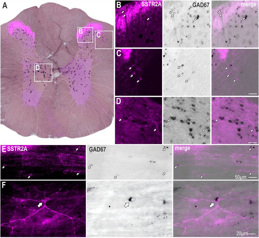
Bowman et al.                                                                                                     SSTR2 Receptors in Spinal Cord

sensory information and the intermediolateral cell column                 Anatomical Experiments
(IML), the major source of sympathetic preganglionic neurons              Immunohistochemistry
(SPN) (Krukoff, 1987; Patel, 1999; Schulz et al., 2000). SSTR1-           Immunohistochemistry was conducted as described previously
4 are present in the spinal cord (Segond von Banchet et al.,              (Bowman et al., 2013; Parker et al., 2013). Briefly, pentobarbitone
1999; Schulz et al., 2000). The spinal distribution of SST and            anesthetized rats (80 mg/kg, n = 4) were perfused transcardially
somatostatin receptor (SSTR) suggests involvement in modifying            with saline followed by 4% paraformaldehyde (PFA) in phosphate
afferent information entering the dorsal horn and the activity of         buffer (pH 7.4, fixative). Spinal cords (C8-T3 and T4-T10), placed
the SPN. Although effects at the dorsal horn are well established         in fixative overnight, then sectioned parasagittally or coronally
(Kuraishi et al., 1985; Sandkühler et al., 1990; Shi et al., 2014;        (50 µm) using a vibrating microtome. Free-floating sections
Takahashi et al., 2014), there has been no investigation into the         were washed 3 × 30 min in TPBS (Tris-HCl 10 mM, sodium
role SST exerts at the sympathetic outflow in the thoracic spinal         phosphate buffer 10 mM, 0.9% NaCl, pH 7.4) then incubated
cord that may modify cardiovascular and/or metabolic activity.            48 h at 4◦ C in TPBS with 0.01% merthiolate (TPBSm) containing
    Somatostatin modulates cardiovascular and metabolic                   5% normal horse serum and primary antibodies to detect SPN
functions at higher levels of the neuraxis (Burke et al., 2008;           (goat anti-choline acetyl transferase [ChAT], 1:800, #AB144P,
Cote-Vélez et al., 2017). For example, inhibiting ventral                 RRID:AB_2079751, Millipore, United States) and SSTR2A
medullary nuclei with SST results in apneusis, bradycardia,               (1:100, #SS-800, RRID:AB_2491103, Biotrend, Germany) or
hypotension sympathoinhibition, and attenuation of chemo-                 SSTR2B receptor (1:750, #SS-810, Biotrend, Germany). After
and somatosympathetic reflexes (Burke et al., 2008, 2010) and             three washes, sections were incubated overnight at 4◦ C in TPBSm
these effects were blocked using selective SSTR2 antagonists              containing 2% normal horse serum and secondary antisera:
(Burke et al., 2008). Furthermore all premotor sympathetic                Alexa Fluor 488-conjugated donkey anti-goat (#A-11055,
                                                                                       R

cell groups contain SST including those in the ventral medulla            RRID:AB_2534102, Invitrogen, Australia) and Cy3-conjugated
(Millhorn et al., 1987; Strack et al., 1989a), serotonergic neurons       donkey anti-rabbit (#711-165-152, RRID:AB_2307443, Jackson
of the raphe (Loewy and McKellar, 1981; Chiba and Masuko,                 ImmunoResearch, United States). Sections were washed,
1989), the A5 cell group (Strack et al., 1989a) and neurons in the        mounted with FluoromountTM mounting medium (#K024,
paraventricular nucleus of the hypothalamus (PVN) (Sawchenko              Diagnostic Biosystems, Pleasanton, CA, United States) and
and Swanson, 1982). These data further suggest that SST in the            coverslipped when almost dry.
spinal cord influences sympathetic function.
    We explored this hypothesis. Our initial targets were                 Retrograde Tracing
SSTR1 and SSTR2 as these are the most abundant SSTR                       Retrograde tracing was carried out as described previously
in rodents (Gunther et al., 2018), however, as SSTR1 is                   (Bowman and Goodchild, 2015) with the tissue used here
found presynaptically, acting often as an autoreceptor,                   acquired from the animals used in this previous study. Rats
we focused our attention on SSTR2. We identified the                      (n = 12) were anesthetized with ketamine (75 mg/kg) and
distribution of SSTR2A and SSTR2B-like immunoreactivity                   medetomidine (0.5 mg/kg, ip). Cephazolin (200 mg, im) and
within the thoracic spinal cord and showed expression in                  carprofen (2.5 mg/kg sc) were administered. Animals were
the dorsal horn and on SPN together with an association                   secured in a stereotaxic frame and a laminectomy exposed
of SSTR2A with GABAergic neurons. We investigated the                     T2. Cholera toxin B (CTB, 1% 2 × 100 nl injections, #103C,
functional effects of SST and an SSTR2 agonist applied                    List Biologicals, United States) was bilaterally microinjected
intrathecally to the upper thoracic spinal cord on sympathetic,           into the spinal cord centered on the lateral horn. The
cardiovascular and thermogenic outflows and determined                    skin wound was closed, povidine-iodine (Betadine, Faulding
the functional consequences of blocking SSTR2. Our data                   Pharmaceuticals, Australia) applied to the area and the
suggest that, within the thoracic spinal cord, SST acts on                animal was administered atipamezole (1 mg/kg sc) and
SSTR2 expressing sympathetic and dorsal horn neurons to                   monitored closely.
tonically suppress sympathetic activity and interscapular                    After 2 days, rats were reanesthetized with sodium
brown adipose tissue (iBAT) thermogenesis. Additionally, we               pentobarbital (80 mg/kg ip), perfused as described above
propose that SST tonically suppresses, via SSTR2, a spinal                and brain and spinal cord were removed and placed fixative
network of GABAergic neurons that modulates sympathetic and               overnight before processing. CTB injection sites within the spinal
cardiovascular outputs.                                                   cord were shown previously (Bowman and Goodchild, 2015).

                                                                          Combined Immunohistochemistry and in situ
MATERIALS AND METHODS                                                     Hybridization
                                                                          Combined immunohistochemistry and in situ hybridization
Adult, male, Sprague Dawley rats (450–550 g; Animal Resources             was carried out on brains and spinal cords cut coronally
Centre, Perth, Australia) were used in accordance with the                using a microtome (VT1200S, Leica, Wetzlar, Germany; 40
guidelines of the Australian Code of Practice for the Care                and 100 µm, respectively). Spinal cord injection sites were
and Use of Animals for Scientific Purposes and all procedures             identified using a modified nickel intensified diaminobenzidine
were approved by the Animal Care and Ethics Committee and                 (DAB) reaction as described and demonstrated previously
Biosafety Committee, of Macquarie University.                             (Bowman and Goodchild, 2015).

Frontiers in Neuroscience | www.frontiersin.org                       2                                     February 2019 | Volume 13 | Article 121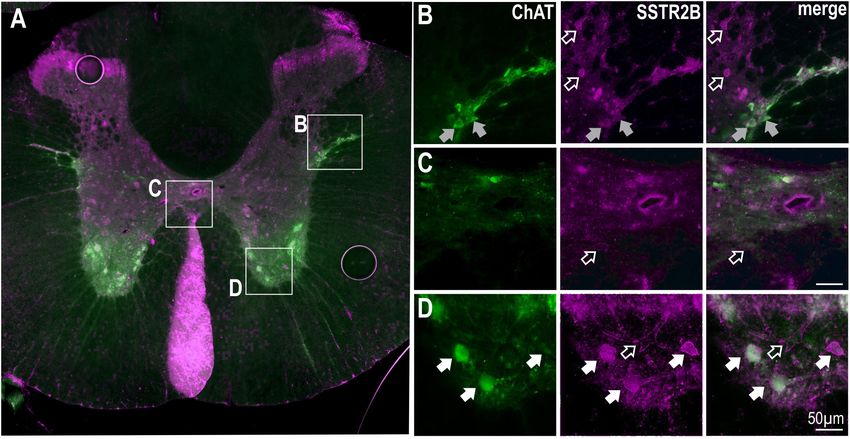
Bowman et al.                                                                                                    SSTR2 Receptors in Spinal Cord

   In brain sections fluorescent immunohistochemical detection           the A5 region analyzed at −10.20 and −9.72 mm and the
of CTB was conducted using a rabbit anti-CTB primary                     PVN analyzed at −2.04 and −1.72 mm. Cells were counted
antibody (1:5,000, #7927, RRID:AB_2313635, ViroStat,                     bilaterally, and the number of CTB-ir neurons expressing
Portland, ME, United States) in conjunction with detection               PPS mRNA was determined. Results were expressed as the
of mRNA using digoxigenin (DIG)-labeled riboprobes as                    average percent (±SEM) of retrogradely labeled cells expressing
described previously (Kumar et al., 2010; Bowman et al.,                 PPS in each region.
2013; Bowman and Goodchild, 2015). Sense (forward) and
antisense (reverse) riboprobes for preprosomatostatin (PPS)              Physiological Experiments
were designed as previously published (Burke et al., 2008), with         Surgical Preparation
a T7 promoter attached to the 50 end of the antisense primer             Surgical preparation was conducted as described previously
and an SP6 promoter attached to the sense primer as follows              (Burke et al., 2008, 2010). Rats were anesthetized with urethane
(promoters in uppercase):                                                (ip, 10% in saline 1.3 g/kg) and core temperature was monitored
                                                                         via flexible rectal thermistor and maintained (36.5–37◦ C)
       Forward: GGATCCATTTAGGTGACACTATAGAAGctca
                                                                         using a heating blanket (Harvard Apparatus, Holliston, MA,
       agctcggctgtctgag
                                                                         United States). Thirty minutes before intrathecal injection, core
       Reverse: GAATTCTAATACGACTCACTATAGGGAGAgg
                                                                         temperature was adjusted to 36◦ C to generate brown adipose
       aggagagggatcagaggt
                                                                         tissue thermogenesis, as previously described (Tupone et al.,
    Detection of GAD67 mRNA in SSTR2A expressing neurons                 2011). The right femoral artery and jugular vein were cannulated
of the spinal cord was conducted as described above, in 40 µm            for measurement of arterial pressure and administration of
thick parasagittal or coronal sections of spinal cord segments C8-       drugs and saline, respectively. Animals were vagotomized and
T3 and T4-T10. A primary antibody raised in rabbit was used              intubated for artificial oxygen-enriched ventilation (rodent
to target SSTR2A (#SS-800, Biotrend, Germany), and a donkey              respirator #7025, UGO Basile, Italy), and secured in a stereotaxic
anti-rabbit secondary antibody (Cy3-conjugated, #711-165-152,            frame. Rats were paralyzed with pancuronium bromide [0.8 mg
Jackson ImmunoResearch, United States) for visualization. The            iv, followed by an infusion (0.1 mg/ml in saline) at 2 ml/h].
following sense and antisense primers were designed for GAD67            End-tidal CO2 (Capstar-100, CWE Inc., PA, United States) was
riboprobe (promoters in uppercase) as described previously               maintained between 3.5 and 4.5% under control conditions.
(Burke et al., 2008; Bowman et al., 2013):                                   Heart rate was calculated using the R wave of ECG recordings.
                                                                         Recordings of efferent sympathetic nerve activity (SNA) were
       Forward: GGATCCATTTAGGTGACACTATAGAAGttatg                         made from the cut, left greater splanchnic nerve using bipolar
       tcaatgcaaccgc                                                     silver wire electrodes. Signals were amplified (BMA-400, #09-
       Reverse: GAATTCTAATACGACTCACTATAGGGAGAcc                          03010, CWE Inc., United States), band pass filtered (0.1 Hz–
       caacctctctatttcctc                                                3 kHz) and digitized for recording using Spike2 software
                                                                         (Cambridge Electronic Design Ltd., Cambridge, England). Data
Imaging and Analysis                                                     were normalized for comparison by taking the activity present
All imaging was conducted using an AxioImager Z1 (Carl                   at the nadir following euthanasia (see below) as zero nerve
Zeiss, Germany). Spinal cord sections were scanned for ChAT-             activity and 100% nerve activity was taken as the average
ir neurons within the lateral horn or close to the central canal.        of activity 5 min prior to intrathecal drug injection. The
ChAT-ir nests were imaged and quantified in C8-T3 sections, by           temperature of left and right iBAT deposits was measured
counting the number of ChAT-immunoreactive (-ir) cells which             using T2 thermocouple probes (#BT-1, Physitemp, Clifton, NJ,
expressed SSTR2A in each animal. Results were expressed as               United States) attached to a T-type Pod (#ML312 & #ML305,
mean percentage of ChAT-ir cells expressing SSTR2A ± SEM.                ADInstruments, Sydney, Australia) connected to a PowerLab
    The presence of CTB-ir and PPS mRNA expression were                  (30 series, ADInstruments, Sydney, Australia) then digitized for
identified in brain sections containing the presympathetic cell          recording. Thus, three sympathetic outflows were monitored: HR
groups: the caudal raphe (pallidus, obscurus and magnus),                (vagotomised), splanchnic SNA and iBAT temperature.
RVMM (paragigantocellular and parapyramidal groups), RVLM,                   In two animals the adrenal glands were removed bilaterally via
A5 region and PVN. After scanning the rostrocaudal extent of             the retroperitoneal cavity and the adrenal blood vessels ligated.
each premotor group, the Bregma level which had the highest
expression of CTB was identified. One or two additional sections         Intrathecal Drug Administration
were also selected for inclusion in analysis, according to the           Dura beneath the atlanto-occipital membrane was incised, and
longitudinal spread of CTB labeling within a cell group. Thus            a catheter (od 0.61; id 0.28 mm) inserted in the sub-arachnoid
cell groups with a longer rostrocaudal extent of CTB-ir were             space and advanced to spinal level T5–T6, as described previously
allocated three Bregma levels for analysis, while those with             (Bowman and Goodchild, 2015).
a smaller rostrocaudal CTB-ir distribution were allocated two               Intrathecal injections of drug (agonists (SST 1 mM [#H-
levels. Sections from the following Bregma levels were analyzed          1490.0005, Auspep, Australia], seglitide 1 mM [#S1316, Sigma
for double labeling: the raphe was analyzed at −13.90, −12.12            Aldrich, United States]) and antagonists (BIM-23627 (45 µM,
and −11.52 mm; the RVMM analyzed at −13.56, −12.96 and                   150 µM, 450 µM and 1.5 mM) [#H-5886.1000, Bachem,
−12.24 mm; the RVLM analyzed at −12.36 and −12.00 mm;                    Switzerland], CYN-154806 1 mM [D Tyr form, #1843, Tocris,

Frontiers in Neuroscience | www.frontiersin.org                      3                                     February 2019 | Volume 13 | Article 121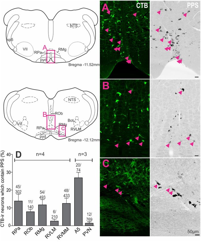
Bowman et al. SSTR2 Receptors in Spinal Cord
United Kingdom])) or vehicle (saline or phosphate buffered SSTR2B immunolabeling was also present in lamina II
saline, PBS, pH 7.4) were conducted using a Hamilton syringe. of the dorsal horn and in the ventral horn (Figure 3)
Five microliter of drug or vehicle, flushed with 7 µl of vehicle, as previously described (Schulz et al., 1998). More diffuse
was injected over 10–15 s. MAP, HR, splanchnic SNA, iBAT SSTR2B immunolabeling was observed in other parts of
temperature, end tidal CO2 and core temperature were recorded the gray matter [as previously described (Schulz et al.,
for at least 60 min following injection. The concentration of SST 1998)] with some ChAT labeled SPN expressing SSTR2B-like
was the same as used previously (Burke et al., 2008). immunoreactivity (Figure 3B). Accurate quantification was not
The animals were then euthanized with 3 M KCl (0.3 ml iv), possible due to the diffuse nature of the SSTR2B expression
and the level of nerve activity recorded, and a laminectomy was as previously described (Schulz et al., 1998). ChAT positive
performed to confirm the position of the catheter tip. motoneurons in the ventral horn clearly expressed SSTR2B-
like immunoreactivity (Figures 3A,D) but not SSTR2A-like
Data Analysis immunoreactivity (see Figure 1A).
Electrophysiological data were analyzed as the peak change Thus, immunolabeling for SSTR2 is present in the spinal
from baseline (pre-injection) values. Data are presented as the cord: in the dorsal horn (SSTR2A & SSTR2B) and ventral horn
mean ± the SEM. GraphPad Prism and/or SPSS (Statistical (SSTR2B); on some GABAergic, likely, interneurons and on SPN
Package for the Social Sciences) were used for statistical analysis (SSTR2A & SSTR2B), with the latter suggesting their activation
and the data were considered significant at p < 0.05. Where peak influences sympathetic outputs.
changes due to drug were compared to vehicle, Student’s t-test
was used to compare responses. Where the effects of the different PPS mRNA Is Found in all Sympathetic
doses of antagonist were compared, mean peak and trough Premotor Cell Groups
responses across treatment groups were analyzed separately via To assess supraspinal sources of SST that could directly influence
analysis of variance (ANOVA). Dunnett’s t-test adjustments were SPN, premotor sympathetic cell groups were examined to
used to compare individual responses to control (saline). A determine if they expressed PPS mRNA (n = 3–4). Examples of
priori polynomial contrasts were used to assess systematic trends double labeling in the raphe pallidus (Figure 4A), raphe magnus
(linear, quadratic, etc.) across treatment means for the main (Figure 4B) and RVMM (Figure 4C) reflect the larger proportion
dependent variable. of CTB-labeled cells double labeled for PPS mRNA in these
regions (Figure 4D): raphe pallidus (13.9 ± 3.8% of 302 CTB-
ir neurons), raphe magnus, (11.7 ± 2.9% of 493 CTB-ir neurons),
RESULTS raphe obscurus (7.9 ± 1.4% of 140 CTB-ir neurons), the RVMM
(12.5 ± 3.0% of 433 CTB-ir neurons) and the region containing
SSTR2 Are Densely Expressed in the the A5 group (26.9 ± 3.0% CTB-ir of 74 neurons), compared to
the RVLM (2.6 ± 1.0% of 210 CTB-ir neurons) and the PVN
Dorsal Horn, on SPN and on GABAergic
(1.4 ± 0.7% of 769 CTB-ir neurons).
Neurons in the Thoracic Spinal Cord Thus, all major premotor sympathetic cell groups have the
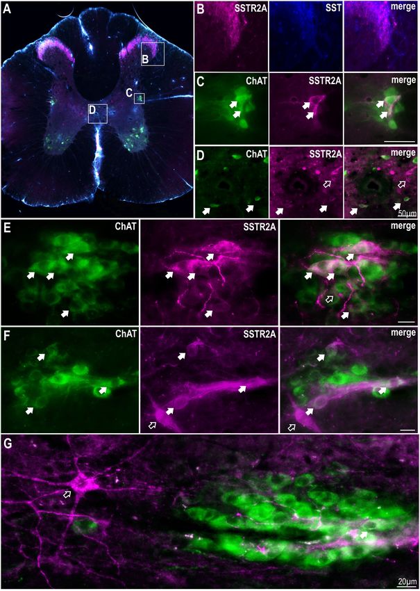
Although SST terminals have been described around SPN no potential to release SST at SPN with the greatest innervation
SSTR have been described here thus, we sought to identify the arising from the brainstem. The predominant SST projection,
location of SSTR2A and SSTR2B in thoracic spinal cord. Dense representing 81% of all PPS mRNA + CTB-ir neurons, arises
immunolabeling of SSTR2A and SST was found in lamina II of from the midline raphe/RVMM regions of the brainstem.
the dorsal horn (Figures 1A,B) as has been previously described
(Schulz et al., 1998; Todd, 2017). Some, but not all, ChAT labeled SST or Selective Activation of SSTR2 in
SPN in the IML and around the central canal expressed SSTR2A-
like immunoreactivity (Figures 1C–F). About 10% of SPN in the
the Thoracic Spinal Cord Reduces
IML expressed SSTR2A-like immunoreactivity in C8-T3 spinal Cardiovascular, Sympathetic and
cord segments (10.1 ± 1.2% of 582 SPN counted, n = 3) with some Thermogenic Activity
nests having multiple SSTR2A expressing neurons (Figures 1E,F) As some SPN expressed SST2R-like immunoreactivity and
but many having none. Non-ChAT labeled neurons both close potentially receive SST input from raphe/RVMM regions the
to, and more distant, from the IML, also expressed SSTR2A-like effects of intrathecal administration of SST (total volume 5 µl,
immunoreactivity (Figures 1F,G). n = 8), at a dose previously used within the ventral medulla
To determine whether non-ChAT SSTR2A positive neurons to reduce autonomic outflows [1 mM; (Burke et al., 2008)],
were GABAergic, in situ hybridization for GAD67 mRNA was was determined. Sympathetic, cardiovascular and thermogenic
combined with SSTR2A immunolabeling (Figure 2A). SSTR2A- parameters were decreased (Figure 5A). SST evoked significant
like immunoreactivity was co-expressed with GAD67 mRNA sympathoinhibition (−15.1 ± 3.8 vs −0.3 ± 1.1%, t (8) = 3.71;
in some neurons in the dorsal horn, as previously described p = 0.006), modest hypotension (−7.9 ± 1 vs −0.1 ± 1.0 mmHg,
(Todd, 2017) (Figure 2B) including lamina III (Figure 2E), t (10) = 5.38; p = 0.0003), and bradycardia (−41 ± 6 vs
the dorsolateral funiculus (Figure 2C), adjacent to central canal 0.5 ± 2 bpm, t (14) = 6.32; p < 0.0001), compared to
(Figure 2D), as well as in some star shaped neurons close to the vehicle control (Figures 5A,C). Similarly, metabolic outflows
IML (Figure 2F). were reduced (Figures 5A,C), with a peak decrease of
Frontiers in Neuroscience | www.frontiersin.org 4 February 2019 | Volume 13 | Article 121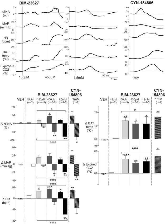
Bowman et al.                                                                                                                         SSTR2 Receptors in Spinal Cord

  FIGURE 1 | SSTR2A are expressed in dorsal horn, on SPN and non-ChAT labeled neurons in the thoracic spinal cord. (A) Coronal section of thoracic spinal cord
  showing distribution of SSTR2A-like (magenta), SST (blue) and ChAT immunolabeling (green). (B) Dorsal horn showing overlapping SSTR2-like and SST labeling.
  (C) Intermediolateral cell column (IML) containing ChAT labeled SPN with some exhibiting SSTR2A-like immunoreactivity (filled arrows). (D) Central autonomic area
  containing some SSTR2A expressing ChAT labeled SPN (filled arrows). (E–G) “Nests” of ChAT labeled SPN some of which co-express SSTR2A-like labeling (filled
  arrows). Scale bars = 20 µm. Open arrows indicate non-ChAT SSTR2A labeled neurons close to or adjacent to SPN.

−0.38 ± 0.11◦ C in iBAT temperature and −0.24 ± 0.09%                                  change any parameters measured, as previously observed
in expired CO2 compared to vehicle (iBAT = 0.06 ± 0.05◦ C,                             (Bowman and Goodchild, 2015).
t (6) = 4.14; p = 0.0061, and expired CO2 = 0.00 ± 0.01%,                                 To determine whether SSTR2 contributed to the responses
t (6) = 2.61; p = 0.04, respectively). Note that core temperature                      observed, the SSTR2 selective agonist Seglitide was administered
was maintained at 36◦ ensuring iBAT thermogenesis was                                  intrathecally (5 µl, 1 mM, n = 7) and decreased cardiovascular
active (Tupone et al., 2011). Administration of 45 µM                                  and thermogenic outflows measured (Figures 5B,C). As observed
SST (n = 2) elicited a smaller bradycardic response (−19                               with SST, application of Seglitide resulted in sympathoinhibition
bpm) but did not alter any other parameter measured                                    (−14.0 ± 4.1 vs −2.54 ± 0.77%; t (12) = 2.73; p = 0.018),
(data not shown). PBS used as a vehicle control did not                                bradycardia (−30 ± 4.7 vs −2.6 ± 1.2 bpm; t (12) = 5.68;

Frontiers in Neuroscience | www.frontiersin.org                                    5                                           February 2019 | Volume 13 | Article 121
Bowman et al.                                                                                                                     SSTR2 Receptors in Spinal Cord

  FIGURE 2 | Some SSTR2A expression colocalises with GAD67 mRNA in thoracic spinal cord. (A) Coronal section of spinal cord showing SSTR2A-like
  immunoreactivity (magenta) and GAD67 mRNA (black). Some neurons in dorsal horn (B), dorsolateral funiculus (C), central autonomic area (D) and lamina III (E)
  co-express both markers. (F) Neuron adjacent to IML expressing SSTR2A-like immunoreactivity and GAD67 mRNA. Filled arrows indicate double-labeled neurons.
  Scale bars for B–E = 50 µm and for F = 20 µm.

p = 0.0001) and hypotension (−8.7 ± 1.5 vs 1.3 ± 1.4 mmHg;                          affinity for SSTR2 (Tulipano et al., 2002). A low dose of
t (12) = 4.85; p = 0.0004) when compared to vehicle. Moreover,                      45 µM BIM-23627 had little effect on any parameter (data
decreases in iBAT temperature (−0.41 ± 0.10 vs 0.01 ± 0.03◦ C;                      not shown) whereas 150 µM BIM-23627 increased sympathetic,
t (6) = 3.91; p = 0.0079) and expired CO2 (−0.20 ± 0.03 vs                          cardiovascular (HR, MAP) and thermogenic (iBAT, expired CO2 )
0.001 ± 0.01%; t (12) = 6.61; p < 0.0001) also resulted from                        parameters with 450 µM amplifying these effects (Figure 6A).
selective SSTR2 activation.                                                         Higher concentrations of BIM-23627 (450 µM and 1.5 mM)
     These data indicate that activation of SSTR, and specifically                  produced a biphasic response, where the initial increase in
SSTR2, in the spinal cord reduces sympathetic, cardiovascular                       sympathetic and cardiovascular outflows was followed by a
and thermogenic parameters.                                                         secondary decrease in these parameters (Figure 6A).
                                                                                        Very similar effects in all parameters were evoked by CYN-
                                                                                    154806 (1 mM) (Figure 6B), also a peptide antagonist with a
Blockade of SSTR2 in the Spinal Cord                                                high affinity for SST2R. Importantly with both antagonists all
Increases Sympathetic, Cardiovascular                                               parameters returned to baseline within ∼75 min.
and Thermogenic Parameters                                                              The dose response grouped data are shown in Figure 6C,
We then sought to determine whether SSTR2 expressing                                with the peak increase and subsequent nadir shown for each
spinal neurons tonically influence sympathetic, cardiovascular or                   dose of BIM-23627 [45 µM n = 2 (not shown), 150 µM n = 4–5,
thermogenic activity.                                                               450 µM n = 5–7, 1.5 mM n = 4–5] and for CYN-154806 (1 mM,
   Figure 6A shows representative responses to increasing                           n = 3). These were compared to the largest change evoked
doses of the peptide antagonist BIM-23627 which exhibits high                       by vehicle (VEH).

Frontiers in Neuroscience | www.frontiersin.org                                 6                                           February 2019 | Volume 13 | Article 121
Bowman et al. SSTR2 Receptors in Spinal Cord FIGURE 3 | SSTR2B in dorsal and ventral horns and diffusely in the IML in the thoracic spinal cord. (A) Coronal section of thoracic cord showing SSTR2B-like (magenta) and ChAT (green) immunoreactivity. Clear membrane associated SSTR2B-like labeling is present dorsal and ventral (D) horns with diffuse labeling in intermediolateral cell column (B) and central autonomic areas (C). Filled arrows indicate double-labeled neurons (white indicates SSTR2B-like immunoreactivity appears membrane-bound, gray indicates diffuse SSTR immunoreactivity). Scale bars = 50 µm. In relation to the peak sympathetic and cardiovascular BIM-23627 when compared to control (iBAT: 1.38 ± 0.25 vs responses ANOVA revealed significant dose-dependent effects −0.06 ± 0.03◦ C, p < 0.01, n = 4; expired CO2 : 0.32 ± 0.07 on sSNA and HR (F (3,14) = 3.44, p = 0.044, and F (3,18) = 9.03, vs −0.02 ± 0.03%, p < 0.0001, n = 5, respectively), p = 0.0007, respectively), but not MAP (F (3,14) = 2.58, as increasing doses of 450 µM (iBAT: 1.17 ± 0.16◦ C, p = 0.095). A significant quadratic effect was observed on p = 0.01, n = 5; expired CO2 : 0.24 ± 0.03%, p < 0.01, peak sSNA (F (1,15) = 6.43, p < 0.05) and HR (F (1,18) = 17.53, n = 7, respectively) and 1.5 mM (iBAT: 1.16 ± 0.48◦ C, p < 0.0005), suggesting that selective SSTR2 inhibition p < 0.05; n = 4, expired CO2 : 0.27 ± 0.04%, n = 4, increased cardiovascular outflows before decreasing them. The p < 0.01) did not significantly alter the peak responses maximum increase occurred at 450 µM BIM-23627, where the reached. Only peak increases were measured for iBAT and sympathoexcitatory (42.5 ± 11.3 vs 0.70 ± 3.30%, p < 0.05, n = 5), expired CO2 as later changes could arise because of evoked and tachycardic (43.2 ± 4.5 vs 3.6 ± 3.0 bpm, p < 0.001, n = 7) hemodynamic alterations. responses peaked, before returning toward baseline. Significant To further corroborate SSTR2 involvement in setting tachycardia was also observed at the other doses (150 µmM; sympathetic, cardiovascular and thermogenic tone, the effect 32.5 ± 8.5 bpm, 1.5 mM; 27.0 ± 6.9 bpm, p < 0.05, n = 5 of CYN-154806 (1 mM, n = 3) was determined (Figure 6C). and 6, respectively). Although CYN-154806 evoked initial sympathoexcitatory With respect to the secondary decreased responses, and hypertensive effects, these did not reach significance. ANOVA revealed significant dose-dependent effects on The sympathoinhibition and hypotension that followed, sSNA (F (3,14) = 11.84, p = 0.0004), MAP (F (3,14) = 4.972, however, reached significance when compared to control p = 0.012), and HR (F (3,15) = 8.24, p = 0.0018). In contrast (sSNA: −80.8 ± 18.6%, p < 0.05, MAP: −35.8 ± 6.03 mmHg, to the initial peaks, a linear effect was seen in the secondary p < 0.01, respectively). CYN-154806 administration also trough responses of sSNA (F (3,14) = 36.843, p < 0.0001), resulted in significant tachycardia (37.3 ± 5.7 bpm, p < 0.01), HR (F (1,14) = 20.49, p < 0.0001), and MAP (F (1,14) = 14.67, increased iBAT temperature (1.64 ± 0.34◦ C, p < 0.01), p < 0.005), suggesting that more SSTR2 were influenced or and increased expired CO2 (0.38 ± 0.19%, p < 0.05) when were impacted for longer, resulting in significant cardiovascular compared to control. decline. Significant sympathoinhibition and hypotension were In order to ascertain that the tachycardic and thermogenic evoked by 450 µM and 1.5 mM BIM-23627 (sSNA: −49.15 ± 13.0 effects of BIM-23627 were not due to activation of SPN and −79.72 ± 3.10%, respectively, p < 0.01, MAP: −22.7 ± 7.7 innervating the adrenal gland resulting in catecholamine release and −24.0 ± 4.3 mmHg, respectively, p < 0.01), whereas heart potentially activating adrenergic receptors that regulate the heart rate reached significance only at 1.5 mM (−71.7 ± 14.0 bpm, and iBAT (Cannon and Nedergaard, 2004) the adrenal glands p < 0.01), when compared to control. were removed in two experiments. The effect of intrathecal With respect to thermogenesis, peak increases in iBAT BIM-23627 at 1 mM was identical to the effects seen without temperature and expired CO2 appear saturated at 150 µM adrenalectomy (data not shown). Frontiers in Neuroscience | www.frontiersin.org 7 February 2019 | Volume 13 | Article 121
Bowman et al.                                                                                                                      SSTR2 Receptors in Spinal Cord

  FIGURE 4 | Premotor sympathetic neurons express preprosomatostatin (PPS) mRNA. (A–C) Premotor sympathetic regions (magenta boxes) showing CTB-ir
  neurons retrogradely labeled from the spinal cord (green) and PPS mRNA expression (black). Double-labeled cells are indicated by arrowheads. (A) Raphe pallidus
  (RPa) and raphe magnus (RMg), (B) Raphe obscurus (ROb) and raphe magnus (RMg), (C) Rostral ventromedial medulla (RVMM). Scale bars = 50 µm.
  (D) Percentage of CTB immunoreactive cells containing PPS mRNA within each presympathetic region examined. Data are shown as mean ± SEM. Cell counts per
  region are shown above each bar. Schematic diagrams adapted from Paxinos and Watson (2005).

DISCUSSION                                                                           Schulz et al., 2000). Novel data reveal that SSTR2A-like
                                                                                     immunoreactivity and possibly SSTR2B-like immunoreactivity
We demonstrate that SST or SSTR2 agonist injected intrathecally                      were present on a subset of SPN, in line with the description
in thoracic spinal cord, modulates sympathetic, cardiovascular                       of SST terminals around SPN (Johansson et al., 1984; Krukoff,
and iBAT thermogenic tone. SSTR2-like immunoreactivity                               1987). These data strongly support the idea that SST, released
was localized to subsets of SPN, dorsal horn neurons and                             at SSTR2, reduces sympathetic activity. Intrathecal injection
inhibitory interneurons. Importantly, we show that SSTR2                             of SST, or the SSTR2 selective agonist Seglitide, did evoke
antagonists increase sympathetic, cardiac and iBAT thermogenic                       sympathoinhibition, hypotension, bradycardia, and decreased
tone suggesting that SST, tonically activating SST2R, in the                         iBAT temperature and expired CO2 . These effects are in keeping
thoracic cord is required for the maintenance of sympathetic,                        with the well-described inhibitory effects of SST on neurons
cardiovascular and thermogenic tone (at least in the anesthetized                    (Bou Farah et al., 2016; Gunther et al., 2018). We also found
preparation used here). SST potentially released from the RVMM                       PPS mRNA in subsets of sympathetic premotor neurons in all
and raphe premotor nuclei may contribute to this.                                    major loci, predominantly from the midline raphe and RVMM
   Our data confirms and extends previous studies exploring                          regions. This also corroborates earlier studies describing SST-
the distribution of SSTR2 in the spinal cord (Todd et al., 1998;                     like immunoreactivity in the medullary raphe, RVMM and A5

Frontiers in Neuroscience | www.frontiersin.org                                  8                                           February 2019 | Volume 13 | Article 121
Bowman et al. SSTR2 Receptors in Spinal Cord
FIGURE 5 | Intrathecal SST and SSTR2 agonist decrease sympathetic, cardiovascular and thermogenic outflows. (A) Representative responses show that SST
(1 mM) elicited decreases in sSNA, MAP, HR, iBAT temperature and expired CO2 . (B) The SSTR2 agonist seglitide evoked similar effects to SST. (C) Grouped data
showing peak responses evoked by intrathecal SST (n = 7), seglitide (n = 8) or vehicle (n = 8). Data are expressed as mean ± SEM. Effect of each drug compared to
vehicle ∗ = p < 0.05, ∗∗ = p < 0.01, ∗∗∗ = p < 0.001, ∗∗∗∗ = p < 0.0001.
noradrenergic neurons (Strack et al., 1989b; Jansen et al., 1995). effects in only these parameters and, such a response may
Taken together these data suggest it is possible that inhibitory SST represent thermogenesis, as described previously (Morrison
is released from sympathetic premotor neurons (thought to be and Madden, 2014). This may be produced directly at
primarily excitatory) directly at SPN. SPN altering both HR and iBAT temperature, however, it
Surprisingly, we have demonstrated that SST acts tonically is likely that SSTR2 blockade prevents the actions of SST
at SSTR2 in the spinal cord, as intrathecal administration of tonically released in the dorsal horn, which occurs even
two selective SSTR2 antagonists evoked sympathoexcitation, when no peripheral afferent stimulus is applied (Morton
tachycardia, thermogenesis and an increase in end tidal CO2 . et al., 1988). Thus, it is possible that temperature sensitive
Administration of high doses of both antagonists produced peripheral inputs to dorsal horn neurons are gated by SST
biphasic responses, where initial excitatory responses in all released intraspinally and, when removed thermogenesis occurs.
outflows were followed by rapid and sustained decreases in The spinoparabrachial cold defense pathway (Morrison and
sympathetic and cardiovascular outflows before returning to Madden, 2014) may be disinhibited by SSTR2 antagonists as
baseline. Several mechanisms are possible. although SSTR2A are expressed on inhibitory dorsal horn
It is plausible that the SSTR2 antagonist induced excitatory neurons (Todd et al., 1998) a spinoparabrachial population
responses are evoked by removal of inhibitory SST directly at of excitatory SSTR2A expressing neurons has been described
the different pools of SPN regulating splanchnic SNA, the heart (Cameron et al., 2015).
and iBAT. Our data and that of others demonstrate that SST The increase in splanchnic SNA generated by SSTR2 blockade
terminals are present in the IML and that there are premotor is unlikely related to thermogenesis as the effective antagonist
sympathetic and spinal (Jung et al., 2008) sources of SST that may doses do not align and innervation of splanchnic and BAT
be tonically released. preganglionic neurons are independent. Therefore, a sympathetic
It is also possible that the SSTR2 antagonist evoked increases pathway driving functions activated by the splanchnic nerve
in HR, iBAT temperature and expired CO2 may be coordinated, appears to be tonically inhibited by SST. As the splanchnic
as low doses of BIM-23627 induced significant and large nerve controls both vasomotor and gastrointestinal functions this
Frontiers in Neuroscience | www.frontiersin.org 9 February 2019 | Volume 13 | Article 121Bowman et al. SSTR2 Receptors in Spinal Cord FIGURE 6 | Effects of intrathecal SSTR2 antagonists on sympathetic, cardiovascular and thermogenic outflows. (A) Representative responses show that 150 and 450 µM of the SSTR2 antagonist BIM-23627 evoked increases in sSNA, MAP, HR, iBAT temperature and expired CO2 . Higher doses (1.5 mM) of BIM-23627 elicited smaller early increases in sSNA, MAP, HR, iBAT temperature and expired CO2 followed immediately by large falls below baseline in sSNA, MAP and HR. IBAT temperature also declined but not below baseline. (B) CYN-154806 (1 mM) evoked similar effects to BIM-23627. (C) Grouped data showing peak changes relative to baseline with little to no effect seen with vehicle or 45 µM BIM-23627 (n = 2). In contrast 150 or 450 µM BIM-23627 evoked large early increases in all parameters. Biphasic responses in SNA, MAP and HR were evident at doses greater than 150 µM BIM-23627. All doses of BIM-23627 (except 45 µM) and CYN-154806 increased iBAT temperature and expired CO2 . Data are expressed mean ± SEM, asterisks denote significant difference compared to PBS control (∗ = p < 0.05, ∗∗ = p < 0.01, ∗∗∗ = p < 0.001, ∗∗∗∗ = p < 0.0001), and the octothorp denotes either significant linear or quadratic trends (# = p < 0.05, #### = p < 0.0001). Frontiers in Neuroscience | www.frontiersin.org 10 February 2019 | Volume 13 | Article 121
Bowman et al.                                                                                                                                           SSTR2 Receptors in Spinal Cord

action may contribute to the increase in MAP that also occurs                                        CONCLUSION
following SST2R blockade.
    Drug movement when applied intrathecally has been assessed                                       The findings demonstrate that SSTR2-like immunoreactivity
(Yaksh and Rudy, 1976; Nishio et al., 1989) and it is clear                                          is present on SPN, GABAergic interneurons and dorsal horn
that spinal cord regions impacted are influenced by catheter                                         neurons of the thoracic spinal cord and that exogenously applied
placement, drug-type, concentration, speed of application                                            SST, or an SSTR2 agonist, reduce sympathetic, cardiovascular
and time. Significant sympathoinhibition, hypotension and                                            and thermogenic activity. Blockade of endogenous SST signaling,
bradycardia followed the early excitatory responses when higher                                      using selective SSTR2 antagonists, increased all parameters
doses of SSTR2 antagonists were administered. These robust                                           measured with higher doses of antagonists then robustly reducing
responses recover within about 70 min, indicating a non-                                             sympathetic and cardiovascular parameters. Thus, acting at
neurotoxic effect. While there is some evidence for constitutive                                     multiple sites within the spinal cord, SST appears to act as a
activity in SST receptors in vitro (Ben-Shlomo et al., 2007,                                         “brake,” tonically inhibiting sympathetic and iBAT thermogenic
2009) there is no evidence that the antagonists used here block                                      tone via SSTR2. The data are also consistent with the notion
constitutive activity. Studies well suited to detecting inverse                                      that SST tonically modulates, via SSTR2, a diffuse GABAergic
agonists (i.e., GTPyS) do not report effects of the antagonists,                                     network that impacts sympathetic and cardiovascular function
so constitutive activation of SSTR is a formal possibility,                                          with the source/s of spinal SST having supra- and/or intra-
however, available evidence is more consistent with reversal                                         spinal origins.
of endogenous SST tone (Gunther et al., 2018). Both peptide
antagonists used here act competitively with high binding affinity
at SSTR2 (Bass et al., 1996; Feniuk et al., 2000; Tulipano
et al., 2002) and although previous data suggest an interaction
                                                                                                     AUTHOR CONTRIBUTIONS
with opioid receptors (Ghosh et al., 1997), CYN-154806 does                                          BB, AG, and PGB conceived and designed the study.
not block µ-opioid mediated inhibition (Feniuk et al., 2000),                                        BB and PGB acquired the data. BB, PB, and AG
indicating that the observed effects are due to blockade of SSTR2.                                   analyzed the data. All authors (BB, PB, SM, AG, and
The effects are consistent with the hypothesis that increasing                                       PGB) contributed to the interpretation of the data,
blockade of SSTR2 results in potent disinhibition by recruiting                                      preparation of the figures and writing and or revising
a spinal GABAergic network that counteracts the enhanced                                             of the manuscript.
sympathetic and cardiovascular function. The delayed effect
and a higher dose of antagonists may be required to access
and disinhibit such a diffuse network, as blockade at more
than a few neurons in the network may be needed to drive                                             FUNDING
sympathoinhibition. A tonic GABAergic network is present in the
spinal cord regulating sympathetic and cardiovascular functions                                      This work was supported by the National Health
(Deuchars et al., 2005; Goodchild et al., 2008; Bowman and                                           and Medical Research Council of Australia (NHMRC
Goodchild, 2015) possibly via α5 GABA-A receptors (Wang                                              APP1028183 and APP1127817), the Australian Research
et al., 2008) and perhaps, this is regulated by SST. In keeping                                      Council (DP120100920), and the Hillcrest Foundation
with this idea GABAergic interneurons are responsible for about                                      (IPAP201600725, Perpetual).
half (Llewellyn-Smith, 2002) of the GABAergic boutons which
comprise about 50% of the innervation of SPN (Llewellyn-Smith
et al., 1995) and as demonstrated here and elsewhere (Todd, 2017)                                    ACKNOWLEDGMENTS
GABAergic interneurons express SSTR2A. As primary afferents
only contribute about 40% of SST even in the dorsal horn                                             We wish to thank Sophie Fletcher for her assistance with
(Morton et al., 1989) the remainder arises from local intraspinal                                    histological work, and Travis Wearne for assistance with some
(Proudlock et al., 1993) or supraspinal sources as described here.                                   statistical analysis.

REFERENCES                                                                                           Bou Farah, L., Bowman, B. R., Bokiniec, P., Karim, S., Le, S., Goodchild, A. K.,
                                                                                                        et al. (2016). Somatostatin in the rat rostral ventrolateral medulla: origins
Bass, R. T., Buckwalter, B. L., Patel, B. P., Pausch, M. H., Price, L. A., Strnad, J., et al.           and mechanism of action. J. Comp. Neurol. 524, 323–342. doi: 10.1002/cne.
   (1996). Identification and characterization of novel somatostatin antagonists.                       23846
   Mol. Pharmacol. 50, 709–715.                                                                      Bowman, B. R., and Goodchild, A. K. (2015). GABA and enkephalin tonically
Ben-Shlomo, A., Pichurin, O., Barshop, N. J., Wawrowsky, K. A., Taylor, J., Culler,                     alter sympathetic outflows in the rat spinal cord. Auton Neurosci. 193, 84–91.
   M. D., et al. (2007). Selective regulation of somatostatin receptor subtype                          doi: 10.1016/j.autneu.2015.08.006
   signaling: evidence for constitutive receptor activation. Mol. Endocrinol. 21,                    Bowman, B. R., Kumar, N. N., Hassan, S. F., McMullan, S., and Goodchild, A. K.
   2565–2578. doi: 10.1210/me.2007-0081                                                                 (2013). Brain sources of inhibitory input to the rat rostral ventrolateral medulla.
Ben-Shlomo, A., Zhou, C., Pichurin, O., Chesnokova, V., Liu, N. A., Culler,                             J. Comp. Neurol. 521, 213–232. doi: 10.1002/cne.23175
   M. D., et al. (2009). Constitutive somatostatin receptor activity determines tonic                Burbach, J. P. (2010). Neuropeptides from concept to online database
   pituitary cell response. Mol. Endocrinol. 23, 337–348. doi: 10.1210/me.2008-                         www.neuropeptides.nl. Eur. J. Pharmacol. 626, 27–48. doi: 10.1016/j.ejphar.
   0361                                                                                                 2009.10.015

Frontiers in Neuroscience | www.frontiersin.org                                                 11                                               February 2019 | Volume 13 | Article 121
Bowman et al. SSTR2 Receptors in Spinal Cord Burke, P. G., Abbott, S. B., McMullan, S., Goodchild, A. K., and Pilowsky, Llewellyn-Smith, I. J., Minson, J. B., Pilowsky, P. M., Arnolda, L. F., and Chalmers, P. M. (2010). Somatostatin selectively ablates post-inspiratory activity after J. P. (1995). The one hundred percent hypothesis: glutamate or GABA in injection into the Bötzinger complex. Neuroscience 167, 528–539. doi: 10.1016/ synapses on sympathetic preganglionic neurons. Clin. Exp. Hypertens. 17, j.neuroscience.2010.01.065 323–333. doi: 10.3109/10641969509087074 Burke, P. G., Li, Q., Costin, M. L., McMullan, S., Pilowsky, P. M., and Goodchild, Loewy, A. D., and McKellar, S. (1981). Serotonergic projections from the ventral A. K. (2008). Somatostatin 2A receptor-expressing presympathetic neurons in medulla to the intermediolateral cell column in the rat. Brain Res. 211, 146–152. the rostral ventrolateral medulla maintain blood pressure. Hypertension 52, doi: 10.1016/0006-8993(81)90074-3 1127–1133. doi: 10.1161/hypertensionaha.108.118224 Millhorn, D. E., Seroogy, K., Hökfelt, T., Schmued, L. C., Terenius, L., Buchan, A., Cameron, D., Polgar, E., Gutierrez-Mecinas, M., Gomez-Lima, M., Watanabe, M., et al. (1987). Neurons of the ventral medulla oblongata that contain both and Todd, A. J. (2015). The organisation of spinoparabrachial neurons in the somatostatin and enkephalin immunoreactivities project to nucleus tractus mouse. Pain 156, 2061–2071. doi: 10.1097/j.pain.0000000000000270 solitarii and spinal cord. Brain Res. 424, 99–108. doi: 10.1016/0006-8993(87) Cannon, B., and Nedergaard, J. (2004). Brown adipose tissue: function and 91197-8 physiological significance. Physiol. Rev. 84, 277–359. doi: 10.1152/physrev. Morrison, S. F., and Madden, C. J. (2014). Central nervous system regulation of 00015.2003 brown adipose tissue. Compr. Physiol. 4, 1677–1713. doi: 10.1002/cphy.c140013 Chiba, T., and Masuko, S. (1989). Coexistence of varying combinations of Morton, C. R., Hutchison, W. D., and Hendry, I. A. (1988). Release of neuropeptides with 5-hydroxytryptamine in neurons of the raphe pallidus et immunoreactive somatostatin in the spinal dorsal horn of the cat. Neuropeptides obscurus projecting to the spinal cord. Neurosci. Res. 7, 13–23. doi: 10.1016/ 12, 189–197. doi: 10.1016/0143-4179(88)90054-6 0168-0102(89)90033-3 Morton, C. R., Hutchison, W. D., Hendry, I. A., and Duggan, A. W. (1989). Cote-Vélez, A., Martínez Báez, A., Lezama, L., Uribe, R. M., Joseph-Bravo, P., Somatostatin: evidence for a role in thermal nociception. Brain Res. 488, 89–96. and Charli, J. L. (2017). A screen for modulators reveals that orexin-A doi: 10.1016/0006-8993(89)90696-3 rapidly stimulates thyrotropin releasing hormone expression and release in Nishio, Y., Sinatra, R. S., Kitahata, L. M., and Collins, J. G. (1989). Spinal hypothalamic cell culture. Neuropeptides 62, 11–20. doi: 10.1016/j.npep.2017. cord distribution of 3H-morphine after intrathecal administration: relationship 01.005 to analgesia. Anesth. Analg. 69, 323–327. doi: 10.1213/00000539-198909000- Deuchars, S. A., Milligan, C. J., Stornetta, R. L., and Deuchars, J. (2005). GABAergic 00009 neurons in the central region of the spinal cord: a novel substrate for Parker, L. M., Kumar, N. N., Lonergan, T., and Goodchild, A. K. (2013). sympathetic inhibition. J. Neurosci. 25, 1063–1070. doi: 10.1523/JNEUROSCI. Neurochemical codes of sympathetic preganglionic neurons activated by 3740-04.2005 glucoprivation. J. Comp. Neurol 521, 2703–2718. doi: 10.1002/cne.23310 Feniuk, W., Jarvie, E., Luo, J., and Humphrey, P. P. A. (2000). Selective Patel, Y. C. (1999). Somatostatin and its receptor family. Front. Neuroendocrinol. somatostatin sst2 receptor blockade with the novel cyclic octapeptide, CYN- 20, 157–198. doi: 10.1006/frne.1999.0183 154806. Neuropharmacology 39, 1443–1450. doi: 10.1016/S0028-3908(00) Paxinos, G., and Watson, C. (2005). The Rat Brain in Stereotaxic Coordinates. 00035-6 Amsterdam, Boston, MA: Elsevier Academic Press. Ghosh, S., Geller, E. B., and Adler, M. W. (1997). Interaction of cholecystokinin Proudlock, F., Spike, R. C., and Todd, A. J. (1993). Immunocytochemical study and somatostatin with a selective µ-opioid agonist and µ-and κ-antagonists in of somatostatin, neurotensin, GABA, and glycine in rat spinal dorsal horn. thermoregulation. Brain Res. 745, 152–157. doi: 10.1016/S0006-8993(96)01144- J. Comp. Neurol. 327, 289–297. doi: 10.1002/cne.903270210 4 Sandkühler, J., Fu, Q. G., and Helmchen, C. (1990). Spinal somatostatin Goodchild, A. K., van Deurzen, B. T., Hildreth, C. M., and Pilowsky, P. M. (2008). superfusion in vivo affects activity of cat nociceptive dorsal horn neurons: Control of sympathetic, respiratory and somatomotor outflow by an intraspinal comparison with spinal morphine. Neuroscience 34, 565–576. doi: 10.1016/ pattern generator. Clin. Exp. Pharmacol. Physiol. 35, 447–453. doi: 10.1111/j. 0306-4522(90)90165-Z 1440-1681.2008.04913.x Sawchenko, P. E., and Swanson, L. W. (1982). Immunohistochemical identification Gunther, T., Tulipano, G., Dournaud, P., Bousquet, C., Csaba, Z., Kreienkamp, of neurons in the paraventricular nucleus of the hypothalamus that project to H. J., et al. (2018). International union of basic and clinical pharmacology. the medulla or to the spinal cord in the rat. J. Comp. Neurol. 205, 260–272. cv. somatostatin receptors: structure, function, ligands, and new nomenclature. doi: 10.1002/cne.902050306 Pharmacol. Rev. 70, 763–835. doi: 10.1124/pr.117.015388 Schulz, S., Händel, M., Schreff, M., Schmidt, H., and Höllt, V. (2000). Localization Jansen, A. S., Wessendorf, M. W., and Loewy, A. D. (1995). Transneuronal of five somatostatin receptors in the rat central nervous system using subtype- labeling of CNS neuropeptide and monoamine neurons after pseudorabies virus specific antibodies. J. Physiol. Paris 94, 259–264. doi: 10.1016/S0928-4257(00) injections into the stellate ganglion. Brain Res. 683, 1–24. doi: 10.1016/0006- 00212-6 8993(95)00276-V Schulz, S., Schmidt, H., Händel, M., Schreff, M., and Höllt, V. (1998). Differential Johansson, O., Hökfelt, T., and Elde, R. (1984). Immunohistochemical distribution distribution of alternatively spliced somatostatin receptor 2 isoforms (sst2A of somatostatin-like immunoreactivity in the central nervous system and sst2B) in rat spinal cord. Neurosci. Lett. 257, 37–40. doi: 10.1016/S0304- of the adult rat. Neuroscience 13, 265–339. doi: 10.1016/0306-4522(84) 3940(98)00803-9 90233-1 Segond von Banchet, G., Schindler, M., Hervieu, G. J., Beckmann, B., Emson, P. C., Jung, S. J., Jo, S. H., Lee, S., Oh, E., Kim, M. S., Nam, W. D., et al. (2008). Effects and Heppelmann, B. (1999). Distribution of somatostatin receptor subtypes of somatostatin on the responses of rostrally projecting spinal dorsal horn in rat lumbar spinal cord examined with gold-labelled somatostatin and anti- neurons to noxious stimuli in cats. Korean J. Physiol. Pharmacol. 12, 253–258. receptor antibodies. Brain Res. 816, 254–257. doi: 10.1016/S0006-8993(98) doi: 10.4196/kjpp.2008.12.5.253 01226-8 Krukoff, T. L. (1987). Peptidergic inputs to sympathetic preganglionic neurons. Shi, T. J., Xiang, Q., Zhang, M. D., Barde, S., Kai-Larsen, Y., Fried, K., et al. (2014). Can. J. Physiol. Pharmacol. 65, 1619–1625. doi: 10.1139/y87-254 Somatostatin and its 2A receptor in dorsal root ganglia and dorsal horn of Kumar, N. N., Allen, K., Parker, L., Damanhuri, H., and Goodchild, A. K. (2010). mouse and human: expression, trafficking and possible role in pain. Mol. Pain Neuropeptide coding of sympathetic preganglionic neurons; focus on adrenally 10:12. doi: 10.1186/1744-8069-10-12 projecting populations. Neuroscience 170, 789–799. doi: 10.1016/j.neuroscience. Strack, A. M., Sawyer, W. B., Hughes, J. H., Platt, K. B., and Loewy, A. D. (1989a). 2010.07.047 A general pattern of CNS innervation of the sympathetic outflow demonstrated Kuraishi, Y., Hirota, N., Sato, Y., Hino, Y., Satoh, M., and Takagi, H. (1985). by transneuronal pseudorabies viral infections. Brain Res. 491, 156–162. Evidence that substance P and somatostatin transmit separate information Strack, A. M., Sawyer, W. B., Platt, K. B., and Loewy, A. D. (1989b). CNS cell related to pain in the spinal dorsal horn. Brain Res. 325, 294–298. doi: 10.1016/ groups regulating the sympathetic outflow to adrenal gland as revealed by 0006-8993(85)90326-9 transneuronal cell body labelling with pseudorabies virus. Brain Res. 491, Llewellyn-Smith, I. J. (2002). GABA in the control of sympathetic preganglionic 274–296. neurons. Clin. Exp. Pharmacol. Physiol. 29, 507–513. doi: 10.1046/j.1440-1681. Takahashi, M., Takeda, M., and Matsumoto, S. (2014). Somatostatin enhances 2002.03664.x tooth-pulp-evoked cervical dorsal horn neuronal activity in the rat via Frontiers in Neuroscience | www.frontiersin.org 12 February 2019 | Volume 13 | Article 121
Bowman et al.                                                                                                                                SSTR2 Receptors in Spinal Cord

   inhibition of GABAergic interneurons. Brain Res. Bull. 100, 76–83. doi: 10.1016/        Wang, L., Spary, E., Deuchars, J., and Deuchars, S. A. (2008). Tonic GABAergic
   j.brainresbull.2013.11.008                                                                 inhibition of sympathetic preganglionic neurons: a novel substrate for
Todd, A. J. (2017). Identifying functional populations among the interneurons                 sympathetic control. J. Neurosci. 28, 12445–12452. doi: 10.1523/JNEUROSCI.
   in laminae I-III of the spinal dorsal horn. Mol. Pain 13:1744806917693003.                 2951-08.2008
   doi: 10.1177/1744806917693003                                                           Yaksh, T. L., and Rudy, T. A. (1976). Chronic catheterization of the spinal
Todd, A. J., Spike, R. C., and Polgar, E. (1998). A quantitative study of                     subarachnoid space. Physiol. Behav. 17, 1031–1036. doi: 10.1016/0031-9384(76)
   neurons which express neurokinin-1 or somatostatin sst2a receptor in rat                   90029-9
   spinal dorsal horn. Neuroscience 85, 459–473. doi: 10.1016/S0306-4522(97)
   00669-6                                                                                 Conflict of Interest Statement: The authors declare that the research was
Tulipano, G., Soldi, D., Bagnasco, M., Culler, M. D., Taylor, J. E., Cocchi, D.,           conducted in the absence of any commercial or financial relationships that could
   et al. (2002). Characterization of new selective somatostatin receptor                  be construed as a potential conflict of interest.
   subtype-2 (sst2) Antagonists, BIM-23627 and BIM-23454. Effects of BIM-
   23627 on GH release in anesthetized male rats after short-term high-dose                Copyright © 2019 Bowman, Bokiniec, McMullan, Goodchild and Burke. This is an
   dexamethasone treatment. Endocrinology 143, 1218–1224. doi: 10.1210/en.143.             open-access article distributed under the terms of the Creative Commons Attribution
   4.1218                                                                                  License (CC BY). The use, distribution or reproduction in other forums is permitted,
Tupone, D., Madden, C. J., Cano, G., and Morrison, S. F. (2011). An orexinergic            provided the original author(s) and the copyright owner(s) are credited and that the
   projection from perifornical hypothalamus to raphe pallidus increases rat               original publication in this journal is cited, in accordance with accepted academic
   brown adipose tissue thermogenesis. J. Neurosci. 31, 15944–15955. doi: 10.1523/         practice. No use, distribution or reproduction is permitted which does not comply
   JNEUROSCI.3909-11.2011                                                                  with these terms.

Frontiers in Neuroscience | www.frontiersin.org                                       13                                              February 2019 | Volume 13 | Article 121
You can also read


