STRUCTURAL BASIS OF MISMATCH RECOGNITION BY A SARS-COV-2 PROOFREADING ENZYME - SCIENCE
←
→
Page content transcription
If your browser does not render page correctly, please read the page content below
REPORTS

                                                                                                                                   Cite as: C. Liu et al., Science
                                                                                                                                10.1126/science.abi9310 (2021).

Structural basis of mismatch recognition by a SARS-CoV-2
proofreading enzyme
Chang Liu1*, Wei Shi2, Scott T. Becker3, David G. Schatz1, Bin Liu2*, Yang Yang3*
1Department of Immunobiology, Yale School of Medicine, New Haven, CT, USA. 2Section of Transcription & Gene Regulation, The Hormel Institute, University of Minnesota,
Austin, MN, USA. 3Roy J. Carver Department of Biochemistry, Biophysics and Molecular Biology, Iowa State University, Ames, IA, USA.
*Corresponding author. Email: c.liu@yale.edu (C.L.); liu00794@umn.edu (B.L.); yan9yang@iastate.edu (Y.Y.)

Coronavirus 3′–5′ exoribonuclease (ExoN), residing in the nonstructural protein (nsp) 10-nsp14 complex,
boosts replication fidelity by proofreading RNA synthesis and is critical for the virus life cycle. ExoN also
recognizes and excises nucleotide analog inhibitors incorporated into the nascent RNA, undermining the
effectiveness of nucleotide analog-based antivirals. Here, we present cryo-electron microscopy structures
of both wild-type and mutant SARS-CoV-2 nsp10-nsp14 in complex with an RNA substrate bearing a 3′-end

                                                                                                                                                                         Downloaded from http://science.sciencemag.org/ on July 28, 2021
mismatch at resolutions ranging from 2.5 Å to 3.9 Å. The structures reveal the molecular determinants of
ExoN substrate specificity and give insight into the molecular mechanisms of mismatch correction during
coronavirus RNA synthesis. Our findings provide guidance for rational design of improved anti-coronavirus
therapies.

SARS-CoV-2, the causative agent of the COVID-19 pandemic,                             subgenomic messenger RNAs (16–18). The ExoN activity of
has infected over 160 million people and led to over 3 million                        nsp14 is stimulated by nsp10, which binds to the ExoN do-
deaths worldwide (https://covid19.who.int). Although several                          main and helps stabilize the architecture of the ExoN active
SARS-CoV-2 vaccines are now available (1), there are no                               site (18). Previous studies of the SARS-CoV nsp10-nsp14 com-
highly effective antiviral agents to treat the disease. One of                        plex defined the nsp14 ExoN domain as a DED/EDh-type ex-
the most important druggable targets for SARS-CoV-2 is its                            onuclease and identified the five active site residues through
replication/transcription complex (RTC), a multi-subunit                              structural comparison and mutagenesis analyses (19). How-
machine that carries out viral genome replication and                                 ever, the molecular details of substrate binding by corona-
transcription and plays an essential role in the virus life cycle                     virus nsp10-nsp14 ExoN remain unclear. In addition, how the
(2, 3). Central to the coronavirus RTC is the core RNA-                               viral ExoN recognizes and excises mis-incorporated nucleo-
dependent RNA polymerase (RdRp), nsp12 (4), and two                                   tide or nucleotide analog inhibitor at the 3′ end of the newly
associated accessory proteins, nsp7 and nsp8 (5). SARS-CoV-                           synthesized RNA is poorly understood.
2 RdRp is a promising target for nucleotide analog antivirals,                            To understand the substrate recognition and catalytic
such as remdesivir (6, 7). However, the efficacy of nucleotide                        mechanism of SARS-CoV-2 ExoN, we constructed a hairpin
analog inhibitors on coronavirus RdRp is compromised by                               RNA substrate (hereafter referred to as T35P31) that contains
the presence of the viral nsp14 exoribonuclease (ExoN) (8, 9),                        a template strand (T-strand, which is also the non-scissile
an RNA proofreader specific to coronaviruses and a few other                          strand for ExoN) with three initiating guanosines followed
closely related virus families of the Nidovirales order and                           by the 3′-end 32 nucleotides (nt) of the SARS-CoV-2 genome
crucial to maintain the integrity of their unusually large RNA                        (excluding the poly(A) tail) and a 31-nt product strand (P-
genome (9–11). In addition, ExoNs from coronaviruses and                              strand, which is also the scissile strand for ExoN) ending with
other RNA viruses play an important role in the evasion of                            a cytidine-5′-monophosphate (CMP), resulting in a C-U mis-
host immune responses by degrading the viral double-                                  match at the 3′ end (Fig. 1C). The pre-formed SARS-CoV-2
stranded RNA (dsRNA) intermediates that would otherwise                               nsp10-nsp14 complex digests the T35P31 RNA substrate in the
be recognized by host pathogen recognition receptors (12–15).                         presence of MgCl2 (Fig. 1B). To obtain a stable nsp10-nsp14-
    Nsp14 is a bi-functional enzyme that harbors both 3′–5′                           RNA complex, we substituted MgCl2 with CaCl2 in the recon-
ExoN and mRNA cap guanine-N7 methyltransferase (N7-                                   stitution buffer, or introduced an ExoN active site mutation,
MTase) activities (16, 17) (Fig. 1A). The N-terminal ExoN do-                         E191A, to nsp14. Both measures retained the RNA-binding ca-
main of nsp14 improves RNA synthesis fidelity by removing                             pability but abolished the RNA cleavage activity of the nsp10-
mis-incorporated nucleotides or nucleotide analogs from the                           nsp14 complex (Fig. 1B and figs. S1 and S2).
nascent RNA, while the C-terminal N7-MTase domain is in-                                  The reconstituted nsp10-nsp14-RNA complexes were puri-
volved in the 5′ capping processes of the viral genomic and                           fied by size-exclusion chromatography (SEC) and analyzed by

First release: 27 July 2021                                            www.sciencemag.org                (Page numbers not final at time of first release) 1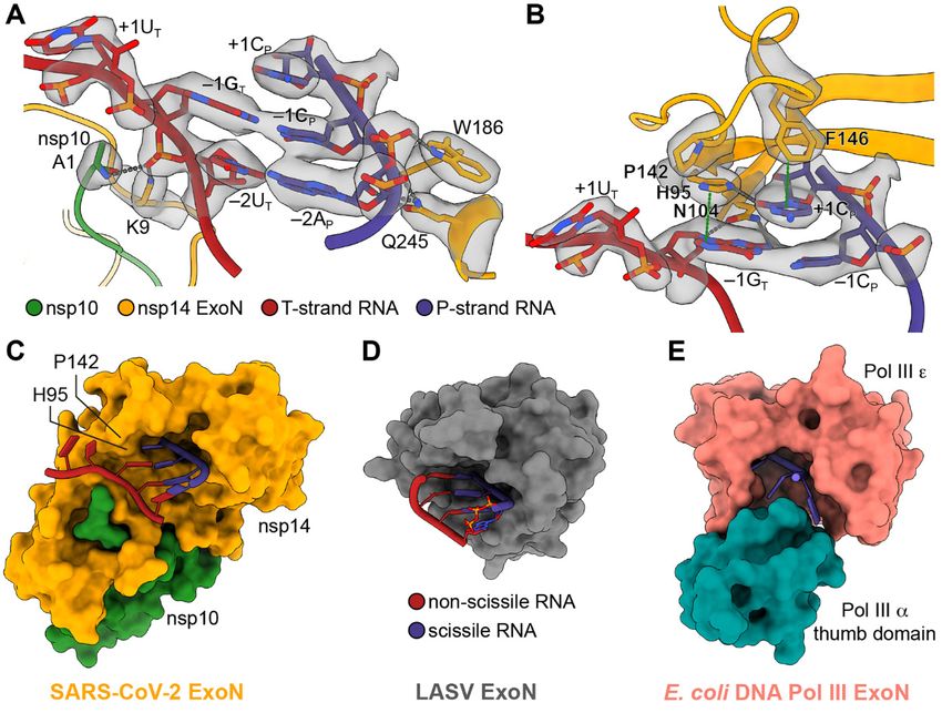
single-particle cryo-EM. The final cryo-EM maps for the WT          nsp10-nsp14-RNA complex dataset also reveal particles likely
and mutant nsp10-nsp14-RNA complexes were refined to 3.9            representing the tetrameric form of the complex (fig. S1B),
Å (figs. S1 and S3) and 3.4 Å (figs. S2 and S3), respectively.      the limited quantity of such particles precluded a meaningful
With the exception of minor differences in the conformations        3D reconstruction. Unless otherwise indicated, we will use
of the RNA substrate and protein residue side chains, the two       the tetramer form of the nsp10-nsp14-RNA complexes for
structures are almost identical with an RMSD of 0.39 Å across       subsequent structural analyses of the ExoN active site and its
all protein Cα atoms (fig. S4). The ExoN active site, which is      interactions with RNA substrate because of its higher resolu-
located in nsp14 ExoN domain and supported by the N termi-          tion.
nus of nsp10, binds the 3′ end of the RNA, separating it from           Compared with the apo form of the SARS-CoV nsp10-
the 5′ overhang (Fig. 1D). The majority of the RNA helix re-        nsp14 complex (19), the structure of SARS-CoV-2 WT nsp10-
mains freely accessible in the solvent-exposed space (Fig. 1D).     nsp14-RNA complex displays local conformational changes in
    To explore the possible link between SARS-CoV-2 RdRp            the α4-α5 and α2-α3 loops, resulting in a slightly narrowed
and ExoN, SARS-CoV-2 nsp8 was included in the reconstitu-           RNA-binding pocket (Fig. 2A). Substrate binding also leads to
tion of the complex and was found to be co-eluted with the          full assembly of the ExoN active site. While apo ExoN cap-
nsp10-nsp14-RNA complex on the SEC column (figs. S1A and            tures only one divalent metal ion (19), the RNA-bound ExoN

                                                                                                                                         Downloaded from http://science.sciencemag.org/ on July 28, 2021
S2A). However, it did not form a stable complex with nsp10-         contains two metal ion binding sites in its catalytic center
nsp14-RNA in the cryo-EM sample and was only observed in            (Fig. 2B and fig. S7A). Metal ion A, coordinated by carboxylate
a small fraction of the particles (fig. S2C), indicating that as-   oxygens of D90, E92 and D273, activates a water molecule for
sociation of nsp8 with the nsp10-nsp14-RNA complex is weak          nucleophilic attack. Metal ion B is coordinated by D90 and
and dynamic. Although further in silico classification of the       E191 and stabilizes the O3′ leaving group of –1CP (nucleotide
nsp8-bound class did not yield a map with high-resolution           numbering shown in Fig. 1C) (Fig. 2B and fig. S7A). In the
features of nsp8, the 6 Å low-pass filtered map showed strong       E191A mutant nsp10-nsp14-RNA complex, metal ion B is
extra density along the solvent exposed region of the RNA           poorly coordinated due to the absence of E191 side chain car-
duplex (fig. S2C). When docking nsp8 from the SARS-CoV-2            boxylate and is out of the coordination distance from the O3′
RdRp complex structure (20) into the density as a rigid body,       leaving group of –1CP (Fig. 2C and fig. S7B). The fifth catalytic
its N-terminal extended helices fit generally well and its ori-     residue H268, which functions as a general base and depro-
entation relative to the RNA backbone matched that in the           tonates the catalytic water during the phosphoryl transfer re-
SARS-CoV-2 RdRp complex (20, 21) (fig. S5A). The docking            action (24, 25) (Fig. 2B and fig. S7A), is located in the nsp14
places the C-terminal domain of nsp8 outside of the cryo-EM         α4-α5 loop and shifts 2.6 Å toward the scissile phosphate,
density, but there is unoccupied cryo-EM density adjoining          completing the active site in the presence of the RNA sub-
the N-terminal helices (fig. S5A), suggesting nsp8 likely           strate (Fig. 2A).
adopts a different conformation than when it is in the RdRp             The nucleoprotein (NP) of Lassa virus (LASV) in the Are-
complex. This is consistent with previous structural studies,       naviridae family represents the only other group of ExoNs
demonstrating extensive structural plasticity of nsp8 (21–23).      found in RNA viruses (14, 15, 26). Although the coronavirus
The binding mode of nsp8 to nsp10-nsp14-RNA complex sug-            nsp14 and arenaviruses NP have evolved divergent additional
gests nsp8 may help stabilize substrate binding for ExoN-           domains to address different functions (15), the overall fold
mediated RNA cleavage. Indeed, exoribonuclease activity as-         and active site conformation of their ExoN domains are sim-
say shows nsp8 enhances RNA digestion by the nsp10-nsp14            ilar (fig. S7, C and D). The major difference is that D466 of
complex (fig. S5B). As a common component in both ExoN              LASV NP undertakes the role of E191 in nsp14 to coordinate
and RdRp complexes, nsp8 may play a role in RNA substrate           metal ion B, presumably through an intermediate water mol-
transfer between the two enzymes. However, the detailed             ecule due to its shorter side chain (fig. S7D).
function of nsp8 in mismatch correction in vivo needs further           The shallow SARS-CoV-2 ExoN substrate-binding pocket
investigation.                                                      encompasses only base pairs (bp) –1 and –2 of the dsRNA,
    The cryo-EM sample reconstituted using mutant ExoN              interacting with the RNA backbone through the A1 of nsp10
contained a class that represents a tetrameric form of the          and K9, W186 and Q245 in nsp14 (Fig. 3A). At the 3′ end of
nsp10-nsp14-RNA complex (Fig. 1E and fig. S2C). The tetram-         the dsRNA substrate, nsp14 separates the mismatched C-U
erization improved the resolution of 3D reconstruction to 2.5       pair and flips +1UT out of the RNA double helix (Fig. 3, A and
Å without affecting the architecture of the complex (figs. S2C      B). As a result, binding in the SARS-CoV-2 ExoN active site is
and S6A). However, tetramerization of nsp10-nsp14-RNA               a dsRNA with 1-nt 3′ overhang comprising +1CP (Fig. 3, A and
complex likely blocks nsp8 binding (fig. S6B). As a result,         B), a substrate structure different from that observed in other
nsp8-like density is not observed along the RNA duplex in the       RNA virus and proofreading DED/EDh exonucleases (26–28)
tetramer map. Although 2D class averages from the WT                and from previously predicted for SARS-CoV ExoN (8, 18).

First release: 27 July 2021                             www.sciencemag.org       (Page numbers not final at time of first release)   2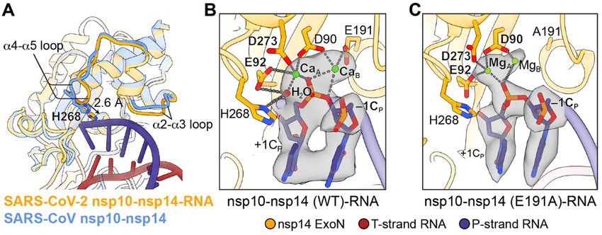
The substrate specificity of SARS-CoV-2 ExoN is contributed        dsRNA substrate with the same P-strand sequence (fig. S9).
by many interactions between nsp14 and the RNA substrate           This is likely due to the weaker binding of ssRNA to SARS-
(Fig. 3, A and B). F146 at the bottom of the SARS-CoV-2 ExoN       CoV-2 ExoN resulting from the loss of protein-RNA interac-
substrate-binding pocket stacks against the 3′-end unpaired        tions on the T-strand side (Fig. 3, A and B). The ability of
+1CP. N104 inserts into the minor groove of the dsRNA and          ExoN to accept both ssRNA and dsRNA substrates suggest
establishes two hydrogen bonds with the nucleobase and 2′-         two possible modes of mismatch correction in vivo. ExoN
OH group of –1GT, respectively. H95, which is approximately        may bind to and cleave the 3′-end single-stranded region of
co-planar with the unpaired +1CP, is hydrogen bonded with          P-strand RNA resulted from RdRp backtracking, as proposed
the cytidine base and stacks against –1GT (Fig. 3B). The ability   by previous studies (21, 29). Alternatively, the dsRNA sub-
of H95 to act as both hydrogen bond donor and acceptor             strates containing a 3′-end mismatch may dissociate from
probably allows it to accommodate all four types of nucleo-        RdRp and are subsequently recognized by ExoN for mis-
tides, explaining the relative insensitivity of nsp14 to sub-      match excision.
strate sequence (18). Digestion of dsRNA substrates by SARS-           Removing the 2′- or 3′-OH groups of the 3′-end nucleotide
CoV-2 ExoN may slow at a C-G base pair due to the higher           either reduces or almost abolishes nucleolytic degradation by
energy required to break this base pair. Additionally, P142,       SARS-CoV-2 ExoN within the range of tested enzyme concen-

                                                                                                                                        Downloaded from http://science.sciencemag.org/ on July 28, 2021
situated at the rim of the ExoN RNA-binding pocket, works          trations (Fig. 4B), consistent with the previous findings on
together with H95 to restrict the depth of the substrate-bind-     SARS-CoV ExoN (18) and reflecting the important roles of 2′-
ing pocket on the T-strand side and likely forces the strand       and 3′-oxygens in coronavirus ExoN catalysis. On the other
separation of the RNA substrate 3′-end C-U mismatched pair         hand, 2′-O-methylation of the 3′-end cytidine does not signif-
(Fig. 3, B and C). The lower energy for separating a mis-          icantly affect the substrate cleavage by SARS-CoV-2 nsp10-
matched base pair could explain the preference of corona-          nsp14 (Fig. 4B), likely because some interactions between the
virus ExoN for dsRNA substrate with a 3′-end mismatch over         2′-oxygen and nsp14 are retained.
a perfectly matched substrate (18). By contrast, the LASV              Remdesivir is the only FDA-approved nucleotide analog
ExoN RNA-binding pocket has a slightly deeper opening on           antiviral to treat COVID-19. To assess if remdesivir can be ef-
the non-scissile strand side and therefore is able to accom-       fectively excised by SARS-CoV-2 ExoN, we modeled the incor-
modate a fully base-paired dsRNA substrate (26) (Fig. 3D).         porated form of the inhibitor, remdesivir monophosphate
This is consistent with its role as an dsRNA-degrading im-         (RMP), at the +1 position of the P-strand (Fig. 4C). The mod-
mune suppressor, rather than an RNA synthesis proofreader          eled RMP maintains most of the favorable interactions
(14, 15). At the other end of the spectrum, are DNA polymer-       formed between nsp14 and the 3′-end CMP. In addition, the
ase-associated proofreading ExoNs, such as the E. coli DNA         1′-cyano group of RMP, the determinant of its delayed RdRp
polymerase III (Pol III) ε subunit. It has a much narrower         stalling activity (6, 7), snugly fits in the space between H95
DNA-binding pocket, partially due to its tight association         and N104 and forms hydrogen bonds with the side chain ni-
with the Pol III α subunit, and can only fit a single-stranded     trogen atoms from the two residues (Fig. 4C). These observa-
DNA substrate (27) (Fig. 3E). All the RNA-contacting residues      tions indicate that product RNA containing RMP could be a
in nsp14 are highly conserved among different coronavirus          substrate for coronavirus ExoN, consistent with the findings
genera (fig. S8), indicating a shared RNA substrate recogni-       that RNA terminated with RMP does not display significant
tion mechanism of coronavirus ExoN.                                resistance to ExoN excision (30) and that coronaviruses lack-
    As a 3′–5′ exoribonuclease, SARS-CoV-2 nsp10-nsp14 spe-        ing ExoN proofreading activity was significantly more sensi-
cifically recognizes the 2′- and 3′-OH groups of the 3′-end nu-    tive to remdesivir (31).
cleotide. The 2′-OH of +1CP forms two hydrogen bonds with              Our study gives insights into the mechanism of mismatch
H95 and the carbonyl oxygen of G93, respectively, whereas          correction during SARS-CoV-2 RNA synthesis and reveals the
the 3′-OH of the nucleotide is hydrogen bonded with the G93        structural features in the substrate that are essential for ExoN
main chain nitrogen and catalytic residue E92 (Fig. 4A). To        recognition and catalysis, providing a basis for structural-
examine the effects of the 2′- and 3′-OH groups of the 3′-end      guided design of specific and potent ExoN inhibitors. Coad-
nucleotide on RNA cleavage efficiency by SARS-CoV-2 ExoN,          ministration of such ExoN inhibitors with nucleotide analog-
we performed the exonuclease assays using 32-nt single-            based viral RdRp antivirals could constitute a more effective
stranded RNA (ssRNA) substrates (referred to as P32 RNAs)          treatment for COVID-19. Additionally, our study sheds light
ending with either a standard ribonucleotide or a nucleotide       on the development of ExoN-resistant nucleotide analog in-
with modifications at the 2′ or 3′ position (Fig. 4B). SARS-       hibitors. In particular, we show that a free 3′-OH of the RNA
CoV-2 nsp10-nsp14 efficiently cleaves the unmodified ssRNA,        substrate is critical for exonucleolytic degradation by ExoN.
although significantly higher enzyme concentrations are            It has been shown that 3′-deoxy ribonucleotides can be effi-
needed to obtain cleavage comparable to that achieved on a         ciently incorporated into nascent RNA by RdRp from other

First release: 27 July 2021                            www.sciencemag.org       (Page numbers not final at time of first release)   3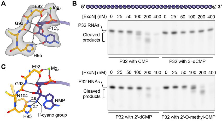
positive-strand RNA viruses, such as HCV and poliovirus,                                           17. Y. Chen, H. Cai, J. Pan, N. Xiang, P. Tien, T. Ahola, D. Guo, Functional screen reveals
and subsequently block RNA extension (32, 33). Therefore,                                               SARS coronavirus nonstructural protein nsp14 as a novel cap N7
                                                                                                        methyltransferase. Proc. Natl. Acad. Sci. U.S.A. 106, 3484–3489 (2009).
3′-deoxy nucleotide analogs can potentially act as effective                                            doi:10.1073/pnas.0808790106 Medline
coronavirus RdRp chain terminators that also resist ExoN ex-                                       18. M. Bouvet, I. Imbert, L. Subissi, L. Gluais, B. Canard, E. Decroly,
cision. Nonetheless, modifications at other positions on the                                            RNA 3′-end mismatch excision by the severe acute respiratory syndrome
                                                                                                        coronavirus nonstructural protein nsp10/nsp14 exoribonuclease complex. Proc.
ribose ring are also worth further exploration.
                                                                                                        Natl. Acad. Sci. U.S.A. 109, 9372–9377 (2012). doi:10.1073/pnas.1201130109
REFERENCES AND NOTES                                                                                    Medline
1. A. Mullard, COVID-19 vaccines buoy hope. Nat. Rev. Drug Discov. 20, 8 (2021).                   19. Y. Ma, L. Wu, N. Shaw, Y. Gao, J. Wang, Y. Sun, Z. Lou, L. Yan, R. Zhang, Z. Rao,
     doi:10.1038/d41573-020-00215-9 Medline                                                             Structural basis and functional analysis of the SARS coronavirus nsp14-nsp10
2. J. Ziebuhr, The coronavirus replicase. Curr. Top. Microbiol. Immunol. 287, 57–94                     complex. Proc. Natl. Acad. Sci. U.S.A. 112, 9436–9441 (2015).
     (2005). doi:10.1007/3-540-26765-4_3 Medline                                                        doi:10.1073/pnas.1508686112 Medline
3. I. Sola, F. Almazán, S. Zúñiga, L. Enjuanes, Continuous and Discontinuous RNA                   20. H. S. Hillen, G. Kokic, L. Farnung, C. Dienemann, D. Tegunov, P. Cramer, Structure
     Synthesis in Coronaviruses. Annu. Rev. Virol. 2, 265–288 (2015).                                   of replicating SARS-CoV-2 polymerase. Nature 584, 154–156 (2020).
     doi:10.1146/annurev-virology-100114-055218 Medline                                                 doi:10.1038/s41586-020-2368-8 Medline
4. A. J. te Velthuis, J. J. Arnold, C. E. Cameron, S. H. van den Worm, E. J. Snijder, The          21. J. Chen, B. Malone, E. Llewellyn, M. Grasso, P. M. M. Shelton, P. D. B. Olinares, K.
     RNA polymerase activity of SARS-coronavirus nsp12 is primer dependent. Nucleic                     Maruthi, E. T. Eng, H. Vatandaslar, B. T. Chait, T. M. Kapoor, S. A. Darst, E. A.
     Acids Res. 38, 203–214 (2010). doi:10.1093/nar/gkp904 Medline                                      Campbell, Structural Basis for Helicase-Polymerase Coupling in the SARS-CoV-2

                                                                                                                                                                                                 Downloaded from http://science.sciencemag.org/ on July 28, 2021
5. Y. Gao, L. Yan, Y. Huang, F. Liu, Y. Zhao, L. Cao, T. Wang, Q. Sun, Z. Ming, L. Zhang,               Replication-Transcription Complex. Cell 182, 1560–1573.e13 (2020).
     J. Ge, L. Zheng, Y. Zhang, H. Wang, Y. Zhu, C. Zhu, T. Hu, T. Hua, B. Zhang, X. Yang,              doi:10.1016/j.cell.2020.07.033 Medline
     J. Li, H. Yang, Z. Liu, W. Xu, L. W. Guddat, Q. Wang, Z. Lou, Z. Rao, Structure of the        22. Y. Zhai, F. Sun, X. Li, H. Pang, X. Xu, M. Bartlam, Z. Rao, Insights into SARS-CoV
     RNA-dependent RNA polymerase from COVID-19 virus. Science 368, 779–782                             transcription and replication from the structure of the nsp7-nsp8 hexadecamer.
     (2020). doi:10.1126/science.abb7498 Medline                                                        Nat. Struct. Mol. Biol. 12, 980–986 (2005). doi:10.1038/nsmb999 Medline
6. G. Kokic, H. S. Hillen, D. Tegunov, C. Dienemann, F. Seitz, J. Schmitzova, L. Farnung,          23. Y. Xiao, Q. Ma, T. Restle, W. Shang, D. I. Svergun, R. Ponnusamy, G. Sczakiel, R.
     A. Siewert, C. Höbartner, P. Cramer, Mechanism of SARS-CoV-2 polymerase                            Hilgenfeld, Nonstructural proteins 7 and 8 of feline coronavirus form a 2:1
     stalling by remdesivir. Nat. Commun. 12, 279 (2021). doi:10.1038/s41467-020-                       heterotrimer that exhibits primer-independent RNA polymerase activity. J. Virol.
     20542-0 Medline                                                                                    86, 4444–4454 (2012). doi:10.1128/JVI.06635-11 Medline
7. J. P. K. Bravo, T. L. Dangerfield, D. W. Taylor, K. A. Johnson, Remdesivir is a delayed         24. Y. Zuo, M. P. Deutscher, Exoribonuclease superfamilies: Structural analysis and
     translocation inhibitor of SARS-CoV-2 replication. Mol. Cell 81, 1548–1552.e4                      phylogenetic distribution. Nucleic Acids Res. 29, 1017–1026 (2001).
     (2021). doi:10.1016/j.molcel.2021.01.035 Medline                                                   doi:10.1093/nar/29.5.1017 Medline
8. F. Ferron, L. Subissi, A. T. Silveira De Morais, N. T. T. Le, M. Sevajol, L. Gluais, E.         25. S. Hamdan, P. D. Carr, S. E. Brown, D. L. Ollis, N. E. Dixon, Structural basis for
     Decroly, C. Vonrhein, G. Bricogne, B. Canard, I. Imbert, Structural and molecular                  proofreading during replication of the Escherichia coli chromosome. Structure 10,
     basis of mismatch correction and ribavirin excision from coronavirus RNA. Proc.                    535–546 (2002). doi:10.1016/S0969-2126(02)00738-4 Medline
     Natl. Acad. Sci. U.S.A. 115, E162–E171 (2018). doi:10.1073/pnas.1718806115                    26. X. Jiang, Q. Huang, W. Wang, H. Dong, H. Ly, Y. Liang, C. Dong, Structures of
     Medline                                                                                            arenaviral nucleoproteins with triphosphate dsRNA reveal a unique mechanism of
9. F. Robson, K. S. Khan, T. K. Le, C. Paris, S. Demirbag, P. Barfuss, P. Rocchi, W.-L.                 immune suppression. J. Biol. Chem. 288, 16949–16959 (2013).
     Ng, Coronavirus RNA Proofreading: Molecular Basis and Therapeutic Targeting.                       doi:10.1074/jbc.M112.420521 Medline
     Mol. Cell 79, 710–727 (2020). doi:10.1016/j.molcel.2020.07.027 Medline                        27. R. Fernandez-Leiro, J. Conrad, J.-C. Yang, S. M. V. Freund, S. H. W. Scheres, M. H.
10. A. E. Gorbalenya, L. Enjuanes, J. Ziebuhr, E. J. Snijder, Nidovirales: Evolving the                 Lamers, Self-correcting mismatches during high-fidelity DNA replication. Nat.
     largest RNA virus genome. Virus Res. 117, 17–37 (2006).                                            Struct. Mol. Biol. 24, 140–143 (2017). doi:10.1038/nsmb.3348 Medline
     doi:10.1016/j.virusres.2006.01.017 Medline                                                    28. L. S. Beese, V. Derbyshire, T. A. Steitz, Structure of DNA polymerase I Klenow
11. N. S. Ogando, J. C. Zevenhoven-Dobbe, Y. van der Meer, P. J. Bredenbeek, C. C.                      fragment bound to duplex DNA. Science 260, 352–355 (1993).
     Posthuma, E. J. Snijder, The Enzymatic Activity of the nsp14 Exoribonuclease Is                    doi:10.1126/science.8469987 Medline
     Critical for Replication of MERS-CoV and SARS-CoV-2. J. Virol. 94, e01246-20                  29. B. Malone, J. Chen, Q. Wang, E. Llewellyn, Y. J. Choi, P. D. B. Olinares, X. Cao, C.
     (2020). doi:10.1128/JVI.01246-20 Medline                                                           Hernandez, E. T. Eng, B. T. Chait, D. E. Shaw, R. Landick, S. A. Darst, E. A.
12. M. Becares, A. Pascual-Iglesias, A. Nogales, I. Sola, L. Enjuanes, S. Zuñiga,                       Campbell, Structural basis for backtracking by the SARS-CoV-2 replication-
     Mutagenesis of Coronavirus nsp14 Reveals Its Potential Role in Modulation of the                   transcription complex. Proc. Natl. Acad. Sci. U.S.A. 118, e2102516118 (2021).
     Innate Immune Response. J. Virol. 90, 5399–5414 (2016).                                            doi:10.1073/pnas.2102516118 Medline
     doi:10.1128/JVI.03259-15 Medline                                                              30. S. Jockusch, C. Tao, X. Li, M. Chien, S. Kumar, I. Morozova, S. Kalachikov, J. J.
13. J. B. Case, Y. Li, R. Elliott, X. Lu, K. W. Graepel, N. R. Sexton, E. C. Smith, S. R. Weiss,        Russo, J. Ju, Sofosbuvir terminated RNA is more resistant to SARS-CoV-2
     M. R. Denison, Murine Hepatitis Virus nsp14 Exoribonuclease Activity Is Required                   proofreader than RNA terminated by Remdesivir. Sci. Rep. 10, 16577 (2020).
     for Resistance to Innate Immunity. J. Virol. 92, e01531-17 (2017).                                 doi:10.1038/s41598-020-73641-9 Medline
     doi:10.1128/JVI.01531-17 Medline                                                              31. M. L. Agostini, E. L. Andres, A. C. Sims, R. L. Graham, T. P. Sheahan,
14. K. M. Hastie, C. R. Kimberlin, M. A. Zandonatti, I. J. MacRae, E. O. Saphire, Structure             X. Lu, E. C. Smith, J. B. Case, J. Y. Feng, R. Jordan, A. S. Ray, T. Cihlar,
     of the Lassa virus nucleoprotein reveals a dsRNA-specific 3′ to 5′ exonuclease                     D. Siegel, R. L. Mackman, M. O. Clarke, R. S. Baric, M. R. Denison, Coronavirus
     activity essential for immune suppression. Proc. Natl. Acad. Sci. U.S.A. 108, 2396–                Susceptibility to the Antiviral Remdesivir (GS-5734) Is Mediated by the Viral
     2401 (2011). doi:10.1073/pnas.1016404108 Medline                                                   Polymerase and the Proofreading Exoribonuclease. mBio 9, e00221-18 (2018).
15. X. Qi, S. Lan, W. Wang, L. M. L. Schelde, H. Dong, G. D. Wallat, H. Ly, Y. Liang, C.                doi:10.1128/mBio.00221-18 Medline
     Dong, Cap binding and immune evasion revealed by Lassa nucleoprotein                          32. J. Shim, G. Larson, V. Lai, S. Naim, J. Z. Wu, Canonical 3′-deoxyribonucleotides as
     structure. Nature 468, 779–783 (2010). doi:10.1038/nature09605 Medline                             a chain terminator for HCV NS5B RNA-dependent RNA polymerase. Antiviral Res.
16. E. Minskaia, T. Hertzig, A. E. Gorbalenya, V. Campanacci, C. Cambillau, B. Canard,                  58, 243–251 (2003). doi:10.1016/S0166-3542(03)00007-X Medline
     J. Ziebuhr, Discovery of an RNA virus 3′->5′ exoribonuclease that is critically               33. G. Campagnola, P. Gong, O. B. Peersen, High-throughput screening identification
     involved in coronavirus RNA synthesis. Proc. Natl. Acad. Sci. U.S.A. 103, 5108–                    of poliovirus RNA-dependent RNA polymerase inhibitors. Antiviral Res. 91, 241–
     5113 (2006). doi:10.1073/pnas.0508200103 Medline                                                   251 (2011). doi:10.1016/j.antiviral.2011.06.006 Medline

First release: 27 July 2021                                                       www.sciencemag.org                  (Page numbers not final at time of first release)                     4
34. C. Kao, M. Zheng, S. Rüdisser, A simple and efficient method to reduce complex), 7N0C (SARS-CoV-2 nsp10-nsp14 (E191A)-RNA complex, monomer)
nontemplated nucleotide addition at the 3 terminus of RNAs transcribed by T7 and 7N0D (SARS-CoV-2 nsp10-nsp14 (E191A)-RNA complex, tetramer). The
RNA polymerase. RNA 5, 1268–1272 (1999). doi:10.1017/S1355838299991033 cryo-EM maps have been deposited in the Electron Microscopy Data Bank with
Medline accession number EMD-24102 (SARS-CoV-2 nsp10-nsp14 (WT)-RNA complex),
35. S. Q. Zheng, E. Palovcak, J.-P. Armache, K. A. Verba, Y. Cheng, D. A. Agard, EMD-24103 (SARS-CoV-2 nsp10-nsp14 (E191A)-RNA complex, monomer) and
MotionCor2: Anisotropic correction of beam-induced motion for improved cryo- EMD-24104 (SARS-CoV-2 nsp10-nsp14 (E191A)-RNA complex, tetramer).
electron microscopy. Nat. Methods 14, 331–332 (2017). doi:10.1038/nmeth.4193 Materials are available from Y.Y. upon request and may require a material
Medline transfer agreement. This work is licensed under a Creative Commons Attribution
36. J. Zivanov, T. Nakane, S. H. W. Scheres, Estimation of high-order aberrations and 4.0 International (CC BY 4.0) license, which permits unrestricted use,
anisotropic magnification from cryo-EM data sets in RELION-3.1. IUCrJ 7, 253– distribution, and reproduction in any medium, provided the original work is
267 (2020). doi:10.1107/S2052252520000081 Medline properly cited. To view a copy of this license, visit
37. A. Punjani, J. L. Rubinstein, D. J. Fleet, M. A. Brubaker, cryoSPARC: Algorithms for https://creativecommons.org/licenses/by/4.0/. This license does not apply to
rapid unsupervised cryo-EM structure determination. Nat. Methods 14, 290–296 figures/photos/artwork or other content included in the article that is credited
(2017). doi:10.1038/nmeth.4169 Medline to a third party; obtain authorization from the rights holder before using such
38. A. Punjani, H. Zhang, D. J. Fleet, Non-uniform refinement: Adaptive regularization material.
improves single-particle cryo-EM reconstruction. Nat. Methods 17, 1214–1221
(2020). doi:10.1038/s41592-020-00990-8 Medline SUPPLEMENTARY MATERIALS
39. T. D. Goddard, C. C. Huang, E. C. Meng, E. F. Pettersen, G. S. Couch, J. H. Morris, science.sciencemag.org/cgi/content/full/science.abi9310/DC1
T. E. Ferrin, UCSF ChimeraX: Meeting modern challenges in visualization and Materials and Methods
Downloaded from http://science.sciencemag.org/ on July 28, 2021
analysis. Protein Sci. 27, 14–25 (2018). doi:10.1002/pro.3235 Medline Figs. S1 to S9
40. Y. Z. Tan, P. R. Baldwin, J. H. Davis, J. R. Williamson, C. S. Potter, B. Carragher, D. Table S1
Lyumkis, Addressing preferred specimen orientation in single-particle cryo-EM References (34–47)
through tilting. Nat. Methods 14, 793–796 (2017). doi:10.1038/nmeth.4347 MDAR Reproducibility Checklist
Medline
41. A. Casañal, B. Lohkamp, P. Emsley, Current developments in Coot for 9 April 2021; accepted 20 July 2021
macromolecular model building of Electron Cryo-microscopy and Published online 27 July 2021
Crystallographic Data. Protein Sci. 29, 1069–1078 (2020). doi:10.1002/pro.3791 10.1126/science.abi9310
Medline
42. J. A. Pleiss, M. L. Derrick, O. C. Uhlenbeck, T7 RNA polymerase produces 5′ end
heterogeneity during in vitro transcription from certain templates. RNA 4, 1313–
1317 (1998). doi:10.1017/S135583829800106X Medline
43. P. V. Afonine, B. K. Poon, R. J. Read, O. V. Sobolev, T. C. Terwilliger, A. Urzhumtsev,
P. D. Adams, Real-space refinement in PHENIX for cryo-EM and crystallography.
Acta Crystallogr. D 74, 531–544 (2018). doi:10.1107/S2059798318006551
Medline
44. V. B. Chen, W. B. Arendall 3rd, J. J. Headd, D. A. Keedy, R. M. Immormino, G. J.
Kapral, L. W. Murray, J. S. Richardson, D. C. Richardson, MolProbity: All-atom
structure validation for macromolecular crystallography. Acta Crystallogr. D 66,
12–21 (2010). doi:10.1107/S0907444909042073 Medline
45. F. Madeira, Y. M. Park, J. Lee, N. Buso, T. Gur, N. Madhusoodanan, P. Basutkar, A.
R. N. Tivey, S. C. Potter, R. D. Finn, R. Lopez, The EMBL-EBI search and sequence
analysis tools APIs in 2019. Nucleic Acids Res. 47, W636–W641 (2019).
doi:10.1093/nar/gkz268 Medline
46. X. Robert, P. Gouet, Deciphering key features in protein structures with the new
ENDscript server. Nucleic Acids Res. 42, W320–W324 (2014).
doi:10.1093/nar/gku316 Medline
47. C. A. Schneider, W. S. Rasband, K. W. Eliceiri, NIH Image to ImageJ: 25 years of
image analysis. Nat. Methods 9, 671–675 (2012). doi:10.1038/nmeth.2089
Medline
ACKNOWLEDGMENTS
We thank S. Wu for help with cryo-EM data collection at Yale Titan Krios cryo-EM
facility. We particularly thank Dr. Ronald Breaker at Yale University for his
support during the early phases of this work. Funding: This work was supported
by NVIDIA GPU Grant Program (C.L.), the start-up funding granted to B.L. from
the Hormel Institute, University of Minnesota and the start-up funding granted to
Y.Y. from Iowa State University. Author contributions: Y.Y. and C.L. conceived
and designed the experiments. Y.Y. performed the biochemical
characterizations. Y.Y., B.L, and W.S. prepared the cryo-EM samples and
collected cryo-EM data. Y.Y., C.L. and B.L. processed the cryo-EM data. C.L. and
Y.Y. performed modeling building and structural analyses. Y.Y. and C.L. wrote
the manuscript with input from S.T.B., D.G.S and B.L. Competing interests: The
authors declare no competing interests. Data and materials availability: Atomic
coordinates of three structures determined in this study have been deposited in
PDB with accession codes 7N0B (SARS-CoV-2 nsp10-nsp14 (WT)-RNA
First release: 27 July 2021 www.sciencemag.org (Page numbers not final at time of first release) 5Downloaded from http://science.sciencemag.org/ on July 28, 2021
Fig. 1. Structural and functional overview of SARS-CoV-2 nsp10-nsp14-
RNA complexes. (A) Domain organization of SARS-CoV-2 nsp10 and nsp14.
Domain boundary residues are numbered. Five catalytic residues in the
nsp14 ExoN domain are indicated as red dots and are labeled. (B) Cleavage
of T35P31 RNA substrate by SARS-CoV-2 nsp10-nsp14 ExoN complex. The
concentrations (in nM) of wild-type (WT) or E191A mutant ExoN are
indicated. The RNAs were resolved by denaturing polyacrylamide gel
electrophoresis (PAGE) and stained by SYBR Gold. A representative result
from three biological replicates is shown. (C) Sequence and numbering
of the T35P31 RNA substrate used in biochemical characterization and
structural determination. T-strand, template strand; P-strand, product
strand. T-strand and P-strand RNAs are connected by a UUCG tetraloop.
(D) Cryo-EM map and atomic model of the monomeric form of SARS-CoV-2
nsp10-nsp14-RNA complex. (E) Cryo-EM map and atomic model of the
tetrameric form of SARS-CoV-2 nsp10-nsp14-RNA complex.
First release: 27 July 2021 www.sciencemag.org (Page numbers not final at time of first release) 6Fig. 2. Active site conformation and catalytic mechanism of SARS-CoV-2
ExoN. (A) Superimposition of SARS-CoV nsp10-nsp14 complex (cornflower
blue, PDB 5C8U) and SARS-CoV-2 nsp10-nsp14-RNA complex (orange)
illustrates the conformational changes of α2-α3 and α4-α5 loops and a 2.6 Å
Downloaded from http://science.sciencemag.org/ on July 28, 2021
shift of H268 toward the RNA upon substrate binding. (B) Active site
structure of SARS-CoV-2 nsp10-nsp14 (WT)-RNA complex. Ca2+ ions, green
spheres; catalytic water, red sphere. Nucleotide residues in P-strand RNA
are indicated with subscript “P.” +1CP, –1CP, the catalytic water and two
active site metal ions are superimposed with their cryo-EM densities
contoured at 10σ. (C) Active site structure of SARS-CoV-2 nsp10-nsp14
(E191A)-RNA complex. Mg2+ ions, green spheres. +1CP, –1CP, and two active
site metal ions are superimposed with their cryo-EM densities contoured at
7σ.
First release: 27 July 2021 www.sciencemag.org (Page numbers not final at time of first release) 7Downloaded from http://science.sciencemag.org/ on July 28, 2021
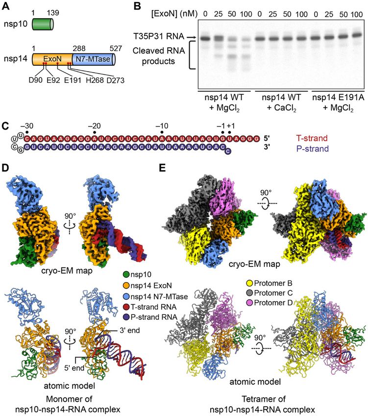
Fig. 3. Mechanism of substrate recognition by SARS-CoV-2 ExoN. (A) Interactions
between SARS-CoV-2 nsp10-nsp14 ExoN and T35P31 RNA backbone. Nucleotide
residues in T-strand RNA are indicated with subscript “T,” nucleotide residues in
P-strand RNA are indicated with subscript “P.” Hydrogen bonds and salt bridges are
shown as gray dotted lines. Interacting nucleotide and protein residues are
superimposed with their cryo-EM densities contoured at 7σ. (B) Interactions between
SARS-CoV-2 nsp10-nsp14 ExoN and T35P31 RNA at +1 and –1 nucleobases positions.
Hydrogen bonds and salt bridges are shown as gray dotted lines. π-π stacking
interactions are indicated by green dotted lines. Interacting nucleotide and protein
residues are superimposed with their cryo-EM densities contoured at 7σ. (C) Surface
representation of SARS-CoV-2 nsp10-nsp14 ExoN substrate-binding pocket shows a
restricted opening on the T-strand side that prevents base-pairing at substrate RNA +1
position. For clarity, the N7-MTase domain of nsp14 is not shown. (D) Surface
representation of LASV NP ExoN domain. A fully base-paired dsRNA substrate (shown
as cartoons) is bound in the substrate-binding pocket of LASV ExoN. (E) Surface
representation of E. coli DNA Pol III ExoN complex. The narrow substrate-binding
pocket allows the entry of ssDNA only.
First release: 27 July 2021 www.sciencemag.org (Page numbers not final at time of first release) 8Downloaded from http://science.sciencemag.org/ on July 28, 2021
Fig. 4. Structural insights into antiviral design. (A) Interactions between
SARS-CoV-2 nsp10-nsp14 ExoN and 3′-end nucleotide mediated by its
2′- and 3′-OH groups. Mg2+ ion, green sphere. Hydrogen bonds are shown
as gray dotted lines. +1CP and its interacting protein residues are
superimposed with cryo-EM densities contoured at 7σ. (B) Cleavage of
various P32 ssRNA substrates by SARS-CoV-2 nsp10-nsp14 complex. The
3′-end CMP that bears different modifications at its 2′- or 3′-OH groups is
shown as a black letter in white circles. The concentrations (in nM) of ExoN
are indicated. The RNAs were resolved by denaturing polyacrylamide gel
electrophoresis (PAGE) and stained by SYBR Gold. A representative result
from three biological replicates is shown. (C) Predicted interactions between
SARS-CoV-2 nsp10-nsp14 ExoN and remdesivir monophosphate (RMP),
which is modeled at the 3′-end +1 position of P-strand RNA.
First release: 27 July 2021 www.sciencemag.org (Page numbers not final at time of first release) 9Structural basis of mismatch recognition by a SARS-CoV-2 proofreading enzyme
Chang Liu, Wei Shi, Scott T. Becker, David G. Schatz, Bin Liu and Yang Yang
published online July 27, 2021
ARTICLE TOOLS http://science.sciencemag.org/content/early/2021/07/26/science.abi9310
Downloaded from http://science.sciencemag.org/ on July 28, 2021
SUPPLEMENTARY http://science.sciencemag.org/content/suppl/2021/07/26/science.abi9310.DC1
MATERIALS
REFERENCES This article cites 47 articles, 17 of which you can access for free
http://science.sciencemag.org/content/early/2021/07/26/science.abi9310#BIBL
PERMISSIONS http://www.sciencemag.org/help/reprints-and-permissions
Use of this article is subject to the Terms of Service
Science (print ISSN 0036-8075; online ISSN 1095-9203) is published by the American Association for the Advancement of
Science, 1200 New York Avenue NW, Washington, DC 20005. The title Science is a registered trademark of AAAS.
Copyright © 2021 The Authors, some rights reserved; exclusive licensee American Association for the Advancement of Science.
No claim to original U.S. Government Works. Distributed under a Creative Commons Attribution License 4.0 (CC BY).You can also read