MILD SARS-COV-2 INFECTIONS AND NEUTRALIZING ANTIBODY TITERS
←
→
Page content transcription
If your browser does not render page correctly, please read the page content below
Prepublication Release

 Mild SARS-CoV-2 Infections and
 Neutralizing Antibody Titers
 Francesco Bonfante, DVM, Paola Costenaro, MD, DTM&H, Anna Cantarutti, PhD,
 Costanza Di Chiara, MD, Alessio Bortolami, DVM, PhD, Maria Raffaella Petrara, PhD,
 Francesco Carmona, BSc, Matteo Pagliari, PhD, Chiara Cosma, MD, Sandra Cozzani, MD,
 Eva Mazzetto, DVM, Giovanni Di Salvo, MD, Prof, Liviana Da Dalt, MD, Prof,
 Paolo Palma,MD, Prof, Luisa Barzon, MD, Prof, Giovanni Corrao, Prof,
 Calogero Terregino,MD, Prof, Andrea Padoan, MD, Prof, Mario Plebani, MD, Prof,
 Anita De Rossi, PhD, Prof, Daniele Donà, MD, PhD, Carlo Giaquinto , MD, Prof

DOI: 10.1542/peds.2021-052173
Journal: Pediatrics
Article Type: Regular Article

Citation: Bonfante F, Costenaro P, Cantarutti A, et al. Mild SARS-CoV-2 infections and
neutralizing antibody titers. Pediatrics. 2021; doi: 10.1542/peds.2021-052173

This is a prepublication version of an article that has undergone peer review and been accepted
for publication but is not the final version of record. This paper may be cited using the DOI and
date of access. This paper may contain information that has errors in facts, figures, and
statements, and will be corrected in the final published version. The journal is providing an early
version of this article to expedite access to this information. The American Academy of
Pediatrics, the editors, and authors are not responsible for inaccurate information and data
described in this version.

 ©2021 American Academy of Pediatrics
 Downloaded from www.aappublications.org/news by guest on September 9, 2021
Prepublication Release

 Mild SARS-CoV-2 Infections and Neutralizing Antibody Titers

 Francesco Bonfante1†, DVM, Paola Costenaro2†, MD, DTM&H, Anna Cantarutti3, PhD,
 Costanza Di Chiara2, MD, Alessio Bortolami1, DVM, PhD, Maria Raffaella Petrara4, PhD,
 Francesco Carmona5, BSc, Matteo Pagliari1, PhD, Chiara Cosma6, MD, Sandra Cozzani2, MD,
 Eva Mazzetto1, DVM, Giovanni Di Salvo7, MD, Prof, Liviana Da Dalt7, MD, Prof,
 Paolo Palma8,9, MD, Prof, Luisa Barzon10, MD, Prof, Giovanni Corrao3,11, Prof,
 Calogero Terregino1, MD, Prof, Andrea Padoan12, MD, Prof, Mario Plebani6,12, MD, Prof,
 Anita De Rossi4,5, PhD, Prof, Daniele Donà2*, MD, PhD, Carlo Giaquinto2*, MD, Prof

† contributed equally as co-first authors
*contributed equally as co-senior authors

Affiliations:
1
 Division of Comparative Biomedical Sciences, Istituto Zooprofilattico Sperimentale delle
Venezie, Padua, Italy.
2
 Division of Pediatric Infectious Diseases, Department for Women’s and Children’s Health,
University of Padua, Italy.
3
 Department of Statistics and Quantitative Methods, Division of Biostatistics, Epidemiology and
Public Health, Laboratory of Healthcare Research and Pharmacoepidemiology, University of
Milano-Bicocca, Milan, Italy.
4
 Department of Surgery, Oncology and Gastroenterology, Section of Oncology and Immunology,
University of Padua, Padua, Italy.
5
 Istituto Oncologico Veneto (IOV) - IRCCS, Padua, Italy.
6
 Department of Laboratory Medicine, University-Hospital of Padua, Italy.
7
 Department for Women's and Children's Health, University of Padua, Italy.
8
 Research Unit of Congenital and Perinatal Infections, Bambino Gesù Children's Hospital, Rome;
9
 Chair of Pediatrics, Department of Systems Medicine, University of Rome "Tor Vergata", Rome.
10
 Department of Molecular Medicine, University of Padua, Italy.
11
 National Centre for Healthcare Research and Pharmacoepidemiology, University of Milano-
Bicocca, Milan, Italy
12
 Department of Medicine-DIMED, University of Padua, Italy.

 ©2021 American Academy of Pediatrics
 Downloaded from www.aappublications.org/news by guest on September 9, 2021
Prepublication Release

Corresponding author:
Francesco Bonfante, Istituto Zooprofilattico Sperimentale delle Venezie, viale dell’Università
10, 35020 Legnaro, Padua (Italy); FBonfante@izsvenezie.it ; ORCID: 0000-0002-1276-2710

Conflict of interest disclosures: The authors declare no conflicts of interest relevant to this article
to disclose.
Funding: This work is partially supported by the ORCHESTRA project, Horizon 2020 research
and innovation programme under Grant Agreement Number 101016167 to CG. In addition, this
work has received funding from the EU Horizon 2020 (RECOVER), Grant Agreement Number
101003589 to CG, and Fondazione Cassa di Risparmio di Padova e Rovigo, Progetti di Ricerca
COVID-19 (ADR participant).

Abbreviations:
CLIA, chemiluminescence immunoassay
COVID-19, Coronavirus disease 2019
CovFC, COVID-19 Family Cluster Follow-up Clinic
ddPCR, digital droplet Polymerase Chain Reaction
FP, family pediatrician
GMT, geometric mean titer
hCoV, human coronaviruses
nAbs, neutralizing antibodies
NPS, nasal-pharyngeal swab
PRNT, Plaque Reduction Neutralizing Test
RT-PCR, real-time Polymerase Chain Reaction

Article summary: High levels of SARS-CoV-2 neutralizing antibodies (nAbs) are found up to 7-
8 months after asymptomatic/mild COVID-19, particularly in children aged less than 6 years.

What’s Known on This Subject:
Children/adolescents usually present with asymptomatic/mild COVID-19 diseases; however, they
are key in transmitting SARS-CoV-2 infection. Recent findings showed that neutralizing
antibodies (nAbs) persist up to 6 months in convalescent adults, however little is known about
nAbs kinetics in children.
What This Study Adds:
Younger children develop higher levels of nAbs during the first 7-8 months after
asymptomatic/mild symptomatic COVID-19, compared to older siblings/adults. The long-lasting

 ©2021 American Academy of Pediatrics
 Downloaded from www.aappublications.org/news by guest on September 9, 2021
Prepublication Release

levels of nAbs may lead to durable protection and higher viral clearance, reducing shedding and
transmission.
Abstract

Background. Recent evidence suggests that neutralizing antibodies to SARS-CoV-2 may persist
over time; however, knowledge regarding pediatric subjects is limited.

Methods. A single-center, prospective observational study was conducted on 57 family clusters
of COVID-19, including children of neonatal and pediatric age attending the University Hospital
of Padua (Italy). For each patient, blood samples were collected for both the quantification of
neutralizing antibodies (nAbs) through a Plaque Reduction Neutralizing Test (PRNT) and the
detection of anti-nucleocapsid-spike protein IgG/IgM.

Results. We analyzed 283 blood samples collected from 152 confirmed COVID-19 cases (82
parents and 70 children/older siblings of median age of 8 years, IQR 4-13), presenting
asymptomatic or with mildly symptomatic disease. Despite the decrease of IgG over time, nAbs
were found to persist up to 7-8 months in children while adults recorded a modest declining trend.
Interestingly, children under 6 years of age, and in particular under 3 years developed higher long-
lasting levels of nAbs compared to older siblings and/or adults.

Conclusion. Mild and asymptomatic SARS-CoV-2 infections in family clusters elicited higher
neutralizing antibodies among children.

Introduction

European countries have been facing a “third wave” of the novel Coronavirus disease 2019

(COVID-19) pandemic and the spread of several SARS-CoV-2 variants. With the advent of

vaccines 1, longitudinal studies of both convalescent and vaccinated patients are of fundamental

importance to understand the kinetics of humoral response and infer correlates of protection for

both infection and disease. In this respect, the titration of neutralizing antibodies (nAbs) is key to

determine the concentration of antibodies preventing cells to be infected by SARS-CoV-2 2.

 ©2021 American Academy of Pediatrics
 Downloaded from www.aappublications.org/news by guest on September 9, 2021
Prepublication Release

Studies including convalescent adults reported that humoral immunity against SARS-CoV-2 may
 3–5
be short-lived, particularly in persons with mild illness . However recent findings provided
 6–10
evidences of nAbs persisting up to 6 months , as after seasonal and SARS-like coronavirus

infection, where nAbs can persist, respectively up to one or several years 11,12.

SARS-CoV-2 infection in children is less severe than in adults 13, resulting in underdiagnosis given
 14
the mild or asymptomatic clinical course . However, children and adolescents are key in the

transmission of infection 15. Little is known about the kinetics of SARS-CoV-2 nAbs in pediatric

populations. Understanding the differences in the antibody response between adults and children

has important scientific and public health implications, including design of risk-based surveillance

programs, cost-effective vaccination campaigns and mathematical modelling of clinical outcome.

In this study, we evaluated the role of age as a determinant of the production and persistence of

naturally acquired nAbs among a cohort of family clusters of COVID-19, including adults and

children who recovered from asymptomatic/mild symptomatic infections.

METHODS

Study design and population

A single-center, prospective study was conducted on Italian family clusters of COVID-19

attending the COVID-19 Family Cluster Follow-up Clinic (CovFC), at the Department of

Women’s and Children’s Health of the University Hospital of Padua (Veneto Region, Italy). From

March 1st to September 4th 2020, 57 families were enrolled meeting the following inclusion

criteria: a) having children of pediatric age (
Prepublication Release program 4-8 weeks after the end of either isolation or hospitalization, and after referral from the family pediatrician (FP). Evaluation of children and relatives included data collection on demographic parameters and past medical history, clinical evaluation and the collection of a blood sample for a characterization of the immune response to SARS-CoV-2. All subjects older than 18 years of age, including older siblings and parents, and legally authorized representatives of subjects under 18 years of age, were informed of the research proposal and provided written consent for the collection and use of biological specimens and routine patient-based data for research purposes. Families were invited to return to the clinic for longitudinal blood collection. The protocol was communicated to the Ethical Committee according to the national regulation (Prot. N° 0070714 of November 24th, 2020; amendment N°71779 of November 26th, 2020). Data collection and definitions Information collected during the clinic were entered into a web-based database using the REDCap platform (Vanderbilt University, Tennessee) hosted in the server of the University of Padova. For this study, data were collected retrospectively from the existing clinical files and analyzed anonymously. Subjects were considered confirmed COVID-19 cases if they had a record of virological positivity for SARS-CoV-2 by real-time RT-PCR according to routine diagnostic molecular protocols16 and/or resulted positive by either of the two serological tests adopted in this study. For each confirmed COVID-19 case, a baseline date was defined as follows: 1) for symptomatic cases: the first date between the onset of symptoms or the date of first positive SARS- CoV-2 molecular assay; 2) for asymptomatic cases: the date of the first positive molecular assay or, in those with only serologically confirmed COVID-19 and with negative/undetermined nasal- pharyngeal swab (NPS), by the family outbreak temporal sequence, coinciding with the date of ©2021 American Academy of Pediatrics Downloaded from www.aappublications.org/news by guest on September 9, 2021
Prepublication Release

symptoms onset in a virologically confirmed SARS-CoV-2 family outbreak (Supplementary

Figure S2). Subjects that were asymptomatic and had no analytical evidence of SARS-CoV-2

infection were considered non-COVID-19 cases. The severity of COVID-19 was scored as mild,
 17
moderate, severe, critical, following the WHO classification . For stratification purposes

individuals were divided, based on both social and biological development, in toddlers (
Prepublication Release

Statistical analyses

Descriptive statistics were used for comparing the distribution of gender, age, disease-related

symptoms and pediatric comorbidities between COVID-19 infected and uninfected patients.

The humoral response was assessed comparing the geometric mean titer (GMT), and the 95%

confidence interval (95% CI), of IgM, IgG, and PRNT50 values, in the overall dataset including

both independent and subject-paired samples, stratified by age classes and by time between

serological sampling and baseline, categorizing subjects into three intervals, namely 1-2, 3-6 and

7-8 months. The one-way ANOVA and the independent samples t-test were performed, where

appropriate. Associations between antibody titers, baseline intervals and age, were assessed with

linear regression models. Strength of associations between variables was assessed by Pearson

correlation coefficient, using the logarithm (base 10) of the antibody titers given data skew.

Use of the robust variance estimator to account for correlations within patients with multiple blood

samplings did not change the confidence intervals considerably in the unadjusted analyses, so

correlation structures were omitted from all analyses. Among a sub-cohort of subjects that agreed

to be sampled again after enrolment, a dependent t-test for subject-paired samples was used to

compare the GMT and 95% CI.

To test the robustness of our datasets against selection bias, we conducted a chi-square test and

verified the homogeneity within each age class and time window of a) the temporal distribution of

serological samplings (p=0.4363) and b) the proportion of cases identified by

virological/serological methods (p=0.6568). Moreover, we conducted a chi-square test to verify

 ©2021 American Academy of Pediatrics
 Downloaded from www.aappublications.org/news by guest on September 9, 2021
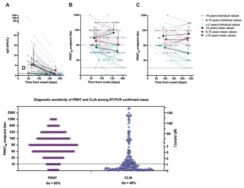
Prepublication Release

among subjects who contributed with either 1, 2 or three samples the homogeneity of gender

(p=0.6082), age (p=0.0973), family position (p=0.3971) and severity of symptoms (p=0.6947).

The diagnostic sensitivity of the CLIA and PRNT assays were assessed on subjects with a positive

NPS. Considering the PRNT assay as reference method for the validation of immunoassays for

SARS-CoV-2, we calculated measures of diagnostic accuracy of the CLIA assay.

Analyses were performed using the Statistical Analysis System software (version 9.4; SAS

Institute, Cary, North Carolina). Statistical significance was set at the .05 level. All P values were

2‐sided. Graphs were made using GraphPad Prism version 9 (GraphPad Software, La Jolla, CA).

RESULTS

From March 1st to December 3rd 2020, we prospectively evaluated 57 family clusters of COVID-

19 (Supplementary Figure S1). A serological assessment was performed at least once on 209

recruited subjects. Subjects who had previously tested positive for SARS-CoV-2 by real-time RT-

PCR (111/209) were considered confirmed COVID-19 cases, together with individuals that had

no record of virological positivity but showed evidence of seropositivity by either of the two

serological tests adopted in this study (44/209). Descriptive analysis and additional information on

baseline identification are provided as Supplementary Material (Table S1; Supplementary Figure

S2). Three out of 73 children were excluded from the analyses (see supplement legend to Figure

S1). In total 152 confirmed COVID-19 cases were studied: 70 children/older siblings and 82

parents with median ages of 8 (interquartile range (IQR), 4-13) and 42 years (IQR, 34-46),

respectively. Out of 152 cases, 38, 97 and 17 were sampled once, twice and three times,

respectively.

 ©2021 American Academy of Pediatrics
 Downloaded from www.aappublications.org/news by guest on September 9, 2021
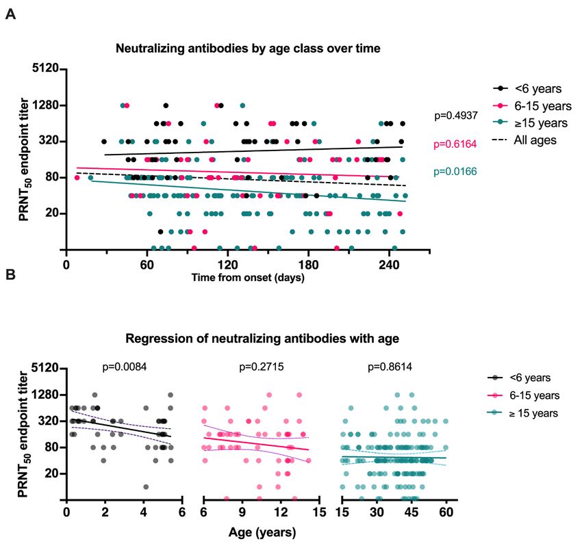
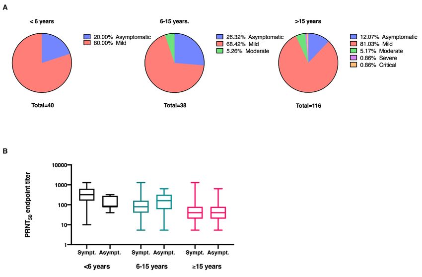
Prepublication Release

Analyzing all 283 blood samples collected from confirmed COVID-19 cases, we observed that

nAbs persisted in the population, (Figure 1A) recording a modest non-significant decline

(p=0.1062) over a median period of 132 days (IQR, 79-187) from baseline. When samples were

stratified by age, children under 6 years where the only class with a slightly increasing trend over

time, as opposed to children of 6-15 years and adults, although only for subjects ≥15 years of age

we recorded a statistical support for the regression line (p=0.0166). A further correlation analysis

confirmed that nAbs inversely correlated with age (Pearson =-0.4144, p
Prepublication Release

Tables 1 and S3) (time window 2). In time window 1, we observed an increase of nAbs titers for

children under 6 years (slope 0.0076), while children of 6-
Prepublication Release

We further explored whether nAbs correlated with either clinical presentation or viral load.

Differences in the distribution of clinical presentations between age classes were non-significant

(Figure 4A) and nAbs titers did not significantly differ between subjects showing mild or no

symptoms (Figure 4B).

For 63/111 COVID-19 confirmed cases that had recorded virological positivity, the original swab

was available for viral load quantification by ddPCR. In order to select a biologically relevant

period of infection and standardize comparisons, we focused on a subgroup of 32/63 subjects for

whom swabs had been collected within 4 days from symptom onset and serological samplings had

been taken within 1-2 months. We observed that adults recorded a mean viral load of 107.88 copies,

while children under 6 and those aged 6-
Prepublication Release

The current study describes a longitudinal comparison of the magnitude/persistence of nAbs

against SARS-CoV-2, among asymptomatic and mildly symptomatic toddlers, pre-school

children, school-age subjects and parents, in family clusters of COVID-19. In our cohort,

antibodies neutralizing SARS-CoV-2 virus persisted over a period of 2-8 months from infection,

recording only a modest decline. This result is in line with previous studies using PRNT and
 7–10,21,22
surrogate-neutralization based-assays describing a minimal decline of nAbs in adult

populations. Surprisingly, nAbs inversely correlated with age and children under 6 years, and in

particular toddlers under 3 years, had the highest titers throughout early, intermediate and late

times from infection onset. Our data strengthens and expands recent work published by Yang et

al. 23 who described higher surrogate neutralizing ability and avidity of antibodies in children aged

1-10 years, proving these features to be age-dependent, in a cohort of subjects aged 1-24 years,

early after recovery. In contrast with our findings, other studies indicated that nAbs in children

were lower than in adults 24,25. However, in one study 24 stratification by age was done below/above

24 years and children and adults were sampled around 5 and 12 days from hospital admission,
 25
respectively; in the other study , authors compared children with mildly affected adults

previously selected as plasma donors at the hospital. We believe these selection and sampling

biases might account for discrepancies with data reported in our study. Interestingly, in the latter

study 25, anti-S IgG and nAbs inversely correlated with age among children.

Strains encountered in childhood imprint adaptive immunity. Subsequent exposure to

antigenically-related viruses directs the antibody response largely towards known conserved

epitopes and less against novel immunodominant proteins, blunting the neutralizing potential 26.

Recently, this mechanism has been explored for influenza, proving that children under 6 years of

age have a narrow strain-specific hemagglutinating inhibition activity, while adults have a back-

 ©2021 American Academy of Pediatrics
 Downloaded from www.aappublications.org/news by guest on September 9, 2021
Prepublication Release

boost response to past infections 27. In light of this, we hypothesize that an original antigenic sin

driven by repeat exposure to endemic human coronaviruses (hCoV) might impair the response to

SARS-CoV-2 in adults, while the less experienced immune repertoire of children could favor a
 28
prompt selective response. Recent work published by Selva et al. supports this hypothesis

proving that infection in elderly patients associates with antibodies targeting the cross-reactive S2

and NP proteins, while in children the response is dominated by antibodies with high Fc-effector

function targeting the immunodominant S1 protein of SARS-CoV-2. In addition, Westerhuis et al.
29
 proved that in adult patients, an expansion of B-cell clones against seasonal hCoVs dominates

the response, generating antibodies poorly reactive with SARS-CoV-2.

Another relevant result of our study is the persistence of nAbs in children. We demonstrate for the

first time that mildly affected children under 6 years displayed increasing nAbs levels, over a

period of 236 days from infection. Interestingly, children aged 6-
Prepublication Release

with magnitude of nAbs evaluated after 1-2 months, suggesting that a higher exposure to the

antigen results in stronger humoral responses.

 30,31
In line with other reports , we observed a dramatic drop in the sensitivity of a CLIA assay

targeting a spike-nucleoprotein fused antigen, confirming the importance of selecting

immunoassays that are specifically validated for assessing antibodies over long periods of time.

Our study has several limitations. The processes of enrollment, case definition and identification

of timelines were not coincidental, since we relied on retrospective heterogeneous diagnostic

evaluations related to the structure of the clinic. This potentially led to biases in the identification

of baseline intervals, especially for pediatric cases with no virological record of positivity, for

whom mild symptoms reported by parents were the only temporal reference to infection.

Nonetheless, information from other family members and the long duration of the study potentially

reduced the weight of these indeterminate values; moreover, sensitivity analyses confirmed our

conclusions against the exclusion of few cases.

In the absence of correlates of protection for nAbs acquired after infection, it is not advisable to

translate our data into predictions of a superior immunity of children to re-infection. According to

clinical studies and experimental animal work, superior nAbs for SARS-CoV-2 might translate

into protection from COVID-19 disease and higher viral clearance in the upper respiratory tract,

leading to a reduction in shedding and transmission19,32. It is of the utmost importance to identify

age and time-matched correlates of protection to finally translate serological data into useful

elements for the design of vaccines and immunization campaigns for SARS-CoV-2.

Acknowledgments

 ©2021 American Academy of Pediatrics
 Downloaded from www.aappublications.org/news by guest on September 9, 2021
Prepublication Release

The authors are grateful to Doctor Franco Pisetta and to all the Family Pediatricians of Veneto
Region for collaborating in this study. Moreover, the authors are grateful to all families that
participated to the study.
References
1. Hodgson SH, Mansatta K, Mallett G, et al. What defines an efficacious COVID-19 vaccine?
 A review of the challenges assessing the clinical efficacy of vaccines against SARS-CoV-2.
 Lancet Infect Dis. 2021;21(2):e26-e35. doi:10.1016/S1473-3099(20)30773-8
2e. Verkerke HP, Maier CL. Towards characterized convalescent plasma for COVID-19: The
 dose matters. EClinicalMedicine. 2020;26:100545. doi:10.1016/j.eclinm.2020.100545
3. Moshe M, Brown JC, Flower B, et al. Declining prevalence of antibody positivity to SARS-
 CoV-2: a community study of 365,000 adults. medRxiv. 2020.
 doi:10.1101/2020.10.26.20219725
4. Seow J, Graham C, Merrick B, et al. Longitudinal evaluation and decline of antibody
 responses in SARS-CoV-2 infection. medRxiv. July 2020:1-13.
 doi:10.1101/2020.07.09.20148429
5. Ibarrondo FJ, Fulcher JA, Goodman-Meza D, et al. Rapid Decay of Anti-SARS-CoV-2
 Antibodies in Persons with Mild Covid-19. N Engl J Med. 2020;383(11):1085-1087.
 doi:10.1056/NEJMc2025179
6. Ripperger TJ, Uhrlaub JL, Watanabe M, et al. Orthogonal SARS-CoV-2 Serological Assays
 Enable Surveillance of Low Prevalence Communities and Reveal Durable Humoral
 Immunity. Immunity. 2020. doi:10.1016/j.immuni.2020.10.004
7. Wajnberg A, Amanat F, Firpo A, et al. Robust neutralizing antibodies to SARS-CoV-2
 infection persist for months. Science. 2020;7728(October):1-7. doi:10.1126/science.abd7728
8. Isho B, Abe KT, Zuo M, et al. Persistence of serum and saliva antibody responses to SARS-
 CoV-2 spike antigens in COVID-19 patients. Sci Immunol. 2020;5(52):1-21.
 doi:10.1126/sciimmunol.abe5511
9. Chia WN, Zhu F, Wei S, et al. Articles Dynamics of SARS-CoV-2 neutralising antibody
 responses and duration of immunity : a longitudinal study. The Lancet Microbe.
 2021;5247(21). doi:10.1016/S2666-5247(21)00025-2
10. Lau EHY, Tsang OTY, Hui DSC, et al. Neutralizing antibody titres in SARS-CoV-2
 infections. Nat Commun. 2021;12(1):1-7. doi:10.1038/s41467-020-20247-4
11. Edridge AWD, Kaczorowska J, Hoste ACR, et al. Seasonal coronavirus protective immunity
 is short-lasting. Nat Med. 2020;26:1691-1693. doi:10.1038/s41591-020-1083-1
12. Liu W, Fontanet A, Zhang PH, et al. Two-year prospective study of the humoral immune
 response of patients with severe acute respiratory syndrome. J Infect Dis. 2006;193(6):792-
 795. doi:10.1086/500469

 ©2021 American Academy of Pediatrics
 Downloaded from www.aappublications.org/news by guest on September 9, 2021
Prepublication Release

13. Dong Y, Dong Y, Mo X, et al. Epidemiology of COVID-19 among children in China.
 Pediatrics. 2020;145(6). doi:10.1542/peds.2020-0702
14. Di Nardo M, van Leeuwen G, Loreti A, et al. A literature review of 2019 novel coronavirus
 (SARS-CoV2) infection in neonates and children. Pediatr Res. 2020;(April):1-8.
 doi:10.1038/s41390-020-1065-5
15. Li F, Li Y-Y, Liu M-J, et al. Household transmission of SARS-CoV-2 and risk factors for
 susceptibility and infectivity in Wuhan: a retrospective observational study. Lancet Infect
 Dis. 2021;3099(20):1-11. doi:10.1016/s1473-3099(20)30981-6
16. Lavezzo E, Franchin E, Ciavarella C, et al. Suppression of a SARS-CoV-2 outbreak in the
 Italian municipality of Vo’. Nature. 2020;584(7821):425-429. doi:10.1038/s41586-020-
 2488-1
17. WHO. Clinical management of COVID-19. Interim Guid 27 May 2020. 2020:1-62.
 WHO/2019-nCoV/clinical/2020.5.
18. Padoan A, Bonfante F, Pagliari M, et al. Analytical and clinical performances of five
 immunoassays for the detection of SARS-CoV-2 antibodies in comparison with
 neutralization activity. EBioMedicine. 2020;62(August):103101.
 doi:10.1016/j.ebiom.2020.103101
19. Cotugno N, Ruggiero A, Bonfante F, et al. Virological and immunological features of
 SARS-CoV-2-infected children who develop neutralizing antibodies. Cell Rep. 2021;34(11).
 doi:10.1016/j.celrep.2021.108852
20. Garcia-Beltran WF, Lam EC, Astudillo MG, et al. COVID-19-neutralizing antibodies
 predict disease severity and survival. Cell. 2021;184(2):476-488.e11.
 doi:10.1016/j.cell.2020.12.015
21. Ripperger TJ, Uhrlaub JL, Watanabe M, et al. Detection, prevalence, and duration of
 humoral responses to SARS-CoV-2 under conditions of limited population exposure.
 medRxiv Prepr Serv Heal Sci. 2020. doi:10.1101/2020.08.14.20174490
22. Gudbjartsson DF, Norddahl GL, Melsted P, et al. Humoral Immune Response to SARS-
 CoV-2 in Iceland. N Engl J Med. 2020;383(18):1724-1734. doi:10.1056/nejmoa2026116
23. Yang HS, Costa V, Racine-Brzostek SE, et al. Association of Age With SARS-CoV-2
 Antibody Response. JAMA Netw open. 2021;4(3):e214302.
 doi:10.1001/jamanetworkopen.2021.4302
24. Pierce CA, Preston-Hurlburt P, Dai Y, et al. Immune responses to SARS-CoV-2 infection in
 hospitalized pediatric and adult patients. Sci Transl Med. 2020;12(564).
 doi:10.1126/scitranslmed.abd5487
25. Weisberg SP, Connors TJ, Zhu Y, et al. Distinct antibody responses to SARS-CoV-2 in
 children and adults across the COVID-19 clinical spectrum. Nat Immunol. 2020.
 doi:10.1038/s41590-020-00826-9

 ©2021 American Academy of Pediatrics
 Downloaded from www.aappublications.org/news by guest on September 9, 2021
Prepublication Release

26. Kim JH, Skountzou I, Compans R, Jacob J. Original Antigenic Sin Responses to Influenza
 Viruses. J Immunol. 2009;183(5):3294-3301. doi:10.4049/jimmunol.0900398
27. Meade P, Kuan G, Strohmeier S, et al. Influenza virus infection induces a narrow antibody
 response in children but a broad recall response in adults. MBio. 2020;11(1):1-15.
 doi:10.1128/mBio.03243-19
28. Selva KJ, van de Sandt CE, Lemke MM, et al. Systems serology detects functionally distinct
 coronavirus antibody features in children and elderly. Nat Commun. 2021;12(1):2037.
 doi:10.1038/s41467-021-22236-7
29. Westerhuis BM, Aguilar-Bretones M, Raadsen MP, et al. Severe COVID-19 patients display
 a back boost of seasonal coronavirus-specific antibodies. medRxiv.
 2020;4550:2020.10.10.20210070.
30. Bolotin S, Tran V, Osman S, et al. SARS-CoV-2 Seroprevalence Survey Estimates Are
 Affected by Anti-Nucleocapsid Antibody Decline. J Infect Dis. Published online 2021.
 doi:10.1093/infdis/jiaa796
31. Long Q, Tang X, Shi Q, et al. Clinical and immunological assessment of asymptomatic
 SARS-CoV-2 infections. Nat Med. 2020;26:1200-1204. doi:10.1038/s41591-020-0965-6
32. Corbett KS, Flynn B, Foulds KE, et al. Evaluation of the mRNA-1273 Vaccine against
 SARS-CoV-2 in Nonhuman Primates. N Engl J Med. 2020;383(16):1544-1555.
 doi:10.1056/nejmoa2024671

 ©2021 American Academy of Pediatrics
 Downloaded from www.aappublications.org/news by guest on September 9, 2021
Prepublication Release

Table 1. Subject-paired serological data of 76 subjects who were sampled twice around periods of 72 days
(SD, ± 22) and 169 days (SD, ± 26) from baseline and data from 50 subjects, for whom paired samples
were available around 99 days (SD, ± 35) and 234 days (SD, ± 10) from baseline.

 Age < 6 years (n= 16) Age < 6 years (n= 11)

 Second sample Latest sample
 First sample p-value§ First sample p-value§
 (5-6 months) (7-9 months)

 Mean days from
 64.2 (13.1) 156.6 (20.8) 92.2 (43.8) 236.7 (9.3)
 baseline (STD)

 GMT (95% CI) GMT (95% CI) GMT (95% CI) GMT (95% CI)

 IgM (kAU/L) ¥ 0.7 (0.6 - 1) 0.7 (0.5 - 1.1) 0,5856 0.8 (0.4 - 1.3) 0.7 (0.4 - 1.3) 0,234

 IgG (kAU/L) ¥ 4.7 (2.9 - 7.5) 1.1 (0.7 - 1.8) < 0.0001 3.2 (1.3 - 7.8) 0.2 (0.1 - 0.4) < 0.0001

 PRNT (endpoint titer) 146.7 (83 - 259.5) 246.8 (146.7 - 415.1) 0,1246 193.3 (106.9 - 349.5) 233.5 (138.1 - 394.9) 0,5175

 Age 6-
Prepublication Release

Figure 1. Stability of SARS-CoV-2 neutralizing antibodies titers over time. (A) PRNT50 titers from 283
serum samples collected at a median time of 132 days (IQR, 79-187) from infection onset, overall and
stratified by three age classes including children
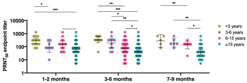
Prepublication Release

Figure 2. Differences in neutralizing antibodies (PRNT50) titers observed among four classes of age.
PRNT50 titers from 194 serum samples were stratified by age (< 3, ≥3 age
Prepublication Release

Figure 3. Performance of SARS-CoV-2 CLIA IgG and PRNT titers over time. (A) Decreasing levels
of SARS-CoV-2 CLIA IgG levels observed for all classes of age (
Prepublication Release

Figure 4. Neutralizing antibodies titers according to COVID-19 disease severity. (A) Clinical
presentation of COVID-19 in children aged
Prepublication Release Supplementary Materials Serological assays Blood samples were collected in EDTA-coated tubes to further separate cells and plasma by Ficoll procedure. Plasma and cellular samples were appropriately store at -80°C and liquid nitrogen, respectively, until use. A high-throughput method for Plaque Reduction Neutralizing Test (PRNT) was used for the quantification of neutralizing antibodies in plasma samples [16]. Samples were heat-inactivated by incubation at 56°C for 30 min and 2-fold dilutions were prepared in Dulbecco modified Eagle medium (DMEM). The dilutions, mixed to a 1:1 ratio with a virus solution containing approximately 25 focus- forming units (FFUs) of SARS-CoV-2, were incubated for 1 h at 37 °C. Fifty microliters of the virus-serum mixtures were added to confluent monolayers of Vero E6 cells, in 96-wells plates and incubated for 1 h at 37 °C, in a 5% CO2 incubator. The inoculum was removed and 100 ml of overlay solution of Minimum essential medium (MEM), 2% fetal bovine serum (FBS), penicillin (100 U/ml), streptomycin (100 U/ml) and 0.8% carboxy methyl cellulose was added to each well. After a 26-h incubation, cells were fixed with a 4% paraformaldehyde (PFA) solution. Visualization of plaques was obtained with an immunocytochemical staining method using an anti-dsRNA monoclonal antibody (J2, 1:10,000; Sci- cons) for 1 hour, followed by 1 h incubation with peroxidase-labeled goat anti-mouse antibodies (1:1000; DAKO) and a 7 min incubation with the True Blue (KPL) peroxidase substrate. FFUs were counted after acquisition of pictures on a flatbed scanner. Biosafety Level 3 laboratory setting was used for PRNT tests. The neutralization titer was defined as the reciprocal of the highest dilution resulting in a reduction of the control plaque count >50% (PRNT50). Samples recording titers equal to or above 1:10 were considered as positive according to a previous validation conducted on a panel of archive samples collected in 2018 in Italy1. Sera from the same donors were analyzed with the chemiluminescence immunoassay (CLIA) MAGLUMI™ 2019-nCoV IgM/IgG on the analytical system MAGLUMI™ 2000 Plus (New Industries ©2021 American Academy of Pediatrics Downloaded from www.aappublications.org/news by guest on September 9, 2021
Prepublication Release

Biomedical Engineering Co., Ltd [Snibe], Shenzhen, China). IgG/IgM immunocomplexes are formed upon

addition of a recombinant antigen expressing the full-length spike and nucleocapsid proteins of SARS-

CoV-2. According to the manufacturer’s inserts (271 2019-nCoV IgM, V2.0, 2020-03 and 272 2019-nCoV

IgG, V1.2, 2020-02), the 2019-nCoV IgM cut-off is 1.0 AU/mL, while the 2019-nCoV IgG cut-off is 1.1

AU/mL. The assay is intended for qualitative detection and differentiation of IgM and IgG antibodies. The

combined sensitivity and specificity of IgG/IgM is declared to be 95.6% and 96.0%, respectively.

SARS-CoV-2 viral load measurement

A selection of nasopharyngeal (NP) swabs of enrolled subjects that had been originally screened at the

Padova University Hospital were made available for quantification of the viral load. NP swabs tested were

collected by using flocked swabs in liquid-based collection and transport systems. Total nucleic acids were

purified from 200µl media and eluted in a final volume of 100µl. Copies of SARS-CoV-2 were quantified

by a home-made multiplex quantitative assay based on One-Step digital droplet PCR (ddPCR). The reaction

mixture consisted of 5µl of supermix (Bio-Rad, CA, USA), 2μl of reverse transcriptase, 2μl of DTT final

concentration 300mM, forward and reverse primers of SARS-CoV-2 E gene to a final concentration of

400nM each and probe to a final concentration of 200nM and 5µl of nucleic acids were eluted from

nasopharyngeal swab samples into a final volume of 20 µl. Housekeeping GAPDH was employed to verify

the good quality of RNA extracted and amplified under the same conditions using the GAPDH Kit (PE

Applied Biosystems, Waltham, MA, USA) 2. Each well of the prepared mix was loaded into an 8-channel

cartridge and 70μl of the Droplet Generation Oil for Probes (Bio-Rad) were added. Droplets were formed

in the QX200TM Droplet Generator (Bio-Rad). Droplets in the oil suspension were transferred into a 96

well plate and placed into a Mastercycler (Eppendorf, Hamburg, Germany) with the following cycling

parameters: 42-50°C for 60 min; 95°C for 10min; 95°C for 30sec and 60°C for 1 min; the last two passages

were repeated for 40 cycles followed by 98°C for 10 min. The droplets were then read by the QX200TM

Droplet Reader (Bio-Rad) and the results were analyzed with the QuantaSoftTM Analysis Software

 ©2021 American Academy of Pediatrics
 Downloaded from www.aappublications.org/news by guest on September 9, 2021
Prepublication Release

1.7.4.0917 (Bio-Rad) 2. Wells with less than 10000 droplets were discarded from the analysis. Each sample

was run at least in duplicate. Results were expressed as SARS-CoV-2 copies/5µl.

References
 1. Padoan A, Bonfante F, Pagliari M, et al. Analytical and clinical performances of five
 immunoassays for the detection of SARS-CoV-2 antibodies in comparison with neutralization
 activity. EBioMedicine. 2020; 62:103101. Available at:
 https://linkinghub.elsevier.com/retrieve/pii/S2352396420304771.
 2. Cotugno N, Ruggiero A, Bonfante F, et al. Virological and immunological features of SARS-
 CoV-2-infected children who develop neutralizing antibodies. Cell Rep. 2021; 34:108852.

 ©2021 American Academy of Pediatrics
 Downloaded from www.aappublications.org/news by guest on September 9, 2021
Prepublication Release

Supplementary Figure S1.
Flow chart of family clusters of COVID-19 observed from March 1st to the September 4th 2020, at
the COVID-19 follow-up clinic of the Pediatric Department, Department of Women’s and
Children’s Health, University of Padua.

 * 3 children with positive SARS-CoV-2 neutralizing antibodies (PRNT) were further excluded
from the analysis as they constituted “peculiar cases” if compared to the general cohort: in fact, 2 children
presented MIS-C, 4-6 weeks after Covid-19 onset and 1 newborn of a Covid-19 positive mother presented
positive SARS-CoV-2 neutralizing antibodies (PRNT) detected 51 days after birth that could be related to
maternal immunity and not seroconversion (SARS-CoV-2 molecular assay was never performed at birth).

 ©2021 American Academy of Pediatrics
 Downloaded from www.aappublications.org/news by guest on September 9, 2021
Prepublication Release

Supplementary Figure S2. Identification of cases and criteria for the definition of the baseline
time, defined as the most likely onset of infection, for confirmed COVID-19 cases.

 ©2021 American Academy of Pediatrics
 Downloaded from www.aappublications.org/news by guest on September 9, 2021
Prepublication Release

Supplementary Table S1. Descriptive analysis of the 57 families observed at the Department of
Women’s and Children’s Health of the University Hospital of Padua (Italy), overall (n=209) and stratified
by familiar status as children/older siblings (n=103) and parents (n=106).

 OVERALL CHILDREN/OLDER SIBLINGS PARENTS
 COVID- COVID- COVID- COVID- COVID-
 COVID-19
 19 19 p-value 19 19 19
 negative p-value § p-value§
 positive negative § positive positive negative
 (n=30)
 (n=155) (n=54) (n=73) (n=82) (n=24)
 Female 81 23 0.22 36 12 0.39 45 11 0.44
 (percentage) (52.3%) (42.6%) (49.3%) (40%) (54.9%) (45.8%)
 Mean age 25.8 23.4 0.37 8.75 (6.3) 7.12 0.26 40.9 43.7 (7.4) 0.13
 (SD) (17.7) (19.5) (5.7) (8.3)
Age classes (n,
 %):
 28 15 0.28 28 15 (50%) 0.63 0 (0%) 0 (0%)
 < 6 years (18.1%) (27.8%) (38.4%)
 34 12 34 12 (40%) 0 (0%) 0 (0%)
 6≤ age
Prepublication Release
Supplementary Table S2. Serological data of 283 plasma samples obtained from 152 confirmed COVID-19 cases (38
independent samples, 245 dependent samples obtained from 114 cases) among age classes, overall and stratified by time
from baseline.
 All data, irrespective of onset

 Age Classes (years) < 3 (n=30) 3 -
Prepublication Release

Supplementary Table S3.
Temporal distribution of sample collection among subjects who contributed to the study with either one,
two or three plasma samples.

 First sample Second sample Third sample

 1-2 3-6 1-2 3-6 7-9 3-6 7-9
 Time from baseline
 months months months months months months months

 Subjects with only one

 sample 21 17 0 0 0 0 0

 (n=38)

 Subjects with only two
 samples 52 45 0 62* 35§ 0 0
 (n=97)

 Subjects with three
 samples 17 0 3 14* 0 2 15§
 (n=17)

Total number of samples
 90 62 3 76 35 0 17
 per period

Total number of samples
 152 114 17
 (n=283)

* second samples included in subject-paired analyses of time window 1 (total of 76)
§ second/third samples included in subject-paired analyses of time window 2 (total of 50)

 ©2021 American Academy of Pediatrics
 Downloaded from www.aappublications.org/news by guest on September 9, 2021
Prepublication Release

Supplementary Table S4. Distribution of plasma samples across age classes and baseline intervals.

 Age classes

 Baseline < 3 years 3 -
Prepublication Release

Supplementary Table S6. Correlation between SARS-CoV-2 viral load (genome copies)
detected by means of ddPCR in NP swabs collected within 4 days from symptom onset and
PRNT titers assessed 1-2 months later, overall and stratified for classes of age.

 NP swabs collected within 4 days from
 symptom onset

 p-
 N Pearson coef.
 value

 All ages 32 -0.00796 0.9655
Mild SARS-CoV-2 Infections and Neutralizing Antibody Titers
 Francesco Bonfante, Paola Costenaro, Anna Cantarutti, Costanza Di Chiara, Alessio
Bortolami, Maria Raffaella Petrara, Francesco Carmona, Matteo Pagliari, Chiara Cosma,
Sandra Cozzani, Eva Mazzetto, Giovanni Di Salvo, Liviana Da Dalt, Paolo Palma, Luisa
Barzon, Giovanni Corrao, Calogero Terregino, Andrea Padoan, Mario Plebani, Anita De
 Rossi, Daniele Donà and Carlo Giaquinto
 Pediatrics originally published online June 22, 2021;

Updated Information & including high resolution figures, can be found at:
Services http://pediatrics.aappublications.org/content/early/2021/06/18/peds.2021-05
 2173.citation
Permissions & Licensing Information about reproducing this article in parts (figures, tables) or in its
 entirety can be found online at:
 http://www.aappublications.org/site/misc/Permissions.xhtml
Reprints Information about ordering reprints can be found online:
 http://www.aappublications.org/site/misc/reprints.xhtml

 Downloaded from www.aappublications.org/news by guest on September 9, 2021
Mild SARS-CoV-2 Infections and Neutralizing Antibody Titers
 Francesco Bonfante, Paola Costenaro, Anna Cantarutti, Costanza Di Chiara, Alessio
 Bortolami, Maria Raffaella Petrara, Francesco Carmona, Matteo Pagliari, Chiara Cosma,
 Sandra Cozzani, Eva Mazzetto, Giovanni Di Salvo, Liviana Da Dalt, Paolo Palma, Luisa
 Barzon, Giovanni Corrao, Calogero Terregino, Andrea Padoan, Mario Plebani, Anita De
 Rossi, Daniele Donà and Carlo Giaquinto
 Pediatrics originally published online June 22, 2021;

The online version of this article, along with updated information and services, is located on
 the World Wide Web at:
 http://pediatrics.aappublications.org/content/early/2021/06/18/peds.2021-052173.citation

 Data Supplement at:
 http://pediatrics.aappublications.org/content/suppl/2021/08/04/peds.2021-052173.DCSupplemental

 Pediatrics is the official journal of the American Academy of Pediatrics. A monthly publication, it has
 been published continuously since 1948. Pediatrics is owned, published, and trademarked by the
 American Academy of Pediatrics, 345 Park Avenue, Itasca, Illinois, 60143. Copyright © 2021 by the
 American Academy of Pediatrics. All rights reserved. Print ISSN: 1073-0397.

 Downloaded from www.aappublications.org/news by guest on September 9, 2021
You can also read