FLAVONOIDS AND NUCLEOTIDE ANALOGS SHOW HIGH ANITY FOR VIRAL PROTEINS OF SARS-COV-2 BY IN SILICO ANALYSIS: NEW CANDIDATES FOR THE TREATMENT OF ...
←
→
Page content transcription
If your browser does not render page correctly, please read the page content below
Flavonoids and Nucleotide Analogs Show High
A nity for Viral Proteins of SARS-CoV-2 by in silico
Analysis: New Candidates for the Treatment of
COVID-19.
Luis Adrián De Jesús-González ( luis.dejesus@cinvestav.mx )
CINVESTAV https://orcid.org/0000-0003-1415-6260
Juan Fidel Osuna-Ramos
CINVESTAV
José Manuel Reyes-Ruiz
CINVESTAV
Carlos Noe Farfan-Morales
CINVESTAV
Selvin Noé Palacios-Rápalo
CINVESTAV
Carlos Daniel Cordero-Rivera
CINVESTAV
Arianna M. Hurtado-Monzón
CINVESTAV
Ana Lorena Gutiérrez-Escolano
CINVESTAV
Rosa María Del Ángel
CINVESTAV
Short Report
Keywords: SARS-CoV-2, COVID-19, antiviral, drugs, molecular docking
DOI: https://doi.org/10.21203/rs.3.rs-67272/v1
License: This work is licensed under a Creative Commons Attribution 4.0 International License.
Read Full License
Page 1/18
Abstract
The recent epidemic of COVID-19 caused by SARS-CoV-2 was declared by the World Health Organization
as a public health emergency of international concern. The absence of an approved vaccine or a speci c
antiviral drug has made bioinformatic tools crucial for the identi cation of potential therapeutic targets
and drugs for its control. As in other RNA viruses, the protease 3C-like and the RNA-polymerase are two of
the SARS-CoV-2 targets to test drugs that can be analyzed in silico. In the present study, compounds
derived from plants, fungi, and nucleoside 5'-triphosphate or uridine nucleotide analogs, with anti-DENV
activity in vitro or in vivo, were analyzed by molecular docking as potential anti-SARS-CoV-2 drugs.
Anthraquinone, with a DENV NS3 protease inhibitory activity; Balapiravir, Fisetin, Hyperoside, and
Sofosbuvir, with a DENV NS5 RNA-polymerase inhibitory activity; and Quercetin, with both anti-NS3-NS5
activities, were tested against 3C-like protease and RNA-polymerase of SARS-CoV-2. All these drugs
demonstrated a high a nity for the corresponding SARS-CoV-2 proteins, representing excellent
candidates for the treatment of COVID-19. Therefore, in vitro or in vivo studies should be carried out using
these compounds on models for SARS-CoV-2 infection.

Introduction
Coronavirus disease 2019 (COVID-19) is currently affecting millions of people worldwide. According to
the World Health Organization (WHO), the COVID-19 pandemic has been declared as a public health
emergency of international concern; causing more than 500,000 deaths worldwide [1,2]. Therefore; it is a
priority to search antivirals that reduce or prevent COVID-19 mortality. COVID-19 is caused by the severe
acute respiratory syndrome coronavirus-2 (SARS-CoV-2), a spherical virus with a positive-sense RNA
genome [3]. SARS-CoV-2 viral proteins include the 3C- like protease (NSP5) and the RNA-polymerase
(NSP12), two of the proposed RNA virus targets tested for different drugs [4–8]. The NSP5 protein is a
chymotrypsin-like protease; which processes the polyproteins ppa1a and pp1ab; generating unique viral
proteins [9]. It consists of three domains: domain I (8-100 aa), II (101-183), and III (200-302 aa). The
protease catalytic site is located in the ssure between domains I and II; with a catalytic dyad of His41
and Cys145 [10]. On the other hand; the NSP12 protein is the RNA-dependent RNA polymerase (RdRp or
RNA-polymerase), responsible for the replication and transcription of the genomic RNA [6,11]. This
enzyme has the preserved architecture of viral RNA-polymerases. It is composed of the ngers; palm;
thumb; NiRAN (nidovirus RdRp-associated nucleotidyltransferase), present only in nidoviruses such as
SARS-CoV-2; and interface domains (Figure 1B) [6,11]. Therefore; it has been described that some
antivirals, whose principal functions are to inhibit these two enzymes, may have a broad spectrum
against RNA viruses [12,13].

Antiviral drugs have been developed to target either host cell proteins required for viral replication or viral
proteins. The rst approach may provide a broader spectrum of activity and less chance of developing
resistance; however, inhibiting host cell proteins has a more signi cant impact on cell viability. For this
reason, the use of antiviral drugs that target viral proteins is more speci c and less toxic [14,15]. The
recent closure of research laboratories worldwide as part of the containment measures to prevent the
                                                   Page 2/18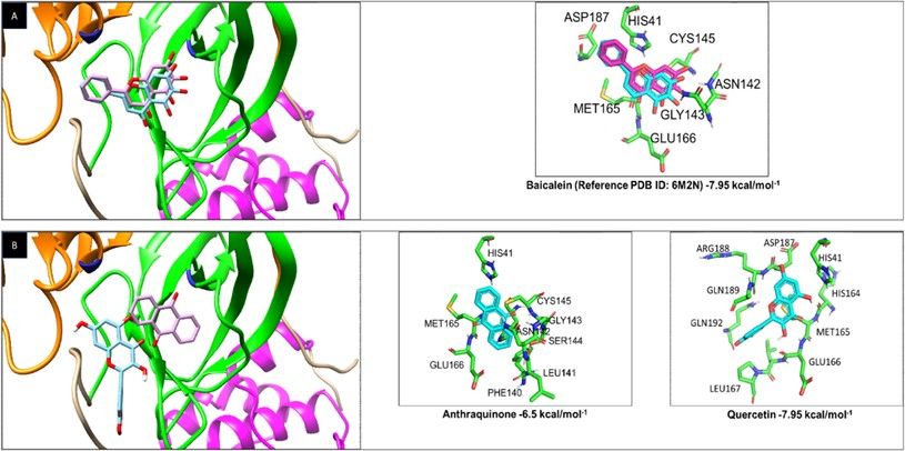
spread of the SARS-CoV-2 virus has hindered the in vitro and in vivo trials to test the effectiveness of
drugs [4]. However, the integration of computational and experimental strategies in pharmacology has
been of great value in the identi cation and development of new antivirals. Particularly, the molecular
docking analysis is a widely used bioinformatics tool in modern drug design [16].

Currently, there is no speci c antiviral to content SARS-CoV-2 infection, and the expectation of an e cient
vaccine continues to cause concern due to the time required for its approval and distribution. Therefore,
testing the antiviral activity of FDA-approved drugs, using bioinformatic strategies, is today a promising
tool for the selection of drugs that can potentially inhibit the replication of SARS-CoV-2 [7,8,17,18].

In recent years secondary metabolites of plants such as Quercetin, Fisetin, and Hyperoside avonoids,
have demonstrated antiviral effect in vivo and in vitro [19,20], in RNA virus-like dengue virus (DENV) by
acting on NS5 RNA-polymerase. Moreover, its antiviral effects have also been observed in other viral
infections such as with Enterovirus, Rhinovirus, Ebola virus, Chikungunya, and Zika virus [19,21–24]. On
the other hand, Quercetin and Anthraquinone, secondary metabolites of plants and fungi, have
demonstrated activity against the DENV NS3 protease [19,25]. Other examples of antiviral compounds
are the nucleoside and nucleotide analogs such as Balapiravir and Sofosbuvir, respectively, alternative
substrates for viral polymerases that competitively inhibit the synthesis of viral RNA in infections such as
hepatitis C virus (HCV) and DENV [26,27] excellent candidates against the SARS-CoV-2.

In this study, we tested the a nity of Anthraquinone and Quercetin to the SARS-CoV-2 3C-like protease,
and the a nity of Balapiravir, Quercetin, Fisetin, Hyperoside, and the previously tested Sofosbuvir for
SARS-CoV-2 RNA-Polymerase using a molecular docking analysis. Our results showed that these drugs,
previously used as antivirals for other RNA viruses, have an a nity for SARS-CoV-2 viral proteins and,
therefore a potential inhibitory activity on viral replication.

Results
2.1 Validation of the three-dimensional structures and sequences of the SARS-CoV-2 3C-like protease and
RNA-polymerase,

Previous to the molecular docking analyses, the crystalline structures of the SARS-CoV-2 3C- like protease
(PDB ID: 6M2N) (Figure 1A) and RNA-polymerase (PDB ID: 6NUR) (Figure 1B) were validated by amino
acid sequence comparison with those reported in the UniProtKB database (ID: P0C6×7). The comparison
was performed using the EMBOSS Needle software (https://www.ebi.ac.uk/Tools/psa/emboss_needle/)
[28], obtaining a sequence identity of 96.1% and 97.5%, respectively.

Furthermore, the quality of the protein structures was evaluated using ERRAT software
(https://servicesn.mbi.ucla.edu/ERRAT/) which differentiates regions of protein structures based on
atomic interaction, generating a structure quality factor in a percentage of 0-100% [29], and the Verify 3D
software (https://servicesn.mbi.ucla.edu/Verify3D/) that determines the compatibility of the 3D atomic

                                                     Page 3/18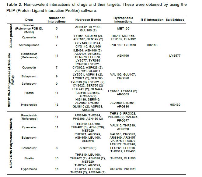
model with its amino acid sequence and its atomic coordinates, obtaining a high score when the
structure was correct [30].

The 3C-like protease structure of SARS-CoV-2 showed a score of 90.43% with the Verify3D software and a
structured quality factor of 94.5% with the ERRAT software. The structure of the RNA-polymerase showed
a quality factor of 96.68% with ERRAT software and 83. 81% with Verify3D. Once the three-dimensional
structures of both proteins and their amino acid sequences were validated, the molecular docking assays
were carried out with the candidate drugs.

2.2 Screening of DENV NS3 protease inhibitors (Quercetin and Anthraquinone) against the 3C-like
protease of SARS-CoV-2

First, the molecular docking assays were validated by recreating the experimental data on the binding of
the drug 5,6,7-trihydroxy-2-phenyl-4H-chromen-4-one (Baicalein) found in the same structure as the PDB
ID: 6M2N with the 3C-like protease of SARS-CoV-2. The analysis found that the drug in both traditional
and computational experiments joined in the same protease structure position (Figure 2A), validating our
conditions. The binding energy of the drug and protease obtained from the computational data was -7.95
kcal/mol−1.

Quercetin and Anthraquinone inhibitors of DENV NS3 protease bound with a high binding a nity (-6.5
and -7.95 kcal/mol−1, respectively) to the 3C-like protease of SARS-CoV-2 (Figure 2B), both drugs bound
to the active site of the protease, particularly to the HIS41 which belongs to the catalytic dyad. The
a nity of the 3C-like protease for Quercetin and Anthraquinone was compared with the one for Baicalein,
and the drug interactions with the highest a nity for the active site were graphed based on an RMSD
interactions using the PLIP software, it was found that Hyperoside has the maximum possible number of
non-covalent interactions with RNA-polymerase (Table 2).

Besides, to the high a nity of the tested drugs to the active RNA polymerase site, a high a nity for the
NiRAN sub-domain was also found. The NiRAN subdomain is present only in nidoviruses such as SARS-
CoV-2, it has a nucleotidylation activity and is essential for viral replication; its mutation generates non-
infective viral particles. It has also been involved as a protein primer for RNA synthesis, as the Vpg primer
activity exhibited in Picornaviruses [34,35]; reasons why it might be an excellent target to inhibit viral
replication.

Fisetin, Balapiravir, Quercetin, and Sofosbuvir exhibited a higher a nity (-8.1, -8.06, -7.79, -7.74
kcal/mol−1) for the RNA polymerase NiRAN-Fingers site, compared to the Remdesivir control (-7.67
kcal/mol−1) (Figure 3C and 3D). Hyperoside showed a lower ΔG (-7.43 kcal/mol−1), compared to the
control; however, it exhibits the highest number of non-covalent interactions (Table 2).

Finally, to compare the a nity of drugs for the RNA polymerase, the drug interactions with the highest
a nity for the active and NiRAN sites were graphed based on an RMSD
Page 6/18
Discussion
The World Health Organization has declared the recent epidemic of COVID-19 caused by SARS-CoV-2, as
a public health emergency of international interest [2]. Approximately 17 million cases and more than
650,000 deaths for COVID-19 have been reported until mid-2020. America, the continent most affected
had reported 7 million new cases in 24 hours [1]. Given this, it is crucial to nd drugs as antivirals to
inhibit viral replication. However, the closure of research laboratories to prevent the contagion and spread
of the virus has made it di cult to conduct trials to test the effectiveness of drugs [37]. Therefore, an
alternative strategy is a search for drugs against SARS- CoV-2 using computational techniques, whose in
vitro and in vivo action could be later corroborated by traditional experimental methods.

Viral proteins such as 3C-like protease and RNA-polymerase of SARS-CoV-2 are two of the proposed
targets for preventing viral replication [4,5]. 3C-like protease (NSP5) is a chymotrypsin- like protease,
which processes the polyproteins ppa1a and pp1ab, generating unique viral proteins involved in the rst
steps of the virus replication [9]. The RNA-polymerase (NSP12) is responsible for the replication and
transcription of the genomic RNA, together with the NSP7 and NSP8 cofactors required to stimulate its
polymerase activity [11]. Given the critical functions of both proteins, they have been widely proposed as
targets for pharmacological action.

Remdesivir is a drug that inhibits the action of different viral RNA polymerases, and Inhibitory activity
against SARS-CoV-2 has been demonstrated. It has been tested as a treatment option in phase III clinical
trials with patients, showing an improvement; however, there is still mortality of 13% of the patients, and
in 60% of those treated, side effects such as liver problems, diarrhea, skin rash, kidney failure, and
hypotension occurred [38,39].

Therefore, the search for new effective treatments for COVID19 disease with fewer side effects, which can
be used in severe patients and which are available to the entire population, must be studied.

In this work, we analyzed utilizing molecular docking techniques, drugs with a previous in vitro, or in vivo
anti-DENV activity. Anthraquinone with a DENV NS3 protease inhibitory activity, and Balapiravir, Fisetin,
Hyperoside, and Sofosbuvir with a DENV NS5 RNA-polymerase inhibitory activity. Also, Quercetin with
anti-DENV NS3 and NS5 activity was tested [40,41]. Here, the ability of these compounds to bind to
SARS-CoV2 NSP5 protease and RNA-polymerase NSP12 were tested.

Fisetin, Quercetin, Hyperoside, and Anthraquinone are drugs derived from plants and fungi. Fisetin and
Quercetin are the drugs that exhibited the highest a nity for the SARS-CoV-2 RNA- polymerase active site
(-7.08 kcal/mol-1 and -8.1 and -7.26 kcal/mol-1 respectively) and the NiRAN- Fingers domain, and
concerning Remdesivir control (-6.81 kcal/mol-1 for the active and 7.67 kcal/mol-1 and the NiRAN-sites),
suggesting that so they may be good anti-SARS-CoV-2 drug candidates.

Fisetin and Quercetin are avonols and secondary metabolites from a diversity of plants (e.g., Rhus
cotinus and Quercus spp.), with an antibiotic, antiviral, antioxidant, anti-in ammatory, and anti-cancer

                                                  Page 7/18
activities [42,43]. The DENV2 activity of Quercetin was demonstrated in experimental studies with a half-
maximal inhibitory concentration (IC50) of 28.9 µg/ml-1 and the half-maximal cytotoxic concentration
(CC50) of 252.6 µg/ml-1 in Vero cells [19]. In DC-SIGN infected cells, Quercetin showed an IC50 of 24.5
µM and a CC50 of 340 µM. It was also found that both Quercetin and Fisetin inhibit DENV2 and DENV3
infection in the absence or presence of antibodies (anti- DENV) and negatively regulate the production of
pro-in ammatory cytokines induced during severe DENV infection [44]. Quercetin has been described as a
candidate in the prevention and treatment of COVID-19, due to its e cacy in other antiviral diseases, low
cost, bioavailability, and low toxicity [45]. Our study would reinforce the experimentally use of this drug in
patients with COVID-19.
Hyperoside showed the greatest number of interactions with the NiRAN domain and the active site of
RNA-polymerase (Figure 3 and Table 1B); this metabolite comes from the extract of Houttuynia cordata,
which is consumed in Thailand. It has shown an anti-DENV activity by slowing down viral RNA synthesis
and inactivating infection by blocking the virus entry into HepG2 cells. In DENV-infected LLC-MK2 cells,
an EC50 = 0.8 µg/ml and a CC50 of 1.24 mg/ml were obtained [46,47]
In this study, the drug with the lowest binding score to the NSP5 protease was Anthraquinone with -6.5
kcal/mol-1; however, this binding energy is comparable to that exhibited by Remdesivir (-6.4 kcal/mol-1) in
other studies, using Autodock Vina [48]. Therefore, its use and effectiveness for Covid-19 cannot be ruled
out. Anthraquinones are plants (Rheum palmatum, Cassia obtusifolia, Morinda panamensis, etc.) [49]
and fungal (Monascus spp.) metabolites with pharmaceutical, food, and clothing dye use [50].
Experimental studies with both computational and traditional DENV2 have shown that it reduces viral
replication by a concentration of 4.2 µM (EC50) and a CC50 of 69 uM in LLC-MK2 cells [25].
Anthraquinone also has been tested on DENV infected Huh7 cells, where an EC50 was of 2.69 µM, and a
CC50 of 106.6 µM were found, in which there was an absence of cytotoxicity during three days of
incubation [51]. Besides, anthraquinone derivatives have been shown to have anti-Herpetic, anti-DENV,
anti-human immunode ciency virus (HIV), and antineoplastic effects [41,52,53].
The RNA-polymerase inhibitors Balapiravir and Sofosbuvir demonstrated a high a nity for the active and
the NiRAN sites of SARS-CoV-2 RNA-polymerase, even higher than the drug Remdesivir, suggesting they
can be excellent candidates for the treatment of COVID-19.
Balapiravir is a nucleoside 5'-triphosphate analog and Sofosbuvir is a uridine nucleotide analog, which
are alternative substrates for viral polymerase and competitively inhibits viral RNA synthesis [54,55].
Balapiravir was developed for the treatment of hepatitis C virus (HCV) and was tested in humans with
chronic phase 1b HCV infection, exhibiting a viral suppression of 3.7 log(10) in 14 days of treatment at its
highest dose of 4500 mg, where reversible hematological changes occurred in the patients. The tolerable
dose of 300 mg reduced the infection by 2.6 log(10) [56]. Because of its similarity between the NS5
polymerases of both HCV and DENV, Balapiravir was tested in phase II human trials, in which it showed
no effect on viremia or fever elimination [57]. However, another study reported that Balapiravir in its
Page 8/18triphosphate form acts during DENV infection, but cell activation may decrease the conversion of the
drug by requiring a host enzyme and losing its activity [58]. Therefore, the conversion and effectiveness
of the drug in infections with SARS-CoV-2 should be analyzed.
As Balapiravir, Sofosbuvir is a prodrug that must be triphosphorylated in the cell for its action. Clinical
studies have been done in phases I and II, and it currently has an FDA approval for the treatment for
chronic HCV infections [55]. In Huh7 cells infected with DENV2, Sofosbuvir has been shown to have an
a nity for NS5 polymerase, with an EC50 of 4.9 µM and a CC50> 100 µM [59]. Currently, it has been
proposed as a drug for the treatment of COVID-19, using a comparative analysis of the binding site in the
structure of the RNA-polymerase of SARS-CoV2 and that of HCV [60]. The results presented here and in
those previous results from Jácome et al., 2020, promote the study and use of Sofosbuvir, as well as
plant and mushroom derivatives Quercetin, Fisetin, Anthraquinone and Hyperoside as a COVID-19
treatment.
Materials And Methods
4.1 Structure of NSP5 (3C-like protease) and NSP12 (RNA-polymerase) from SARS-CoV-2
The structure of 3C-like protease (X-Ray diffraction, 2.20 Å resolution) and RNA polymerase (Electron
Microscopy, 3.10 Å resolution) of SARS-CoV-2 were obtained from the Protein Data Bank library (RCSB
PDB) with PDB ID: 6M2N and 6NUR, respectively. The quality of the protein structures and the sequence
was evaluated using ERRAT [29], Verify 3D [30], and EMBOSS Needle [28] software.
4.2 Drugs candidates tested
Six drug candidates with inhibitory activity against 3C-like protease and SARS-CoV-2 RNA- polymerase
were selected through literature reviews of drugs with inhibitory activity of viral proteins against DENV
[40,41]. They were selected based on in vitro and/or in vivo experimental background, in addition to
previous Food and Drug Administration (FDA) development and approval processes.
The selected drugs were obtained from the PubChem database, and are listed below with their PubChem
CID: Anthraquinone (6780) against 3C-like protease and Balapiravir (11691726), Fisetin (5281614),
Hyperoside (5281643) against RNA-polymerase from SARS-CoV-2. Quercetin (5280343) was tested
against both viral proteins from SARS-CoV-2, as it showed inhibitory activity against NS3 protease and
NS5 RNA-polymerase from DENV.
4.3 Molecular docking of drugs against 3C-like protease and RNA polymerase of SARS-CoV-2
The Autodock4 and AutoGrid4 software [61–63], were used for the molecular docking analysis of drug
candidates against 3C-like protease and RNA-polymerase from SARS-CoV-2.
Previously, the crystalline structures of 3C-like protease and RNA-polymerase were obtained from the PDB
and minimized with the PyMOL 2.3.3 software [64] and the text editor Kate 2.2 [65], removing water and
Page 9/18other molecules associated with the PDB le [66]. Then, the ligand and target structures were prepared with the AutoDockTools 1.5.6 software, adding polar atoms and charges [62]. The grid box parameters used were 126 Å, 126 Å and 126 Å (X, Y, and Z), grid spacing of 0.375 Å and center grid box (X, Y, and Z) of -35.667 Å, -47.402 Å and 37.332 Å (3C-like protease) and 97.115 Å, 97.809 Å and 94.063 Å (RNA-polymerase). The genetic algorithm parameters used were: Number of GA Runs 100, Population size 150, Maximum Number of evals medium 10000000, the other values were used by default. The best interaction model was chosen based on the lowest calculated ΔG. The validation of the molecular docking was based on recreating the experimental data of the crystalline structures of 3C-like protease (PDB ID: 6M2N) (Su, H.X., et al, to be published) and RNA polymerase (PDB ID: 6NUR) [11] linked to its inhibitor 5,6,7-trihydroxy-2-phenyl-4H-chromen-4-one and Remdesivir, respectively. 4.4 Visualization of molecular docking results The molecular docking results were analyzed with the AutoDockTools 1.5.6, PyMOL 2.3.3, and UCSF Chimera 1.14 software [62,64,67], to visualize the best target-ligand bond formation with lower ΔG. The number and type of non-covalent interactions between the drug and the viral protein were obtained using the PLIP software [36]. 4.5 Statistical analysis The higher a nity interactions between the candidate drugs directed to the active sites or the NiRAN domain of 3C-like protease and the RNA-polymerase of SARS-CoV-2, with RMSD
Funding: This research was supported by CONACYT (Mexico), grants: CB-220824 and A1-S-9005 from
RMDA and CB-250696 from ALGE, and Fundación Miguel Alemán. The funders had no role in study
design, data collection, and analysis, decision to publish, or preparation of the manuscript.

Acknowledgments: LADJ-G: JFO-R, JMR-R, CNF-M, SNP-R, CDC-R, AMH-M received scholarships from
CONACYT.

Con icts of Interest: The authors declare no con ict of interest.

References
1. World Health Organization (WHO) Coronavirus disease (COVID-19) Situation Report-141 Situation in
numbers (by WHO Region); 2020. 2. Zheng, J. SARS-coV-2: An emerging coronavirus that causes a global
threat. Int. J. Biol. Sci. 2020, 16, 1678–1685, doi:10.7150/ijbs.45053. 3. Li, X.; Luk, H.K.H.; Lau, S.K.P.;
Woo, P.C.Y. Human Coronaviruses: General Features. Ref. Module Biomed. Sci. 2019, doi:10.1016/B978-0-
12-801238-3.95704-0. 4. Quimque, M.T.J.; Notarte, K.I.R.; Fernandez, R.A.T.; Mendoza, M.A.O.; Liman,
R.A.D.; Lim, J.A.K.; Pilapil, L.A.E.; Ong, J.K.H.; Pastrana, A.M.; Khan, A.; et al. Virtual screening-driven drug
discovery of SARS-CoV2 enzyme inhibitors targeting viral attachment, replication, post-translational
modi cation and host immunity evasion infection mechanisms. J. Biomol. Struct. Dyn. 2020, 1–18,
doi:10.1080/07391102.2020.1776639. 5. De Clercq, E. Potential antivirals and antiviral strategies against
SARS coronavirus infections. Expert Rev. Anti Infect. Ther. 2014, 4, 291–302,
doi:10.1586/14787210.4.2.291. 6. Peng, Q.; Peng, R.; Yuan, B.; Zhao, J.; Wang, M.; Wang, X.; Wang, Q.;
Sun, Y.; Fan, Z.; Qi, J.; et al. Structural and Biochemical Characterization of the nsp12-nsp7-nsp8 Core
Polymerase Complex from SARS-CoV-2. Cell Rep. 2020, 31, 107774, doi:10.1016/j.celrep.2020.107774. 7.
Yin, W.; Mao, C.; Luan, X.; Shen, D.-D.; Shen, Q.; Su, H.; Wang, X.; Zhou, F.; Zhao, W.; Gao, M.; et al. Structural
basis for inhibition of the RNA-dependent RNA polymerase from SARS-CoV-2 by remdesivir. Science 2020,
368, 1499–1504, doi:10.1126/science.abc1560. 8. Lung, J.; Lin, Y.-S.; Yang, Y.-H.; Chou, Y.-L.; Shu, L.-H.;
Cheng, Y.-C.; Liu, H.T.; Wu, C.-Y. The potential chemical structure of anti-SARS-CoV-2 RNA-dependent RNA
polymerase. J. Med. Virol. 2020, 92, 693– 697, doi:10.1002/jmv.25761. 9. Kneller, D.W.; Phillips, G.; O’Neill,
H.M.; Jedrzejczak, R.; Stols, L.; Langan, P.; Joachimiak, A.; Coates, L.; Kovalevsky, A. Structural plasticity of
SARS-CoV-2 3CL M pro active site cavity revealed by room temperature X-ray crystallography. Nat.
Commun. 2020, 11, 3202, doi:10.1038/s41467-020-16954-7. 10. Kumar, Y.; Singh, H.; Patel, C.N. In silico
prediction of potential inhibitors for the Main protease of SARS- CoV-2 using molecular docking and
dynamics simulation based drug-repurposing. J. Infect. Public Health 2020,
doi:10.1016/j.jiph.2020.06.016. 11. Kirchdoerfer, R.N.; Ward, A.B. Structure of the SARS-CoV nsp12
polymerase bound to nsp7 and nsp8 co-factors. Nat. Commun. 2019, 10, 2342, doi:10.1038/s41467-019-
10280-3. 12. FURUTA, Y.; KOMENO, T.; NAKAMURA, T. Favipiravir (T-705), a broad spectrum inhibitor of
viral RNA polymerase. Proc. Jpn. Acad. Ser. B Phys. Biol. Sci. 2017, 93, 449–463,
doi:10.2183/pjab.93.027. 13. Gordon, C.J.; Tchesnokov, E.P.; Feng, J.Y.; Porter, D.P.; Götte, M. The antiviral
compound remdesivir potently inhibits RNA-dependent RNA polymerase from Middle East respiratory
syndrome coronavirus. J. Biol. Chem. 2020, 295, 4773–4779, doi:10.1074/jbc.AC120.013056. 14. Clercq,
                                                    Page 11/18
E.D. Antivirals and antiviral strategies. Nat. Rev. Microbiol. 2004, 2, 704–720, doi:10.1038/nrmicro975.
15. De Clercq, E. Strategies in the design of antiviral drugs. Nat. Rev. Drug Discov. 2002, 1, 13–25,
doi:10.1038/nrd703. 16. Ferreira, L.G.; dos Santos, R.N.; Oliva, G.; Andricopulo, A.D. Molecular Docking
and Structure-Based Drug Design Strategies. Molecules 2015, 20, 13384–13421,
doi:10.3390/molecules200713384. 17. El ky, A.A.; Ribavirin, Remdesivir, Sofosbuvir, Galidesivir, and
Tenofovir against SARS-CoV-2 RNA dependent RNA polymerase (RdRp): A molecular docking study. Life
Sci. 2020, 253, 117592, doi:10.1016/j.lfs.2020.117592. 18. El ky, A.A. Anti-HCV, nucleotide inhibitors,
repurposing against COVID-19. Life Sci. 2020, 248, 117477, doi:10.1016/j.lfs.2020.117477. 19. Zandi, K.;
Teoh, B.T.; Sam, S.S.; Wong, P.F.; Mustafa, M.; Abubakar, S. Antiviral activity of four types of bio avonoid
against dengue virus type-2. Virol. J. 2011, 8, 560, doi:10.1186/1743-422X-8-560. 20. Zakaryan, H.;
Arabyan, E.; Oo, A.; Zandi, K. Flavonoids: Promising natural compounds against viral infections. Arch.
Virol. 2017, 162, 2539–2551, doi:10.1007/s00705-017-3417-y. 21. Lim, H.; Nguyen, T.T.H.; Kim, N.M.; Park,
J.-S.; Jang, T.-S.; Kim, D. Inhibitory effect of avonoids against NS2B-NS3 protease of ZIKA virus and their
structure activity relationship. Biotechnol. Lett. 2017, 39, 415–421, doi:10.1007/s10529-016-2261-6. 22.
Yao, C.; Xi, C.; Hu, K.; Gao, W.; Cai, X.; Qin, J.; Lv, S.; Du, C.; Wei, Y. Inhibition of enterovirus 71 replication
and viral 3C protease by quercetin. Virol. J. 2018, 15, doi:10.1186/s12985-018-1023-6. 23. Qiu, X.; Kroeker,
A.; He, S.; Kozak, R.; Audet, J.; Mbikay, M.; Chrétien, M. Prophylactic E cacy of Quercetin 3-β-O-d-
Glucoside against Ebola Virus Infection. Antimicrob. Agents Chemother. 2016, 60, 5182–5188,
doi:10.1128/AAC.00307-16. 24. Lin, Y.-J.; Chang, Y.-C.; Hsiao, N.-W.; Hsieh, J.-L.; Wang, C.-Y.; Kung, S.-H.;
Tsai, F.-J.; Lan, Y.-C.; Lin, C.-W. Fisetin and rutin as 3C protease inhibitors of enterovirus A71. J. Virol.
Methods 2012, 182, 93–98, doi:10.1016/j.jviromet.2012.03.020. 25. Tomlinson, S.M.; Malmstrom, R.D.;
Russo, A.; Mueller, N.; Pang, Y.P.; Watowich, S.J. Structure-based discovery of dengue virus protease
inhibitors. Antiviral Res. 2009, 82, 110–114, doi:10.1016/j.antiviral.2009.02.190. 26. Zhong, D.; Liu, M.;
Cao, Y.; Zhu, Y.; Bian, S.; Zhou, J.; Wu, F.; Ryu, K.-C.; Zhou, L.; Ye, D. Discovery of Metal Ions Chelator
Quercetin Derivatives with Potent Anti-HCV Activities. Molecules 2015, 20, 6978– 6999,
doi:10.3390/molecules20046978. 27. Nelson, D.R.; Zeuzem, S.; Andreone, P.; Ferenci, P.; Herring, R.;
Jensen, D.M.; Marcellin, P.; Pockros, P.J.; Rodríguez-Torres, M.; Rossaro, L.; et al. Balapiravir plus
peginterferon alfa-2a (40KD)/ribavirin in a randomized trial of hepatitis C genotype 1 patients(). Ann.
Hepatol. 2012, 11, 15–31. 28. F., M.; Ym, P.; J., L.; N., B.; T., G.; N., M.; P., B.; Arn, T.; Sc, P.; Rd, F.; et al. The
EMBL-EBI search and sequence analysis tools APIs in 2019. Nucleic Acids Res. 2019, 47, W636–W641,
doi:10.1093/nar/gkz268. 29. Colovos, C.; Yeates, T.O. Veri cation of protein structures: Patterns of
nonbonded atomic interactions. Protein Sci. Publ. Protein Soc. 1993, 2, 1511–1519,
doi:10.1002/pro.5560020916. 30. Lüthy, R.; Bowie, J.U.; Eisenberg, D. Assessment of protein models with
three-dimensional pro les. Nature 1992, 356, 83–85, doi:10.1038/356083a0. 31. Jamwal, S.; Gautam, A.;
Elsworth, J.; Kumar, M.; Chawla, R.; Kumar, P. An updated insight into the molecular pathogenesis,
secondary complications and potential therapeutics of COVID-19 pandemic. Life Sci. 2020,
doi:10.1016/j.lfs.2020.118105. 32. Pizzorno, A.; Padey, B.; Dubois, J.; Julien, T.; Traversier, A.; Dulière, V.;
Brun, P.; Lina, B.; Rosa- Calatrava, M.; Terrier, O. In vitro evaluation of antiviral activity of single and
combined repurposable drugs against SARS-CoV-2. Antiviral Res. 2020,
doi:10.1016/j.antiviral.2020.104878. 33. Drożdżal, S.; Rosik, J.; Lechowicz, K.; Machaj, F.; Kot s, K.;
Page 12/18Ghavami, S.; Łos, M.J. FDA approved drugs with pharmacotherapeutic potential for SARS-CoV-2 (COVID-
19) therapy. Drug Resist. Updat. 2020, doi:10.1016/j.drup.2020.100719. 34. Posthuma, C.C.; te Velthuis,
A.J.W.; Snijder, E.J. Nidovirus RNA polymerases: Complex enzymes handling exceptional RNA genomes.
Virus Res. 2017, 234, 58–73, doi:10.1016/j.virusres.2017.01.023. 35. Lehmann, K.C.; Gulyaeva, A.;
Zevenhoven-Dobbe, J.C.; Janssen, G.M.C.; Ruben, M.; Overkleeft, H.S.; van Veelen, P.A.; Samborskiy, D.V.;
Kravchenko, A.A.; Leontovich, A.M.; et al. Discovery of an essential nucleotidylating activity associated
with a newly delineated conserved domain in the RNA polymerase- containing protein of all nidoviruses.
Nucleic Acids Res. 2015, 43, 8416–8434, doi:10.1093/nar/gkv838. 36. Salentin, S.; Schreiber, S.; Haupt,
V.J.; Adasme, M.F.; Schroeder, M. PLIP: Fully automated protein– ligand interaction pro ler. Nucleic Acids
Res. 2015, 43, W443–W447, doi:10.1093/nar/gkv315. 37. The New York Times When Coronavirus Closes
Your Lab, Can Science Go On? N. Y. Times 2020. 38. Srinivas, P.; Sacha, G.; Koval, C. Antivirals for COVID-
19. Cleve. Clin. J. Med. 2020, doi:10.3949/ccjm.87a.ccc030. 39. Grein, J.; Ohmagari, N.; Shin, D.; Diaz, G.;
Asperges, E.; Castagna, A.; Feldt, T.; Green, G.; Green, M.L.; Lescure, F.-X.; et al. Compassionate Use of
Remdesivir for Patients with Severe Covid-19. N. Engl. J. Med. 2020, doi:10.1056/NEJMoa2007016. 40.
Ganji, L. V.; Kanyalkar, M.A. Non-Structural Proteases as a Target of Dengue Virus. J. Antivir. Antiretrovir.
2019, 11, 1–15, doi:10.35248/1948-5964.19.11.188. 41. Tian, Y.S.; Zhou, Y.; Takagi, T.; Kameoka, M.;
Kawashita, N. Dengue virus and its inhibitors: A brief review. Chem. Pharm. Bull. (Tokyo) 2018, 66, 191–
206, doi:10.1248/cpb.c17-00794. 42. Grynkiewicz, G.; Demchuk, O.M. New Perspectives for Fisetin. Front.
Chem. 2019, 7, doi:10.3389/fchem.2019.00697. 43. Li, Y.; Yao, J.; Han, C.; Yang, J.; Chaudhry, M.T.; Wang,
S.; Liu, H.; Yin, Y.; Quercetin, In ammation and Immunity. Nutrients 2016, 8, doi:10.3390/nu8030167. 44.
Jasso-Miranda, C.; Herrera-Camacho, I.; Flores-Mendoza, L.K.; Dominguez, F.; Vallejo-Ruiz, V.; Sanchez-
Burgos, G.G.; Pando-Robles, V.; Santos-Lopez, G.; Reyes-Leyva, J. Antiviral and immunomodulatory effects
of polyphenols on macrophages infected with dengue virus serotypes 2 and 3 enhanced or not with
antibodies. Infect. Drug Resist. 2019, 12, 1833–1852, doi:10.2147/IDR.S210890. 45. Colunga Biancatelli,
R.M.L.; Berrill, M.; Catravas, J.D.; Marik, P.E. Quercetin and Vitamin C: An Experimental, Synergistic
Therapy for the Prevention and Treatment of SARS-CoV-2 Related Disease (COVID-19). Front. Immunol.
2020, 11, doi:10.3389/ mmu.2020.01451. 46. Teixeira, R.R.; Pereira, W.L.; Oliveira, A.F.C. da S.; da Silva,
A.M.; de Oliveira, A.S.; da Silva, M.L.; da Silva, C.C.; de Paula, S.O. Natural Products as Source of Potential
Dengue Antivirals. Molecules 2014, 19, 8151–8176, doi:10.3390/molecules19068151. 47.
Leardkamolkarn, V.; Sirigulpanit, W.; Phurimsak, C.; Kumkate, S.; Himakoun, L.; Sripanidkulchai, B. The
Inhibitory Actions of Houttuynia Cordata Aqueous Extract on Dengue Virus and Dengue-Infected Cells. J.
Food Biochem. 2012, 36, 86–92, doi:10.1111/j.1745-4514.2010.00514.x. 48. Beck, B.R.; Shin, B.; Choi, Y.;
Park, S.; Kang, K. Predicting commercially available antiviral drugs that may act on the novel coronavirus
(SARS-CoV-2) through a drug-target interaction deep learning model. Comput. Struct. Biotechnol. J. 2020,
18, 784–790, doi:10.1016/j.csbj.2020.03.025. 49. Friedman, M.; Xu, A.; Lee, R.; N.; Nguyen, D.; A.; Phan, T.;
M.; Hamada, S.; Panchel, R.; C.; Tam, C.; H.; Kim, J.; W.; Cheng, L.; et al. The Inhibitory Activity of
Anthraquinones against Pathogenic Protozoa, Bacteria, and Fungi and the Relationship to Structure.
Molecules 2020, 25, 3101, doi:10.3390/molecules25133101. 50. Fouillaud, M.; Venkatachalam, M.;
Girard-Valenciennes, E.; Caro, Y.; Dufossé, L. Anthraquinones and derivatives from marine-derived fungi:
Structural diversity and selected biological activities. Mar. Drugs 2016, 14, doi:10.3390/md14040064. 51.
Page 13/18Chu, J.J.H.; Lee, R.C.H.; Ang, M.J.Y.; Wang, W.L.; Lim, H.A.; Wee, J.L.K.; Joy, J.; Hill, J.; Brian Chia, C.S.
Antiviral activities of 15 dengue NS2B-NS3 protease inhibitors using a human cell-based viral
quanti cation assay. Antiviral Res. 2015, 118, 68–74, doi:10.1016/j.antiviral.2015.03.010. 52. Roa-
Linares, V.C.; Miranda-Brand, Y.; Tangarife-Castaño, V.; Ochoa, R.; García, P.A.; Castro, M.Á.; Betancur-
Galvis, L.; Feliciano, A.S. Anti-herpetic, anti-dengue and antineoplastic activities of simple and
heterocycle-fused derivatives of terpenyl-1,4-naphthoquinone and 1,4-anthraquinone. Molecules 2019, 24,
doi:10.3390/molecules24071279. 53. Sosic, A.; Saccone, I.; Carraro, C.; Kenderdine, T.; Gamba, E.;
Caliendo, G.; Corvino, A.; Di Vaio, P.; Fiorino, F.; Magli, E.; et al. Non-Natural Linker Con guration in 2,6-
Dipeptidyl-Anthraquinones Enhances the Inhibition of TAR RNA Binding/Annealing Activities by HIV-1 NC
and Tat Proteins. Bioconjug. Chem. 2018, 29, 2195–2207, doi:10.1021/acs.bioconjchem.8b00104. 54.
Klumpp, K.; Lévêque, V.; Pogam, S.L.; Ma, H.; Jiang, W.-R.; Kang, H.; Granycome, C.; Singer, M.; Laxton, C.;
Hang, J.Q.; et al. The Novel Nucleoside Analog R1479 (4′-Azidocytidine) Is a Potent Inhibitor of NS5B-
dependent RNA Synthesis and Hepatitis C Virus Replication in Cell Culture. J. Biol. Chem. 2006, 281,
3793–3799, doi:10.1074/jbc.M510195200. 55. Bhatia, H.K.; Singh, H.; Grewal, N.; Natt, N.K. Sofosbuvir: A
novel treatment option for chronic hepatitis C infection. J. Pharmacol. Pharmacother. 2014, 5, 278–284,
doi:10.4103/0976-500X.142464. 56. Roberts, S.K.; Cooksley, G.; Dore, G.J.; Robson, R.; Shaw, D.; Berns, H.;
Hill, G.; Klumpp, K.; Najera, I.; Washington, C. Robust antiviral activity of R1626, a novel nucleoside
analog: A randomized, placebo- controlled study in patients with chronic hepatitis C. Hepatology 2008,
48, 398–406, doi:10.1002/hep.22321. 57. Nguyen, N.M.; Tran, C.N.B.; Phung, L.K.; Duong, K.T.H.; Huynh,
H. le A.; Farrar, J.; Nguyen, Q.T.H.; Tran, H.T.; Nguyen, C.V.V.; Merson, L.; et al. A Randomized, Double-Blind
Placebo Controlled Trial of Balapiravir, a Polymerase Inhibitor, in Adult Dengue Patients. J. Infect. Dis.
2013, 207, 1442–1450, doi:10.1093/infdis/jis470. 58. Chen, Y.-L.; Abdul Ghafar, N.; Karuna, R.; Fu, Y.; Lim,
S.P.; Schul, W.; Gu, F.; Herve, M.; Yokohama, F.; Wang, G.; et al. Activation of Peripheral Blood Mononuclear
Cells by Dengue Virus Infection Depotentiates Balapiravir. J. Virol. 2014, 88, 1740–1747,
doi:10.1128/JVI.02841-13. 59. Xu, H.-T.; Colby-Germinario, S.P.; Hassounah, S.A.; Fogarty, C.; Osman, N.;
Palanisamy, N.; Han, Y.; Oliveira, M.; Quan, Y.; Wainberg, M.A. Evaluation of Sofosbuvir (β-D-2′-deoxy-2′-α-
uoro-2′-β-C- methyluridine) as an inhibitor of Dengue virus replication#. Sci. Rep. 2017, 7,
doi:10.1038/s41598-017- 06612-2. 60. Jácome, R.; Campillo-Balderas, J.A.; Ponce de León, S.; Becerra, A.;
Lazcano, A. Sofosbuvir as a potential alternative to treat the SARS-CoV-2 epidemic. Sci. Rep. 2020, 10,
9294, doi:10.1038/s41598- 020-66440-9. 61. Morris, G.M.; Goodsell, D.S.; Halliday, R.S.; Huey, R.; Hart,
W.E.; Belew, R.K.; Olson, A.J. Automated Docking Using a Lamarckian Genetic Algorithm and an Empirical
Binding Free Energy Function. J. Comput. Chem. 19, 24. 62. Morris, G.M.; Huey, R.; Lindstrom, W.; Sanner,
M.F.; Belew, R.K.; Goodsell, D.S.; Olson, A.J. AutoDock4 and AutoDockTools4: Automated Docking with
Selective Receptor Flexibility. J. Comput. Chem. 2009, 30, 2785–2791, doi:10.1002/jcc.21256. 63. Huey,
R.; Morris, G.M.; Olson, A.J.; Goodsell, D.S. A semiempirical free energy force eld with charge- based
desolvation. J. Comput. Chem. 2007, 28, 1145–1152, doi:10.1002/jcc.20634. 64. Rigsby, R.E.; Parker, A.B.
Using the PyMOL application to reinforce visual understanding of protein structure. Biochem. Mol. Biol.
Educ. Bimon. Publ. Int. Union Biochem. Mol. Biol. 2016, 44, 433–437, doi:10.1002/bmb.20966. 65. Team,
T.K. Kate | Get an Edge in Editing Available online: https://kate-editor.org/(accessed on Jul 19, 2020). 66.
Cosconati, S.; Forli, S.; Perryman, A.L.; Harris, R.; Goodsell, D.S.; Olson, A.J. Virtual Screening with
Page 14/18AutoDock: Theory and Practice. Expert Opin. Drug Discov. 2010, 5, 597–607,
doi:10.1517/17460441.2010.484460. 67. Pettersen, E.F.; Goddard, T.D.; Huang, C.C.; Couch, G.S.;
Greenblatt, D.M.; Meng, E.C.; Ferrin, T.E. UCSF Chimera--a visualization system for exploratory research
and analysis. J. Comput. Chem. 2004, 25, 1605–1612, doi:10.1002/jcc.20084.
Figures
Figure 1
Overview of the structure of NSP5 (3C-like protease) and NSP12 (RNA-Polymerase) from SARS-CoV-2. A)
Structure of the 3C-like protease (Doman I-orange, Domain II-green, Domain III-purple) and its catalytic
dyad (blue). B) Structure of RNA-Polymerase and its different domains (Fingers-blue, Thumb-green, Palm-
red, Interface-orange, NiRAN-yellow).
Page 15/18Figure 2
Baicalein, Quercetin, and Anthraquinone bind with a high a nity to the active site of the 3C-like protease
of SARS-CoV-2. A) Recreating experimental results by Molecular Docking (Rosa. Experimental reference
by crystallography assays of the binding of 5,6,7-trihydroxy-2-phenyl-4H- chromen-4-one (PDB ID: 6M2N)
to the active site of the 3C-like protease; Blue. Binding results by Molecular Docking Assays.). B)
Molecular Docking of Anthraquinone and Quercetin to the active site of the protease. In both cases, the
domains I (orange), II (green), III (purple), and the catalytic dyad (blue) of the 3C-like proteases are shown.
Besides, Amino acids are shown to interact with different drugs and biding energy.
Page 16/18Figure 3
Remdesivir, Quercetin, Fisetin, Hyperoside, Balapiravir, and Sofosbuvir bind with high a nity to the active
site and the NiRAN domain of the SARS-CoV-2 RNA-polymerase. A) Molecular Docking of Remdesivir
(blue), Quercetin (pink), and Balapiravir (green) to the active site of the RNA- Polymerase. B) Molecular
Docking of Sofosbuvir (blue), Fisetin (pink), Hyperoside (green) to the active site of the RNA-Polymerase.
C) Molecular Docking of Remdesivir (blue), Quercetin (pink), and Balapiravir (green) to the NiRAN domain
of the RNA-Polymerase. D) Molecular Docking of Sofosbuvir (blue), Fisetin (pink), Hyperoside (green) to
the RNA-Polymerase NiRAN domain site. RNA-Polymerase Fingers domain (blue), Thumb (green), Palm
(red), Interface (orange), and NiRAN (yellow) are shown. Also, amino acids are shown to interact with
different drugs and binding energy.
Page 17/18Figure 4
Comparison of drug a nities by 3C-like protease and SARS-CoV-2 RNA polymerase. A) Drug a nities by
the active site of the 3C-like protease. B) Drug a nities for the active site of RNA polymerase. C) A nities
of drugs by the NiRAN site.
Page 18/18You can also read