Up-Regulation of Glycogen Synthesis and Degradation Enzyme Level Maintained Myocardial Glycogen in Huddling Brandt's Voles Under Cool Environments ...
←
→
Page content transcription
If your browser does not render page correctly, please read the page content below
ORIGINAL RESEARCH
                                                                                                                                    published: 26 March 2021
                                                                                                                             doi: 10.3389/fphys.2021.593129

                                                Up-Regulation of Glycogen
                                                Synthesis and Degradation Enzyme
                                                Level Maintained Myocardial
                                                Glycogen in Huddling Brandt’s Voles
                                                Under Cool Environments
                                                Jin-Hui Xu* † , Zhe Wang † , Jun-Jie Mou, Chuan-Li Wang, Wei-Mei Huang, Hui-Liang Xue,
                                                Ming Wu, Lei Chen and Lai-Xiang Xu
                                                College of Life Sciences, Qufu Normal University, Qufu, China

                                                Small mammals exhibit limited glucose use and glycogen accumulation during
                                                hypothermia. Huddling is a highly evolved cooperative behavioral strategy in social
                                                mammals, allowing adaptation to environmental cooling. However, it is not clear whether
                             Edited by:         this behavior affects the utilization of glycogen in cold environments. Here, we studied
                        Claudia Penna,
                 University of Turin, Italy
                                                the effects of huddling on myocardial glycogen content in Brandt’s voles (Lasiopodomys
                        Reviewed by:
                                                brandtii) under a mild cold environment (15◦ C). Results showed that (1) Compared to
                  Tommaso Angelone,             the control (22◦ C) group (CON), the number of glycogenosomes more than tripled in
            University of Calabria, Italy
                                                the cool separated group (CS) in both males and females; whereas the number of
                      Claudio Molinari,
  University of Eastern Piedmont, Italy         glycogenosomes increased in females but was maintained in males in the cool huddling
                   *Correspondence:             group (CH). (2) Glycogen synthase (GS) activity in the CS group remained unchanged,
                             Jin-Hui Xu         whereas glycogen phosphorylase (GYPL) activity decreased, which mediated the
                  xujinhui@qfnu.edu.cn
                                                accumulation of glycogen content of the CS group. (3) Both GS and GYPL activity
     † These   authors have contributed
                    equally to this work
                                                increased which may contribute to the stability of glycogen content in CH group. (4)
                                                The expression levels of glucose transporters GLUT1 and GLUT4 increased in the CS
                   Specialty section:           group, accompanied by an increase in glucose metabolism. These results indicate that
         This article was submitted to
                Integrative Physiology,         the reduced glycogen degradation enzyme level and enhanced glucose transport may
               a section of the journal         lead to an increase in myocardial glycogen content of the separated voles under cool
               Frontiers in Physiology
                                                environment; while the up-regulation of glycogen synthesis and degradation enzyme
          Received: 09 August 2020
                                                level maintained myocardial glycogen content in the huddling vole.
          Accepted: 04 March 2021
          Published: 26 March 2021              Keywords: huddling, low temperature, heart, glycogenosomes, glycogen synthetase, glycogen phosphorylase
                            Citation:
          Xu J-H, Wang Z, Mou J-J,
   Wang C-L, Huang W-M, Xue H-L,                INTRODUCTION
   Wu M, Chen L and Xu L-X (2021)
Up-Regulation of Glycogen Synthesis             Low temperature is a stress stimulus for mammals, especially for small mammals as their energy
     and Degradation Enzyme Level
                                                requirements are high due to the large surface area to volume ratio. Moreover, when environmental
  Maintained Myocardial Glycogen in
 Huddling Brandt’s Voles Under Cool
                                                stressors persist for prolonged periods, small animal tissues and organs are more vulnerable to the
                      Environments.             impact of external environmental temperature (Gilbert et al., 2010; Wei et al., 2018). Hypothermia
          Front. Physiol. 12:593129.            can lead to a slowed heart rate, decreased blood flow output, and decreased myocardial contraction
   doi: 10.3389/fphys.2021.593129               and relaxation function (Polderman, 2009; Kelly and Nolan, 2010; Tessier and Storey, 2012;

Frontiers in Physiology | www.frontiersin.org                                          1                              March 2021 | Volume 12 | Article 593129
Xu et al.                                                                                                                   Huddling Maintained Myocardial Glycogen

Chavez et al., 2017). As above, the cardiac muscle of small                           in the liver and β-cells of the pancreas and admits glucose
mammals is more susceptible to low external temperatures.                             based on a positive glucose gradient between blood and tissue
Our previous study showed that, in comparison to warm                                 (Roach et al., 2012). Research on hibernating Daurian ground
environmental conditions, Brandt’s voles (Lasiopodomys                                squirrels (Spermophilus dauricus) has shown that the increase in
brandtii) under cool (15◦ C) conditions exhibit myocardial                            glycogen content in skeletal muscle in winter is mainly due to the
mitochondrial swelling and crista disruption, as well as decreased                    maintenance of P-GS and decrease in GYPL protein expression
adenosine triphosphate (ATP) synthase activity (Wang et al.,                          (Wang et al., 2019). Thus, studies on the above factors could help
2020b). Glucose is the energy supply of mitochondria, and                             reveal the mechanism related to changes in myocardial glycogen
thus changes in mitochondrial function may involve changes in                         content under cool environments.
glycogen content in tissues (Hall and Mackay, 1933; Tarnopolsky,                         Huddling is a social thermoregulatory behavior, defined as
2016; Xu et al., 2020). Altered carbohydrate metabolism during                        the active aggregation of nestled animals. It is a cooperative
hypothermia in mammals is accompanied by abnormalities                                group behavior, permitting individuals involved in social
in glucose metabolism (Baum et al., 1968; Curry and Curry,                            thermoregulation to minimize heat loss and thereby lower energy
1970; Helman et al., 1984). For example, in rats (Popovic,                            expenditure, possibly allowing reallocation of saved energy to
1960; Fuhrman and Fuhrman, 1963) and rabbits (Bickford                                other functions (Gilbert et al., 2010; Douglas et al., 2017).
and Mottram, 1960), metabolism of both endogenously and                               It is commonly exhibited in small mammals and birds to
exogenously administered glucose is substantially reduced                             reduce heat and energy loss under cold environments (Jefimow
during hypothermia. Furthermore, exposure to only 4 h of                              et al., 2011; Wojciechowski et al., 2011; Sukhchuluun et al.,
cold temperature (15◦ C) can lead to an increase in myocardial                        2018; Zhang et al., 2018). Research has shown that many
glycogen content in rats (Steffen, 1988), suggesting that the                         mammals, such as degu (Octodon degus), Damaraland mole-
effects of hypothermia on cardiac muscle may involve the                              rat (Cryptomys damarensis), and Natal mole-rat (C. hottentotus
balance between glycogen synthesis and degradation.                                   natalensis), huddle when the ambient temperature is lower than
    Glycogen is a branched polymer of glucose and stores energy                       15–20◦ C, with an energy saving of up to 30% (Kotze et al.,
in times of nutritional sufficiency for utilization in times of need.                 2008; Nunez-Villegas et al., 2014). Research on Eastern pygmy
Glycogen synthase (GS), a key enzyme for synthesis, polymerizes                       possums (Cercartetus nanus) has shown that huddling in mild
UDP-glucose to form glycogen granules, with phosphorylated                            low temperatures (14◦ C) can reduce energy consumption by up
GS (P-GS) being its active state (Palm et al., 2013; Zeqiraj                          to 50% (Namekata and Geiser, 2009). The benefits of huddling
and Sicheri, 2015; Wang et al., 2019). Glycogen phosphorylase                         in energy conservation (Scantlebury et al., 2006; Kotze et al.,
(GYPL) is a rate-limiting enzyme that breaks down glycogen                            2008), local environmental heating (Nowack and Geiser, 2016),
granules to glucose (Agius, 2010; Mavrokefalos et al., 2015). The                     and survival (Sealander, 1952) have also been studied in several
direct pathway of glycogen synthesis requires the transport of                        species. Overall, huddling individuals exhibit increased survival,
glucose into cells by one or several glucose transporters (GLUTs)                     lower food intake, decreased body mass loss, increased growth
(Thorens and Mueckler, 2010). GLUT1 is widely distributed                             rate, more constant body temperature, and reduced metabolic
and provides basal glucose transport; GLUT4 is up-regulated                           rate (Gilbert et al., 2010). To date, previous studies have primarily
by insulin and is important in insulin-sensitive tissues, such as                     focused on morphological and physiological changes in animal
skeletal muscle and adipose tissue; and GLUT2 is prominent                            bodies under various temperatures. However, no studies have

  FIGURE 1 | Ultrastructural distribution of myocardial glycogenosomes in Brandt’s voles. Arrow indicates glycogenosome. Muscle filaments (see asterisk) was well
  arranged. Scale bar = 0.5 µm.

Frontiers in Physiology | www.frontiersin.org                                     2                                          March 2021 | Volume 12 | Article 593129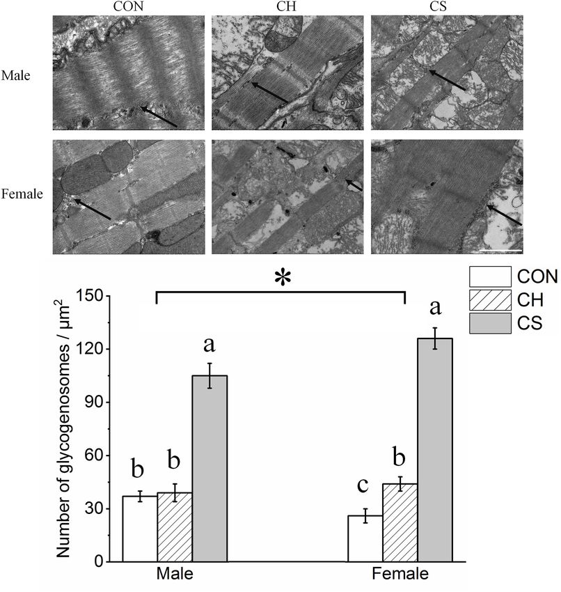
Xu et al.                                                                                                                     Huddling Maintained Myocardial Glycogen

reported on changes in myocardial glycogen in mammals under                            cause an increase in myocardial glycogen content in Brandt’s
different temperatures.                                                                voles. We also hypothesized that huddling could effectively
   Brandt’s voles are small non-hibernating herbivorous rodents                        alleviate this change. To test these hypotheses, we observed
widely distributed among the Inner Mongolian grasslands of                             the ultrastructure of cardiac muscle in huddling and individual
Northern China, dry steppe zone of Mongolia, and southeast                             (separated) Brandt’s voles under mild temperature differences
Baikal region of Russia. They are highly socialized animals                            (normal: 22◦ C; cool: 15◦ C) in autumn. We also determined the
that huddle in winter as an adaptation to their harsh habitats                         protein expression levels of glucose transport glycogen synthesis,
(Zhang et al., 2018), which differs substantially from model                           and glycogen degradation-related signals. We further explored
animals. Research has shown that mild cooling can significantly                        the underlying molecular mechanism related to the effects of a
change the morphology of mitochondria in the cardiac muscle of                         mild cold environment and huddling on changes in myocardial
Brandt’s voles (Xu et al., 2019; Wang et al., 2020a). Furthermore,                     glycogen content.
their metabolic rate and thermogenic capacity decrease but
activity increases compared with separated individuals under low
temperatures, suggesting that huddling is a good strategy for
small mammals to cope with cold environments (Sukhchuluun                              MATERIALS AND METHODS
et al., 2018). Glycogen is one of the most important energy
supply substances in muscles. However, the role of myocardial                          Ethics Statement
glycogen in adaptive huddling has not yet been reported.                               All procedures followed the Laboratory Animal Guidelines for
Therefore, we hypothesized that a cool environment could                               the Ethical Review of Animal Welfare (GB/T 35892-2018) and

  FIGURE 2 | Changes in number of myocardial glycogenosomes in Brandt’s voles. (A) Myocardial glycogenosomes in three treatment groups. Arrow indicates
  glycogenosome. Scale bar = 1 µm. (B) Bar graph depicting changes in number of glycogenosomes. Values are mean ± SD. Six figures were analyzed in each
  sample; eight samples were analyzed in each group. CON, control group; CH, cool huddling group; CS, cool separated group. Different letters identify statistically
  significant differences among temperature treatment groups (P < 0.05). *P < 0.05 significant differences between males and females.

Frontiers in Physiology | www.frontiersin.org                                      3                                           March 2021 | Volume 12 | Article 593129
Xu et al. Huddling Maintained Myocardial Glycogen
were approved by the Animal Care and Use Committee of Qufu males (28–50 g, average 38 g) and 24 female (27–54 g, average
Normal University (Permit Number: dwsc 2019012). 33 g) adult voles were randomly divided into three groups,
respectively. Control group (CON): Voles were continuously
Animals and Groups housed under an ambient temperature of 22 ± 2◦ C, with four
Forty-eight adult voles were captured and housed as described animals in each cage (two males and two females), similar to their
previously (Wang et al., 2020b). The voles were acclimated to normal state in autumn. Cool huddling group (CH): Voles were
laboratory conditions for 2 weeks. They were housed four animals housed together in a cage (two males and two females) under
per cage (28 × 18 × 12 cm) at an ambient temperature of an ambient temperature of 15◦ C. The group size (four voles in
22 ± 2◦ C, relative humidity of 55 ± 5%, and light/dark regime each cage) ensured most animals remained inactive in a huddle
of 12 h:12 h (light on from 06:00 to 18:00). Food (standard (Sukhchuluun et al., 2018). Cool separated group (CS): Voles
rabbit chow, Pengyue Experimental Animal Breeding Co., Ltd., were housed individually in cages at an ambient temperature
China) and water was provided ad libitum and wood shavings of 15◦ C. The three treatment groups were maintained under
were used as bedding. Based on body weight, a total of 24 the same relative humidity (55 ± 5%) and light regime (12 h:
12 h light /dark, light on from 06:00 to 18:00). Animal treatment
started in late September and lasted 8 weeks (Wang et al., 2020b).
Sample Preparation
All animals were sacrificed by CO2 asphyxiation between 08:00
and 11:00 a.m. on the last day of the experiment (Sukhchuluun
et al., 2018; Wang et al., 2020b). After the rapid removal of
cardiac muscle, portions of the ventricles were immediately
excised and fixed in glutaraldehyde. Specimens were fixed in 1%
osmium tetroxide in the same buffer, dehydrated with a graded
series of ethanol, and embedded in epoxy resin. The remaining
cardiac muscle was frozen in liquid nitrogen and stored at
−80◦ C. All procedures were carried out in accordance with the
approved guidelines.
Transmission Electron Microscopy (TEM)
The cardiac muscle samples were cut into blocks and immersed
FIGURE 3 | Changes in content of myocardial glycogenosomes in Brandt’s in 3% glutaraldehyde-paraformaldehyde. The blocks were then
voles. Glycogen levels were normalized by cell protein concentration dehydrated in a graded series of ethanol and embedded in epoxy
measured using the BCA assay. Different letters identify statistically significant
resin, with TEM then performed as described previously (Wang
differences among temperature treatment groups (P < 0.05). CON, control
group; CH, cool huddling group; CS, cool separated group.
et al., 2020a). Semi-thin sections of the tissue samples were
stained with methylene blue (Biazik et al., 2015), then adjusted
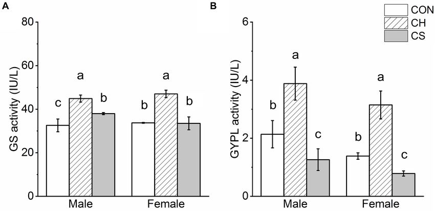
FIGURE 4 | Glycogen synthase (GS) and glycogen phosphorylase (GYPL) activity in cardiac muscle of voles. (A) GS activity. (B) GYPL activity. Values are
mean ± SD. n = 8. CON, control group; CH, cool huddling group; CS, cool separated group. Different letters indicate significant differences among temperature
treatment groups (P < 0.05).
Frontiers in Physiology | www.frontiersin.org 4 March 2021 | Volume 12 | Article 593129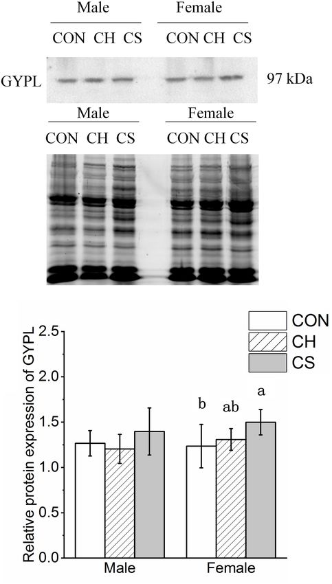
Xu et al.                                                                                                                     Huddling Maintained Myocardial Glycogen

  FIGURE 5 | Changes in protein expression levels of glycogen synthesis-related factors in cardiac muscle of Brandt’s voles. (A) Representative immunoblots of GS
  and P-GS in cardiac muscle. (B) Representative polyacrylamide gel of total protein. (C) Relative protein expression of GS. (D) Relative protein expression of P-GS.
  (E) Ratio of P-GS to GS. Values are mean ± SD. n = 8. CON, control group; CH, cool huddling group; CS, cool separated group. Different letters identify statistically
  significant differences among temperature treatment groups (P < 0.05). ∗ P < 0.05 significant differences between males and females.

under the microscope and sliced with an ultramicrotome (LKB-                            Glycogen Quantification
NOVA, United States). The ultrathin sections were double                                Samples stored at −80◦ C were used to detect glycogen content.
stained with Reynolds’ lead citrate and ethanolic uranyl acetate                        The amount of glycogen in the myocardia from the three groups
(Reynolds, 1963) and then examined via TEM (Hitachi, HT7800,                            was determined with a Glycogen Assay Kit (BC0340, Solarbio,
Japan). Images were processed with NIH Image-Pro Plus 6.0.                              Beijing, China). Glycogen levels were normalized by cell protein
Images were analyzed using the measurement tools provided by                            concentration measured using the BCA assay (Zhao et al., 2017).
the software. Glycogenosome densities were determined within
a defined region (4 µm2 area) at a minimum of three locations
within an image taken at 25,000 × magnification.                                        Western Blotting
                                                                                        Total protein was extracted from the tissues and solubilized in
GS and GYPL Activity                                                                    sample buffer (100 mM Tris, pH 6.8, 5% 2-β-mercaptoethanol,
Samples stored at −80◦ C were used to detect GS and GYPL                                5% glycerol, 4% SDS, and bromophenol blue), with the extracts
activity. GS activity was determined by measuring the rate of                           of cardiac protein then resolved via SDS-PAGE [10% Laemmli
NADH decline at 450 nm using a Glycogen Synthase Assay Kit                              gel with an acrylamide/bisacrylamide ratio of 29:1 and 98% 2,2,2-
(20E10Y14, Shanghai Hengyuan Biological Technology Co., Ltd.,                           trichloroethanol (Aladdin, JI522028, China)]. To study protein
China) according to the manufacturer’s instructions (Ouyang                             expression in different tissues, we used total protein content as a
et al., 2018). GYPL activity was determined by measuring the rate                       reference. After electrophoresis, the gel was irradiated on the UV
of NADPH increase at 450 nm with a Glycogen Phosphorylase                               platform of the electrophoresis gel imaging analysis system (Bio-
Activity Assay Kit (20H10L15, Shanghai Hengyuan Biological                              Rad, California, United States) for 5 min, with the signal then
Technology Co., Ltd., China) according to the manufacturer’s                            collected. As described previously (Li and Shen, 2013; Posch et al.,
instructions (Song et al., 2018).                                                       2013), the original image captured with no gain was stored. The

Frontiers in Physiology | www.frontiersin.org                                       5                                           March 2021 | Volume 12 | Article 593129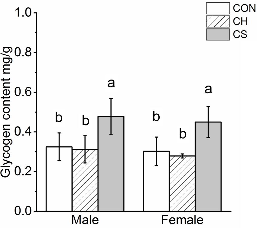
Xu et al.                                                                                                        Huddling Maintained Myocardial Glycogen

fluorescence intensity of each lane (after removal of background
fluorescence intensity) was determined with Image-Pro Plus 6.0,
which contains an internal reference to correct the fluorescence
intensity of the target protein. The proteins were then electrically
transferred to polyvinylidene fluoride (PVDF) membranes (0.45
µm pore size) using a Bio-Rad wet transfer apparatus. The blotted
membranes were blocked with 5% skimmed milk powder in Tris-
buffered saline (TBS; 150 mM NaCl, 50 mM Tris-HCl, pH 7.5)
and incubated with rabbit anti-glycogen phosphorylase (1:1,000,
#ab198268, Abcam, Cambridge, United Kingdom), rabbit anti-
glycogen synthase (1:1,000, #3886, Cell Signaling Technology
CST, Danvers, MA, United States), rabbit anti-phospho glycogen
synthase (1:1,000, #3891, CST), rabbit anti-glucose transporter
type 1 (1:500, #21829, Proteintech, China), rabbit anti-glucose
transporter type 2 (1:500, #20436, Proteintech, China), and
rabbit anti-glucose transporter type 4 (1:500, #21048, Proteintech,
China) in TBS containing 0.1% BSA at 4◦ C overnight. The
membranes were then incubated with IRDye 800 CW goat anti-
rabbit secondary antibodies (1:5,000, #31460, Thermo Fisher
Scientific, Rockford, IL, United States) for 90 min at room
temperature and visualized with an Odyssey scanner (Bio-Rad,
CA, United States). Quantification of blots was performed using
NIH Image-Pro Plus 6.0.

Statistical Analyses
The normality of data and homogeneity of variance were tested
by Shapiro-Wilk and Levene tests, respectively. All data exhibited
normal distribution and homogeneous variance. Double-factor
variance analysis [two-way analysis of variance (ANOVA)]                    FIGURE 6 | Changes in protein expression levels of glycogen
was used to compare differences between treatment and sex.                  degradation-related factors in cardiac muscle of Brandt’s voles.
Results were significant at P < 0.05. Data are expressed as                 (A) Representative immunoblots of GYPL in cardiac muscle.
                                                                            (B) Representative polyacrylamide gel of total protein. (C) Relative protein
mean ± standard deviation (Mean ± SD). All statistical analyses             expression of GYPL. Values are mean ± SD. n = 8. CON, control group; CH,
were conducted using SPSS 19.0.                                             cool huddling group; CS, cool separated group. Different letters identify
                                                                            statistically significant differences among temperature treatment groups
                                                                            (P < 0.05).

RESULTS
                                                                           Changes in GS and GYPL
Ultrastructural Changes in Number of                                       Results showed that GS activity in the CH group was
Glycogenosomes                                                             significantly higher than that in the CON and CS groups
Glycogenosome clusters were observed, with each                            (P < 0.05), but there were no significant differences
glycogenosome showing a diameter of ∼30 nm. Most                           between the CON and CS group in females. Furthermore,
glycogenosomes were distributed between the muscle                         among the three groups, GYPL activity was highest in
filaments, with a small number distributed around the                      the CH group (P < 0.05) and lowest in the CS group
mitochondria (Figure 1).                                                   (P < 0.05) (Figure 4).
    Figure 2A shows the distribution of glycogenosomes at low
magnification. In the CS group, the number of glycogenosomes               Changes in Protein Expression of
was more than triple that in the CON and CH groups (P < 0.05).
                                                                           Glycogen Synthesis-Related Proteins
In addition, the number was significantly higher (P < 0.05) in
                                                                           The GS and P-GS concentrations were detected by western blot
females than in males (Figure 2B).
                                                                           analysis, as shown in Figure 5. Representative polyacrylamide
                                                                           gels of total protein are shown in Figure 5B.
Glycogen Quantification                                                       The relative protein expression levels of GS and P-GS showed
Glycogen quantification showed significant accumulation in                 different trends among the three treatment groups. Specifically,
the CS group (P < 0.05), but no significant differences were               the protein expression levels of GS in the CS group were
observed between the CON and CH groups in either males or                  lower than the levels in the CH and CON groups, whereas
females (Figure 3).                                                        protein expression levels of P-GS in the CH and CS groups

Frontiers in Physiology | www.frontiersin.org                          6                                          March 2021 | Volume 12 | Article 593129
Xu et al. Huddling Maintained Myocardial Glycogen
FIGURE 7 | Changes in protein expression levels of glucose transporter proteins in cardiac muscle of Brandt’s voles. (A) Representative immunoblots of GLUT1,
GLUT2, and GLUT4 in cardiac muscle. (B) Representative polyacrylamide gel of total protein. (C) Relative protein expression of GLUT1. (D) Relative protein
expression of GLUT2. (E) Relative protein expression of GLUT4. Values are mean ± SD. n = 8. CON, control group; CH, cool huddling group; CS, cool separated
group. Different letters identify statistically significant differences among temperature treatment groups (P < 0.05).
were higher than levels in the CON group (P < 0.05). Levels Changes in Protein Expression of
of P-GS was higher (P < 0.05) in females than in males Glucose Transporter Proteins
(Figures 5C,D).
The contents of GLUT1, GLUT2, and GLUT4 were detected
The P-GS to GS ratio is one of the most direct indicators
by western blot analysis, as shown in Figure 7. Representative
of glycogen synthesis. Here, the ratio trend among the three
polyacrylamide gels of total protein are shown in Figure 7B.
treatment groups was CON < CH < CS (P < 0.05).
The relative protein expression of GLUT1 increased in the
The ratio was also higher (P < 0.05) in females than in
CS group compared with the other groups in both males
males (Figure 5E).
and females (P < 0.05). The relative protein expression of
GLUT2 showed the same trend, i.e., CON > CH > CS,
but there was a significant difference between males and
Changes in Protein Expression of females. The relative protein expression of GLUT4 in males
Glycogen Decomposition-Related was markedly higher in the CS group than in the CON group
Proteins (P < 0.05), but there were no differences in females among the
The content of GYPL was detected by western blot analysis, as three groups.
shown in Figure 6. Representative polyacrylamide gels of total
protein are shown in Figure 6B.
The relative protein expression of GYPL showed a slight DISCUSSION
change among the three treatment groups. Specifically, levels
were higher in CS group females than in CON group We studied the effects of a cool environment on the number
females (Figure 6C). of cardiac glycogenosomes and glycogen content in huddling
Frontiers in Physiology | www.frontiersin.org 7 March 2021 | Volume 12 | Article 593129Xu et al. Huddling Maintained Myocardial Glycogen
Brandt’s voles, as well as the underlying mechanism related effect of low temperature on glycogen synthesis enzyme
to the regulation of glycogenosome number. One of the can be significantly alleviated by huddling behavior. Here,
most important findings of this study is the ultrastructural huddling behavior completely or partially alleviated the
observation of a significant increase in the number of cardiac increase in glycogen content caused by the decrease in
glycogenosomes in the CS group, as verified by the glycogen glycogen degradation enzyme in the myocardium of voles
content results. following cold environment exposure by increasing glycogen
Changes in myocardial glycogen in mammals during decomposition. Normal glycogen metabolism is the basis
long-term cool exposure have not been reported previously, of exercise in mammals (Consitt et al., 2019; Moniz et al.,
although our results are consistent with those of myocardium 2020). Earlier studies on Brandt’s voles showed that activity
under short-term hypothermia and skeletal muscle under is higher in huddling groups than separated groups under
long-term hypothermia, as the major types of muscle cool environments (Sukhchuluun et al., 2018). Thus, we
fibers in ventricles are similar to those in soleus muscle speculated that the similar level of glycogen metabolism
(Schaub et al., 1989). Research on rats has shown that in the myocardium of the CH group and CON group
glycogen content in the myocardium is significantly compared to that in the CS group may be the one of the
increased after only 4 h of exposure to 15◦ C (Steffen, underlying reasons.
1988). Furthermore, Daurian ground squirrels experience Glycogen synthesis and decomposition also depend
an increase in glycogen concentration in the soleus muscle on changes in glucose metabolism (Chen and Phelix,
after 2 months of low temperature exposure in winter 2019). In this study, the protein expression levels of
(Wang et al., 2019). Excessive glycogen accumulation in glucose transporters GLUT1 and GLUT4 in the CS group
the heart can lead to degenerative changes such as arrhythmia, males were significantly higher than in the CON group
cardiac hypertrophy, and hypotonia (Kanungo et al., 2018). males, which may contribute to intracellular glucose
In this study, glycogen content in the myocardium of accumulation and glycogen content increase. In female
the CS group was significantly higher than that of the voles, the protein expression of GLUT1 was significantly
CON group. This indicates that hypothermia may cause higher in the CS group than in the CH group, which
significant degenerative damage to the myocardium of may be one of the reasons why glycogen content in
small mammals and may involve disrupting the balance the myocardium of the CS group was higher than that
between glycogen synthesis and decomposition. In addition, of the CH group.
our previous study indicated that ATP synthase activity in In summary, we explored the regulatory mechanism related
the myocardial mitochondria of Brandt’s voles under cool to the balance between glycogen synthesis and degradation
conditions is significantly lower than that observed under on the number in myocardial glycogenosomes of huddling
warm environments, which may lead to a decrease in glucose and separated Brandt’s voles under cool environments.
utilization in the mitochondria (Wang et al., 2020b). Thus, Results showed that a cool environment led to an increase
this may be one of the reasons for glycogen accumulation in myocardial glycogen content in voles, which could
in the CS group. be alleviated by huddling behavior, and may be a good
Here, compared with the CON group, GS activity in consequence of the collective overwintering behavior of
the myocardium increased in the CS group males but socialized animals. The activity of glycogen phosphorylase
remained stable in the CS group females, indicating decreased, and the protein expression of GLUT1 and GLUT2
that the level of glycogen synthesis did not decrease. In increased in CS group, indicating that the glycogen degradation
addition, in the CS group, GYPL activity decreased in the enzyme decreased and glucose transport increased in the
myocardium of both males and females, indicating that CS group. The activities of glycogen synthase and glycogen
glycogen decomposition was weakened. Therefore, the phosphorylase increased in the CH group, suggesting that
maintenance of glycogen synthesis enzyme and reduction the synthesis and decomposition of glycogen were increased
of glycogen degradation enzyme in the CS group may in the CH group. These results indicate that the reduced
be one of the main reasons for the increase in glycogen glycogen degradation enzyme level and enhanced glucose
content/glycogen particle accumulation in the myocardium. transport may lead to an increase in myocardial glycogen
One thing to note is that the expression of GS protein content in the separated voles under cool environment; while
was significantly decreased in the CS group, but its the up-regulation of glycogen synthesis and degradation
phosphorylation rate, the active state of GS (Greenberg enzyme level maintained myocardial glycogen content in
et al., 2006) was significantly increased, which may be the huddling voles.
a major mechanism related to the unchanged enzyme
activity level of GS.
Surprisingly, compared with the CON group, the content DATA AVAILABILITY STATEMENT
of glycogen in the myocardium of the CH group remained
unchanged, with the synchronous increase in glycogen The original contributions presented in the study are included
synthesis and degradation enzyme likely responsible for the in the article/Supplementary Material, further inquiries can be
maintenance of glycogen stability. This suggests that the directed to the corresponding author/s.
Frontiers in Physiology | www.frontiersin.org 8 March 2021 | Volume 12 | Article 593129Xu et al. Huddling Maintained Myocardial Glycogen
ETHICS STATEMENT FUNDING
All procedures followed the Laboratory Animal Guidelines for This work was supported by the funds from the National
the Ethical Review of Animal Welfare (GB/T 35892-2018) and Natural Science Foundation of China (Nos. 31770455,
were approved by the Animal Care and Use Committee of Qufu 32072436, and 31570377).
Normal University (Permit Number: dwsc 2019012).
AUTHOR CONTRIBUTIONS ACKNOWLEDGMENTS
J-HX and ZW conceived and designed the research, edited We thank two reviewers for excellent criticism of the article.
the manuscript, approved the final version, and drafted the
manuscript. J-HX, ZW, J-JM, C-LW, and W-MH performed the
experiments. J-JM and ZW analyzed the data and prepared the SUPPLEMENTARY MATERIAL
figures. J-JM interpreted the experimental results. H-LX, MW,
LC, and L-XX provided experimental guidance and suggestions The Supplementary Material for this article can be found
for revision. All authors contributed to the article and approved online at: https://www.frontiersin.org/articles/10.3389/fphys.
the submitted version. 2021.593129/full#supplementary-material
REFERENCES Jefimow, M., Glabska, M., and Wojciechowski, M. S. (2011). Social
thermoregulation and torpor in the siberian hamster. J. Exp. Biol. 214(Pt
Agius, L. (2010). Physiological control of liver glycogen metabolism: lessons from 7), 1100–1108. doi: 10.1242/jeb.050823
novel glycogen phosphorylase inhibitors. Mini Rev. Med. Chem. 10, 1175–1187. Kanungo, S., Wells, K., Tribett, T., and El-Gharbawy, A. (2018). Glycogen
doi: 10.2174/1389557511009011175 metabolism and glycogen storage disorders. Ann. Transl. Med. 6:9.
Baum, D., Dillard, D. H., and Porte, D. Jr. (1968). Inhibition of insulin release in Kelly, F. E., and Nolan, J. P. (2010). The effects of mild induced hypothermia on
infants undergoing deep hypothermic cardiovascular surgery. N. Engl. J. Med. the myocardium: a systematic review. Anaesthesia 65, 505–515. doi: 10.1111/j.
279, 1309–1314. doi: 10.1056/nejm196812122792404 1365-2044.2009.06237.x
Biazik, J., Vihinen, H., Anwar, T., Jokitalo, E., and Eskelinen, E. L. (2015). Kotze, J., Bennett, N. C., and Scantlebury, M. (2008). The energetics of huddling in
The versatile electron microscope: an ultrastructural overview of autophagy. two species of mole-rat (Rodentia: Bathyergidae). Physiol. Behav. 93, 215–221.
Methods 75, 44–53. doi: 10.1016/j.ymeth.2014.11.013 doi: 10.1016/j.physbeh.2007.08.016
Bickford, A. F., and Mottram, R. F. (1960). Glucose metabolism during induced Li, R., and Shen, Y. (2013). An old method facing a new challenge: re-visiting
hypothermia in rabbits. Clin. Sci. 19, 345–359. housekeeping proteins as internal reference control for neuroscience research.
Chavez, L. O., Leon, M., Einav, S., and Varon, J. (2017). Editor’s choice- inside the Life Sci. 92, 747–751. doi: 10.1016/j.lfs.2013.02.014
cold heart: a review of therapeutic hypothermia cardioprotection. Eur. Heart J. Mavrokefalos, N., Myrianthopoulos, V., Chajistamatiou, A. S., Chrysina, E. D., and
Acute Cardiovas. Care 6, 130–141. doi: 10.1177/2048872615624242 Mikros, E. (2015). Discovery of the glycogen phosphorylase-modulating activity
Chen, L. Y., and Phelix, C. F. (2019). Extracellular gating of glucose transport of a resveratrol glucoside by using a virtual screening protocol optimized for
through GLUT 1. Biochem. Biophys. Res. Commun. 511, 573–578. doi: 10.1016/ solvation effects. Planta Med. 81, 507–516. doi: 10.1055/s-0035-1545910
j.bbrc.2019.02.067 Moniz, S. C., Islam, H., and Hazell, T. J. (2020). Mechanistic and methodological
Consitt, L. A., Dudley, C., and Saxena, G. (2019). Impact of endurance and perspectives on the impact of intense interval training on post-exercise
resistance training on skeletal muscle glucose metabolism in older adults. metabolism. Scand. J. Med. Sci. Sports 30, 638–651. doi: 10.1111/sms.13610
Nutrients 11:2636. doi: 10.3390/nu11112636 Namekata, S., and Geiser, F. (2009). Effects of nest use, huddling, and torpor
Curry, D. L., and Curry, K. P. (1970). Hypothermia and insulin secretion. on thermal energetics of eastern pygmy-possums. Aust. Mammal. 31, 31–34.
Endocrinology 87, 750–755. doi: 10.1210/endo-87-4-750 doi: 10.1071/AM08114
Douglas, T. K., Cooper, C. E., and Withers, P. C. (2017). Avian torpor or alternative Nowack, J., and Geiser, F. (2016). Friends with benefits: the role of huddling in
thermoregulatory strategies for overwintering? J. Exp. Biol. 220, 1341–1349. mixed groups of torpid and normothermic animals. J. Exp. Biol. 219, 590–596.
doi: 10.1242/jeb.154633 doi: 10.1242/jeb.128926
Fuhrman, G. J., and Fuhrman, F. A. (1963). Utilization of glucose by the Nunez-Villegas, M., Bozinovic, F., and Sabat, P. (2014). Interplay between group
hypothermic rat. Am. J. Phys. 205, 181–183. doi: 10.1152/ajplegacy.1963.205. size, huddling behavior and basal metabolism: an experimental approach in the
1.181 social degu. J. Exp. Biol. 217, 997–1002. doi: 10.1242/jeb.096164
Gilbert, C., Mccafferty, D., Le Maho, Y., Martrette, J. M., Giroud, S., Blanc, S., Ouyang, Q., Tao, N., and Zhang, M. (2018). A damaged oxidative phosphorylation
et al. (2010). One for all and all for one: the energetic benefits of huddling mechanism is involved in the antifungal activity of citral against
in endotherms. Biol. Rev. Camb. Philos. Soc. 85, 545–569. doi: 10.1111/j.1469- Penicillium digitatum. Front. Microbiol. 9:239. doi: 10.3389/fmicb.2018.
185X.2009.00115.x 00239
Greenberg, C. C., Jurczak, M. J., Danos, A. M., and Brady, M. J. (2006). Glycogen Palm, D. C., Rohwer, J. M., and Hofmeyr, J. H. (2013). Regulation of glycogen
branches out: new perspectives on the role of glycogen metabolism in the synthase from mammalian skeletal muscle–a unifying view of allosteric and
integration of metabolic pathways. Am. J. Physiol. Endocrinol. Metab. 291, covalent regulation. FEBS J. 280, 2–27. doi: 10.1111/febs.12059
E1–E8. doi: 10.1152/ajpendo.00652.2005 Polderman, K. H. (2009). Mechanisms of action, physiological effects, and
Hall, E. M., and Mackay, E. M. (1933). The relation between the mitochondria and complications of hypothermia. Crit. Care Med. 37(7 Suppl), S186–S202. doi:
glucose-glycogen equilibrium in the liver. Am. J. Pathol. 9, 205–220.1. 10.1097/CCM.0b013e3181aa5241
Helman, A., Gilbert, M., Pfister-Lemaire, N., Reach, G., and Assan, R. (1984). Popovic, V. (1960). Physiological characteristics of rats and ground squirrels
Glucagon and insulin secretion and their biological activities in hypothermic during prolonged lethargic hypothermia. Am. J. Physiol. 199, 467–471. doi:
rats. Endocrinology 115, 1722–1728. doi: 10.1210/endo-115-5-1722 10.1152/ajplegacy.1960.199.3.467
Frontiers in Physiology | www.frontiersin.org 9 March 2021 | Volume 12 | Article 593129Xu et al. Huddling Maintained Myocardial Glycogen Posch, A., Kohn, J., Oh, K., Hammond, M., and Liu, N. (2013). V3 stain-free Wang, Z., Xu, J.-H., Mou, J.-J., Kong, X.-T., Zou, J.-W., Xue, H.-L., et al. (2020b). workflow for a practical, convenient, and reliable total protein loading control Novel ultrastructural findings on cardiac mitochondria of huddling Brandt’s in western blotting. J. Vis. Exp. 82:50948. doi: 10.3791/50948 voles in mild cold environment. Comp. Biochem. Physiol. A Mol. Integr. Physiol. Reynolds, E. S. (1963). The use of lead citrate at high pH as an electron-opaque 249:110766. doi: 10.1016/j.cbpa.2020.110766 stain in electron microscopy. J. Cell Biol. 17:208. doi: 10.1083/jcb.17.1.208 Wei, Y., Zhang, J., Xu, S., Peng, X., Yan, X., Li, X., et al. (2018). Controllable Roach, P. J., Depaoli-Roach, A. A., Hurley, T. D., and Tagliabracci, V. S. (2012). oxidative stress and tissue specificity in major tissues during the torpor-arousal Glycogen and its metabolism: some new developments and old themes. cycle in hibernating daurian ground squirrels. Open Biol. 8:180068. doi: 10. Biochem. J. 441, 763–787. doi: 10.1042/bj20111416 1098/rsob.180068 Scantlebury, M., Bennett, N. C., Speakman, J. R., Pillay, N., and Schradin, C. (2006). Wojciechowski, M. S., Jefimow, M., and Pinshow, B. (2011). Heterothermy, and the Huddling in groups leads to daily energy savings in free-living African four- energetic consequences of huddling in small migrating passerine birds. Integr. striped grass mice, Rhabdomys pumilio. Funct. Ecol. 20, 166–173. doi: 10.1111/ Comp. Biol. 51, 409–418. doi: 10.1093/icb/icr055 j.1365-2435.2006.01074.x Xu, D.-L., Xu, M.-M., and Wang, D.-H. (2019). Effect of temperature Schaub, M. C., Brunner, U. T., Von Schulthess, C., Neidhart, M., and Baumann, H. on antioxidant defense and innate immunity in Brandt’s (1989). Adaptation of contractile proteins in human heart and skeletal muscles. voles. Zool. Res. 40, 305–316. doi: 10.24272/j.issn.2095-8137.20 Biomed. Biochim. Acta 48, S306–S312. 19.045 Sealander, J. A. Jr. (1952). The relationship of nest protection and huddling to Xu, W., Fu, Y., Jiang, L. Y., Yang, Z. F., Wang, Y., Tang, W. C., et al. survival of Peromyscus at low temperature. Ecology 33, 63–71. doi: 10.2307/ (2020). Cardiopulmonary resuscitation ameliorates myocardial mitochondrial 1931252 dysfunction in a cardiac arrest rat model. Am. J. Emer. Med. 38, 65–72. doi: Song, M., Chen, F. F., Li, Y. H., Zhang, L., Wang, F., Qin, R. R., et al. (2018). 10.1016/j.ajem.2019.04.024 Trimetazidine restores the positive adaptation to exercise training by mitigating Zeqiraj, E., and Sicheri, F. (2015). Getting a handle on glycogen synthase–its statin-induced skeletal muscle injury. J. Cachexia Sarcopenia Muscle 9, 106–118. interaction with glycogenin. Mol. Aspects Med. 46, 63–69. doi: 10.1016/j.mam. doi: 10.1002/jcsm.12250 2015.08.004 Steffen, J. M. (1988). Glucose, glycogen, and insulin responses in the hypothermic Zhang, X. Y., Sukhchuluun, G., Bo, T. B., Chi, Q. S., Yang, J. J., Chen, B., et al. rat. Cryobiology 25, 94–101. doi: 10.1016/0011-2240(88)90002-8 (2018). Huddling remodels gut microbiota to reduce energy requirements in a Sukhchuluun, G., Zhang, X. Y., Chi, Q. S., and Wang, D. H. (2018). Huddling small mammal species during cold exposure. Microbiome 6:103. doi: 10.1186/ conserves energy, decreases core body temperature, but increases activity in s40168-018-0473-9 Brandt’s Voles (Lasiopodomys brandtii). Front. Physiol. 9:563. doi: 10.3389/ Zhao, X., Sun, K., Lan, Z., Song, W., Cheng, L., Chi, W., et al. (2017). fphys.2018.00563 Tenofovir and adefovir down-regulate mitochondrial chaperone Tarnopolsky, M. A. (2016). Metabolic myopathies. Continuum (Minneap. Minn) TRAP1 and succinate dehydrogenase subunit B to metabolically 22, 1829–1851. reprogram glucose metabolism and induce nephrotoxicity. Sci. Rep. Tessier, S. N., and Storey, K. B. (2012). Myocyte enhancer factor-2 and cardiac 7:46344. muscle gene expression during hibernation in thirteen-lined ground squirrels. Gene 501, 8–16. doi: 10.1016/j.gene.2012.04.004 Conflict of Interest: The authors declare that the research was conducted in the Thorens, B., and Mueckler, M. (2010). Glucose transporters in the 21st Century. absence of any commercial or financial relationships that could be construed as a Am. J. Phys. Endocrinol. Metab. 298, E141–E145. potential conflict of interest. Wang, Z., Jiang, S. F., Cao, J., Liu, K., Xu, S. H., Arfat, Y., et al. (2019). Novel findings on ultrastructural protection of skeletal muscle fibers during Copyright © 2021 Xu, Wang, Mou, Wang, Huang, Xue, Wu, Chen and Xu. This is an hibernation of daurian ground squirrels: mitochondria, nuclei, cytoskeleton, open-access article distributed under the terms of the Creative Commons Attribution glycogen. J. Cell Physiol. 234, 13318–13331. doi: 10.1002/jcp.28008 License (CC BY). The use, distribution or reproduction in other forums is permitted, Wang, Z., Xu, J.-H., Mou, J.-J., Kong, X.-T., Wu, M., Xue, H.-L., et al. (2020a). provided the original author(s) and the copyright owner(s) are credited and that the Photoperiod affects harderian gland morphology and secretion in female original publication in this journal is cited, in accordance with accepted academic Cricetulus barabensis: autophagy, apoptosis, and mitochondria. Front. Physiol. practice. No use, distribution or reproduction is permitted which does not comply 11:408. doi: 10.3389/fphys.2020.00408 with these terms. Frontiers in Physiology | www.frontiersin.org 10 March 2021 | Volume 12 | Article 593129
You can also read