A Portable Smartphone-Based Laryngoscope System for High-Speed Vocal Cord Imaging of Patients With Throat Disorders: Instrument Validation Study ...
←
→
Page content transcription
If your browser does not render page correctly, please read the page content below
JMIR MHEALTH AND UHEALTH                                                                                                                        Kim et al

     Original Paper

     A Portable Smartphone-Based Laryngoscope System for
     High-Speed Vocal Cord Imaging of Patients With Throat Disorders:
     Instrument Validation Study

     Youngkyu Kim1,2, BSc; Jeongmin Oh1, BSc; Seung-Ho Choi3, MD, PhD; Ahra Jung4, MD; June-Goo Lee1,2, PhD;
     Yoon Se Lee3, MD, PhD; Jun Ki Kim1,2, PhD
     1
      Biomedical Engineering Research Center, Asan Institute for Life Sciences, Asan Medical Center, Seoul, Republic of Korea
     2
      Department of Convergence Medicine, College of Medicine, University of Ulsan, Seoul, Republic of Korea
     3
      Department of Otorhinolaryngology-Head and Neck Surgery, Asan Medical Center, Seoul, Republic of Korea
     4
      Department of Otorhinolaryngology-Head and Neck Surgery, Eulji Medical Center, Eulji University School of Medicine, Seoul, Republic of Korea

     Corresponding Author:
     Jun Ki Kim, PhD
     Biomedical Engineering Research Center
     Asan Institute for Life Sciences
     Asan Medical Center
     88, Olympic-ro, 43-gil
     Seoul, 05505
     Republic of Korea
     Phone: 82 230108619
     Email: kim@amc.seoul.kr

     Abstract
     Background: Currently, high-speed digital imaging (HSDI), especially endoscopic HSDI, is routinely used for the diagnosis
     of vocal cord disorders. However, endoscopic HSDI devices are usually large and costly, which limits access to patients in
     underdeveloped countries and in regions with inadequate medical infrastructure. Modern smartphones have sufficient functionality
     to process the complex calculations that are required for processing high-resolution images and videos with a high frame rate.
     Recently, several attempts have been made to integrate medical endoscopes with smartphones to make them more accessible to
     people in underdeveloped countries.
     Objective: This study aims to develop a smartphone adaptor for endoscopes, which enables smartphone-based vocal cord
     imaging, to demonstrate the feasibility of performing high-speed vocal cord imaging via the high-speed imaging functions of a
     high-performance smartphone camera, and to determine the acceptability of the smartphone-based high-speed vocal cord imaging
     system for clinical applications in developing countries.
     Methods: A customized smartphone adaptor optical relay was designed for clinical endoscopy using selective laser melting–based
     3D printing. A standard laryngoscope was attached to the smartphone adaptor to acquire high-speed vocal cord endoscopic images.
     Only existing basic functions of the smartphone camera were used for HSDI of the vocal cords. Extracted still frames were
     observed for qualitative glottal volume and shape. For image processing, segmented glottal and vocal cord areas were calculated
     from whole HSDI frames to characterize the amplitude of the vibrations on each side of the glottis, including the frequency, edge
     length, glottal areas, base cord, and lateral phase differences over the acquisition time. The device was incorporated into a
     preclinical videokymography diagnosis routine to compare functionality.
     Results: Smartphone-based HSDI with the smartphone-endoscope adaptor could achieve 940 frames per second and a resolution
     of 1280 by 720 frames, which corresponds to the detection of 3 to 8 frames per vocal cycle at double the spatial resolution of
     existing devices. The device was used to image the vocal cords of 4 volunteers: 1 healthy individual and 3 patients with vocal
     cord paralysis, chronic laryngitis, or vocal cord polyps. The resultant image stacks were sufficient for most diagnostic purposes.
     The cost of the device including the smartphone was lower than that of existing HSDI devices. The image processing and analytics
     demonstrated the successful calculation of relevant diagnostic variables from the acquired images. Patients with vocal pathologies
     were easily differentiable in the quantitative data.

     https://mhealth.jmir.org/2021/6/e25816                                                             JMIR Mhealth Uhealth 2021 | vol. 9 | iss. 6 | e25816 | p. 1
                                                                                                                      (page number not for citation purposes)
XSL• FO
RenderX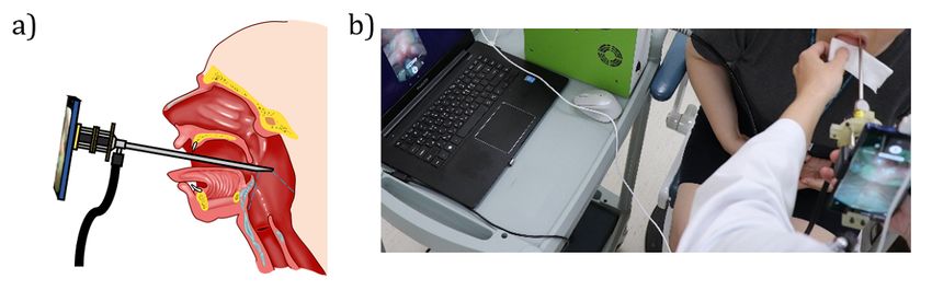
JMIR MHEALTH AND UHEALTH                                                                                                                 Kim et al

     Conclusions: A smartphone-based HSDI endoscope system can function as a point-of-care clinical diagnostic device. The
     resulting analysis is of higher quality than that accessible by videostroboscopy and promises comparable quality and greater
     accessibility than HSDI. In particular, this system is suitable for use as an accessible diagnostic tool in underdeveloped areas with
     inadequate medical service infrastructure.

     (JMIR Mhealth Uhealth 2021;9(6):e25816) doi: 10.2196/25816

     KEYWORDS
     smartphone; mobile phone; endoscope; high-speed imaging; vocal cord; low-cost device; mHealth; otorhinolaryngology; head
     and neck; throat

                                                                          insufficient endoscopic gastroscope and colonoscope
     Introduction                                                         accessories, HSDI devices are difficult to furnish because of
     The endoscope enables optical visualization of internal organs       their high cost and additional technology requirements. This
     and is a fundamental diagnostic device in clinical fields; unique    seriously restricts clinical voice evaluation in underdeveloped
     properties of endoscopic systems are indispensable in                countries since HSDI has become an important part in the
     diagnostics [1], biopsy [2], and minimally invasive surgery [3].     diagnosis of voice disorders. As with many problems regarding
     To acquire more accurate and specific diagnoses, endoscope           endoscopy in developing countries, affordable and more
     systems have been developed with higher resolution [4], greater      manageable endoscopic HSDI systems are urgently required.
     imaging speed [5,6], quantitative image analysis [7,8], and          Several point-of-care diagnostic devices have been invented in
     higher performance. With these advantages, endoscopy is now          response to the demand in developing and underdeveloped
     considered to be an essential procedure for a variety of clinical    countries. Prior to the age of integrated electronics, handheld
     applications, including clinical voice evaluation.                   point-of-care devices were largely restricted to diagnostic strips,
     Imaging of vocal cord vibrations is one of many techniques           which relied heavily on color changes due to chemical reactions
     used for clinical voice assessment. It is commonly accepted that     [18]. With the ubiquity of smartphones, the rate of invention of
     vocal cord vibration irregularities strongly correlate with voice    electronic point-of-care devices has drastically increased.
     disorders. However, since the frequencies of vocal cord              Modern smartphones offer a computing performance comparable
     vibrations are approximately 80-240 Hz, standard                     to that of personal computers, and imaging quality similar to
     videostroboscopy at 60 frames per second (fps) does not permit       that of professional camera devices. Consequently, there have
     the capture of vocal cord movement aside from stable and             been many attempts to use smartphones for point-of-care
     periodic states [9-11]. Alternative methods have been studied        diagnosis. For example, smartphone cameras can be used to
     to overcome the limitations of videostroboscopy for imaging          distinguish reaction intensities, and several smartphone
     vocal cord vibrations. The most promising approach is                attachments have been introduced for biological analysis similar
     high-speed digital imaging (HSDI) [12], which typically              to that performed by enzyme-linked immunosorbent assay
     captures images between 4000 and 8000 fps [13]. The high             (ELISA) [19], fecal hemoglobin detection [20], and
     frame rate of HSDI allows for the capture of vocal vibrations        electrochemical monitoring of blood contents [21]. This
     from 80-240 Hz, which cannot be observed in standard 60 fps          represents an evolution of traditional point-of-care strip
     imaging systems. Furthermore, HSDI provides a sufficient             diagnosis, in which a smartphone can be used to distinguish the
     resolution of more than 256 × 256 pixels [14]. Although the          intensity of a biological reaction. Other proposed applications
     resolution is hardly comparable to that of HD (high definition),     of the smartphone camera for point-of-care diagnosis include
     it is sufficient for medical analysis. Use of HSDI for clinical      clinical    microscopy,      endoscopic       imaging,      video
     voice evaluation has been extensively demonstrated [10,15,16].       nasolaryngoscopy, flexible robotic endoscopy, etc [22-28].
     However, despite several advantages, HSDI is not widely used         In this study, we introduce a smartphone-based endoscopic
     in clinical diagnosis because systems with better performance        imaging device and validate its imaging performance relative
     than laryngostroboscopy are generally expensive, require             to videostroboscopy and HSDI. The smartphone adaptor was
     specialized technology, and have large data footprints [13].         designed and manufactured using 3D design software and
     Endoscopic diagnosis is mostly inaccessible for patients in          selective laser melting (SLM)–based 3D printing. This
     underdeveloped and developing countries. Indeed, even in the         customized smartphone adaptor for the endoscope has minimal
     most developed and well-equipped hospitals in Nigeria, there         and low-cost optical components for easy maintenance and
     may be only one functional gastroscope and/or colonoscope,           control. Unlike previous studies that only used smartphones for
     for which the accessories are often deficient. Furthermore, the      acquisition of regular images and videos, in this study, the
     majority of the teaching hospitals in Nigeria have no facilities     specialized high-speed imaging function of the smartphone was
     for therapeutic endoscopy [17]; this leads to a lack of experience   applied for HSDI of vocal cord vibrations. With this
     in endoscopy for medical students, which can lead to                 functionality, high-speed vocal cord videos were acquired at
     unsatisfactory or even fatal endoscopy procedures. Furthermore,      940 fps following standard clinical protocols and analyzed using
     more serious issues, including inadequate maintenance,               postprocessed imaging techniques such as segmentation and
     cross-infections, and insufficient performance of devices, have      registration to simplify diagnosis. The resultant images are
     also been found in developing countries. In facilities with          quantitatively more revealing than videostroboscopy, and

     https://mhealth.jmir.org/2021/6/e25816                                                      JMIR Mhealth Uhealth 2021 | vol. 9 | iss. 6 | e25816 | p. 2
                                                                                                               (page number not for citation purposes)
XSL• FO
RenderX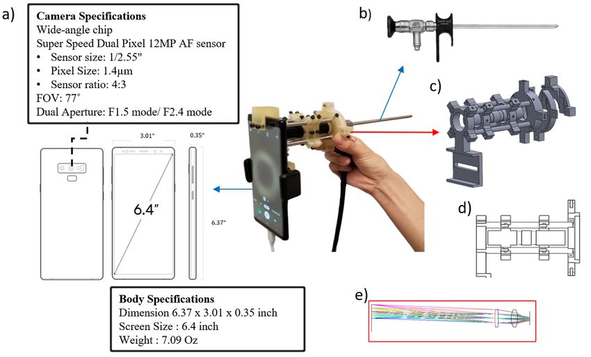
JMIR MHEALTH AND UHEALTH                                                                                                                        Kim et al

     approach the quality of HSDI due to their high resolution and             lenses, a lens tube, and mounts for the lens tube and for
     frame rate. In preclinical image analysis, several diagnostic             connection to the endoscope. The holders and connectors were
     variables were determined likely to assist in the diagnosis of            designed using 3D modeling software (Solid Works) and printed
     common vocal cord pathologies. This custom simple, low-cost               by an SLM 3D printer (Objet260, Stratasys Ltd). A lens system
     adaptor promises to reduce the cost of high-speed clinical                was used to magnify the endoscope probe eyepiece image
     imaging by an order of magnitude when it can be used as a                 between the endoscope eyepiece and the smartphone camera
     substitute for standard HSDI. The device demonstrates the                 and was held by 3D-printed parts. To make the system more
     promise and emerging capabilities of clinical diagnostic tools            cost-effective, a combination of 2 biconvex lenses was used
     that incorporate commodity smartphone technology to bring                 instead of expensive achromatic lenses for magnification of the
     point-of-care diagnosis to remote locations.                              endoscopic image. The focal lengths of the 2 biconvex lenses
                                                                               were 50 mm and 15 mm, respectively; this optical setup could
     Methods                                                                   acquire images with approximately 1.5× magnification.
                                                                               Illumination was coupled into the illumination port on the
     Development of a Smartphone-Adapted Endoscope                             endoscope. For endoscopic illumination, we used a broadband
     A smartphone adaptor was developed for use in clinical                    (360-770 nm) LED light source (X-Cite XYLIS, Excelitas
     endoscopy (Figure 1) with a standard 70° rigid laryngoscope               Technologies Corp), which was connected to the illumination
     (Karl Storz Co). The adaptor consists of a smartphone holder,             port of the endoscope with a liquid light guide.

     Figure 1. Schematics of a customized smartphone-endoscope adaptor that enables high-speed laryngoscopy. (A) Smartphone camera and body
     specifications. (B) A photograph of the demonstrated smartphone adaptor incorporating a rigid clinical endoscope. (C) 3D models of the customized
     smartphone-endoscope adaptor. (D) Cross-sectional view of the customized smartphone-endoscope adaptor. (E) Schematic and lens simulations of the
     magnification lens system. AF: autofocus, FOV: field of view.

     A specific smartphone model (Galaxy Note 9, Samsung) was                  Statistical and Data Analysis
     used to capture endoscopic images at a high frame rate. The               The acquired high-speed vocal cord vibration images were
     catalog specification provided by the manufacturer of the                 segmented using the seeded region growing algorithm [30] to
     smartphone claims that this specific smartphone model can                 determine the amplitude of the vibrations on each side of the
     acquire high-speed images at 960 fps, for a maximum duration              glottis [31]. Image analysis software was developed in
     of 0.4 seconds, at 1280 × 720 pixel resolution [29]. The                  MATLAB (MathWorks) to calculate the diagnostic parameters,
     complementary metal-oxide-semiconductor sensor of the                     including the vocal cord vibration frequencies, glottal edge
     smartphone is specified for 12.0 megapixels (MP) and 1/2.55               phase shifts, and total glottal area changes.
     inches (1.4 μm pixel size), and is packaged with a dual aperture
     mode.                                                                     Clinical Experiment
                                                                               The developed smartphone-based imaging system was used to
                                                                               acquire a high-speed video of the vocal cords of 4 human

     https://mhealth.jmir.org/2021/6/e25816                                                             JMIR Mhealth Uhealth 2021 | vol. 9 | iss. 6 | e25816 | p. 3
                                                                                                                      (page number not for citation purposes)
XSL• FO
RenderX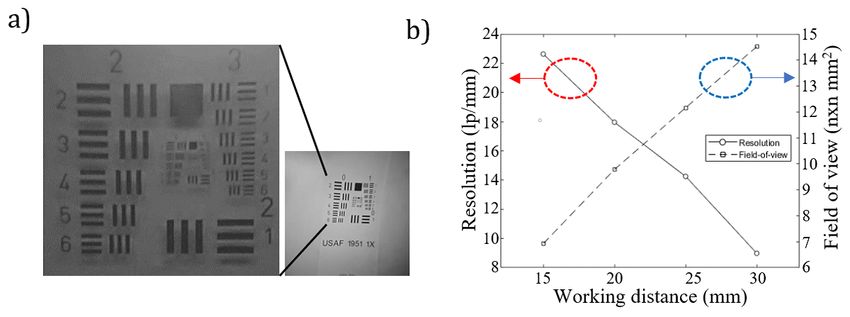
JMIR MHEALTH AND UHEALTH                                                                                                                          Kim et al

     subjects—1 healthy individual and 3 patients with known vocal               the patient and assess their symptoms and voice status, and the
     pathologies. A board-certified otolaryngologist at Asan Medical             second step was videostrobolaryngoscopy or videokymography
     Center with 15 years of experience followed common                          with an endoscope. Depending on the patient’s condition,
     videokymography routines [11] to capture high-speed diagnostic              additional tests such as computed tomography (CT) scans or a
     videos in a clinical environment (Figure 2). The routine vocal              blood test may be required.
     cord examination was as follows: the first step was to interview
     Figure 2. (A) A schematic representation of laryngeal imaging performed by the smartphone-based high-speed imaging system. (B) A clinical
     demonstration of the smartphone-based high-speed imaging system.

     Before imaging human subjects, the smartphone-based HSDI
     system was adapted to the clinical endoscope by adjusting the
                                                                                 Results
     fine focus and fixing the endoscope in place. Following                     System Characterization
     confirmation that the received images were clear and focused,
     the clinician performed routine vocal cord evaluation using the             The US Air Force resolution target was used to evaluate the
     endoscope-smartphone device. When the diagnosis routine was                 imaging performance of the smartphone-endoscope system
     completed, the endoscope probe was wiped with a disinfectant                (Figure 3A). The distance between the end of the endoscope
     to avoid cross-contamination between human subjects [32]. The               probe and the resolution target was adjusted from 15 mm to
     whole procedure took less than a minute, thus avoiding undue                30 mm in order to determine the proper optical field of view
     burden on volunteers.                                                       (FOV) and resolution. At the highest resolution, the system
                                                                                 resolved 23 line pairs per millimeter (lp/mm), which
     The study was approved by the Institutional Review Board (IRB               corresponded to 43.478 μm at a working distance of 15 mm.
     #2020-0798) of Asan Medical Center, Seoul, under the Korean                 The resolution of the system was observed to decrease as the
     Bioethics and Safety Act and the Korean Medical Device Act.                 working distance was increased. In addition, the optical FOV
                                                                                 was 7.8 mm × 7.8 mm at a 15 mm working distance, and 14.6
                                                                                 mm × 14.6 mm at a 30 mm working distance; thus the optical
                                                                                 FOV increased approximately linearly with the working distance
                                                                                 (Figure 3B).
     Figure 3. (A) US Air Force target test image taken at 960 fps. (B) A graph of the measured image resolution and field of view as functions of working
     distance.

     https://mhealth.jmir.org/2021/6/e25816                                                               JMIR Mhealth Uhealth 2021 | vol. 9 | iss. 6 | e25816 | p. 4
                                                                                                                        (page number not for citation purposes)
XSL• FO
RenderX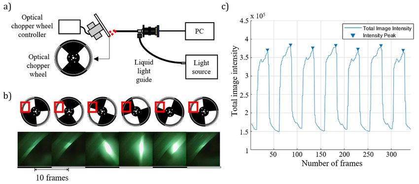
JMIR MHEALTH AND UHEALTH                                                                                                                       Kim et al

     A custom frame rate test was designed to confirm the video               captured by the high-speed imaging system. The total intensity
     frame rate of the customized high-speed system. An optical               of light reflected from the 2 quadrants of the chopper wheel
     chopper wheel was used to create a time-dependent moving                 resulted in a series of image frames with a periodically
     sample of known frequency. Two quadrants of a 4-quadrant                 modulated intensity signal that could be plotted frame by frame.
     optical chopper wheel mask were optically blocked, and the               The period of intensity modulation was calculated as the time
     other 2 quadrants were left transparent as shown schematically           interval between frames with peak intensities and was equal to
     in Figure 4. While the light source provided illumination from           half the rotational period of the optical chopper; this was
     the endoscope, high frame rate videos were acquired with the             measured to be 46.972 frames. Since the optical chopper was
     optical chopper wheel rotating at 10 Hz. The acquired high               rotating at 10 Hz, the duration of a half-rotation was
     frame rate videos were analyzed by image processing in                   0.05 seconds; therefore, the actual frame rate of the smartphone
     MATLAB, as light reflected from the chopper wheel was                    was 939.44 fps.
     Figure 4. (A) Schematic of the system for high-speed imaging assessment. (B) Image series acquired over 50 frames. (C) A plot of the total image
     intensity by chopper wheel rotation. PC: personal computer.

                                                                              glottal area changes are shown in Figure 6A. Using the fast
     Clinical Vocal Cord Vibration HSDI                                       Fourier transform, we were able to remove motion error and
     The obtained HSDI vocal cord vibration videos were taken                 extract the base frequency of the healthy vocal cord. The
     through a clinical diagnosis process. The results are presented          fundamental frequency of the healthy subject, a 24-year-old
     in Figures 5 and 6. As depicted in Figure 5, the 940-fps HSDI            female, was 224 Hz. The average fundamental frequency of
     video obtained from the clinical experiments was segmented               adult females of this age is reported to be similar, at around
     into vocal cord area and glottal area for further analysis; this         217 Hz [33]. Figure 6B shows the normalized total and lateral
     allowed us to obtain the parameters of the vocal cord with the           (left and right) glottal area changes in the vocal cords of a patient
     largest contribution to clinical diagnosis. The parameters               with left vocal cord paralysis. These data show that the
     obtained from the vocal cord vibration HSDI data included the            amplitudes of the left vocal cord vibrations are significantly
     base vibration frequency of the vocal cord, the difference               lower than those of the right vocal cord. The normalized total
     between the left glottal area and the right glottal area, and the        and lateral glottal areas of the patient with chronic laryngitis
     total glottal area.                                                      are shown in Figure 6C. Although the maximum amplitudes
     As shown in Figure 5, the data were obtained from the healthy            are similar to those of the healthy subject in both vocal cords,
     subject, and patients with left vocal cord paralysis, chronic            the minimum amplitude of each cycle differs from those of the
     laryngitis, and right vocal cord polyp. The original RGB                 healthy subject. The minimum amplitude value of each cycle
     (red-green-blue) color image data were converted to grayscale,           in the healthy subject had almost zero area; this is because the
     and the contrast was adjusted for glottal area segmentation using        vocal cord closes completely when the vocalizations are
     MATLAB. The seeded region growing method [30] was used                   generated in a healthy subject. However, in the patient with
     to segment the glottal area from the image processed data. In            chronic laryngitis, the minimum glottal area was larger than
     addition, the anatomical midline of the vocal cord was drawn,            that in the healthy subject, because in chronic laryngitis the
     after consultation with a clinician, to divide the left and right        vocal cords do not close completely as sound is generated.
     glottal areas.                                                           Figure 6D shows the normalized total and lateral glottal areas
                                                                              for the vocal cords of a patient with a polyp on the right vocal
     The segmented glottal areas were analyzed in order to determine          cord. In this case, the vibrations of the right vocal cord were
     any differences between the data of the healthy subject and              restricted by the polyp; therefore, the vibration amplitude of the
     those of patients. From healthy subject data, normalized total           right glottal area is less than that of the left glottal area. Similar

     https://mhealth.jmir.org/2021/6/e25816                                                            JMIR Mhealth Uhealth 2021 | vol. 9 | iss. 6 | e25816 | p. 5
                                                                                                                     (page number not for citation purposes)
XSL• FO
RenderX
JMIR MHEALTH AND UHEALTH Kim et al
to chronic laryngitis, the patient with the right vocal cord polyp phenomenon is caused by imperfect closure of the vocal cords
had much larger minimum areas than the healthy subject; this due to the polyp.
Figure 5. High-speed images of the glottis and their segmentation. Raw vocal cord image (left), preprocessed image for segmentation (center), and
area of the normal vocal cord by image segmentation (right). (A) The glottis of a healthy volunteer. (B) Images taken from a patient with left vocal cord
paralysis. (C) Vocal analysis from a patient with chronic laryngitis. (D) Images from a patient with a right vocal cord polyp.
https://mhealth.jmir.org/2021/6/e25816 JMIR Mhealth Uhealth 2021 | vol. 9 | iss. 6 | e25816 | p. 6
(page number not for citation purposes)
XSL• FO
RenderXJMIR MHEALTH AND UHEALTH Kim et al
Figure 6. Plots of the normalized total glottal area (left) and a comparison of the normalized left and right glottal areas separated by the anatomical
midline of the glottis over the course of a 0.4-second video at 940 fps (right) plotted for each patient: (A) normal healthy subject, (B) patient with left
vocal cord paralysis, (C) patient with chronic laryngitis, and (D) patient with right vocal cord polyp.
including blood, saliva, sweat, and urine [18,34-36], and usage
Discussion of smartphone functionalities are restricted to recognition of
Principal Findings color changes or receiving electric signals generated by
biochemical reactions. Most point-of-care devices are invasive,
Although many point-of-care devices have been developed require bodily fluids, and their methods have limited
based on smartphone systems, the full use of smartphone applicability for noninvasive diagnostics. In these cases, the
capabilities has been limited. Most smartphone-based function of the camera is restricted to basic image acquisition
point-of-care systems use chemical reactions of bodily fluids,
https://mhealth.jmir.org/2021/6/e25816 JMIR Mhealth Uhealth 2021 | vol. 9 | iss. 6 | e25816 | p. 7
(page number not for citation purposes)
XSL• FO
RenderXJMIR MHEALTH AND UHEALTH Kim et al
[34]. Another way to integrate the smartphone into a frequencies were successfully captured with 3- to 4-fold frame
point-of-care device would be by using the built-in functionality rates, demonstrating the suitability of this technology for clinical
of a smartphone. Recently, consumer demand has resulted in diagnostics.
rapid upgrades to embedded smartphone camera technology,
Several clinically relevant features that differentiated the healthy
resulting in unique functionality that can be incorporated into
control from the patients with voice pathologies were
a special purpose imaging device.
successfully observed. As shown in Figure 6, diagnosis of glottal
Although commercialized HSDI devices enable unparalleled pathologies can be easily obtained from plots of the vocal cord
diagnoses, they also have several limitations. Their high cost areas as calculated by segmentation of the glottal area in the
and need for additional connection to video transmission images. In all patients, vocal impairments were shown by the
processors like PCI-e (Peripheral Component Interconnect limited amplitude of glottal vibration. In the patient with the
Express) frame grabbers make their application difficult for one-sided vocal cord paralysis, the diagnosis presented as
users in underdeveloped countries. In comparison, our severely limited vibrational amplitude on one side, while chronic
smartphone-based HSDI system combines a printed laryngitis presented with limited closure of the vocal cords. A
endoscope-smartphone adapter and a low-cost commercial lens, polyp on one side of the focal cord displayed both limited
demonstrating that even with low-cost components, it is possible closure and limited vibrational amplitude. Although our system
to acquire high-speed digital images of vocal cord vibrations does not achieve the high frame rate of clinical
with existing clinical endoscopes. videokymography [37], the data analysis achieved from a 0.4
second clip is comparable in quality to that of videostroboscopy
Normally, images of vocal cord vibrations obtained by HSDI
[38,41], capturing phase shifts of vibration, amplitude,
are postprocessed before being used in actual clinical practice.
vibrational asymmetry, and characteristic immobilization for
First, masking areas are drawn manually by the clinician, and
diagnosis.
the area is then plotted or processed for clinically relevant
parameters. While this is traditionally performed by examining Commercially available HSDI systems, as shown in Table 1,
a series of individual frames superimposed on a laryngeal have list prices of more than $10,000, and require installation
paralysis stroboscopic recording, or digital kymograms of PCI cards to acquire an image series. Conversely, the
superimposed on wavefronts [37-39], modern computing makes smartphone-based HSDI system developed in this study can be
it possible to extract glottal areas frame by frame using configured for less than $500, which includes manufacturing
semiautomated image segmentation. As a result, the vocal cord of all 3D-printed parts and lens optics. The manufacturing costs
areas and the glottal areas are divided and segmented as shown of plastic parts could be further reduced by mass production.
in Figure 5A. The clinical diagnostics obtained by the above Furthermore, no additional connection interfaces are required
process are the base cord and lateral phase difference of the to capture video, since the video interface used is included in
vocal cord. The base cord is defined as the amount of change the smartphone software. These calculations exclude the price
in the area of the entire glottal region, while the lateral phase of the smartphone, as smartphones are widely distributed in
difference is defined as the amount of change in the edge length underserved markets. However, even if specific models of
of the left and right glottal areas [40]. smartphone are required for smartphone-based HSDI systems,
the price of a new smartphone is significantly less than that of
The patient data obtained from our device made it possible to
a standard HSDI system. In addition, since smartphones can be
identify some well-known differences between patients with
connected to both Wi-Fi and mobile internet, users can easily
voice pathologies and the healthy subject. These results suggest
transfer the captured video via wireless communication for
that the smartphone-based HSDI equipment has the potential
analysis in remote regions. In developing countries, large
to be used to diagnose various voice diseases.
hospitals may be significantly removed from the point of care,
We first obtained clinical control data from a healthy female and this mobility can help to distribute medical service more
volunteer. The fundamental frequency appeared to be 224 Hz, equitably. Our system is simple and has intuitive maintenance
which is similar to the average vocal cord fundamental requirements; therefore, it is easier to educate medical students
frequency of adult females in this age range, which has been in countries where medical schools are limited, although
reported to be 217 Hz [33]. Although typical HSDI guidelines requirements for the cleaning of endoscopes complicate training
call for 7 to 10 frames per vibrational period, in this and distribution.
demonstration vocal cord period, amplitudes at normal speaking
https://mhealth.jmir.org/2021/6/e25816 JMIR Mhealth Uhealth 2021 | vol. 9 | iss. 6 | e25816 | p. 8
(page number not for citation purposes)
XSL• FO
RenderXJMIR MHEALTH AND UHEALTH Kim et al
Table 1. Comparison between a commercial high-speed digital imaging (HSDI) system and the smartphone-based HSDI system.
Characteristic Commercial HSDI system (FASTCAM MC2, Photron) Smartphone-based HSDI system
Frame rate • 4000 fps • 940 fps
Pixel count • 512 × 512 • 1280 × 720
Extra connection interface • Gigabit Ethernet • USB 3.0 port
Price • >USD $10,000, excluding computer •JMIR MHEALTH AND UHEALTH Kim et al
1. Rosen CA, Murry T. Diagnostic laryngeal endoscopy. Otolaryngologic Clinics of North America 2000 Aug;33(4):751-757.
[doi: 10.1016/s0030-6665(05)70241-3]
2. van Rossum PS, Goense L, Meziani J, Reitsma JB, Siersema PD, Vleggaar FP, et al. Endoscopic biopsy and EUS for the
detection of pathologic complete response after neoadjuvant chemoradiotherapy in esophageal cancer: a systematic review
and meta-analysis. Gastrointest Endosc 2016 May;83(5):866-879. [doi: 10.1016/j.gie.2015.11.026] [Medline: 26632523]
3. Davila RE, Rajan E, Adler D, Hirota WK, Jacobson BC, Leighton JA, et al. ASGE guideline: the role of endoscopy in the
diagnosis, staging, and management of colorectal cancer. Gastrointestinal Endoscopy 2005 Jan;61(1):1-7. [doi:
10.1016/s0016-5107(04)02391-0]
4. Schlegel P, Kunduk M, Stingl M, Semmler M, Döllinger M, Bohr C, et al. Influence of spatial camera resolution in high-speed
videoendoscopy on laryngeal parameters. PLoS One 2019 Apr 22;14(4):e0215168 [FREE Full text] [doi:
10.1371/journal.pone.0215168] [Medline: 31009488]
5. Deliyski DD, Petrushev PP, Bonilha HS, Gerlach TT, Martin-Harris B, Hillman RE. Clinical implementation of laryngeal
high-speed videoendoscopy: challenges and evolution. Folia Phoniatr Logop 2008 Nov 30;60(1):33-44 [FREE Full text]
[doi: 10.1159/000111802] [Medline: 18057909]
6. Poburka BJ, Patel RR, Bless DM. Voice-Vibratory Assessment With Laryngeal Imaging (VALI) Form: Reliability of Rating
Stroboscopy and High-speed Videoendoscopy. J Voice 2017 Jul;31(4):513.e1-513.e14. [doi: 10.1016/j.jvoice.2016.12.003]
[Medline: 28040342]
7. Mehta DD, Deliyski DD, Quatieri TF, Hillman RE. Automated Measurement of Vocal Fold Vibratory Asymmetry From
High-Speed Videoendoscopy Recordings. J Speech Lang Hear Res 2011 Feb;54(1):47-54. [doi:
10.1044/1092-4388(2010/10-0026)]
8. Bohr C, Kraeck A, Eysholdt U, Ziethe A, Döllinger M. Quantitative analysis of organic vocal fold pathologies in females
by high-speed endoscopy. Laryngoscope 2013 Jul 06;123(7):1686-1693. [doi: 10.1002/lary.23783] [Medline: 23649746]
9. Bless D, Hirano M, Feder RJ. Videostroboscopic evaluation of the larynx. Ear Nose Throat J 1987 Jul;66(7):289-296.
[Medline: 3622324]
10. Hirose H. High-speed digital imaging of vocal fold vibration. Acta Otolaryngol Suppl 1988 Jul 08;458(sup458):151-153.
[doi: 10.3109/00016488809125120] [Medline: 3245422]
11. Schutte HK, Svec JG, Sram F. First results of clinical application of videokymography. Laryngoscope 1998 Aug;108(8 Pt
1):1206-1210. [doi: 10.1097/00005537-199808000-00020] [Medline: 9707245]
12. Schade G, Müller F. [High speed glottographic diagnostics in laryngology]. HNO 2005 Dec;53(12):1085-6, 1088. [doi:
10.1007/s00106-005-1285-3] [Medline: 15951995]
13. Echternach M. High-Speed Digital Imaging. In: Sataloff RT, editor. Neurolaryngology. San Diego, CA: Plural Publishing;
2017:369.
14. Guzman M, Laukkanen A, Traser L, Geneid A, Richter B, Muñoz D, et al. The influence of water resistance therapy on
vocal fold vibration: a high-speed digital imaging study. Logoped Phoniatr Vocol 2017 Oct 02;42(3):99-107. [doi:
10.1080/14015439.2016.1207097] [Medline: 27484690]
15. Lohscheller J, Dollinger M, Schuster M, Schwarz R, Eysholdt U, Hoppe U. Quantitative Investigation of the Vibration
Pattern of the Substitute Voice Generator. IEEE Trans Biomed Eng 2004 Aug;51(8):1394-1400. [doi:
10.1109/tbme.2004.827938]
16. Yamauchi A, Yokonishi H, Imagawa H, Sakakibara K, Nito T, Tayama N, et al. Quantification of Vocal Fold Vibration in
Various Laryngeal Disorders Using High-Speed Digital Imaging. J Voice 2016 Mar;30(2):205-214. [doi:
10.1016/j.jvoice.2015.04.016] [Medline: 26003886]
17. Nwokediuko SC. Challenges of Gastrointestinal Endoscopy in Resource-Poor Countries. In: Gastrointestinal Endoscopy.
Norderstedt, Deutschland: Books on Demand; Nov 21, 2011.
18. St John A, Price C. Existing and Emerging Technologies for Point-of-Care Testing. Clin Biochem Rev 2014
Aug;35(3):155-167 [FREE Full text] [Medline: 25336761]
19. del Rosario M, Redmond S, Lovell N. Tracking the Evolution of Smartphone Sensing for Monitoring Human Movement.
Sensors (Basel) 2015 Jul 31;15(8):18901-18933 [FREE Full text] [doi: 10.3390/s150818901] [Medline: 26263998]
20. Soraya G, Nguyen T, Abeyrathne C, Huynh D, Chan J, Nguyen P, et al. A Label-Free, Quantitative Fecal Hemoglobin
Detection Platform for Colorectal Cancer Screening. Biosensors (Basel) 2017 May 05;7(2):19 [FREE Full text] [doi:
10.3390/bios7020019] [Medline: 28475117]
21. Guo J. Smartphone-Powered Electrochemical Dongle for Point-of-Care Monitoring of Blood β-Ketone. Anal Chem 2017
Sep 05;89(17):8609-8613. [doi: 10.1021/acs.analchem.7b02531] [Medline: 28825471]
22. Breslauer DN, Maamari RN, Switz NA, Lam WA, Fletcher DA. Mobile phone based clinical microscopy for global health
applications. PLoS One 2009 Jul 22;4(7):e6320 [FREE Full text] [doi: 10.1371/journal.pone.0006320] [Medline: 19623251]
23. Mistry N, Coulson C, George A. endoscope-i: an innovation in mobile endoscopic technology transforming the delivery
of patient care in otolaryngology. Expert Rev Med Devices 2017 Nov 17;14(11):913-918. [doi:
10.1080/17434440.2017.1386548] [Medline: 28972409]
https://mhealth.jmir.org/2021/6/e25816 JMIR Mhealth Uhealth 2021 | vol. 9 | iss. 6 | e25816 | p. 10
(page number not for citation purposes)
XSL• FO
RenderXJMIR MHEALTH AND UHEALTH Kim et al
24. Bae JK, Vavilin A, You JS, Kim H, Ryu SY, Jang JH, et al. Smartphone-Based Endoscope System for Advanced Point-of-Care
Diagnostics: Feasibility Study. JMIR Mhealth Uhealth 2017 Jul 27;5(7):e99 [FREE Full text] [doi: 10.2196/mhealth.7232]
[Medline: 28751302]
25. Ha JHI, Sagili SR. Smartphone adaptor use for nasal endoscopy. Eye (Lond) 2019 May 16;33(5):854-855 [FREE Full text]
[doi: 10.1038/s41433-018-0334-6] [Medline: 30651594]
26. Quimby AE, Kohlert S, Caulley L, Bromwich M. Smartphone adapters for flexible Nasolaryngoscopy: a systematic review.
J Otolaryngol Head Neck Surg 2018 May 08;47(1):30-37 [FREE Full text] [doi: 10.1186/s40463-018-0279-6] [Medline:
29739440]
27. Moon Y, Oh J, Hyun J, Kim Y, Choi J, Namgoong J, et al. Cost-Effective Smartphone-Based Articulable Endoscope
Systems for Developing Countries: Instrument Validation Study. JMIR Mhealth Uhealth 2020 Sep 10;8(9):e17057 [FREE
Full text] [doi: 10.2196/17057] [Medline: 32909951]
28. Agu E, Pedersen P, Strong D, Tulu B, He Q, Wang L, et al. The smartphone as a medical device: Assessing enablers,
benefits and challenges. 2013 Presented at: IEEE International Workshop of Internet-of-Things Networking and Control
(IoT-NC); June 24; New Orleans, LA p. 48-52. [doi: 10.1109/IoT-NC.2013.6694053]
29. The Official Samsung Galaxy Site: Specifications. Samsung. 2018. URL: https://www.samsung.com/global/galaxy/
galaxy-note9/specs/ [accessed 2020-09-11]
30. Adams R, Bischof L. Seeded region growing. IEEE Trans. Pattern Anal. Machine Intell 1994 Jun;16(6):641-647. [doi:
10.1109/34.295913]
31. Schlegel P, Kniesburges S, Dürr S, Schützenberger A, Döllinger M. Machine learning based identification of relevant
parameters for functional voice disorders derived from endoscopic high-speed recordings. Sci Rep 2020 Jun 29;10(1):10517
[FREE Full text] [doi: 10.1038/s41598-020-66405-y] [Medline: 32601277]
32. Moses FM, Lee JS. Current GI endoscope disinfection and QA practices. Dig Dis Sci 2004 Nov 1;49(11-12):1791-1797.
[doi: 10.1007/s10620-004-9572-5] [Medline: 15628705]
33. Huss PJ. Vocal Pitch Range and Habitual Pitch Level: The Study of Normal College Age Speakers. Master's Theses.Western
Michigan University 1983:1590 [FREE Full text]
34. Xu X, Akay A, Wei H, Wang S, Pingguan-Murphy B, Erlandsson B, et al. Advances in Smartphone-Based Point-of-Care
Diagnostics. Proc. IEEE 2015 Feb;103(2):236-247. [doi: 10.1109/JPROC.2014.2378776]
35. Roda A, Michelini E, Zangheri M, Di Fusco M, Calabria D, Simoni P. Smartphone-based biosensors: A critical review and
perspectives. TrAC Trends in Analytical Chemistry 2016 May;79:317-325. [doi: 10.1016/j.trac.2015.10.019] [Medline:
25904163]
36. Liu J, Geng Z, Fan Z, Liu J, Chen H. Point-of-care testing based on smartphone: The current state-of-the-art (2017-2018).
Biosens Bioelectron 2019 May 01;132:17-37. [doi: 10.1016/j.bios.2019.01.068] [Medline: 30851493]
37. Eysholdt U, Tigges M, Wittenberg T, Pröschel U. Direct evaluation of high-speed recordings of vocal fold vibrations. Folia
Phoniatr Logop 1996;48(4):163-170. [doi: 10.1159/000266404] [Medline: 8823984]
38. Sercarz JA, Berke GS, Ming Y, Gerratt BR, Natividad M. Videostroboscopy of human vocal fold paralysis. Ann Otol
Rhinol Laryngol 1992 Jul 28;101(7):567-577. [doi: 10.1177/000348949210100705] [Medline: 1626902]
39. Wittenberg T, Tigges M, Mergell P, Eysholdt U. Functional imaging of vocal fold vibration: Digital multislice high-speed
kymography. Journal of Voice 2000 Sep;14(3):422-442. [doi: 10.1016/s0892-1997(00)80087-9]
40. Sulter AM, Schutte HK, Miller DG. Standardized laryngeal videostroboscopic rating: Differences between untrained and
trained male and female subjects, and effects of varying sound intensity, fundamental frequency, and age. Journal of Voice
1996 Jan;10(2):175-189. [doi: 10.1016/s0892-1997(96)80045-2]
41. Banjara H, Mungutwar V, Singh D, Gupta A, Singh S. Demographic and videostroboscopic assessment of vocal pathologies.
Indian J Otolaryngol Head Neck Surg 2012 Jun 6;64(2):150-157 [FREE Full text] [doi: 10.1007/s12070-011-0451-z]
[Medline: 23730576]
Abbreviations
CT: computed tomography
ELISA: enzyme-linked immunosorbent assay
FOV: field of view
HD: high definition
HSDI: high-speed digital imaging
PCI-e: Peripheral Component Interconnect Express
RGB: red-green-blue
SLM: selective laser melting
https://mhealth.jmir.org/2021/6/e25816 JMIR Mhealth Uhealth 2021 | vol. 9 | iss. 6 | e25816 | p. 11
(page number not for citation purposes)
XSL• FO
RenderXJMIR MHEALTH AND UHEALTH Kim et al
Edited by L Buis; submitted 17.11.20; peer-reviewed by W Choi, SL Lee; comments to author 09.02.21; revised version received
17.02.21; accepted 13.05.21; published 18.06.21
Please cite as:
Kim Y, Oh J, Choi SH, Jung A, Lee JG, Lee YS, Kim JK
A Portable Smartphone-Based Laryngoscope System for High-Speed Vocal Cord Imaging of Patients With Throat Disorders: Instrument
Validation Study
JMIR Mhealth Uhealth 2021;9(6):e25816
URL: https://mhealth.jmir.org/2021/6/e25816
doi: 10.2196/25816
PMID:
©Youngkyu Kim, Jeongmin Oh, Seung-Ho Choi, Ahra Jung, June-Goo Lee, Yoon Se Lee, Jun Ki Kim. Originally published in
JMIR mHealth and uHealth (https://mhealth.jmir.org), 18.06.2021. This is an open-access article distributed under the terms of
the Creative Commons Attribution License (https://creativecommons.org/licenses/by/4.0/), which permits unrestricted use,
distribution, and reproduction in any medium, provided the original work, first published in JMIR mHealth and uHealth, is
properly cited. The complete bibliographic information, a link to the original publication on https://mhealth.jmir.org/, as well as
this copyright and license information must be included.
https://mhealth.jmir.org/2021/6/e25816 JMIR Mhealth Uhealth 2021 | vol. 9 | iss. 6 | e25816 | p. 12
(page number not for citation purposes)
XSL• FO
RenderXYou can also read