Diagnosing and sports counselling of athletes with myocarditis - SGSM
←
→
Page content transcription
If your browser does not render page correctly, please read the page content below

Review                                                                          Swiss Sports & Exercise Medicine, 67 (2), 28–36, 2019

Diagnosing and sports counselling of athletes
with myocarditis
Eichhorn C1,2, Gräni C1,2
1   Department of Cardiology, Swiss Cardiovascular Center, University Hospital Berne, Switzerland
2
    Noninvasive Cardiovascular Imaging Section, Cardiovascular Division, Department of Medicine, Brigham and Womens Hospital,
    Harvard Medical School, Boston, USA

Abstract                                                              Zusammenfassung

Myocarditis is defined as an inflammation of the heart mus-           Myokarditis ist eine Herzmuskelentzündung mit heterogener
cle and its presentation, especially in athletes, is heterogene-      klinischer Präsentation, dies insbesondere bei Athleten. Die
ous. Underlying causes include in most of the cases viruses,          häufigsten Ursachen umfassen Virusinfektionen, oder weni-
and less often bacteria, toxins, vasculitic diseases or pharma-       ger häufig bakterielle Infektionen, Vaskulitis, Toxine oder
ceutical agents. Cardiac magnetic resonance (CMR) imaging             Medikamente. Neben den laborchemischen Untersuchungen,
is the primary imaging tool to diagnose myocarditis follow-           dem Elektrokardiogramm und der Echokardiographie, ist die
ing laboratory test, electrocardiogram and echocardiography.          kardiovaskuläre Magnet-Resonanz-Tomographie (MRT) ak-
In certain cases, endomyocardial biopsy is required, espe-            tuell die primäre Untersuchungsmethode in diesem klini-
cially in unclear cases with reduced systolic left ventricular        schen Zusammenhang. Bei gewissen Patienten mit Verdacht
ejection fraction. Although, athletes and sport physicians            auf Myokarditis, insbesondere wenn sich eine unklar redu-
face the dilemma of significant performance decline in com-           zierte systolische Ejektionsfraktion zeigt, ist die endomyo-
petitive athletes against the risk of adverse cardiac events,         kardiale Biopsie ein weiteres wichtiges Diagnoseinstrument.
currently abstinence from competitive sports is recommend-            Aktuell ist bei Athleten mit einer Myokarditis mindestens
ed for at least 3–6 months in myocarditis. Sports recommen-           3–6 Monate kompetitive Trainingspause empfohlen, was der
dations are currently based mainly on autopsy studies and             Wettbewerbsfähigkeit eines Athleten nachhaltig schaden
experts’ opinions and better risk stratification tools are im-        kann. Diese Empfehlungen sind hauptsächlich basierend auf
peratively needed. New tissue characterization methods,               Autopsiestudien und Expertenmeinungen entstanden. Neue
namely T1 mapping and T2 mapping in CMR continue to                   Gewebecharakterisierungs-Methoden im kardiovaskulären
improve sensitivity and specificity of diagnosing myocarditis         MRT, namentlich T1 mapping und T2 mapping, verbessern
and may further enhance individual risk assessment. In the            die Sensitivität und Spezifizität der Myokarditis-Diagnostik.
future, sports physicians may be able to rely more on these           Zukünftig werden Sportmediziner ihre Athleten mit Ver-
novel noninvasive tissue characterisation methods in risk             dacht auf eine Myokarditis besser risikostratifizieren können
stratification and sports restriction recommendations of ath-         und bezüglich Sportverhaltens je nach Befund der nichtinva-
letes with suspected myocarditis.                                     siven kardialen Bildgebung besser beraten können.

Keywords: myocarditis, cardiac magnetic resonance imag-               Schlüsselwörter: Myokarditis, kardiale Magnetresonanz-
ing, sports restriction, risk stratification, athletes, competitive   tomographie, Sportbeschränkung, Risikostratifizierung,
                                                                      Sportler, Wettkampf

28                                                                                                                   Eichhorn, Gräni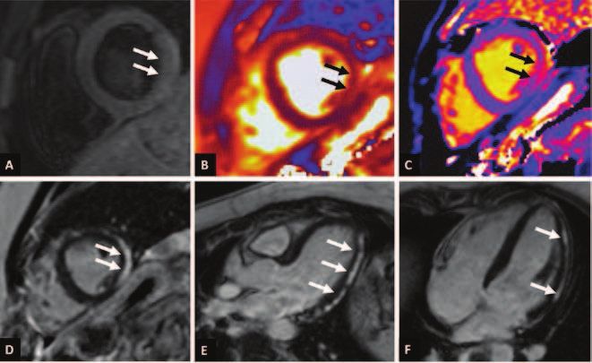
Introduction                                                     cal setting using Cardiac magnetic resonance (CMR) imag-
                                                                 ing (see Figure 1). The presented flow-chart incorporates
Myocarditis, defined as inflammation of the myocardium,          clinical, laboratory and imaging markers with the aim to bet-
presents the third most common cause of sudden cardiac           ter diagnose and risk stratify athletes with suspected myo-
death (SCD) in young athletes after autopsy-negative sudden      carditis.
unexplained death and coronary artery anomalies in the USA
and Italy [1,2]. Also in post-mortem studies of athletes in
Switzerland, we could recently show that myocarditis was         Contributing effect of exercise on pathogenesis
one of the underlying causes of SCD [3,4]. However, wheth-
er exercise is a trigger of fatal arrhythmias is an unanswered   Intense endurance training, compared to moderate exercise,
question as the majority of SCD occurs at rest and not during    which is generally protective, can predispose to viral upper
or immediately after physical activity [1]. While exercise is    respiratory tract infection and increases the chance of a sys-
undeniably beneficial for cardiovascular health [5], excessive   temic inflammatory response [13]. Murine studies have
exercise has been shown to predispose athletes to infection      shown that this inflammatory response in athletes is associ-
and exacerbate it [2]. Commonly, myocarditis is caused by        ated with a higher propensity to arrhythmogenicity [14]. In
viral infection such as enterovirus, Coxsackie B, Parvovirus     addition to that, in athletes affected by myocarditis, infec-
B19 and Human Herpes Virus 6 [6,7]. Non-infectious causes,       tious agents attack the myocardium directly and cause a sub-
which are generally rare, include toxins, vasculitic diseases    sequent autoimmune response that leads to myocardial re-
or pharmaceutical agents and illicit drugs [8]. The question     modelling through three phases: acute viral, subacute
amongst primary care physicians, sports physicians and car-      immune and chronic phase, where severe cases develop into
diologists alike is how we can best identify athletes who may    a dilated cardiomyopathy [15].
be suffering from myocarditis, which inevitably predisposes          In summary, the interaction between an environmental
them to a higher risk of SCD, and how we risk stratify these     trigger and the host’s immune system creates a subacute and
athletes. Currently, based on animal and autopsy studies, ex-    chronic inflammatory state. This leads to death of myocar-
pert opinions recommend an abstinence from competitive           dial tissue and causes scarring of the heart, which further
sports for 3 to 6 months after an episode of myocarditis         predisposes individuals to arrhythmias. Even beyond an ep-
[9–12]. This, however, can be of great inconvenience to ath-     isode of myocarditis, it is believed that chronic myocardial
letes due to deconditioning and an inability to compete. This    inflammation secondary to pathogenic autoimmunity can
review aims to present the current challenges in diagnosing      persist due to the continued presence of cytokines, which
myocarditis and further propose how to approach this clini-      exert pro-arrhythmic effect [16].

Figure 1: Diagnostic flow-chart and sports counselling of athletes with suspected myocarditis. Adapted from Eichhorn C. et
al. JACC Cardiovasc Imaging, 2019.

Diagnosing and sports counselling of athletes with myocarditis                                                              29
Clinical presentation and initial clinical work-up                 Previous studies have shown that viral serology has a very
of athletes with suspected myocarditis                             low sensitivity and intermediate specificity with myocardial
                                                                   biopsy infiltrates confirming myocarditis [17]. Viral serology
The typical clinical scenario is an athlete with atypical chest    may be of use in ruling out hepatitis C, human immunodefi-
pain, aggravated when leaning forward with the upper body,         ciency virus, Lyme disease or rickettsia if there is a high
history of a recent upper respiratory tract infection or gastro-   suspicion. Myocarditis has been shown to be the second most
intestinal infection, elevated inflammatory and myocardial         common cause of high troponin levels in the young and mid-
biomarkers, diffuse elevated ST-elevations and PQ depres-          dle-aged (< 50 years) [18], warranting further investigations
sion in the electrocardiogram (ECG) and absence of coronary        once myocardial infarction has been ruled out. A dilemma is
artery disease. However, this is only in the minority of the       presented by the fact that endurance training can raise tro-
presentations the case. Myocarditis in athletes can present in     ponin levels as demonstrated by a meta-analysis totalling
a variety of ways. Beside chest pain as a leading symptom,         1045 patients where troponin levels were raised above the
patients can present with palpitations, performance drop or        cut-off in 83% of participants [19]. This means that repeat
even be asymptomatic. Biomarkers, ECG and echocardiog-             troponins in athletes are key in making the diagnosis of my-
raphy might be abnormal, however, its sensitivity and speci-       ocarditis as the raised troponin levels due to exercise resolve
ficity is low. As myocarditis may clinically resemble to an        faster and are less prominent as well as monophasic in com-
acute coronary event, it is important to rule out coronary         parison to the troponin profiles found in myocarditis and my-
artery disease (CAD) first in these patients.                      ocardial infarction. Therefore, performing a troponin test in
                                                                   athletes suspected with myocarditis is important but it must
                                                                   be kept in mind that troponin levels do not sufficiently cor-
Biomarkers and Electrocardiogram                                   relate with the level of oedema and scarring or recovery that
                                                                   can be observed on imaging modalities, such as cardiac mag-
There are no specific biomarkers for myocarditis other than        netic resonance (CMR) imaging [20]. ECG abnormalities
possible increased inflammation parameters C-reactive pro-         like PR depression, ST-elevations, T-wave inversion, Q-wave
tein, leucocytes, elevated blood sedimentation rate and ele-       presence, prolonged QRS or QTc may be present but are nei-
vated myocardial biomarkers like troponin/creatin-kinase.          ther sensitive nor specific (see Figure 2) [21].

Figure 2: Case of an athlete with acute myocarditis. This is a case of a 17-year-old athlete with new onset of acute chest pain
and pain in the left shoulder. He complained about a recent gastroenteritis three days ago. His body temperature was 39°C
and C-reactive protein and Leucocytes were mildly elevated. Further, in laboratory tests, creatine kinase and troponin were
elevated. Electrocardiogram showed diffuse ST-elevation over multiple leads. In order to rule out coronary ischemic event an
invasive coronary angiogram was performed which was normal. Cardiac Magnetic resonance imaging was performed (see
Figure 3).

30                                                                                                                Eichhorn, Gräni
Echocardiography Cardiac Magnetic Resonance Imaging
Pericardial effusion, local wall motion abnormalities or im- CMR is a rapidly evolving field and recent evidence suggests
aging consistent with dilated cardiomyopathy can be features that current recommendations about the role of CMR in the
of myocarditis. Fulminant myocarditis, a severe rapidly management of patients with suspected myocarditis have to
evolving form of myocarditis, shows severe impaired systol- be updated. In 2009, the Consensus Criteria for Cardiovas-
ic LV function and is associated with a worse prognosis [22]. cular Magnetic Resonance in Myocardial Inflammation pub-
Echocardiography is an important tool to monitor LV func- lished the Lake Louise Criteria (LLC), which propose oede-
tion and wall motion abnormalities in myocarditis. It is cru- ma by T2-weighted imaging and scar by late gadolinium
cial to differentiate myocarditis from an athlete’s heart enhancement (LGE) as diagnostic features for myocarditis
(Table 1), where the latter is a condition in which LV wall (see Figure 3) [30]. CMR has not only diagnostic value
thickness and/or LV dimensions in athletes change depend- [31,32], but also a prognostic value as we recently could show.
ing on the degree of dynamic and static components of the We analysed the prognostic value of CMR in a cohort that
type of exercise performed [23]. included 670 patients with suspected myocarditis. We could
If there is any doubt about structural changes in an ath- show that any LGE presence doubled the risk for major ad-
lete’s heart, further imaging, such as CMR imaging, is indi- verse cardiovascular outcomes (MACE), while septal and
cated [24]. mid-wall LGE as well as patchy distribution of LGE were
strong prognostic patterns of LGE [21]. In addition, increas-
ing LGE extent resulted in a greater risk for outcome [33].
Endomyocardial Biopsy Further, T2 weighted imaging was also significantly associ-
ated with MACE [21]. Difficulties of CMR are that some
Endomyocardial biopsy (EMB) is not routinely performed in biopsy-proven myocarditis patients do not show evidence of
patients with suspected myocarditis, however, in cases with LGE and subsequently do not fulfil the LLC [25]. Addition-
unclear depressed LV function or in life-threatening pre- ally, it can be the case that the inflammatory process has
sentations, EMB helps to differentiate between infectious, become sufficiently diffuse across the myocardium to such
autoimmune or idiopathic inflammation in myocarditis, an extent that T2 differences are not identifiable, especially
which may guide further therapy [25,26]. While EMB has an if there is concurrent skeletal muscle inflammation in a ref-
outstanding specificity and low complication rates (< 1%) in erence region [34,35]. Novel T1 and T2 mapping sequences
experienced cardiac centres, sensitivity is low and there is a have been shown to provide higher sensitivity and specificity
high chance of false negative results (sampling errors) values than the traditional LLC criteria (see Figure 3)
[25,27]. Whether the diagnostic value of EMB can be in- [36,37]. T1 and T2 mapping values essentially describe mag-
creased by using CMR imaging to guide the biopsy is cur- netic properties on a pixel-by-pixel basis that are determined
rently unclear [25]. However, Baccouche et al. recently by intrinsic characteristics of the tissue, its environment and
showed that using CMR and EMB in combination rather than the measurement parameters, culminating in a global T1 or
individually identifies a greater proportion of patients with T2 value. The rationale behind T1 mapping values is that
myocarditis [28]. Porcine pre-clinical in-vivo models suggest these are able to detect myocarditis throughout multiple stag-
that real-time CMR-guided EMB may also improve sensitiv- es of the disease course since they are affected by both oede-
ity and specificity [29]. In regard to athletes, it is unknown ma and extracellular expansion. T2 mapping can detect tissue
whether EMB adds to risk stratification and sports abstinence free water content which is raised in the acute phase and
counselling. normalizes over time – this can be important in differentiat-
Differentiating Features Differentiating Features
Athletes Heart Myocarditis
Symptoms Asymptomatic Symptomatic
ECG/Holter 1. Specific ECG changes such as early 1. Unspecific ECG changes. Possible
repolarization/ST segment elevation, PQ depression, ST-elevation in mul-
T- wave inversion in V1–V3 in the tiple leads.
young, ST-elevation followed by
T-wave inversion V1–V4 in black
athletes.
Biomarkers/Inflammatory markers 1. Troponin elevation only mild and 1. Troponin elevation mild to high
normalizes quickly. May be present 2. Others: Brain natriuretic peptide
in ultra-endurance athletes. elevation, Creatine-Kinase, Leuco-
2. Others: Brain natriuretic peptide cytosis, elevated C-reactive Protein,
mildly elevated after ultra-endur- elevated Erythrocyte sedimentation
ance exercise rate
Table adapted from Eichhorn C. et al. JACC Cardiovasc Imaging, 2019.
Table 1: Challenges in ECG, biomarkers and noninvasive imaging characteristics in patients with athlete’s heart versus acute
myocarditis.
Fortsetzung auf Seite 34 £
Diagnosing and sports counselling of athletes with myocarditis 31ing myocarditis from other noninflammatory cardiomyopa- Therapy, Follow-up and Sports Restriction thies [38,39]. Furthermore, Spieker et al. have shown that T2 mapping can be used as a prognostic marker in patients with While roughly half of cases of suspected myocarditis resolve myocarditis [40]. Using T1 mapping values pre and post con- spontaneously within a month, there is a lack of evidence for trast agent administration and adjusting for the haematocrit optimal therapy in myocarditis patients with cardiac dysfunc- value, extracellular volume fraction (ECV) can also be esti- tion. In haemodynamically stable patients, management of mated [41]. We have recently shown that there is a significant cardiac dysfunction should be in line with the latest heart association of elevated ECV values with MACE in patients failure guidelines [45] with appropriate step-up of care to with suspected myocarditis, even in those presenting without intensive care units, invasive management and cardiac trans- any LGE [42]. All in all, it has become evident that inflamed plantation when patients become haemodynamically unsta- myocardial tissue shows higher T1, T2 and ECV values, ble. Arrhythmias and the use of implantable cardioverter which compares favourably to other tissue characterisation defibrillator should also follow their respective guidelines. techniques [43]. The recent update of the LLC implemented Current recommendation do not include immunomodulatory current evidence on novel mapping techniques and conclude therapies, while immunosuppressive treatment, in the ab- that CMR provides strong evidence for an active myocardial sence of active infection or other contraindications, is recom- inflammation based on either one T2-based parameter (T2 mended in autoimmune forms of myocarditis. mapping or T2 weighted imaging) or one T1-based parame- In terms of sports restriction for athletes, it is paramount ter (T1 mapping, ECV or LGE), with the specificity being that athletes with acute myocarditis refrain from any com- higher when one of both parameters is present (44). petitive exercise, while there is currently no study that indi- Figure 3: Cardiac Magnetic resonance imaging of an athlete with suspected myocarditis. The CMR of the athlete described in Figure 2 showed in panel A, the Turbo-Inversion Recovery- Magnitude (TIRM), i.e. a T2-weighted (water content) and fat supressed sequence of the midventricular short axis of the heart a hyperintensity signal of the epicardial left myocardial mus- cle inferolateral (white arrows). In the T2 mapping (panel B) sequence, in the same region, a prolonged T2 mapping time of 56ms could be measured consistent with myocardial oedema (black arrows). In the native T1 mapping (panel C), also infe- rolateral (midventricular) a prolonged T1 mapping time (1220ms) was measured (black arrows). Late Gadolinium enhance- ment is seen in a typical myocarditis pattern, i.e. subepicardial inferolateral from the basal to apical segments (panel D, short axis view, panel E, 3 chamber view and F 4 chamber view, white arrows). Left - and right ventricular function was normal without any regional wall motion abnormalities. The patient received non-steroidal anti-inflammatory drugs as was symptom free after discharge. He was counselled to restrict from competitive sports for 3 months and was scheduled for a follow-up CMR and maximal stress-ECG before re-uptake of competitive sports. 34 Eichhorn, Gräni
cates that there is a safe level of exercise in the first months   First author
after a diagnosis of myocarditis. Current guidelines [9–11],
including a recent recommendation by the European Asso-            Christian Eichhorn, MBBS, BSc
ciation of Preventive Cardiology [12], recommend between           Unterassistent
3 and 6 months of competitive sports restriction (Class IIb/       Department of Cardiology
Level C) for athletes diagnosed with myocarditis. Prior to         Swiss Cardiovascular Center
returning to competitive exercise, athletes should undergo         University Hospital Berne
extensive testing, including echocardiography, Holter moni-        Freiburgstrasse
toring and exercise ECG. We believe that a follow-up CMR           CH-3010 Berne, Switzerland
in athletes with diagnosed myocarditis is warranted at 3 to 6
months in order to risk stratify patients based on presence or
absence of inflammation. These recommendations should not
only apply to competitive athletes but also to recreational        Corresponding author
athletes as they form the group who most commonly suffer
from SCD in myocarditis [46]. However, whether moderate            PD Dr. med. Christoph Gräni, PhD
exercise is safe when inflammation subsided to normal or           Director of noninvasive cardiac imaging
whether different sports levels based on dynamic and static        (CMR/CT/Nuclear)
components may need different sport abstinence periods in          Department of Cardiology
athletes with myocarditis are unknown.                             Swiss Cardiovascular Center
    Furthermore, it needs to be evaluated how serial CMR           University Hospital Berne
scans and LGE involvement over time are associated with            Freiburgstrasse
outcome.                                                           CH-3010 Berne, Switzerland
                                                                   Tel: +41 31 632 45 08
                                                                   Email: christoph.graeni@insel.ch
Conclusion

In athletes with myocarditis, currently, 3–6 months sports         References
restriction is recommended for the prevention of adverse car-
diac events. CMR is the primary imaging tool for diagnosing        1.  Harmon KG, Asif IM, Maleszewski JJ, Owens DS, Prutkin JM, Saler-
myocarditis and may also play an important role in risk strat-         no JC, et al. Incidence, Cause, and Comparative Frequency of Sudden
                                                                       Cardiac Death in National Collegiate Athletic Association Athletes:
ifying athletes with suspected myocarditis. Future research            A Decade in Review. Circulation. 2015;132(1):10-9.
is needed to assess the role of novel noninvasive CMR tissue       2. Corrado D, Basso C, Pavei A, Michieli P, Schiavon M, Thiene G.
characterization techniques to better guide treatment and al-          Trends in sudden cardiovascular death in young competitive athletes
low individual sports counseling in this clinical setting.             after implementation of a preparticipation screening program. Jama.
                                                                       2006;296(13):1593-601.
                                                                   3. Asatryan B, Vital C, Kellerhals C, Medeiros-Domingo A, Grani C,
                                                                       Trachsel LD, et al. Sports-related sudden cardiac deaths in the young
Practical implications                                                 population of Switzerland. PloS one.2017;12(3):e0174434.
                                                                   4. Grani C, Chappex N, Fracasso T, Vital C, Kellerhals C, Schmied C,
• The diagnosis of myocarditis in athletes is challenging              et al. Sports-related sudden cardiac death in Switzerland classified by
                                                                       static and dynamic components of exercise. European journal of pre-
• Troponin levels are typically raised in myocarditis but ath-         ventive cardiology. 2016;23(11):1228-36.
  letic activity can also mildly increase troponin levels          5. Myers J. Cardiology patient pages. Exercise and cardiovascular health.
• CMR is the primary tool to diagnose myocarditis and may              Circulation.2003;107(1):e2-5.
  further help to risk stratify athletes                           6. Yilmaz A, Klingel K, Kandolf R, Sechtem U. A geographical mystery:
                                                                       do cardiotropic viruses respect national borders? J Am Coll Cardiol.
• Currently 3–6 months competitive sports abstinence is                52. United States2008. p. 82; author reply -3.
  recommended                                                      7. Breinholt JP, Moulik M, Dreyer WJ, Denfield SW, Kim JJ, Jefferies
                                                                       JL, et al. Viral epidemiologic shift in inflammatory heart disease: the
                                                                       increasing involvement of parvovirus B19 in the myocardium of pe-
$FNQRZOHGJPHQWVFRQëLFWRILQWHUHVWDQG                              diatric cardiac transplant patients. J Heart Lung Transplant. 2010;
                                                                       29(7):739-46.
funding                                                            8. Caforio AL, Pankuweit S, Arbustini E, Basso C, Gimeno-Blanes J,
                                                                       Felix SB, et al. Current state of knowledge on aetiology, diagnosis,
All authors have no conflict of interest. Dr. Gräni received           management, and therapy of myocarditis: a position statement of the
funding support from the Swiss Sports Medicine Society                 European Society of Cardiology Working Group on Myocardial and
                                                                       Pericardial Diseases. Eur Heart J. 2013;34(33):2636-48, 48a-48d.
(SGSM), the SGSM Award 2015.                                       9. Maron BJ, Udelson JE, Bonow RO, Nishimura RA, Ackerman MJ,
                                                                       Estes NAM, et al. Eligibility and Disqualification Recommendations
                                                                       for Competitive Athletes With Cardiovascular Abnormalities: Task
                                                                       Force 3: Hypertrophic Cardiomyopathy, Arrhythmogenic Right Ven-
                                                                       tricular Cardiomyopathy and Other Cardiomyopathies, and Myocar-
                                                                       ditis. Circulation. 2015;132:e273-e80.
                                                                   10. Pelliccia A, Fagard R, Bjørnstad HH, Anastassakis A, Arbustini E,
                                                                       Assanelli D, et al. Recommendations for competitive sports participa-
                                                                       tion in athletes with cardiovascular diseaseA consensus document
                                                                       from the Study Group of Sports Cardiology of the Working Group of
                                                                       Cardiac Rehabilitation and Exercise Physiology and the Working
                                                                       Group of Myocardial and Pericardial Diseases of the European Soci-
                                                                       ety of Cardiology. European heart journal. 2005;26(14):1422-45.

Diagnosing and sports counselling of athletes with myocarditis                                                                             35
11. Pelliccia A, Corrado D, Bjørnstad HH, Panhuyzen-Goedkoop N, Ur- 30. Friedrich MG, Sechtem U, Schulz-Menger J, Holmvang G, Alakija P,
hausen A, Carre F, et al. Recommendations for participation in com- Cooper LT, et al. Cardiovascular magnetic resonance in myocarditis:
petitive sport and leisure-time physical activity in individuals with A JACC White Paper. Journal of the American College of Cardiology.
cardiomyopathies, myocarditis and pericarditis. European Journal of 2009;53:1475-87.
Cardiovascular Prevention & Rehabilitation. 2006;13(6):876-85. 31. Kotanidis CP, Bazmpani MA, Haidich AB, Karvounis C, Antoniades
12. Pelliccia A, Solberg EE, Papadakis M, Adami PE, Biffi A, Caselli S, C, Karamitsos TD. Diagnostic Accuracy of Cardiovascular Magnetic
et al. Recommendations for participation in competitive and leisure Resonance in Acute Myocarditis: A Systematic Review and Me-
time sport in athletes with cardiomyopathies, myocarditis, and peri- ta-Analysis. JACC Cardiovasc Imaging. 2018;11(11):1583-90.
carditis: position statement of the Sport Cardiology Section of the 32. Lagan J, Schmitt M, Miller CA. Clinical applications of multi-para-
European Association of Preventive Cardiology (EAPC). Eur Heart J. metric CMR in myocarditis and systemic inflammatory diseases. Int
2019;40(1):19-33. J Cardiovasc Imaging. 2018;34(1):35-54.
13. Martin SA, Pence BD, Woods JA. Exercise and Respiratory Tract Vi- 33. Grani C, Eichhorn C, Biere L, Kaneko K, Murthy VL, Agarwal V, et
ral Infections. Exerc Sport Sci Rev. 2009;37(4):157-64. al. Comparison of myocardial fibrosis quantification methods by car-
14. Mont L, Elosua R, Brugada J. Endurance sport practice as a risk factor diovascular magnetic resonance imaging for risk stratification of pa-
for atrial fibrillation and atrial flutter. Europace. 2009;11(1):11-7. tients with suspected myocarditis. Journal of cardiovascular magnetic
15. Sagar S, Liu PP, Cooper LT. Myocarditis. The Lancet. resonance : official journal of the Society for Cardiovascular Magnet-
2012;379(9817):738-47. ic Resonance. 2019;21(1):14.
16. Saito J, Niwano S, Niwano H, Inomata T, Yumoto Y, Ikeda K, et al. 34. Laissy JP, Hyafil F, Feldman LJ, Juliard JM, Schouman-Claeys E, Steg
Electrical remodeling of the ventricular myocardium in myocarditis. PG, et al. Differentiating acute myocardial infarction from myocardi-
Circulation journal. 2002;66(1):97-103. tis: diagnostic value of early- and delayed- perfusion cardiac MR im-
17. Mahfoud F, Gartner B, Kindermann M, Ukena C, Gadomski K, Klin- aging. Radiology. 2005;237(1):75-82.
gel K, et al. Virus serology in patients with suspected myocarditis: 35. Ferreira VM, Piechnik SK, Dall'A rmellina E, Karamitsos TD, Francis
utility or futility? Eur Heart J. 2011;32(7):897-903. JM, Ntusi N, et al. T(1) mapping for the diagnosis of acute myocardi-
18. Wu C, Singh A, Collins B, Fatima A, Qamar A, Gupta A, et al. Caus- tis using CMR: comparison to T2-weighted and late gadolinium en-
es of Troponin Elevation and Associated Mortality in Young Patients. hanced imaging. JACC Cardiovasc Imaging. 2013;6(10):1048-58.
Am J Med. 2018;131(3):284-92.e1. 36. Nadjiri J, Nieberler H, Hendrich E, Greiser A, Will A, Martinoff S, et
19. Sedaghat-Hamedani F, Kayvanpour E, Frankenstein L, Mereles D, al. Performance of native and contrast-enhanced T1 mapping to detect
Amr A, Buss S, et al. Biomarker changes after strenuous exercise can myocardial damage in patients with suspected myocarditis: a head-to-
mimic pulmonary embolism and cardiac injury – a metaanalysis of 45 head comparison of different cardiovascular magnetic resonance tech-
studies. Clin Chem. 2015;61(10):1246-55. niques. Int J Cardiovasc Imaging. 2017;33(4):539-47.
20. Berg J, Kottwitz J, Baltensperger N, Kissel CK, Lovrinovic M, Mehra 37. Radunski UK, Lund GK, Saring D, Bohnen S, Stehning C, Schna-
T, et al. Cardiac Magnetic Resonance Imaging in Myocarditis Reveals ckenburg B, et al. T1 and T2 mapping cardiovascular magnetic
Persistent Disease Activity Despite Normalization of Cardiac En- resonance imaging techniques reveal unapparent myocardial injury
zymes and Inflammatory Parameters at 3-Month Follow-Up. Circ in patients with myocarditis. Clin Res Cardiol. 2017;106(1):10-7.
Heart Fail. 2017;10(11). 38. Pan JA, Lee YJ, Salerno M. Diagnostic Performance of Extracellular
21. Grani C, Eichhorn C, Biere L, Murthy VL, Agarwal V, Kaneko K, et Volume, Native T1, and T2 Mapping Versus Lake Louise Criteria by
al. Prognostic Value of Cardiac Magnetic Resonance Tissue Charac- Cardiac Magnetic Resonance for Detection of Acute Myocarditis: A
terization in Risk Stratifying Patients With Suspected Myocarditis. Meta-Analysis. Circ Cardiovasc Imaging. 2018;11(7):e007598.
Journal of the American College of Cardiology. 2017;70(16):1964-76. 39. von Knobelsdorff-Brenkenhoff F, Schuler J, Doganguzel S, Dieringer
22. Felker GM, Boehmer JP, Hruban RH, Hutchins GM, Kasper EK, MA, Rudolph A, Greiser A, et al. Detection and Monitoring of Acute
Baughman KL, et al. Echocardiographic findings in fulminant and Myocarditis Applying Quantitative Cardiovascular Magnetic Reso-
acute myocarditis. Journal of the American College of Cardiology. nance. Circ Cardiovasc Imaging. 2017;10(2):e005242.
2000;36:227-32. 40. Spieker M, Haberkorn S, Gastl M, Behm P, Katsianos S, Horn P, et al.
23. Mitchell JH, Haskell W, Snell P, Van Camp SP. Task Force 8: classi- Abnormal T2 mapping cardiovascular magnetic resonance correlates
fication of sports. J Am Coll Cardiol. 2005;45(8):1364-7. with adverse clinical outcome in patients with suspected acute myo-
24. Galderisi M, Cardim N, D’Andrea A, Bruder O, Cosyns B, Davin L, carditis. J Cardiovasc Magn Reson. 2017;19(1):38.
et al. The multi-modality cardiac imaging approach to the Athlete's 41. Radunski UK, Lund GK, Stehning C, Schnackenburg B, Bohnen S,
heart: an expert consensus of the European Association of Cardiovas- Adam G, et al. CMR in patients with severe myocarditis: diagnostic
cular Imaging. Eur Heart J Cardiovasc Imaging. 2015;16(4):353. value of quantitative tissue markers including extracellular volume
25. Yilmaz A, Kindermann I, Kindermann M, Mahfoud F, Ukena C, imaging. JACC Cardiovasc Imaging. 2014;7(7):667-75.
Athanasiadis A, et al. Comparative evaluation of left and right ven- 42. Grani C, Biere L, Eichhorn C, Kaneko K, Agarwal V, Aghayev A, et
tricular endomyocardial biopsy: differences in complication rate and al. Incremental value of extracellular volume assessment by cardio-
diagnostic performance. Circulation. 2010;122(9):900-9. vascular magnetic resonance imaging in risk stratifying patients with
26. Stiermaier T, Fohrenbach F, Klingel K, Kandolf R, Boudriot E, Sandri suspected myocarditis. The international journal of cardiovascular
M, et al. Biventricular endomyocardial biopsy in patients with suspect- imaging. 2019.
ed myocarditis: Feasibility, complication rate and additional diagnos- 43. Ferreira VM. CMR Mapping For Myocarditis: Coming Soon to a
tic value. Int J Cardiol. 2017;230:364-70. Center Near You. JACC Cardiovasc Imaging. 2018;11(11):1591-3.
27. Holzmann M, Nicko A, Kuhl U, Noutsias M, Poller W, Hoffmann W, 44. Ferreira VM, Schulz-Menger J, Holmvang G, Kramer CM, Carbone
et al. Complication rate of right ventricular endomyocardial biopsy via I, Sechtem U, et al. Cardiovascular Magnetic Resonance in Nonis-
the femoral approach: a retrospective and prospective study analyzing chemic Myocardial Inflammation: Expert Recommendations. Journal
3048 diagnostic procedures over an 11-year period. Circulation. of the American College of Cardiology. 2018;72(24):3158-76.
2008;118(17):1722-8. 45. Ponikowski P, Voors AA, Anker SD, Bueno H, Cleland JGF, Coats
28. Baccouche H, Mahrholdt H, Meinhardt G, Merher R, Voehringer M, AJS, et al. 2016 ESC Guidelines for the Diagnosis and Treatment of
Hill S, et al. Diagnostic synergy of non-invasive cardiovascular mag- Acute and Chronic Heart Failure. Rev Esp Cardiol (Engl Ed).
netic resonance and invasive endomyocardial biopsy in troponin-pos- 2016;69(12):1167.
itive patients without coronary artery disease. Eur Heart J. 46. Bohm P, Scharhag J, Meyer T. Data from a nationwide registry on
2009;30(23):2869-79. sports-related sudden cardiac deaths in Germany. European journal
29. Behm P, Gastl M, Jahn A, Rohde A, Haberkorn S, Krueger S, et al. of preventive cardiology. 2016;23(6):649-56.
CMR-guidance of passively tracked endomyocardial biopsy in an in
vivo porcine model. Int J Cardiovasc Imaging. 2018;34:1917.
36 Eichhorn, GräniYou can also read