Human Melanocyte-Derived Spheroids: A Precise Test System for Drug Screening and a Multicellular Unit for Tissue Engineering - Frontiers
←
→
Page content transcription
If your browser does not render page correctly, please read the page content below
ORIGINAL RESEARCH
                                                                                                                                            published: 04 June 2020
                                                                                                                                     doi: 10.3389/fbioe.2020.00540

                                            Human Melanocyte-Derived
                                            Spheroids: A Precise Test System for
                                            Drug Screening and a Multicellular
                                            Unit for Tissue Engineering
                                            Irina M. Zurina 1,2,3*, Anastasiya A. Gorkun 1,2,3 , Ekaterina V. Dzhussoeva 1 ,
                                            Tamara D. Kolokoltsova 1,3 , Dmitriy D. Markov 4 , Nastasia V. Kosheleva 1,3,5 ,
                                            Sergey G. Morozov 1 and Irina N. Saburina 1,3
                                            1
                                             Laboratory of Cell Biology and Developmental Pathology, FSBSI Institute of General Pathology and Pathophysiology,
                                            Moscow, Russia, 2 Department of Modern Biomaterials, Institute for Regenerative Medicine, Sechenov First Moscow State
                                            Medical University, Moscow, Russia, 3 FSBEI FPE Russian Medical Academy of Continuous Professional Education of the
                                            Russian Ministry of Healthcare, Moscow, Russia, 4 Institute of Molecular Genetics of the Russian Academy of Sciences,
                                            Moscow, Russia, 5 Faculty of Biology, Lomonosov Moscow State University, Moscow, Russia

                            Edited by:
                  Dimitrios I. Zeugolis,    Pigmentation is the result of melanin synthesis, which takes place in melanocytes, and
          National University of Ireland    its further distribution. A dysregulation in melanocytes’ functionality can result in the loss
                       Galway, Ireland
                                            of pigmentation, the appearance of pigment spots and melanoma development. Tissue
                       Reviewed by:
               Elizabeth R. Balmayor,       engineering and the screening of new skin-lightening drugs require the development of
    Maastricht University, Netherlands      simple and reproducible in vitro models with maintained functional activity. The aim of
                     Naresh Mahajan,
    Wake Forest School of Medicine,
                                            the study was to obtain and characterize spheroids from normal human melanocytes
                         United States      as a three-dimensional multicellular structure and as a test system for skin-lightening
                  *Correspondence:          drug screening. Melanocytes are known to lose their ability to synthesize melanin
                        Irina M. Zurina     in monolayer culture. When transferred under non-adhesive conditions in agarose
                   izurina@gmail.com
                                            multi-well plates, melanocytes aggregated and formed spheroids. As a result, the amount
                    Specialty section:      of melanin elevated almost two times within seven days. MelanoDermTM (MatTek)
         This article was submitted to      skin equivalents were used as a comparison system. Cells in spheroids expressed
 Tissue Engineering and Regenerative
                              Medicine,     transcription factors that regulate melanogenesis: MITF and Sox10, the marker of
                a section of the journal    developed melanosomes—gp100, as well as tyrosinase (TYR)—the melanogenesis
       Frontiers in Bioengineering and
                                            enzyme and melanocortin receptor 1 (MC1R)—the main receptor regulating melanin
                        Biotechnology
                                            synthesis. Expression was maintained during 3D culturing. Thus, it can be stated that
       Received: 11 November 2019
           Accepted: 05 May 2020            spheroids maintain melanocytes’ functional activity compared to that in the multi-layered
          Published: 04 June 2020           MelanoDermTM skin equivalents. Culturing both spheroids and MelanoDermTM for seven
                             Citation:      days in the presence of the skin-lightening agent fucoxanthin resulted in a more
                 Zurina IM, Gorkun AA,
    Dzhussoeva EV, Kolokoltsova TD,
                                            significant lowering of melanin levels in spheroids. Significant down-regulation of gp100,
           Markov DD, Kosheleva NV,         MITF, and Sox10 transcription factors, as well as 10-fold down-regulation of TYR
  Morozov SG and Saburina IN (2020)
                                            expression, was observed in spheroids by day 7 in the presence of fucoxanthin, thus
          Human Melanocyte-Derived
 Spheroids: A Precise Test System for       inhibiting the maturation of melanosomes and the synthesis of melanin. MelanoDermTM
   Drug Screening and a Multicellular       samples were characterized by significant down-regulation of only MITF, Sox10 indicating
           Unit for Tissue Engineering.
    Front. Bioeng. Biotechnol. 8:540.
                                            that spheroids formed a more sensitive system allowed for quantitative assays.
      doi: 10.3389/fbioe.2020.00540         Collectively, these data illustrate that normal melanocytes can assemble themselves into

Frontiers in Bioengineering and Biotechnology | www.frontiersin.org              1                                                June 2020 | Volume 8 | Article 540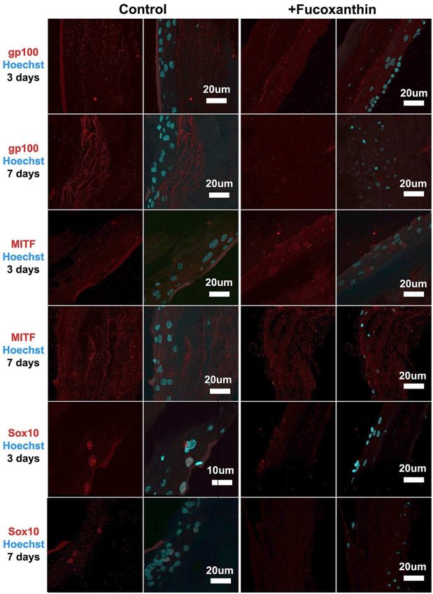
Zurina et al.                                                                                                 Melanocyte-Derived Spheroids as a Test System

                                            spheroids—the viable structures that are able to accumulate melanin and maintain
                                            the initial functional activity of melanocytes. These spheroids can be used as a
                                            more affordable and easy-to-use test system than commercial skin equivalents for
                                            drug screening.

                                            Keywords: melanocyte, melanogenesis, spheroid, 3D culture, drug screening, tissue engineering

INTRODUCTION                                                                      and mechanisms of the effects of active drug substances or
                                                                                  combined drugs.
The pharmaceutical industry is one of the fastest-developing                          The regulation of skin pigmentation, primarily skin
R&D systems. As long as new products are developed, discovery                     lightening, is one of the most common processes in skin
programs require more accurate and high-throughput in vitro                       physiology, which is modified during cosmetological procedures.
systems for preclinical drug screening and testing. Currently,                    Active production of melanin, as well as its transfer from
most in vitro studies are carried out using monolayer cultures                    melanocytes to keratinocytes using special organelles—
of primary cells or immortalized cell lines. However, the efficacy                melanosomes—is one of the protective mechanisms of the
of such approaches is now in doubt as 2D culture conditions do                    skin against UV exposure. Melanin prevents the penetration
not fully reflect the complex 3D microenvironment (intercellular                  of UV into the deeper layers of the skin and blocks the release
junctions, well-organized extracellular matrix, which cells have                  of reactive oxygen species (ROS). Maintaining permanent skin
to make contact with, gradients of oxygen and nutrients) that                     pigmentation depends on a variety of sequential processes:
surrounds cells in vivo (Fitzgerald et al., 2015).                                migration of melanoblasts into tissue during embryogenesis,
   Recently, 3D cell culture systems have been shown to model                     their viability and differentiation into melanocytes, the density
that microenvironment in vitro—they allow for maintaining                         of melanocytes in the skin, expression and functions of the
cell morphology, viability, proliferation rate, and differentiation               enzymatic and structural components of melanosomes, synthesis
processes, as well as long-term culturing without disturbing the                  of various types of melanin (eu- and pheomelanin), maturation
structural integrity (Antoni et al., 2015; Fitzgerald et al., 2015).              and transportation of melanosomes in the dendritic processes of
Overall, this confirms that 3D cultures are more accurate and                     melanocytes and their transfer to keratinocytes, and, finally, the
valuable in terms of predicting the clinical effects of tested                    spread of melanin in the suprabasal layers of the skin (Yamaguchi
drugs as compared to the 2D culture. There are now numerous                       and Hearing, 2007).
technologies with which to obtain 3D cultures that can be divided                     Therefore, to suppress hyperpigmentation and abnormal
into two categories: anchorage-independent and scaffold-based                     melanogenesis, the use of combined drugs that can affect
(Langhans, 2018). The first type of 3D culture is based on the                    different stages and levels of regulation of melanin synthesis
ability of cells to self-organize in aggregates under non-adhesive                is required. For example, fucoxanthin (algae pigment), which
conditions, including the hanging-drop technique, the use of                      belongs to the carotenoid family and is used as a skin protector
low adhesion plates, and the magnetic cell levitation approach                    in cosmetology as a component in different drugs, has been
(Langhans, 2018). The use of multi-well non-adhesive plates                       shown to have multiple protective effects in various pathologies
is a simple way of obtaining a large number of spheroids of                       (Peng et al., 2011).
the same size and controlling their size by simply changing                           Currently, primary cultures of melanocytes and keratinocytes
the cell concentrations (Koudan et al., 2017). In scaffold-based                  (Lei et al., 2002), the commercial tissue equivalents EpiSkin,
technologies, natural (ECM proteins) or synthetic scaffolds are                   MelanoDermTM and others (Costin and Raabe, 2013; Meena
used to provide mechanical support for cells, as well as to enable                and Mohandass, 2019) and tissue equivalents obtained from
them to self-assemble in the preferred manner, similar to the                     cells with induced pluripotency (iPSC) (Gledhill et al., 2015)
native one (Langhans, 2018).                                                      are used to study in vitro the efficacy of drugs against
   Numerous advantages of 3D cultures, mainly spheroids, have                     hyperpigmentation. However, in this case, a simpler 3D
made them a widely used instrument for in vitro drug testing.                     model, namely, spheroids from melanocytes, could also be an
The main area where spheroids are used is in the study of tumor                   effective tool for studying the mechanisms of anti-pigmentation
cells and their resistance to different agents (Huang and Gao,                    drug efficacy. The 3D culturing of melanocytes in different
2018). That said, more researchers are now using 3D cultures                      systems has already been shown to reduce cell proliferation
for studying different pathologies and drug testing in cardiology                 rates and increase their viability and functional activity (Lin
(Figtree et al., 2017), neurology (Hartley and Brennand, 2017;                    et al., 2006; Lee et al., 2015; Hsiao and Young, 2019).
Nzou et al., 2018), pulmonology (Surolia et al., 2017), orthopedics               The purpose of this study was to obtain and characterize
(Zigon-Branc et al., 2018), and endocrinology (Klaka et al., 2017;                spheroids from normal human melanocytes as a cellular
Ribeiro et al., 2018). New highly sensitive, yet easy-to-manipulate               module and as a test system for skin-lightening drug screening
(and corresponding to the norms of bioethics) in vitro models                     and to compare the efficacy of its use with MelanoDermTM
are also required in cosmetic industries to study the efficacy                    tissue equivalents.

Frontiers in Bioengineering and Biotechnology | www.frontiersin.org           2                                            June 2020 | Volume 8 | Article 540
Zurina et al.                                                                                        Melanocyte-Derived Spheroids as a Test System

MATERIALS AND METHODS                                                     (NaCl) buffer was added to the growth medium. The dynamics
                                                                          of spheroids’ formation in agarose plates were monitored by
The study was conducted on the primary culture of                         the Cell-IQ live time-lapse system (CM Technologies, Finland)
human    melanocytes (104-05N, CELL      Applications,                    with photo registration every 20 min. The resulting spheroids
Inc.) and MelanoDermTM tissue equivalents (MEL-300,                       were collected on Days 1, 3, and 7 for melanin concentration
MatTek Corporation).                                                      measurements and real-time PCR, and at Days 3 and 7 for
                                                                          immunocytochemical analysis.
Cultivation of MelanoDermTM Tissue
Equivalents                                                               Photometric Analysis of Melanin
A set of tissue equivalents was transported to the laboratory (at         Concentration in MelanoDermTM Tissue
+4◦ C). The samples located in special holders were transferred           Equivalents and Spheroids From Human
to six-well culture plates in individual wells. Each sample was
placed on a stand (MEL-STND, MatTek Corporation) on a
                                                                          Melanocytes
section between the water and the air phases. Tissue equivalents          Prior to photometric analysis, the samples of MelanoDermTM (3
were cultured in a complete growth medium provided by the                 pieces of tissue equivalent per time point in experimental and
company along with the samples (EPI-100-NMM-113, MatTek                   control groups) and spheroids (256 spheroids per time point
Corporation) under standard conditions (37◦ C, 5% CO2 ); the              in experimental and control groups) were washed three times
medium was replaced daily.                                                from the residues of the medium in PBS (pH = 7.4), after which
   Fucoxanthin powder (3351-86-8, Anhui, China) was dissolved             they were stored at −20◦ C. To extract melanin, the samples
in saline buffer at a concentration of 500 µM (stock solution),           were thawed and dried, and then 250 µl of Solvable solution
then sterilized by filtration through a syringe filter (SLGP033RS,        (6NE9100, PerkinElmer) was added to each sample. Next, the
0.22 µm, Millipore). For this study, the stock solution of                samples were incubated for 18 h in a water bath at +60◦ C.
fucoxanthin mixed with complete growth medium in a volume                 After incubation, the samples were thoroughly mixed in a vortex
ratio of 1:10 (50 µM) was added to the experimental samples. The          mixer, undissolved particles were precipitated by centrifugation
concentration of fucoxanthin was chosen based on the previously           (5 min, 13,000 g), 100 µl of the supernatant was placed into a
reported study on its protective effect on cell culture (Heo and          96-well plate, and samples’ optical densities were measured at
Jeon, 2009) and our study of its cytotoxicity (data not published).       a wavelength of 490 nm on a Multiscan GO plate photometer
An equivalent volume of saline buffer (1:10) was added to the             (Thermo Scientific, USA). A calibration curve was obtained
control samples.                                                          by analyzing standard solutions with a known concentration
                                                                          of melanin prepared from dry matter (M863, Sigma-Aldrich).
2D Cultivation of Human Melanocytes                                       Measurements were performed in triplicate.
The culture of human melanocytes was transported to the
laboratory (at −20◦ C), and the cells were thawed in a water
                                                                          Fixation of MelanoDermTM Tissue
bath at 37◦ C. Cells were resuspended in a complete growth                Equivalents and Spheroids From Human
medium for melanocytes (135–500, CELL Applications, Inc.) and             Melanocytes
placed on Petri dishes (35 mm) at a density of 104 cells/cm2 .            For immunocytochemical analysis of melanocyte monolayer
When the melanocytes reached a confluent state, the culture               culture, cells were seeded on the cover glass. Before fixing the 3D
was passaged with Versene solution (R080p, PanEco) and 0.25%              culture, the spheroids were collected in the tube and centrifuged
trypsin solution (R036p, PanEco). The full growth medium was              (1 min, 100 g). The resulting pellet was washed three times in
replaced every 2 days. For further experimental studies, the              phosphate-saline buffer solution (PBS, pH = 7.4). Cover glasses
culture of melanocytes at passage 4 was used to obtain spheroids.         seeded with melanocytes and spheroids were then fixed in a 4%
                                                                          solution of paraformaldehyde (20 min, +4◦ C). MelanoDermTM
3D Cultivation of Human Melanocytes                                       tissue equivalents were also washed three times with PBS from
Human melanocytes were cultured under 3D non-adhesive                     the remnants of the culture medium (5 min for each wash, pH =
conditions with and without the addition of the drug, to                  7.4). The material was fixed in a 4% solution of paraformaldehyde
obtain three-dimensional spheroids and to study the effect                (one day, +4◦ C).
of fucoxanthin on melanin accumulation by melanocytes
under these conditions, which are closer to the native tissue             Immunocytochemistry
compared to monolayer culture. Agarose plates were obtained               After fixation, the MelanoDermTM tissue equivalents samples
by polymerizing 2% agarose type I (A6013, Sigma) in special               were dehydrated in Isoprep histological processing solution
plastic forms (12–256, 3D Petri Dishes, Microtissue) and were             (06-002/S, Biovitrum) and embedded in paraffin (01-007/1,
then placed in 12-well culture plates. Cell suspension was                Biovitrum). Next, a series of 9 µm sections was made on the
obtained from the monolayer melanocyte culture at passage 4,              microtome; the sections were placed on glass slides. Prior to
resuspended in the full growth medium, and placed in agarose              immunocytochemical staining, the paraffin was removed from
plates at a concentration of 1 × 10³ cells per micro-well.                the sections in three changes of xylene (5 min each) and a
50µM of fucoxanthin was added to experimental spheroids.                  series of alcohols with descending concentrations (1 min each)
In the control group, the same volume of sodium chloride                  to distilled water. Next, the antigens were unmasked in citrate

Frontiers in Bioengineering and Biotechnology | www.frontiersin.org   3                                           June 2020 | Volume 8 | Article 540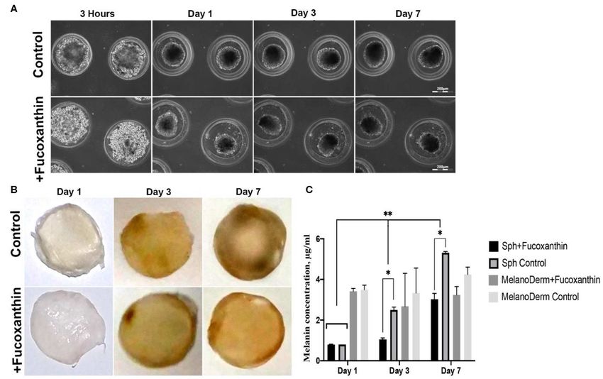
Zurina et al.                                                                                             Melanocyte-Derived Spheroids as a Test System

buffer (pH = 6.0; 06-014, Biovitrum) for 3 min at +100◦ C                 TABLE 1 | Primers used in this research.
under pressure. Cover glasses and spheroids were washed three
                                                                          Primers      Primer sequence (5′ -3′ )           Length Tm◦ C Primer
times with PBS (5 min for each wash, pH = 7.4) from the                                                                                 description
remnants of PFA. All the following staining procedures were
the same for 2D culture, spheroids, and tissue equivalent                 MC1R_For     GTGGTCTTCTTCCTGGCTATGC              22 bp    62.3   MC1R
samples. Melanocytes in monolayer culture were incubated with             MC1R_Rev GGATGGTGAGGGTGACAGCG                    20 bp    63.0
primary antibodies against MEL5 (917801, BioLegend, 1:100).               TYR_For      TTCAAGAAGTTTATCCAGAAGCC             23 bp    57.7   TYR
After preliminary preparations, sections and spheroids were               TYR_Rev      CTTAATGTAGTCTTGAAAAGAGTC            24 bp    53.8
incubated with primary antibodies against gp 100 (ab137078,               HsTBP_For CATGACTCCCGGAATCCCTATCTTT 25 bp                 63.1   HsTBP
Abcam, 1:100), Sox10 (ab155279, Abcam, 1:500), and MITF                   HsTBP_Rev TGTTGCTGCTGCTGCCTTTGTT                 22 bp    63.7
(ab122982, Abcam, 1:300). The result was visualized using
secondary antibodies conjugated with the fluorochromes FITC
(Em = 525 nm) and DyLight594 (Em = 617 nm). The nuclei
were stained with a bisbenzimide fluorescent dye, i.e., Hoechst           studied parameters, and the Pearson χ2 test for the evaluation
33258 (0.002 mg/ml, 10 min, 25◦ C). After staining, the excess            of fucoxanthin influence were tested. Both two-way ANOVA
dye was removed in three changes of PBS (pH = 7.4). The                   assay and multiple t-test were used consecutively to confirm the
resulting preparations were placed in Vitrogel mounting medium            significant difference of data. The p-value was adjusted using the
(12-001, Biovitrum) and analyzed in visible and ultraviolet light         two-stage linear step-up procedure of Benjamini, Krieger, and
ranges using the Olympus Fluoview FV10 laser scanning confocal            Yekutieli, with Q = 5%. All the experiments were reproduced
microscope (Olympus, Japan).                                              in triplicate; data are reported as the means ± SD of at least
   The obtained images of MelanoDermTM sections and                       three experiments.
spheroids from melanocytes were analyzed for mean
fluorescence intensity. Images were imported into ImageJ
software and converted to a 16-bit format. The parameters “area           RESULTS
integrated intensity” and “mean gray value” were measured
in the area limited by the threshold. The corrected total cell
                                                                          Study of Melanin Accumulation in 3D
fluorescence (CTCF) was calculated using the formula: CTCF =              Cultures—Spheroids From Melanocytes
integrated density—(area of selected cell × mean fluorescence             and MelanoDermTM Tissue Equivalent
of background readings). Three images were measured for each              Primary human melanocytes (CELL Applications) were first
marker and time point.                                                    maintained in monolayer culture. During the first passages,
                                                                          melanocytes mostly had spindle-like morphology, with a
Analysis of Gene Expression by Real-Time                                  tendency to the formation of dark aggregates (Figure 1A). By
Polymerase Chain Reaction                                                 the fourth passage, cell morphology slightly changed—cells had
To perform real-time PCR, three pieces of MelanoDermTM and                more dendrites, while no aggregates were observed in the culture
256 spheroids from melanocytes were used for each time point              (Figure 1B). Cells were characterized by the positive staining
in experimental and control groups. Total RNA was isolated                against melanocyte-specific marker MEL5, also known as a
using a standard Trizol method (TRIReagent, Sigma, USA). The              tyrosinase-related protein-1 that is involved in melanin synthesis
RNA was treated with Type I DNase (Fermentas, Germany)                    (Figure 1C).
to remove any DNA contamination, and precipitated in 4M                       The melanocytes at the fourth passage were placed in non-
LiCl. The concentration of the obtained RNA was measured                  adhesive agarose multi-well plates to obtain 3D spheroid culture.
using a Nanodrop 8000 spectrophotometer (Thermo Scientific,               Using the method of live time-lapse microscopy, we observed
USA); 2 µg of total RNA were used to synthesize cDNA. cDNA                the dynamics of spheroid formation in the control culture and
synthesis was performed using M-MLV reverse transcriptase                 in the presence of fucoxanthin (Figure 2A). In the process of
(Evrogen, Russia) and random hexanucleotides (Sileks, Russia).            culturing cells under 3D conditions, general patterns of spheroid
The obtained samples were used to analyze the expression of               formation were observed—initial compaction took place during
the MC1R gene and the TYR gene. The sequences of the used                 the first 24 h of cultivation and lasted up to 7 days. Moreover, by
primers are presented in Table 1. The analysis was performed              Day 7 both control and experimental spheroids became darker
on an automatic 7500 Real-Time PCR System amplifier (Applied              (Figure 2A), but the difference between groups could not be
Biosystems, USA) using a qPCRmix-HSSYBR + ROX mixture                     adequately assessed visually because spheroids were dense and
(Evrogen, Russia). The relative expression of MC1R and TYR                not transparent.
genes was measured using the 11Ct method, normalized to                       MelanoDermTM tissue equivalents are artificially created tissue
HsTBP (Bookout et al., 2006).                                             samples consisting of layers of keratinocytes and melanocytes
                                                                          cultured on a porous membrane. Throughout the cultivation
Statistics                                                                period, the samples accumulated melanin by increasing the
Statistical analysis was performed, and graphs were created using         number of melanocytes and autoregulating the intensity of
the Prism 8.0 GraphPad software package. Using the Shapiro-               pigment synthesis. The visual analysis showed that while on the
Wilk, hypotheses about the normality of the distributions of the          first and third days the dynamics of samples darkening did not

Frontiers in Bioengineering and Biotechnology | www.frontiersin.org   4                                                June 2020 | Volume 8 | Article 540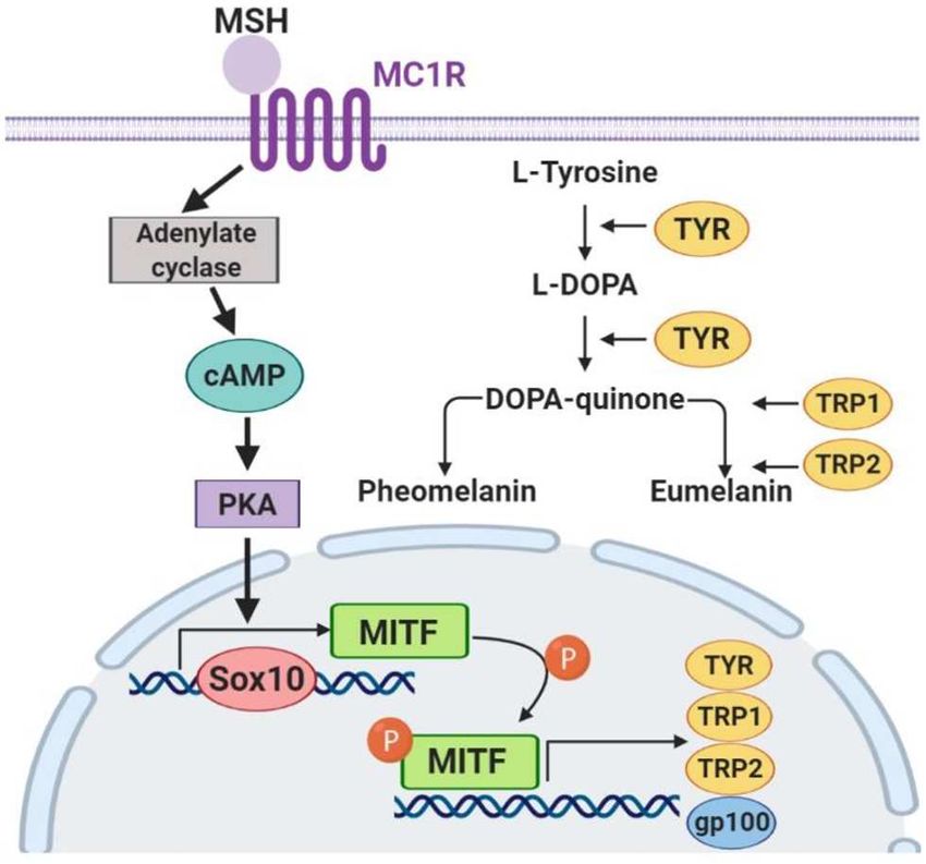
Zurina et al.                                                                                                       Melanocyte-Derived Spheroids as a Test System

  FIGURE 1 | Monolayer culture of human melanocytes at Passages 1 (A) and 4 (B). Cells were shown to express Mel5—the marker of melanocytes (C). Arrowheads
  indicate the sites of aggregates formation. (A,B)—Light phase-contrast microscopy; (C)—laser scanning confocal microscopy.

  FIGURE 2 | Pigmentation of treated and untreated spheroids and skin equivalents. (A) Dynamics of the spheroid formation from a suspension of melanocytes under
  non-adhesive 3D conditions in a standard growth medium (control) and with the addition of fucoxanthin (experimental group). Phase-contrast live time-lapse
  microscopy (Cell-IQ, CM Technologies, Finland). (B) Pigment accumulation in MelanoDermTM tissue equivalents at Days 1, 3, and 7 in the control group and the
  experimental group with the addition of fucoxanthin. All samples had the standard size−9 mm. Photos of samples dried before photometric analysis of melanin
  content. (C) Dynamics of melanin accumulation in melanocyte spheroids and MelanoDermTM tissue equivalents in the presence of the lightening agent fucoxanthin
  (experimental group) and with a solution of sodium chloride (control group). *p < 0.05 for two-way ANOVA assay and multiple t-test. *, **p < 0.05 (*within one
  time-point, **between different time-points).

differ significantly, by Day 7, the experimental skin equivalents                    spectrophotometric analysis of pigment concentration was
cultured in the presence of fucoxanthin were lighter compared to                     performed. The obtained data confirmed the visual assessment
the control group (Figure 2B).                                                       of the intensity of melanin synthesis in control and experimental
   To support the visual data on melanin accumulation in                             spheroids. As shown in Figure 2C, on Day 1 in 3D culture, the
these two types of 3D culture containing melanocytes, the                            average concentration of melanin was similar in both groups, but,

Frontiers in Bioengineering and Biotechnology | www.frontiersin.org              5                                               June 2020 | Volume 8 | Article 540
Zurina et al. Melanocyte-Derived Spheroids as a Test System
on Days 3 and 7, melanin synthesis by spheroids in the presence spheroids and skin equivalents, compared to the corresponding
of fucoxanthin was significantly lower than in the control group, control groups, but, in the case of spheroids, the difference
as confirmed by statistical analysis. between the control and experimental groups was significant
The data from the visual observations of melanin on Days 1 and 7 (Figure 6). The gene expression of the main
accumulation in MelanoDermTM were also confirmed using receptor regulating melanin synthesis, i.e., melanocortin receptor
photometry (Figure 2C). The obtained results showed that 1 (MC1R), was affected by fucoxanthin only in MelanoDermTM
melanin content in control and experimental groups was the culture, which was confirmed by statistical analysis (Figure 6).
same on the first day of cultivation, while, on Day 3, a decrease
in the melanin content was observed in the experimental group
relative to the control group. By Day 7, the final concentration DISCUSSION
of melanin in the experimental samples after cultivation with
fucoxanthin was significantly lower than in the control group, The expanding ozone holes in the atmosphere lead to the growing
but it slightly increased compared to Day 3 in the experimental danger of UV skin damage, making the pharmaceutical industry
group. However, no statistically significant difference was increase the production of new types of sun protection (Smit
observed between the groups. et al., 2009; Zastrow et al., 2017), which requires the new systems
for testing them in vitro. At present, the mainly used test-
Immunocytochemical Analysis of 3D systems are primary cultures of melanocytes and keratinocytes
Cultures (Lei et al., 2002; Lee et al., 2020), melanoma cell lines (Kim
To assess the effect of fucoxanthin on the expression of et al., 2017; Lee et al., 2020), the commercial tissue equivalents
the key factors of melanogenesis gp 100, MITF and Sox10, (EpiSkinTM , MelanoDermTM , and others) (Costin and Raabe,
immunocytochemical analysis of the MelanoDermTM 2013; Kim et al., 2017; Meena and Mohandass, 2019; Lee et al.,
tissue equivalent sections and spheroids from melanocytes 2020), and tissue equivalents obtained from cells with induced
was performed. pluripotency (iPSC) (Gledhill et al., 2015). These approaches
Visual comparison of stained spheroids revealed no difference have some limitations, including non-physiological conditions
in the expression of gp100 on Day 3. However, there was up- (for 2D cultures), difficulties in analysis, and expensiveness
regulation on Day 7 in the control group as compared to the (for commercial tissue equivalents). The current study was
fucoxanthin-present group (Figure 3). On the other hand, MITF aimed to assess the ability of spheroids from normal human
expression was lower on day 3 and showed up-regulation on melanocytes to maintain their functional activity (melanin
the day 7th in the spheroid control group as compared to the synthesis) and to react to the addition of hypopigmentation agent
fucoxanthin present spheroid group (Figure 3). The expression fucoxanthin. Additionally, we compared spheroids’ properties to
of the Sox10 was higher on the 3rd day as well as 7th day in the 3D MelanoDermTM skin tissue equivalents, which we considered
spheroid control group as compared to the fucoxanthin-present as the closest available alternative.
spheroid group (Figure 3). We also analyzed the images using Melanocytes had cell-specific dendritic morphology and
ImageJ for digital analysis (Figure 5). The data obtained were expressed specific marker Mel5 at passage 4 in the standard
consistent with the visual analysis. monolayer system (Figure 1). However, cells were not able
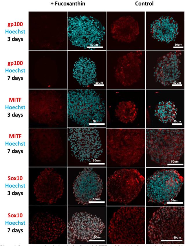
For the MelanoDermTM group, it was shown that the to accumulate melanin in vitro, which was corresponded by
expression level of gp100, which is responsible for the maturation numerous studies (Chung et al., 2019). At the first passages, cells
of melanosomes, was increased in the MelanoDermTM control demonstrated a tendency to the spontaneous formation of dark
group compared to the MelanoDermTM experimental group on aggregates attached to the surface, which has been previously
Day 7 of cultivation (Figure 4). On Days 3 and 7, there was a reported only on specific culture surface (Lin et al., 2005). This
slight increase in the expression level of MITF in the presence effect was no longer observed at the further passages. When
of the drug, as well as in the MelanoDermTM control group with transferred in non-adhesive agarose microplates, melanocytes
a high level of expression of this transcription factor (Figure 4). formed pigmented spheroids that accumulated melanin during
Sox10 on Days 3 and 7 of cultivation was expressed in cell cultivation. The same tendency was observed in commercial skin
nuclei in the control MelanoDermTM samples and practically equivalent MelanoDermTM (Figure 2).
not present in the experimental samples (Figure 4). These In our study, we used the well-known skin-lightening
observations were supported by a quantitative analysis of the compound fucoxanthin, which targets melanocytes, to
mean fluorescence intensity of the obtained images (Figure 5). validate the capability of both spheroids from melanocyte
and skin equivalents of the tissue-specific reaction
Real-Time PCR Analysis of Melanogenesis (the regulation of melanin synthesis). Fucoxanthin is a
naturally occurring brown- or orange-colored pigment
Factor Expression in MelanoDermTM Tissue that belongs to the class of non-provitamin A carotenoids
Equivalents and Spheroids From Human present in the chloroplasts of brown seaweeds (Martin,
Melanocytes 2015). Fucoxanthin is well-known as the compound that
Real-time PCR revealed the effect of fucoxanthin on the affects different cellular pathways providing antioxidative
expression of tyrosinase (TYR)—one of the melanogenesis effects (Martin, 2015) and hypopigmentation (Shimoda
enzymes. That is, it was down-regulated by Day 7 in both et al., 2010). Both spheroids from melanocyte and
Frontiers in Bioengineering and Biotechnology | www.frontiersin.org 6 June 2020 | Volume 8 | Article 540Zurina et al. Melanocyte-Derived Spheroids as a Test System FIGURE 3 | Immunocytochemical analysis of gp100, MITF, and Sox10 (red) expression in spheroids from human melanocytes in the control and experimental groups on Days 3 and 7 in 3D culture. The nuclei are stained with Hoechst 33258 (blue). Laser-scanning confocal microscopy. Frontiers in Bioengineering and Biotechnology | www.frontiersin.org 7 June 2020 | Volume 8 | Article 540
Zurina et al.                                                                                                      Melanocyte-Derived Spheroids as a Test System

  FIGURE 4 | Immunocytochemical analysis of gp100, MITF, and Sox10 (red) expression on sections of MelanoDermTM tissue equivalents in the control and
  experimental groups on Days 3 and 7 of cultivation. The nuclei are stained with Hoechst 33258 (blue). Laser-scanning confocal microscopy.

Frontiers in Bioengineering and Biotechnology | www.frontiersin.org             8                                               June 2020 | Volume 8 | Article 540
Zurina et al.                                                                                                       Melanocyte-Derived Spheroids as a Test System

  FIGURE 5 | The analysis of mean fluorescence intensity on the immunocytochemical staining images of spheroids and MelanoDermTM sections in the experimental (in
  the presence of fucoxanthin) and control groups presenting an expression of gp100, MITF, and Sox10 on Days 3 and 7 in 3D culture. *# p < 0.05 for two-way ANOVA
  assay and multiple t-test.

  FIGURE 6 | Real-time PCR analysis of TYR and MC1R expression in MelanoDermTM tissue equivalents and spheroids from human melanocytes on Days 1, 3, and 7
  in the presence of fucoxanthin and in the control group with growth medium. *p < 0.05 for two-way ANOVA assay and multiple t-test.

MelanoDermTM showed a decrease of melanin synthesis in                              different levels of this melanogenesis signaling pathway
the presence of fucoxanthin, but that difference between                            (Figure 7).
control and experimental groups was significant only for                                The skin pigment synthesis starts with the activation of
spheroids (Figure 2), probably, because they contained                              melanocortin 1 receptor (MC1R) by its agonists—α-melanocyte-
only melanocytes.                                                                   stimulating hormone (αMSH) and adrenocorticotropic
   To confirm the mechanisms of fucoxanthin physiological                           hormone (ACTH) (Slominski et al., 2004). This activation
influence, we studied the expression of key factors that                            stimulates the cAMP/PKA cascade leading to the increased
regulate melanogenesis. Since the synthesis of melanin is                           expression of the master regulator of melanocyte development,
a complex multi-stage process (Figure 7) controlled by                              i.e., microphthalmia-associated transcription factor (MITF)
both endogenous and exogenous factors, and fucoxanthin,                             (Yamaguchi and Hearing, 2007). MITF, in its turn, activates the
as well as other carotenoids, is a multitargeting agent                             expression of the TYR gene (tyrosinase), PMEL17 (or gp100—
(Shimoda et al., 2012), we have analyzed elements from                              premelanosome protein), and Bcl-2 (anti-apoptotic factor),

Frontiers in Bioengineering and Biotechnology | www.frontiersin.org             9                                                June 2020 | Volume 8 | Article 540
Zurina et al.                                                                                             Melanocyte-Derived Spheroids as a Test System

                                                                               (D’Mello et al., 2016). Using this fact, we have also
                                                                               analyzed other crucial factors of melanogenesis—the cellular
                                                                               expression of transcription factors Sox10 and MITF, and
                                                                               the expression of the melanosome maturation regulator—
                                                                               gp100/PMEL17 (Figures 3, 4). Melanocyte spheroids
                                                                               and skin equivalent MelanoDermTM equally showed the
                                                                               significant reduction of expression of the transcription
                                                                               factors MITF and Sox10 in the presence of fucoxanthin
                                                                               (Figure 5). However, only spheroid model has firstly
                                                                               revealed that the application of fucoxanthin can inhibit
                                                                               melanosome formation by significantly decreasing gp100
                                                                               synthesis. Thus, in general, the spheroid model showed
                                                                               higher responsiveness to fucoxanthin, as confirmed by
                                                                               statistical analysis.
                                                                                  The obtained results indicate that, in the case of studies of
                                                                               agents that potentially affect pigment synthesis and physiology
                                                                               of melanocytes, spheroids can be used as a more affordable and
                                                                               reproducible drug-testing system. The preservation of native-like
                                                                               cell morphology and physiology in spheroids makes it possible to
                                                                               conduct a study on isolated cells instead of using expensive tissue
                                                                               equivalents consisting of different types of cells and requiring
  FIGURE 7 | The key components of the melanogenesis signaling pathway.
                                                                               special conditions for culturing.

                                                                               CONCLUSIONS
which leads to an increased synthesis of melanin (Lin and Fisher,              Collectively, these data illustrate that the 3D culture system
2007). The transcription factors Sox10 and PAX3, in synergistic                can maintain the melanin accumulation in vitro compared to
terms, directly regulate the expression of MITF (Bondurand                     the MelanoDermTM skin equivalents. Moreover, the obtained
et al., 2000; Hou et al., 2006). In addition, the intensity of                 spheroids provided a sensitive response to the compound
melanin synthesis in melanocytes directly depends on the                       addition, recapitulated the in vitro and in vivo-like response
amount and activity of the tyrosinase enzyme in the cells, which               (reduction of MITF, Sox10, and TYR expression), and showed
catalyzes the first two stages of melanin formation: hydroxylation             new data (down-regulation of gp100 expression) that has not
of L-tyrosine into L-dihydroxyphenylalanine (L-DOPA) and                       been previously reported using MelanoDermTM or other skin
subsequent oxidation of L-DOPA into L-dopaquinone.                             tissue equivalents. Thus, melanocyte spheroids can be used as
   As for the anti-melanogenic effect of fucoxanthin, it is                    a convenient and affordable test system for screening drugs
shown to reduce TYR activity, the expression of MC1R,                          targeting melanocytes and potentially as building blocks for the
TYR-dependent receptor 1, cyclooxygenase 2 (COX-2),                            tissue engineering of skin equivalents.
prostaglandin receptor 1 (EP1), which also indicates its anti-
inflammatory properties (Shimoda et al., 2010). A general
overview shows that fucoxanthin inhibits TYR and melanin                       DATA AVAILABILITY STATEMENT
production in vitro in B16 melanoma cells and in vivo in
guinea-pigs and mice skin. It also suppressed PGE2, MSH,                       All datasets generated for this study are included in the
TRP1, and melanogenic stimulant receptors—EP1and MC1R                          article/supplementary material.
in vivo (Azam et al., 2017). Based on these findings, we
selected to assay the expression of MC1R and TYR mRNA                          AUTHOR CONTRIBUTIONS
(Figure 6).
   Our study demonstrated significant down-regulating                          IZ, AG, and IS contributed to the conception and the design
of TYR gene expression on days 1 and 7 for melanocyte                          of the study. IZ wrote the first draft of the manuscript and
spheroids and reduction of MC1R gene expression on                             prepared the submitted version together with AG. IZ and ED
day 7—for MelanoDermTM . Thus, melanocyte spheroids                            cultivated Skin Equivalents (MelanoDermTM ) and spheroids,
reproduced results described previously in vitro and in                        collected samples. ED and TK conducted all 2D cell culture
vivo, and MelanoDermTM system showed results matched                           works. AG accomplished the immunocytochemical analysis of all
with in vivo studies. However, neither system showed                           samples. DM performed the molecular assay and the photometric
both results.                                                                  assay of all samples. NK provided statistical analysis and created
   Fucoxanthin is also interacting with other signaling                        the graphs. SM and IS supervised the study and edited the
pathways such as NF-κB and WNT/β-catenin pathways                              manuscript. All authors contributed to manuscript revision, read,
(Martin, 2015), which are also involved in melanogenesis                       and approved the submitted version.

Frontiers in Bioengineering and Biotechnology | www.frontiersin.org       10                                           June 2020 | Volume 8 | Article 540
Zurina et al. Melanocyte-Derived Spheroids as a Test System
FUNDING ACKNOWLEDGMENTS
This work was supported by The Special Federal Programme Part of the work using confocal microscopy was
of the Russian Federation Government, Research Project No. performed at the Faculty of Biology at Lomonosov
0520-2019-0026 (The Study of the Mechanisms of Ageing and Moscow State University (Equipment Complex for Tissue-
Regeneration and the Development of Conditions for Obtaining engineering Technologies, Visualization and Microsurgery;
Tissue-engineered Constructs Using 2D and 3D Cell Cultures). ID: 9351669).
REFERENCES Koudan, E. V., Korneva, J. V., Karalkin, P. A., Gladkaya, I. S., Gryadunova, A. A.,
Mironov, V. A., et al. (2017). THe scalable standardized biofabrication of tissue
Antoni, D., Burckel, H., Josset, E., and Noel, G. (2015). Three-dimensional spheroids from different cell types using nonadhesive technology. 3D Print.
cell culture: a breakthrough in vivo. Int. J. Mol. Sci. 16, 5517–5527. Addit. Manufac. 4, 53–60. doi: 10.1089/3dp.2016.0044
doi: 10.3390/ijms16035517 Langhans, S. A. (2018). Three-dimensional in vitro cell culture models
Azam, M. S., Choi, J., Lee, M.-S., and Kim, H.-R. (2017). Hypopigmenting effects in drug discovery and drug repositioning. Front. Pharmacol. 9:6.
of brown algae-derived phytochemicals: a review on molecular mechanisms. doi: 10.3389/fphar.2018.00006
Marine Drugs 15:297. doi: 10.3390/md15100297 Lee, J., Lee, S., Roh, K., Jung, E., and Park, D. (2015). A novel culture system to
Bondurand, N., Pingault, V., Goerich, D. E., Lemort, N., Sock, E., Caignec, induce melanin synthesis by three-dimensional spheroid culture. Biotechnol.
C. L., et al. (2000). Interaction among SOX10, PAX3 and MITF, three Bioproc. Eng. 20, 194–200. doi: 10.1007/s12257-014-0415-8
genes altered in waardenburg syndrome. Hum. Mol. Genet. 9, 1907–1917. Lee, S. H., Bae, I.-H., Lee, E.-S., Kim, H.-J., Lee, J., and Lee, C. S. (2020).
doi: 10.1093/hmg/9.13.1907 Glucose exerts an anti-melanogenic effect by indirect inactivation of tyrosinase
Bookout, A. L., Cummins, C. L., Mangelsdorf, D. J., Pesola, J. M., and Kramer, M. in melanocytes and a human skin equivalent. Int. J. Mol. Sci. 21:1736.
F. (2006). High-throughput real-time quantitative reverse transcription PCR. doi: 10.3390/ijms21051736
Curr. Protoc. Mol. Biol 73, 15.8.1–15.8.28. doi: 10.1002/0471142727.mb1508s73 Lei, T. C., Virador, V. M., Vieira, W. D., and Hearing, V. J. (2002). A melanocyte–
Chung, S., Lim, G. J., and Lee, J. Y. (2019). Quantitative analysis of melanin keratinocyte coculture model to assess regulators of pigmentation in vitro. Anal.
content in a three-dimensional melanoma cell culture. Sci. Rep 9, 1–9. Biochem. 305, 260–268. doi: 10.1006/abio.2002.5665
doi: 10.1038/s41598-018-37055-y Lin, J. Y., and Fisher, D. E. (2007). Melanocyte biology and skin pigmentation.
Costin, G.-E., and Raabe, H. (2013). Optimizied in vitro pigmentation screening Nature 445:843. doi: 10.1038/nature05660
assay using a reconstructed three dimensional human skin model. Rom. J. Lin, S.-J., Jee, S.-H., Hsaio, W.-C., Lee, S.-J., and Young, T.-H. (2005). Formation
Biochem. 50, 15–27. of melanocyte spheroids on the chitosan-coated surface. Biomaterials 26,
D’Mello, S. A., Finlay, G. J., Baguley, B. C., and Askarian-Amiri, M. E. 1413–1422. doi: 10.1016/j.biomaterials.2004.05.002
(2016). Signaling pathways in melanogenesis. Int. J. Mol. Sci. 17:1144. Lin, S. J., Jee, S. H., Hsiao, W. C., Yu, H. S., Tsai, T. F., Chen, J. S., et al. (2006).
doi: 10.3390/ijms17071144 Enhanced cell survival of melanocyte spheroids in serum starvation condition.
Figtree, G. A., Bubb, K. J., Tang, O., Kizana, E., and Gentile, C. (2017). Vascularized Biomaterials 27, 1462–1469. doi: 10.1016/j.biomaterials.2005.08.031
cardiac spheroids as novel 3D in vitro models to study cardiac fibrosis. Cells. Martin, L. J. (2015). Fucoxanthin and its metabolite fucoxanthinol
Tissues Organs. 204, 191–198. doi: 10.1159/000477436 in cancer prevention and treatment. Mar. Drugs 13, 4784–4798.
Fitzgerald, K. A., Malhotra, M., Curtin, C. M., O’ Brien, F. J., and O’ Driscoll, C. M. doi: 10.3390/md13084784
(2015). Life in 3D is never flat: 3D models to optimise drug delivery. J. Control. Meena, S. N., and Mohandass, C. (2019). “Application of 3D cell culture techniques
Rel. 215, 39–54. doi: 10.1016/j.jconrel.2015.07.020 in cosmeceutical research,” in Advances in Biological Science Research: A
Gledhill, K., Guo, Z., Umegaki-Arao, N., Higgins, C. A., Itoh, M., and Christiano, Practical Approach, eds S. N. Meena and M. M. Naik (London, UK: Academic
A. M. (2015). Melanin transfer in human 3D skin equivalents generated Press), 469–484.
exclusively from induced pluripotent stem cells. PloS ONE 10:e0136713. Nzou, G., Wicks, R. T., Wicks, E. E., Seale, S. A., Sane, C. H., Chen, A., et al.
doi: 10.1371/journal.pone.0136713 (2018). Human cortex spheroid with a functional blood brain barrier for high-
Hartley, B. J., and Brennand, K. J. (2017). Neural organoids for disease throughput neurotoxicity screening and disease modeling. Sci. Rep. 8:7413.
phenotyping, drug screening and developmental biology studies. Neurochem. doi: 10.1038/s41598-018-25603-5
Int. 106, 85–93. doi: 10.1016/j.neuint.2016.10.004 Peng, J., Yuan, J.-P., Wu, C.-F., and Wang, J.-H. (2011). Fucoxanthin, a
Heo, S.-J., and Jeon, Y.-J. (2009). Protective effect of fucoxanthin isolated from marine carotenoid present in brown seaweeds and diatoms: metabolism
Sargassum siliquastrum on UV-B induced cell damage. J. Photochem. Photobiol. and bioactivities relevant to human health. Mar. Drugs 9, 1806–1828.
B Biol. 95, 101–107. doi: 10.1016/j.jphotobiol.2008.11.011 doi: 10.3390/md9101806
Hou, L., Arnheiter, H., and Pavan, W. J. (2006). Interspecies difference in the Ribeiro, D., Kvist, A. J., Wittung-Stafshede, P., Hicks, R., and Forslow, A. (2018).
regulation of melanocyte development by SOX10 and MITF. Proc. Natl. Acad. 3D-Models of insulin-producing beta-cells: from primary islet cells to stem cell-
Sci. U.S.A. 103, 9081–9085. doi: 10.1073/pnas.0603114103 derived islets. Stem Cell Rev. Rep. 14, 177–188. doi: 10.1007/s12015-017-9783-8
Hsiao, W. C., and Young, T. H. (2019). Characteristics of melanocyte spheroids Shimoda, H., Shan, S. J., Tanaka, J., and Maoka, T. (2012). β-Cryptoxanthin
formed through different biomaterial-induced processes. J. Formos. Med. Assoc. suppresses UVB-induced melanogenesis in mouse: involvement of the
118, 152–161. doi: 10.1016/j.jfma.2018.02.010 inhibition of prostaglandin E2 and melanocyte-stimulating hormone pathways.
Huang, B.-W., and Gao, J.-Q. (2018). Application of 3D cultured multicellular J. Pharm. Pharmacol. 64, 1165–1176. doi: 10.1111/j.2042-7158.2012.01495.x
spheroid tumor models in tumor-targeted drug delivery system research. J. Shimoda, H., Tanaka, J., Shan, S. J., and Maoka, T. (2010). Anti-pigmentary
Control. Rel. 270, 246–259. doi: 10.1016/j.jconrel.2017.12.005 activity of fucoxanthin and its influence on skin mRNA expression
Kim, K., Leutou, A. S., Jeong, H., Kim, D., Seong, C. N., Nam, S.-J., et al. of melanogenic molecules. J. Pharm. Pharmacol. 62, 1137–1145.
(2017). Anti-pigmentary effect of (-)-4-hydroxysattabacin from the marine- doi: 10.1111/j.2042-7158.2010.01139.x
derived bacterium Bacillus sp. Mar. Drugs 15:138. doi: 10.3390/md150 Slominski, A., Tobin, D. J., Shibahara, S., and Wortsman, J. (2004). Melanin
50138 pigmentation in mammalian skin and its hormonal regulation. Physiol. Rev.
Klaka, P., Grudl, S., Banowski, B., Giesen, M., Sattler, A., Proksch, P., et al. (2017). 84, 1155–1228. doi: 10.1152/physrev.00044.2003
A novel organotypic 3D sweat gland model with physiological functionality. Smit, N., Vicanova, J., and Pavel, S. (2009). The hunt for natural skin whitening
PLoS ONE 12:e0182752. doi: 10.1371/journal.pone.0182752 agents. Int. J. Mol. Sci. 10, 5326–5349. doi: 10.3390/ijms10125326
Frontiers in Bioengineering and Biotechnology | www.frontiersin.org 11 June 2020 | Volume 8 | Article 540Zurina et al. Melanocyte-Derived Spheroids as a Test System Surolia, R., Li, F. J., Wang, Z., Li, H., Liu, G., Zhou, Y., et al. (2017). spheroid cultures of human osteoarthritic chondrocytes and donor-matched 3D pulmospheres serve as a personalized and predictive multicellular chondrogenically differentiated mesenchymal stem cells. Biotechnol. Prog. 34, model for assessment of antifibrotic drugs. JCI. Insight 2:e91377. 1045–1058. doi: 10.1002/btpr.2629 doi: 10.1172/jci.insight.91377 Yamaguchi, Y., and Hearing, V. J. (2007). “Melanocyte distribution and function in Conflict of Interest: The authors declare that the research was conducted in the himan skin,” in From Melanocytes to Melanoma: The Progression to Malignancy, absence of any commercial or financial relationships that could be construed as a eds V. J. Hearing and S. P. L. Leong (Totowa, NJ: Springer Science & Business potential conflict of interest. Media), 101. Zastrow, L., Meinke, M. C., Albrecht, S., Patzelt, A., and Lademann, Copyright © 2020 Zurina, Gorkun, Dzhussoeva, Kolokoltsova, Markov, Kosheleva, J. (2017). From UV protection to protection in the whole spectral Morozov and Saburina. This is an open-access article distributed under the terms range of the solar radiation: new aspects of sunscreen development. of the Creative Commons Attribution License (CC BY). The use, distribution or Adv. Exp. Med. Biol. 996, 311–318. doi: 10.1007/978-3-319-56 reproduction in other forums is permitted, provided the original author(s) and the 017-5_26 copyright owner(s) are credited and that the original publication in this journal Zigon-Branc, S., Barlic, A., Knezevic, M., Jeras, M., and Vunjak-Novakovic, G. is cited, in accordance with accepted academic practice. No use, distribution or (2018). Testing the potency of anti-TNF-alpha and anti-IL-1beta drugs using reproduction is permitted which does not comply with these terms. Frontiers in Bioengineering and Biotechnology | www.frontiersin.org 12 June 2020 | Volume 8 | Article 540
You can also read