The effects of rivaroxaban, an oral anticoagulant, on human IVD primary cultures
←
→
Page content transcription
If your browser does not render page correctly, please read the page content below
Clinical research

The effects of rivaroxaban, an oral anticoagulant,
on human IVD primary cultures

Tezcan Caliskan1, Hande Akalan2, Ibrahim Yilmaz3, Numan Karaarslan4, Duygu Yasar Sirin2,
Hanefi Ozbek5

 Department of Neurosurgery, School of Medicine, Namik Kemal University, Tekirdag,
1
                                                                                      Corresponding author:
 Turkey                                                                               Ibrahim Yilmaz PhD
 Department of Molecular Biology and Genetics, Faculty of Arts and Sciences, Namik
2
                                                                                      Department of
 Kemal University, Tekirdag, Turkey                                                   Medical Pharmacology
 Department of Medical Pharmacology, School of Medicine, Istanbul Medipol
3
                                                                                      School of Medicine
 University, Istanbul, Turkey                                                         Istanbul Medipol
4
 Department of Neurosurgery, School of Medicine, Halic University, Istanbul, Turkey   University
 Department of Medical Pharmacology, Bakircay University School of Medicine, Izmir,
5
                                                                                      Istanbul, Turkey
 Turkey                                                                               Phone: +90 5327012858
                                                                                      E-mail: ibrahimyilmaz77@
Submitted: 5 November 2020; Accepted: 2 May 2021                                      yahoo.com
Online publication: 9 May 2021

Arch Med Sci
DOI: https://doi.org/10.5114/aoms/136323
Copyright © 2021 Termedia & Banach

Abstract
Introduction: The present study aimed to investigate the potential effects
of rivaroxaban, an oral anticoagulant that inhibits the effects of factor Xa,
on intact intervertebral disc tissue cells and the extracellular matrix (ECM).
Material and methods: Rivaroxaban was applied to primary human cell cul-
tures prepared from tissues of the intervertebral disc. Comparative molec-
ular analyses were performed on non-drug-treated control group samples.
Descriptive statistics were presented as the mean ± standard deviation. An
analysis of variance test was performed to determine whether there were sig-
nificant differences in the mean across the groups. When differences across
groups were observed, Tukey’s honestly significant difference post-hoc test
was used for multiple pairwise comparisons. The significance of the obtained
data was determined statistically. The α significance value was < 0.05.
Results: The cells in the control group and in the rivaroxaban-treated group
were viable, healthy, and proliferated (p < 0.05). However, the expression
levels of the chondroadherin gene (CHAD), cartilage oligo matrix protein
(COMP), matrix metalloproteinase (MMP)-13, and MMP-19 genes were
changed (p < 0.05).
Conclusions: Although rivaroxaban does not suppress cell proliferation due
to morphological, biological, and biochemical changes in the intervertebral
disc tissue, it may change the expression of genes that are related to ECM
maintenance.

Key words: cartilage oligo matrix protein, chondroadherin gene,
cytotoxicity, intervertebral disc cells, matrix metalloproteinases, nucleus
pulposus, rivaroxaban.

Introduction
   Rivaroxaban belongs to a group of medicines called antithrombotic
agents. They are used as an inhibitor of factor Xa, which plays a role in
blood clotting [1]. The administration of this novel antithrombotic agent is
recommended following the use of anticoagulant drugs, such as warfarin,
and it does not require international normalized ratio monitoring [2, 3].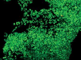
Tezcan Caliskan, Hande Akalan, Ibrahim Yilmaz, Numan Karaarslan, Duygu Yasar Sirin, Hanefi Ozbek

    The half-life of rivaroxaban is between 7 and                   Material and methods
17 h. In addition to its ease of use, it is well ab-
                                                                       The present research was conducted with
sorbed by the gastrointestinal system, its food in-
                                                                    the approval of the Ethics Committee of Univer-
teraction is minimal, and its bioavailability is 80%.
                                                                    sity School of Medicine (ethical permission no.
Peak plasma concentrations are attained within
                                                                    10840098/604.01.01/E.44192, 29). Written in-
approximately 3 h after oral administration. Thus,
                                                                    formed consent was obtained from all patients
it is used for the following: a) to prevent non-valvu-
                                                                    prior to their inclusion in the study.
lar atrial fibrillation [4], b) to prevent stroke [5] and
                                                                       To minimize experimental errors, assays were
blood clots in other veins of the body, c) to treat
                                                                    repeated at least three times. The tissues used in
deep-vein thrombosis and pulmonary embolism,
                                                                    the preparation of the cultures were resected by
and d) to treat deep-vein thrombosis and the pro-
                                                                    the same surgeons, and similar analyses were per-
phylaxis of pulmonary embolism in the lungs [6].
                                                                    formed by the same researchers. The researchers
In addition, rivaroxaban is a direct factor Xa inhib-
                                                                    who examined the results were blind to the drugs
itor used once a day for prevention of thrombotic
                                                                    and dosages utilized in each group.
events in patients with atrial fibrillation [7]. The
recommended dose is 30 mg/day for a minimum
                                                                        Inclusion criteria and preparation of cell
of 21 days. The usage period can be decreased to
                                                                        cultures from human primary intervertebral
12 days for knee replacement surgery, and it can
be increased to 35 days for hip replacement sur-                        disc tissue
gery. Rivaroxaban-related side effects at 20 mg/                        The study included 16 volunteers admitted to
day for the prevention of stroke in atrial fibrilla-                the neurosurgery department of Namik Kemal
tion have been reported in the literature. Recorded                 University School of Medicine after spinal trauma.
adverse side effects include gastrointestinal hem-                  After lumbar magnetic resonance image evalua-
orrhage, urogenital hemorrhage, epistaxis, ulorrha-                 tion, the intervertebral disc tissues of the patients
gia, vitreous hemorrhage, hematoma, hemoptysis,                     who had posterior transpedicular stabilization,
and skin/subcutaneous hemorrhages. In addition,                     lumbar laminectomy, and discectomy as surgical
side effects such as hemorrhage after surgery, limb                 modalities were included in the study.
edema, pain, fever, and orthostatic hypotension or                      Patients suffering from any liver disease with
faintness have been reported. Some studies have                     a risk of bleeding or any other disease that may in-
also reported that fatigue, intracerebral hemor-                    crease the risk of serious bleeding in solid organs
rhage, pseudoaneurysm, or tachycardia may occur                     were excluded from the study. To prevent errors
in patients using rivaroxaban [8].                                  that may arise from drug interactions, patients us-
    However, no previous study has focused on                       ing drugs such as warfarin, heparin, dabigatran, or
the positive and negative effects of this pharma-                   apixaban were excluded from the study. Patients
cological agent on intervertebral disc tissue. For                  administered heparin to keep the venous/arterial
that reason, the effect of this drug, which is fre-                 line open were also excluded from the study. The
quently used before and after the operation, on                     following patients were also excluded from the
the healing process is unknown. This study con-                     study: those with moderate to severe renal insuf-
sidering the effects and toxicity of rivaroxaban on                 ficiency and patients using non-steroidal anti-in-
intervertebral disc (IVD) primary cultures can shed                 flammatory drugs, acetylsalicylic acid, blood clot-
some light on this subject. Therefore, the findings                 ting-inhibiting drugs, or azole antifungal agents,
obtained through the present research could con-                    such as ketoconazole, itraconazole, voriconazole,
tribute to the literature.                                          and posaconazole, within the last 3 weeks. In ad-
    The aim of this study is to investigate the ef-                 dition, patients with acquired immune deficiency
fects of rivaroxaban on primary cell cultures pre-                  syndrome and those using human immunodefi-
pared from human intervertebral disc tissue at the                  ciency virus protease inhibitors, such as ritonavir,
pharmaco-molecular level. To achieve this aim, the                  were excluded from the study.
expression level of chondroadherin (CHAD) [9–14],                       Of the tissues included in the study (n = 16),
an NP-specific marker, was examined. The level of                   tissues of patients using erythromycin (n = 1),
the cartilage oligo matrix protein (COMP) [15–18],                  clarithromycin (n = 1), fluconazole (n = 1), enox-
which plays a role in intervertebral disc catabolic                 aparin (n = 2), clopidogrel (n = 1), or vitamin K
metabolism, was also tested. In addition to these                   antagonists, such as warfarin (n = 3) and naprox-
markers, the levels of matrix metalloproteinase                     en (n = 1), within the last 3 weeks were excluded
(MMP)-13 and MMP-19 [19] were examined. Both                        from the study. The remaining 6 patient tissues
MMP-7 and MMP-19 play roles in remodeling the                       were used in the preparation of the primary cell
intervertebral disc and the extracellular matrix                    culture.
(ECM) structure, as well as in the catabolic path-                      The age range of the patients who met the
ways of these tissues.                                              inclusion criteria was 27–39 years. Patients with

2                                                                                                          Arch Med Sci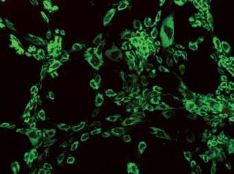
The effects of rivaroxaban, an oral anticoagulant, on human IVD primary cultures

traumatic disc herniation (n = 6, mean age: 33            oxaban-treated groups were performed on days
±5.32 years) underwent discectomy and instru-             10 and 20.
mentation using the posterolateral fixation tech-
nique. The tissues were then transferred to the               Analyses
laboratory at 4°C.
                                                              Cell surface morphology and viability were ex-
   Cell cultures containing intact human inter-
                                                          amined using an inverted light microscope. Micro-
vertebral disc tissue were prepared in accordance
                                                          scopical evaluations were performed under 4×,
with the protocol [9–14]. The tissue samples,
                                                          10×, 20×, and 40× magnifications at 0 h and on
which were irrigated with 0.9% isotonic sodium
                                                          days 10 and 20. In addition, membrane permea-
chloride solution in a laminar flow cabinet (Air
                                                          bility tests were conducted using a fluorescent
Flow-NUVE/NF-800 R, Ankara, Turkey), were then
                                                          microscope and nucleic acid binding dyes (AO and
washed three times consecutively with phosphate
                                                          PI) under similar magnifications. Based on the
buffered saline and clarified from the red blood
                                                          principle [9–14] that live cells produce green fluo-
cells. The tissues were first degraded mechanical-
                                                          rescence and dead cells produce red fluorescence,
ly using a rongeur and then treated with 0.375 µl
                                                          the results of the analyses were evaluated.
of Clostridium histolyticum (collagenase type II en-
                                                              The cell viability tests were conducted using
zyme) (Invitrogen Corp., Sigma Chemical, St. Louis,
                                                          a commercial MTT kit (Vybrant MTT Cell Prolif-
MO) and Hanks’ balanced salt solution (HBSS-1X,
                                                          eration Assay, Cat. No. V-13154; Thermo Fisher
14025, Gibco) to perform the enzymatic degrada-
                                                          Scientific, Waltham, MA, USA). The MTT assay
tion. This cell suspension was incubated overnight
                                                          measures color changes, which occur due to the
in an incubator (NUVE, 06750, Ankara, Turkey)
                                                          production of formazan by the cells undergoing
with 5% CO2 at 37°C. The samples were subse-
                                                          proliferation, using tetrazolium with increasing de-
quently centrifuged three times consecutively
                                                          hydrogenase enzyme activity [21]. The quantity of
at 1,100 rpm and 4°C. The supernatants in the
                                                          formazan was measured by recording the chang-
tubes were discarded. The cell pellets were re-sus-
                                                          es in absorbance at 570 nm using a plate reading
pended with a freshly prepared culture medium
                                                          spectrophotometer. The viability of the cells in the
containing Dulbecco’s modified Eagle’s medium
                                                          control group samples, to which rivaroxaban was
(DMEM; Cat#41965062; Gibco) supplemented
                                                          not initially applied, was designated as 100%. The
with 1% penicillin-streptomycin and 10% fetal
                                                          proliferation and the inhibition of the proliferation
bovine serum (FBS; Gibco). The cells were stained
                                                          were calculated using the following formulas, re-
with trypan blue and counted using an inverted
                                                          spectively: Test OD/Control ODX100 and 1-Test
light microscope (magnification, 10×).
                                                          OD/Control OD. The data were recorded for statis-
   The counted cells were plated at 1.6 × 10⁴ cells
                                                          tical analysis [9–14].
per well in 96-well plates, 1.1 × 10⁴ cells per well
                                                              The total amount of RNA was extracted from
in 24-well plates, and 2.8 × 10⁶ cells per dish in
                                                          cultured primary human intact intervertebral disc
petri dishes (100 mm) for 3-(4,5-dimethylthi-
                                                          cells using the PureLink RNA mini kit. The quanti-
azol-2-yl)-2,5-diphenyltetrazolium bromide (MTT)
                                                          ty of RNA obtained from each sample was mea-
viability and proliferation. Toxicity assays, invert-
                                                          sured using a UV spectrophotometer. To obtain
ed light microscopy, acridine orange (AO), and
                                                          cDNA, 50 ng RNA was reverse transcribed using
propidium iodide (PI) (AO/PI) assays, and histo-
                                                          a high-capacity cDNA reverse transcription kit and
pathological evaluations were conducted. After
                                                          a thermal cycler [22]. All genes were amplified
cell incubation overnight, rivaroxaban was added
                                                          using TaqMan Gene Expression assays for CHAD
to the cell cultures that had become confluent and
                                                          (Cat#4331182, Hs00154382_m1), an endoge-
adhered to the cell culture plates or dishes.
                                                          nous control (ACTβ) (Cat#4331182, Hs03023943_
                                                          g1); COMP (Cat#4331182, Hs00164359_m1);
   Rivaroxaban treatment of the primary cell
                                                          IL-1B (Cat#4331182, Hs01555410_m1); MMP-
   cultures
                                                          13 (Cat#4331182, Hs00942583_g1); MMP-19
   Based on EMEA 2008 [20], the inhibition-caus-          (Cat#4331182, Hs00418247_g1); and MMP-7
ing dose (IC50) for rivaroxaban (20 mg) (Xarelto          (Cat#4331182, Hs01042796_m1). qPCR was per-
Bayer Turk Kimya Sanayi) was 2.1 nM. Therefore,           formed on an Applied Biosystems 7300/7500 re-
rivaroxaban was dissolved in dimethyl sulfoxide           al-time polymerase chain reaction (PCR) system
(DMSO) to obtain a 0.013 µg/ml stock solution.            (Thermo Fisher Scientific, Inc.). The thermocycling
From this obtained stock solution, rivaroxaban            conditions were as follows: 2 min at 50°C, 10 min
in a final concentration of 4.66 µl/ml was dilut-         at 95°C, 15 s at 95°C, and 1 min at 60°C for 40
ed with culture medium and administered to the            cycles. Following the RT-qPCR experiment, the rel-
cultures of the experimental groups. Non-drug-            ative quantity (RQ) values of each sample were
treated cultures were used as the control group.          obtained using the 7500 Fast-SDS program V.2.3
Analyses of the non-drug-treated group and rivar-         (Thermo Fisher Scientific, Inc.). ACTβ was utilized

Arch Med Sci3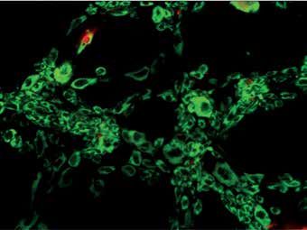
Tezcan Caliskan, Hande Akalan, Ibrahim Yilmaz, Numan Karaarslan, Duygu Yasar Sirin, Hanefi Ozbek

to normalize the target gene expression. To ob-                        Accordingly, the expression of the CHAD, IL-1B,
tain the comparative results, reference (calibrator)                MMP-13, and MMP-7 genes remained unchanged
samples were used, and RQ values were calculat-                     on day 10, while the COMP expression decreased
ed using the 2–ΔΔCq method [10, 11].                                0.4-fold and MMP-19 expression increased 0.4-
                                                                    fold. On day 20, COMP and IL-1B were expressed
     Statistical analysis                                           at levels similar to the control group. However,
                                                                    CHAD expression was 3.3-fold higher, MMP-13 ex-
   The statistical analysis were performed using
                                                                    pression was 36.3-fold higher, MMP-19 expression
SPSS (version 18.0) software, and the data were
                                                                    was 0.7-fold higher, and MMP-7 expression was
evaluated at the 95% confidence interval. De-
                                                                    4.5-fold higher compared with the control (Fig-
scriptive statistics were presented as the mean
                                                                    ure 3). Rivaroxaban application versus expression
± standard deviation. An analysis of variance
                                                                    of genetic markers (CHAD, COMP, IL-1B, MMP-13,
test was performed to determine whether there
                                                                    MMP-19, and MMP-7) and duration of application
were significant differences in the mean across
                                                                    were statistically evaluated. The change in gene
the groups. When differences across groups were
                                                                    expression was statistically significant (p < 0.05)
observed, Tukey’s honestly significant difference
                                                                    (Table I).
post-hoc test was used for multiple pairwise com-
parisons. The α significance value was < 0.05.                      Discussion
Results                                                                A complex set of anabolic and catabolic reac-
                                                                    tions provides continuity to the matrix structure
     Cell morphology, viability, and proliferation                  of intervertebral disc tissue. Matrix-degrading
     remained unchanged in AF/NPC cultures                          enzymes, such as COMP, a member of the disin-
     after rivaroxaban application according to                     tegrin and metalloprotease with thrombospondin
     inverted microscopy, AO/PI staining, and                       motifs (ADAMTS) family, are thought to play a key
     MTT analysis                                                   role during these reactions. MMPs and COMP are
   Morphological evaluation of AF/NPC cultures                      known to cause a decrease in the expression of
demonstrated that the application of rivaroxaban                    both aggrecan and type II collagen genes and in the
for 10 to 20 days did not affect cell morphology                    overall synthesis of aggrecan and collagen as a re-
or proliferation. When inverted microscopy images                   sult of the induction of proinflammatory cytokines,
were examined, the number of cells and the amount                   which are involved in the matrix construction and
of proliferation were similar in both experimental                  destruction cycle in the nucleus pulposus [23].
and control groups. Therefore, ECM formation was                       The absence of lymphatic vessels in the inter-
observed to be weaker in the group treated with ri-                 vertebral disc tissue leads to an increase in the
varoxaban (Figures 1 A–E). AO/PI staining showed                    density of matrix destructive factors and their
that cells from both experimental and control                       effects. In addition to this adverse condition, it
groups continued to proliferate within 10–20 days,                  is known that many drugs that are administered
and no apoptotic cell death was observed as a result                orally or parenterally accumulate in the synovial
of rivaroxaban administration (Figures 1 F–J).                      fluid [9–14, 24, 25]. Because the outer layer of the
   Similarly, with AO/PI staining results, when cell                synovial tissue is thicker, the drugs and/or nutri-
viability was evaluated using the MTT analysis, it                  ents diffuse from the hyaluronan and synovial tis-
was confirmed that rivaroxaban at the adminis-                      sues to the body fluids. They then pass through
tered dose did not suppress cell viability or prolif-               pores in the hyaline membranes located at the
eration (Figure 2). The data obtained from the MTT                  intervertebral disc space and reach the interverte-
analysis were statistically significant (p < 0.05).                 bral disc tissue cells [9–14].
                                                                       The aim of the present study was to investigate
                                                                    the effects of rivaroxaban, a novel oral Xa inhib-
     Gene expression profiles were changed due
                                                                    itor, on primary cell cultures isolated from intact
     to rivaroxaban administration
                                                                    human intervertebral disc tissue. Rivaroxaban is
   To evaluate the gene expression of CHAD, COMP,                   used for the prevention of the risk of early in-hos-
IL-1B, MMP-13, MMP-19, and MMP-7 genes, pre-                        pital death due to acute stroke [4] and for recur-
test cultures without rivaroxaban application (day 0)               rent symptomatic or fatal venous thromboembo-
were used as reference samples. The gene ex-                        lism in patients with acute pulmonary embolism.
pression level in these AF/NPC cultures was des-                    Rivaroxaban is frequently prescribed by many cli-
ignated as 100% (RQ = 1). The expression levels                     nicians, including neurosurgeons, as it is markedly
of CHAD, COMP, IL-1B, MMP-13, MMP-19, and                           effective for a long time, and it does not require
MMP-7 genes on days 10 and 20 were determined                       a constant dosing regimen or routine laboratory
as a fold increase or decrease compared with the                    monitoring [26–29]. In addition to the advantages
control group on the same days.                                     of the drug, however, it has serious side effects.

4                                                                                                          Arch Med Sci
The effects of rivaroxaban, an oral anticoagulant, on human IVD primary cultures
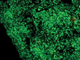
A F
0 d Control
B G
10 d Rivaroxaban
C H
10 d Control
D I
20 d Rivaroxaban
E J
20 d Control
Figure 1. Microscopy of nucleus pulposus and annulus fibrosus cell cultures: A–E – represent inverted light micros-
copy (first column) under 20× magnification, F–J – represent fluorescent microscopy after AO/PI staining (second
column) under 10× magnification. A and F are micrographs of control group cells after first passage incubated for
0 days. B, G, D, and I are rivaroxaban-treated cultures for 10 and 20 days, respectively. C, H, E, and J are micrographs
of non-treated control group cultures at 10 and 20 days, respectively
Arch Med Sci5Tezcan Caliskan, Hande Akalan, Ibrahim Yilmaz, Numan Karaarslan, Duygu Yasar Sirin, Hanefi Ozbek
40
35
0.6
30
0.5 25
Optical density (OD)
0.4 20
15
0.3
10
0.2 5
0
0.1 CHAD COMP IL-1b MMP-13 MMP-19 MMP-7
0 day reference sample
0
0d 10 d 10 d 20 d 20 d 10 days fold change rivaroxaban vs. control
Control Control Rivaroxaban Control Rivaroxaban 20 days fold change rivaroxaban vs. control
Figure 2. Results of the MTT analysis performed through Figure 3. Fold changes in the expression of CHAD,
the spectrophotometric methodology at 570 nm COMP, IL-1b, MMP-13, MMP-19, and MMP-7 genes
on days 10 and 20 compared with the control
group on the same days
Table I. Relationship between incubation period and gene expression in the rivaroxaban-treated samples
Source Adj. SS Adj. MS F-value P-value*
Rivaroxaban 22.850 22.8500 21.54 < 0.001
Markers 95.889 31.9631 30.14 < 0.001
Times 35.971 17.9855 16.96 < 0.001
Rivaroxaban vs. markers 24.041 8.0138 7.56 < 0.001
Markers vs. times 51.210 8.5349 8.05 < 0.001
Adj. SS – adjusted sum of squares; Adj. MS – adjusted mean square; *analysis of variance test (ANOVA, p < 0.05).
Previous studies suggested that the use of rivar- male osteoblastic cell line SaOS2. They treated the
oxaban might exacerbate intracranial hemorrhage cells with different concentrations of rivaroxaban
[30] in patients with mild traumatic brain injuries for 24 h and conducted analyses to determine DNA
and thereby might cause serious and fatal compli- synthesis and creatine kinase- and alkaline phos-
cations, such as intracerebral hematoma, intracra- phatase-specific activities. They then tested bone
nial hemorrhage, and nontraumatic spinal subdu- mineralization on day 21 [20]. They reported that
ral hematoma [31–34]. rivaroxaban (0.01–50 μg/ml) inhibited up to 60%
However, no previous study has focused on the of the DNA synthesis of the cells and that it inhibit-
positive or negative effects of this pharmacologi- ed dose-dependent creatine kinase-specific activi-
cal agent on intervertebral disc tissue. Therefore, ty when it was applied in the same concentrations.
the findings of the present study would contribute They also reported that rivaroxaban inhibited
to the literature. dose-dependent alkaline phosphatase-specific ac-
Dhar et al. [35] investigated the effects of ri- tivity up to 30% [20]. They concluded that rivar-
varoxaban, which is a prodrug and direct Xa in- oxaban inhibited the first stage of bone formation
hibitor, and dabigatran, which is a non-prodrug but did not affect later stages [20].
and direct factor IIa inhibitor, on α-smooth muscle Commercial cell lines or animal tissues have
actin (αSMA) using hepatic stellate cells-LX2 cells. been used in some studies [20, 35, 36]. Howev-
They concluded that the early inhibition of coagu- er, commercial cell lines contain a single type of
lation using rivaroxaban, which is an FXa inhibitor, cell and do not have original phenotypic and ge-
significantly reduced TAA-induced murine liver fi- notypic characteristics. Therefore, the results ob-
brosis [35]. tained in previous studies using a commercial cell
Hashikata et al. [36] investigated the effects of line may be misleading [9–14, 24, 25]. It is well
rivaroxaban on cell migration, proliferation, and known that human and animal tissues differ in
differentiation in cardiac fibroblast cells, which sensitivity. Thus, the results obtained from studies
play a central role in cardiac fibrosis. They report- on live mammals may differ from those obtained
ed that rivaroxaban inhibited Ang II-induced cell in human studies; therefore, such results may be
proliferation and migration and that it blocked misleading [24, 25, 37, 38].
various inflammatory signal pathways [36]. In the present study, we did not use animal
Gigi et al. [20] investigated the direct effects of tissues or commercial cell lines. Instead, we pre-
rivaroxaban on bone biology using the human fe- pared human primary cell cultures from intact and
6 Arch Med SciThe effects of rivaroxaban, an oral anticoagulant, on human IVD primary cultures
healthy human intervertebral disc tissue, and we ic process can be accelerated, and more intensive
evaluated the effects of rivaroxaban on these cul- catabolic effects can be observed.
tures. Our findings contribute to the value of the In addition to these findings, it was determined
present study for the relevant literature. that the expression level of CHAD, an NP-specif-
Our results showed that the expression of ic marker, did not change on the 10th day and
CHAD, IL-1B, MMP-13, and MMP-7 genes remained increased on the 20th day. These data indicate
unchanged on day 10, whereas COMP expression that rivaroxaban does not particularly affect NP
decreased and MMP-19 expression increased. On cell proliferation. Similarly, the fact that IL-1b ex-
day 20, COMP and IL-1B were expressed at levels pression remained unchanged at days 10 and 20
similar to the control group. The expression lev- suggests that the administration does not have
els of CHAD, MMP-13, MMP-19, and MMP-7 were a negative effect in terms of inflammation.
higher compared with the control (p < 0.05). The In conclusion, the results suggest that it is likely
results which were obtained following the statis- that recently released rivaroxaban can reduce ma-
tical evaluations of both proliferation and gene trix synthesis in disc tissue. Therefore, clinicians
expression were markedly significant (p < 0.05). should not forget that rivaroxaban may suppress
Disc degeneration is defined as progressive tissue healing due to drug-use duration. Although
changes that affect the spine and which develop changes in a determined group of genes cannot
due to the natural aging process and multiple oth- always explain the prognosis alone, it should al-
er factors [39]. The leading causes of the degen- ways be carefully evaluated considering genet-
eration of intervertebral disc tissue are degener- ic instability that may be caused by changes in
ative changes related to drug use [9–14, 24, 25]. gene expression level. Rivaroxaban does not have
MMPs are important proteolytic enzymes that are any acute toxic effects, but when the application
members of the endopeptidase family, which can time of rivaroxaban is kept to 20 days or more,
degrade ECM components. The ECM plays a cru- increasing MMP expression can negatively affect
cial role in maintaining cell viability, cell migration, the healing process.
and regulation of the interrelationships among
cells [40]. In our study, it was determined that 10- Conflict of interest
day rivaroxaban administration did not change
MMP-13 and MMP-7 expression, but increased The authors declare no conflict of interest.
MMP-19. However, when the application period
increased to 20 days, an increase was observed in
References
the amount of all MMPs studied. No toxic effect of
1. Ofek F, Barchel D, Perets N, et al. International normal-
rivaroxaban was detected in our treated cultures,
ized ratio as a screening test for assessment of anti-
but it was observed that culture development was coagulant activity for patients treated with rivaroxaban
weaker than the control group, especially in terms or apixaban: a pilot study. Front Pharmacol 2019; 10:
of adhesion and ECM formation. With this finding, 1177.
although it is not possible to claim that MMP in- 2. Li X, Zuo C, Lu W, et al. Evaluation of remote pharma-
crease alone will slow down the clinical recovery cist-led outpatient service for geriatric patients on ri-
process, it shows that the duration of drug use varoxaban for nonvalvular atrial fibrillation during the
COVID-19 pandemic. Front Pharmacol 2020; 11: 1275.
should be carefully evaluated.
3. Hartmann S, Biliouris K, Lesko LJ, Nowak-Göttl U,
Several studies have reported that members of Trame MN. Quantitative systems pharmacology mod-
the MMP and ADAMTS families of enzymes are el-based predictions of clinical endpoints to optimize
prone to damaging cartilage tissues and carti- Warfarin and Rivaroxaban anti-thrombosis therapy.
lage-like tissues [41]. It is well known that COMP is Front Pharmacol 2020; 11: 1041.
a member of the ADAMTS family [40]. It has been 4. Hart RG, Sharma M, Mundl H, et al. Rivaroxaban for
reported in the literature that inflammatory cyto- stroke prevention after embolic stroke of undetermined
kines stimulate ADAMTS-5 and increase COMP tis- source. N Engl J Med 2018; 378: 2191-201.
5. Hurtado-Navarro I, García-Sempere A, Rodríguez-Ber-
sue damage more than MMPs do [42–44]. In our
nal C, Santa-Ana-Tellez Y, Peiró S, Sanfélix-Gimeno G. Es-
study, it was determined that rivaroxaban applica- timating adherence based on prescription or dispensa-
tion decreased COMP expression on day 10, but tion information: impact on thresholds and outcomes.
the expression was normalized on day 20. In this A real-world study with atrial fibrillation patients treat-
context, it can be argued that in terms of COMP ed with oral anticoagulants in Spain. Front Pharmacol
gene expression, AF/NP tissue can resist the appli- 2018; 9: 1353.
cation and will not cause any tissue damage over 6. Naito T, Hayashi H, Kashiwada T, et al. Pulmonary em-
bolism and deep vein thrombosis in eosinophilic gran-
COMP increase. In addition to this increase, drugs
ulomatosis with polyangiitis successfully treated with
accumulated in the tissues increase the effects of rivaroxaban. Respir Med Case Rep 2018; 25: 33-5.
the matrix destructive factors on intervertebral 7. Piotrowski R, Zaborska B, Pilichowska-Paszkiet E, Siko-
disc tissues that are already aliphatic, thereby ra-Frąc M, Baran J, Kułakowski P. RIVAroxaban TWICE
exacerbating catabolic effects. Thus, the catabol- daily for lysis of thrombus in the left atrial append-
Arch Med Sci7Tezcan Caliskan, Hande Akalan, Ibrahim Yilmaz, Numan Karaarslan, Duygu Yasar Sirin, Hanefi Ozbek
age in patients with non-valvular atrial fibrillation: 24. Gumustas SA, Yilmaz İ, Isyar M, et al. Assessing the neg-
the RIVA-TWICE study. Arch Med Sci 2019; 16: 289-96. ative impact of phenyl alkanoic acid derivative, a fre-
8. Koscielny J, Beyer-Westendorf J, von Heymann C, et al. quently prescribed drug for the suppression of pain and
Risk of bleeding and haemorrhagic complication with inflammation, on the differentiation and proliferation of
rivaroxaban: periprocedural management of haemosta- chondrocytes. J Orthop Surg Res 2016; 11: 70.
sis. Hamostaseologie 2012; 32: 287-93. 25. Gumustas F, Yilmaz I, Sirin DY, et al. Chondrocyte prolif-
9. Caliskan T, Sirin DY, Karaarslan N, et al. Effects of etaner- eration, viability and differentiation is declined follow-
cept, a tumor necrosis factor receptor fusion protein, on ing administration of methylphenidate utilized for the
primary cell cultures prepared from intact human inter- treatment of attention-deficit/hyperactivity disorder.
vertebral disc tissue. Exp Ther Med 2019; 18: 69-76. Hum Exp Toxicol 2017; 36: 981-92.
10. Karaarslan N, Yilmaz I, Sirin DY, et al. Pregabalin treat- 26. Chunhong L, Jin L, Qin Z, Guohui X. Anticoagulant effects
ment for neuropathic pain may damage intervertebral of rivaroxaban after surgical fixation of spinal fracture.
disc tissue. Exp Ther Med 2018;16: 1259-65. Pak J Pharm Sci 2018; 31: 1131-5.
11. Kaya YE, Karaarslan N, Sirin DY, Ozbek H, Kaplan N, Yil- 27. Naito T, Hayashi H, Kashiwada T, et al. Pulmonary em-
maz I. Investigation of the effects of methylphenidate, bolism and deep vein thrombosis in eosinophilic gran-
an amphetamine derivative, on intervertebral disc tis- ulomatosis with polyangiitis successfully treated with
sue cell cultures and matrix structures. Turk Neurosurg rivaroxaban. Respir Med Case Rep 2018; 25: 33-5.
2019; 29: 734-42. 28. Wu CT, Chen B, Wang JW, Yen SH, Huang CC. Plasma
12. Akgun FS, Sirin DY, Yilmaz I, et al. Investigation of the D-dimer is not useful in the prediction of deep vein
effect of dipyrone on cells isolated from intervertebral thrombosis after total knee arthroplasty in patients us-
disc tissue. Exp Ther Med 2019; 18: 216-24. ing rivaroxaban for thromboprophylaxis. J Orthop Surg
13. Kaplan N, Karaarslan N, Yilmaz I, et al. Are intervertebral Res 2018; 13: 173.
disc tissue cells damaged when attempting to prevent 29. Bhatnagar UB, Rezkalla J, Sethi P, Stys A. Successful res-
thrombus formation using dabigatran, a new oral anti- olution of a large left ventricular thrombus with rivarox-
coagulant? Turk Neurosurg 2019; 29: 470-7. aban therapy after acute myocardial infarction. S D Med
14. Karaarslan N, Yilmaz I, Ozbek H, et al. Are specific gene 2018; 71: 62-3.
expressions of extracellular matrix and nucleus pul- 30. Beynon C, Potzy A, Sakowitz OW, Unterberg AW. Rivar-
posus affected by primary cell cultures prepared from oxaban and intracranial haemorrhage after mild trau-
intact or degenerative intervertebral disc tissues? Turk matic brain injury: a dangerous combination? Clin Neu-
Neurosurg 2019; 29: 43-52. rol Neurosurg 2015; 136: 73-8.
15. Chen FH, Herndon ME, Patel N, Hecht JT, Tuan RS, Law- 31. Tao J, Bukanova EN, Akhtar S. Safety of 4-factor pro-
ler J. Interaction of cartilage oligomeric matrix protein/ thrombin complex concentrate (4F-PCC) for emergent re-
thrombospondin 5 with aggrecan. J Biol Chem 2007; versal of factor Xa inhibitors. J Intensive Care 2018; 6: 34.
282: 24591-8. 32. Wasserlauf G, Grandi SM, Filion KB, Eisenberg MJ.
16. Veras MA, McCann MR, Tenn NA, Séguin CA. Transcrip- Meta-analysis of rivaroxaban and bleeding risk.
tional profiling of the murine intervertebral disc and Am J Cardiol 2013;112: 454-60.
age-associated changes in the nucleus pulposus. Con- 33. Baschera D, Oberle J, Grubhofer F, Schmid SL. Perioper-
nect Tissue Res 2020; 61: 63-81. ative use of anticoagulant and platelet-inhibiting med-
17. Mao SH, Qian BP, Shi B, Zhu ZZ, Qiu Y. Quantitative eval- ications for elective spine surgery: results of a nation-
uation of the relationship between COMP promoter wide survey. J Neurol Surg A Cent Eur Neurosurg 2018;
methylation and the susceptibility and curve progres- 79: 398-407.
sion of adolescent idiopathic scoliosis. Eur Spine J 2018; 34. Kawabori M, Niiya Y, Iwasaki M, et al. Characteristics
27: 272-7. of symptomatic intracerebral hemorrhage in patient
18. Goode AP, Marshall SW, Kraus VB, et al. Association be- receiving direct oral anticoagulants: comparison with
tween serum and urine biomarkers and lumbar spine warfarin. J Stroke Cerebrovasc Dis 2018; 27: 1338-42.
individual radiographic features: the Johnston County 35. Dhar A, Sadiq F, Anstee QM, Levene AP, Goldin RD,
Osteoarthritis Project. Osteoarthritis Cartilage 2012; 20: Thursz MR. Thrombin and factor Xa link the coagulation
1286-93. system with liver fibrosis. BMC Gastroenterol 2018; 18: 60.
19. Kwon WK, Moon HJ, Kwon TH, Park YK, Kim JH. the role 36. Hashikata T, Yamaoka-Tojo M, Namba S, et al. Rivarox-
of hypoxia in angiogenesis and extracellular matrix reg- aban Inhibits Angiotensin II-induced activation in cul-
ulation of intervertebral disc cells during inflammatory tured mouse cardiac fibroblasts through the modulation
reactions. Neurosurgery 2017; 81: 867-75. of NF-κB pathway. Int Heart J 2015; 56: 544-50.
20. Gigi R, Salai M, Dolkart O, et al. The effects of direct 37. Yilmaz I, Karaarslan N, Ozbek H. Practical performance
factor Xa inhibitor (Rivaroxaban) on the human osteo- of hippocampal tissue resection in rats in pharmacomo-
blastic cell line SaOS2. Connect Tissue Res 2012; 53: lecular research. Turk Neurosurg 2021; 31: 112-8.
446-50. 38. Yilmaz I, Karaarslan N, Ozbek H. Pharmaco-molecular
21. Xu N, Wang L, Fu S, Jiang B. Resveratrol is cytotoxic and assessment of the effects of anandamide and its antag-
acts synergistically with NF-κB inhibition in osteosarco- onists on hippocampal tissue in Wistar albino rats. Eur
ma MG-63 cells. Arch Med Sci 2020; 17: 166-76. Rev Med Pharmacol Sci 2020; 24: 11871-82.
22. Sirin DY, Karaarslan N. Evaluation of the effects of pre- 39. Hu X, Cao D, Wang Z. Metformin alleviates interver-
gabalin on chondrocyte proliferation and CHAD, HIF-1α, tebral disc degeneration by upregulating MMP-1 ex-
and COL2A1 gene expression. Arch Med Sci 2018; 14: pression via the KDM6A/SOX9/miR-202-3p/MMP-1
1340-7. signaling pathway. Arch Med Sci 2021. doi:10.5114/
23. Séguin CA, Pilliar RM, Roughley PJ, Kandel RA. Tumor aoms/117427.
necrosis factor-alpha modulates matrix production and 40. Liang Y, Fu Y, Qi R, et al. Cartilage oligomeric matrix pro-
catabolism in nucleus pulposus tissue. Spine 2005; 30: tein is a natural inhibitor of thrombin. Blood 2015; 126:
1940-8. 905-14.
8 Arch Med SciThe effects of rivaroxaban, an oral anticoagulant, on human IVD primary cultures
41. Min N, Ma J, Shi L, et al. miR-223 promotes cartilage
differentiation of bone marrow-derived mesenchy-
mal stem cells and protects against osteoarthritis by
suppressing NLRP-3 expression. Arch Med Sci 2020.
doi:10.5114/aoms.2020.100640.
42. Yang CY, Chanalaris A, Troeberg L. ADAMTS and ADAM
metalloproteinases in osteoarthritis: looking beyond
the ‘usual suspects’. Osteoarthritis Cartilage 2017; 25:
1000-9.
43. Cal S, Obaya AJ, Llamazares M, Garabaya C, Quesada V,
Lopez-Otin C. Cloning, expression analysis, and struc-
tural characterization of seven novel human ADAMTSs,
a family of metalloproteinases with disintegrin and
thrombospondin-1 domains. Gene 2002; 283: 49-62.
44. Roberts S, Caterson B, Menage J, Evans EH, Jaffray DC,
Eisenstein SM. Matrix metalloproteinases and aggreca-
nase: their role in disorders of the human intervertebral
disc. Spine 2000; 25: 3005-13.
Arch Med Sci9You can also read


