Further Evaluation and Validation of the VETSCAN IMAGYST: In-Clinic Feline and Canine Fecal Parasite Detection System Integrated with a Deep ...
←
→
Page content transcription
If your browser does not render page correctly, please read the page content below
Further Evaluation and Validation of the VETSCAN
IMAGYST: In-Clinic Feline and Canine Fecal Parasite
Detection System Integrated with a Deep Learning
Algorithm
Yoko Nagamori ( yoko.nagamori@zoetis.com )
Northeastern State University
Ruth Hall Sedlak
Zoetis Inc
Andrew DeRosa
Zoetis Inc
Aleah Pullins
Zoetis Inc
Travis Cree
Zoetis Inc
Michael Loenser
Zoetis Inc
Benjamin S. Larson
Techcyte
Richard Boyd Smith
Techcyte
Cory Penn
Zoetis Inc
Richard Goldstein
Zoetis Inc
Research
Keywords: deep learning, fecal egg identi cation, veterinary parasitology diagnostic, Ancylostoma, Toxocara cati,
Cystoisospora, Giardia, oocyst, cyst
DOI: https://doi.org/10.21203/rs.3.rs-52837/v1
License: This work is licensed under a Creative Commons Attribution 4.0 International License. Read Full
License
Page 1/18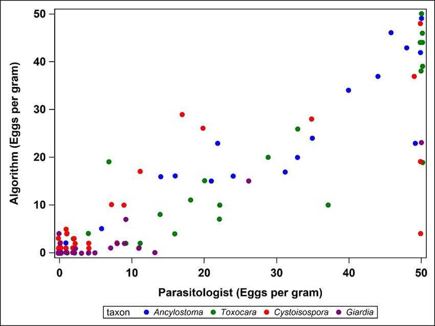
Abstract
Background: Fecal examinations in pet cats and dogs are key components of routine veterinary practice; however, the
accuracy is in uenced by diagnostic methodologies and the experience level of personnel performing the tests. The
VETSCAN IMAGYST system was developed to provide simpler and easier fecal examinations which are less
in uenced by examiners’ skills. This system consists of three components: a sample preparation device, an
automated microscope scanner, and an analysis software. The objectives of this study were to qualitatively evaluate
the performance of the VETSCAN IMAGYST system on feline parasites (Ancylostoma and Toxocara cati) and
protozoan parasites (Cystoisospora and Giardia) and to assess and compare the performance of the VETSCAN
IMAGYST centrifugal otation method to reference centrifugal and passive otation methods.

Methods: To evaluate the diagnostic performance of the scanning and algorithmic components of the VETSCAN
IMAGYST system, fecal slides were prepared by the VETSCAN IMAGYST centrifugal otation technique with pre-
screened fecal samples collected from dogs and cats and examined by both an algorithm and parasitologist. To
assess the performance of the VETSCAN IMAGYST centrifugal otation technique, diagnostic sensitivity and
speci city were calculated and compared to conventional otation techniques.

Results: The performance of the VETSCAN IMAGYST algorithm closely correlated with evaluations by parasitologists,
with sensitivity of 75.8-100% and speci city of 93.1-100% across the targeted parasites. For samples with 50 eggs or
less per slide, Pearson’s correlation coe cients (r) ranged from 0.84 to 0.97 across the targeted parasites. The
VETSCAN IMAGYST centrifugal otation method correlated well with the conventional centrifugal otation method
across the targeted parasites: sensitivity of 65.7-100% and speci city of 97.6-100%. Similar results were observed for
the conventional passive otation method compared to the conventional centrifugal otation method; sensitivity of
56.4-91.7% and speci city of 99.4-100%.

Conclusions: The VETSCAN IMAGYST scanning and algorithmic systems with the VETSCAN IMAGYST fecal
preparation technique demonstrated a similar qualitative performance to the results of parasitologists’ examinations
with conventional fecal otation techniques. Given the deep learning nature of the VETSCAN IMAGYST system, its
performance is expected to improve over time, enabling it to be utilized in veterinary clinics to perform fecal
examinations accurately and e ciently.

Background
Domestic cat ownership in the United States has been increasing over time as overall pet ownership has been
increasing. In 2017–2018, up to 47.1 million households in the U.S. owned at least one cat, with 76% of owners
considering their cats to be family members [1]. However, cats are generally less medicalized or served by
veterinarians compared to dogs [2] even though studies have reported that gastrointestinal parasitism in cats is
common [3–8]. A recent retrospective study demonstrated a signi cant increase in the prevalence of intestinal
parasitic infections in client-owned cats during a 12-year period (19.0% in 2007 vs. 32.5% in 2018; P < 0.0001) [3]. A
majority of people acquire cats from shelter or rescue and adopt stray cats [1] although those cat populations harbor a
higher prevalence of parasitic infections, ranging between 31.8% and 67.2% [4–8]. The most prevalent nematodes
identi ed in cats are Ancylostoma and Toxocara cati [3–9]. The main species of feline Ancylostoma found in North
America are A. tubaeforme and A. braziliense. Since feline Ancylostoma and T. cati are zoonotic parasites [10–17], it is
important to conduct routine fecal examinations and treat cats as needed to maintain wellness of cats and owners.

The most commonly detected protozoan parasites in domestic cats and dogs in North America are Cystoisospora
(formally known as Isospora) and Giardia [3–7, 18–24]. Studies evaluating feline fecal samples have reported the
                                                        Page 2/18
prevalence of Cystoisospora and Giardia to range between 3.8–26.0% and 1.2–9.9%, respectively, with the highest
prevalence in shelter cats [3–7, 18, 19, 21, 24]. The prevalence of Cystoisospora and Giardia in dogs has been reported
to range between 0.5–10.4% and 3.3–13%, respectively [4, 19–23, 25–30]. Cystoisospora oocysts and Giardia
cysts/trophozoites are small in size and can be challenging to detect by microscopy, especially when fecal samples
contain a low number of oocysts, cysts, or trophozoites. In addition, intermittent shedding of Giardia
cysts/trophozoites makes it more di cult to diagnose giardiasis [31–35].
Fecal examinations in dogs and cats detecting evidence of gastrointestinal parasitism are widely recognized as an
important component of routine veterinary cares, both for maintaining pet health and for identifying parasites of
zoonotic signi cance [36]. However, the accuracy and usefulness of fecal testing can be in uenced by many factors,
such as differences in methodology and the experience level of personnel conducting the tests [19, 36, 37]. Of note,
Giardia has been identi ed as a particularly di cult parasite to diagnose by coprology in veterinary practices [36].
Computer-based algorithms to identify parasites in fecal examinations have recently been developed and introduced
with the aim to improve the accuracy and consistency of diagnosing parasitic diseases in dogs and cats [38, 39].
The novel VETSCAN IMAGYST system evaluates fecal samples for evidence of parasitic infections in an organized
and uncomplicated fashion that does not depend greatly on examiners’ level of experience. This system has
demonstrated the ability to reliably detect four targeted parasites (Ancylostoma, Toxocara, Trichuris, Taeniidae) in
fecal samples of 84 dogs and 16 cats, closely correlating with the examination by a parasitologist [Pearson correlation
coe cients (r) 0.83–0.99 across the four targeted parasites] [39]. The VETSCAN IMAGYST system consists of three
main components: a sample preparation device, an automated commercially available microscopic scanner, and a
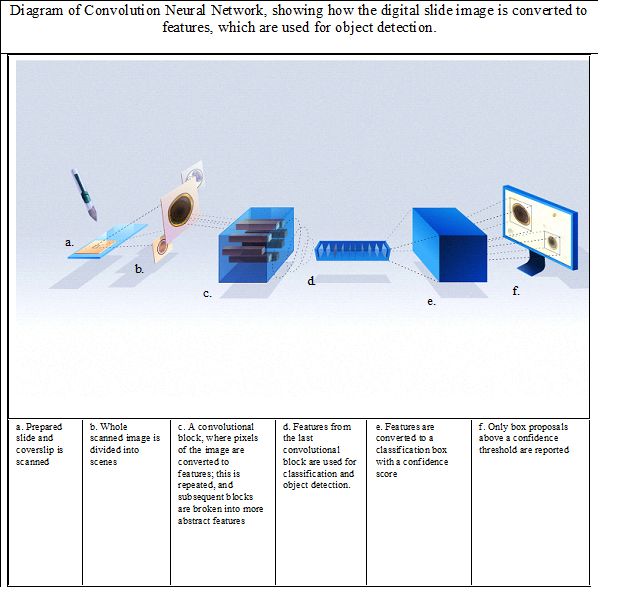
data analysis process using deep neural networks (Fig. 1). The VETSCAN IMAGYST system applies a deep learning
object detection algorithm which uses a convolutional neural network (CNN) to identify convolutional layers that
automatically learn the most discriminating features between classes (Fig. 2). The algorithm assigns a probability
score to each image recognized within each sample preparation as being an egg belonging to a parasite genus/group
that the software has been previously trained/calibrated to recognize. A mature algorithm model, which can perceive
and distinguish morphology of individual parasite eggs and non-parasite objects on fecal otation slides, is developed
through a training process that utilizes samples characterized by an expert. Once the model is su ciently mature, it is
tested against several evaluation datasets to ensure that the nal most promising model will be generalizable to other
similar domains.
The current study had three objectives. First, to qualitatively evaluate the diagnostic performance of the VETSCAN
IMAGYST system to recover and correctly identify eggs from the common feline nematodes, Ancylostoma and
Toxocara cati, in feces of naturally infected cats, compared to manual identi cation by an expert with conventional
sample preparation methods. Second, to qualitatively evaluate the diagnostic performance of the VETSCAN IMAGYST
system to recover and correctly identify protozoan parasites, Cystoisospora oocysts and Giardia cysts, in cat and dog
feces, compared to manual identi cation by an expert with commonly used sample preparation methods. Third, to
compare the diagnostic performance of the VETSCAN IMAGYST centrifugal preparation method to standard
centrifugal and passive otation methods for the four targeted parasites, feline Ancylostoma, Toxocara cati,
Cystoisospora, and Giardia.
Methods
Fecal sample collections
Page 3/18Fecal samples from client-owned and shelter cats and dogs submitted to the veterinary parasitology diagnostic
laboratory at Oklahoma Animal Disease Diagnostic Laboratory of Oklahoma State University were processed for eggs,
oocysts, and cysts recovery using a Wisconsin fecal egg counting method previously described [31, 39]. A total of 100
fecal samples were collected weighing a minimum of 8 grams con rmed positive for at least one of the targeted
parasites: 1) feline Ancylostoma, 2) Toxocara cati, 3) feline Cystoisospora, and 4) canine and feline Giardia, as well as
10 feline samples negative for any targeted parasites. Samples were included as positives or negatives for the relevant
analyses; samples containing multiple targeted parasites were counted as a positive for more than one analyses, and
some samples were counted as a negative for more than one analyses. For the Cystoisospora analysis, 100 additional
fecal samples from a second collection separated by several months, with the same sample acquisition criteria
including canine samples, were included. Table 1 summarizes the number of screened positive and negative samples
assessed for each evaluation. Different reference methods were used for the VETSCAN IMAGYST algorithm and
sample preparation assessments; therefore, a total number of positives and negatives varies between evaluations. To
maintain morphological integrity of the diagnostic forms of parasite elements, all samples were preserved at 4 °C until
the study. For 38 samples, a low level of formalin solution was added as a xative to ensure integrity of the parasite
elements.
Table 1. Summary of fecal samples included in this study by targeted parasites based upon initial characterization.
Targeted Species/N Flotation solution utilized for Sample Numbers
Parasite sample preparation
Positive Negative Total Mixed
Infectionsa
Ancylostoma Feline/80 Sugar 20 60 80 21
Toxocara cati Feline/80 Sugar 34 46 80 21
Cystoisospora Feline/96 Sugar 36 164 200 72
Canine/104
Giardia Feline/80 Zinc sulfate/Sugarb 39 61 100 28
Canine/20
a
Mixed infections are samples that were positive for more than 1 targeted parasites.
bZinc sulfate solution was used for Giardia samples; sugar solution was used for the rest of the samples.
VETSCAN IMAGYST scanner and algorithm
The VETSCAN IMAGYST system has previously been described [39]. Brie y, slides with fecal samples were read by the
Motic EasyScan One® digital slide scanner (Motic, Kowloon Bay, Hong Kong) that provided 40x effective resolution.
The scanned images were then automatically uploaded and analyzed in the cloud with an updated deep learning
objective detection algorithm, version 3033, which was developed based on You Only Look Once (YOLOv3) model
(Techcyte Inc., Lindon, UT, US) [40]. After localization and classi cation of the objects of interest were performed, the
resulting images were then available for viewing on the VETSCAN IMAGYST platform, and a downloadable PDF report
was generated (Figure 1). The VETSCAN IMAGYST analysis software also has the quantitative ability of counting the
parasites of interest; however, this capability was not evaluated in the current study since our objectives were to
Page 4/18qualitatively evaluate the diagnostic performance of the VETSCAN IMAGYST system recovering and correctly
identifying targeted parasites.
VETSCAN IMAGYST algorithm assessment
To qualitatively evaluate the ability of the scanning and algorithmic components of the VETSCAN IMAGYST system in
identifying diagnostic forms of the targeted parasites, slides were prepared with the VETSCAN IMAGYST centrifugal
otation technique on each pre-screened fecal sample as described previously by Nagamori et al. [39]. Brie y, the
VETSCAN IMAGYST fecal preparation device, which was speci cally redesigned and produced from Apacor mini
Parasep® SF (Apacor LTD., Wokingham, United Kingdom), was utilized to perform the VETSCAN IMAGYST centrifugal
method. The VETSCAN IMAGYST fecal preparation device consists of two tubes: the sample tube with a sample
scoop, which ts for approximately 1 gram of feces, and the collection tube containing two types of otation solution,
zinc sulfate solution (speci c gravity, 1.18) for Giardia samples and sugar solution (speci c gravity, 1.25) for the rest
of the samples. The sample tube and collection tube were rmly screwed together; the tube was shaken and
centrifuged for 2 minutes at 300-500 relative centrifugal force (rcf). Following centrifugation, the sample tube was
unscrewed from the collection tube. The transfer loop was used to collect diagnostic forms of parasites from the top
of the oatation solution and transferred to a microscope slide (Figure 3). The coverslip marked with the IMAGYST
label was placed such that the IMAGYST label could be read correctly, ensuring that the marked coverslip was
accurately placed on the slide. The slide was placed in a slide tray. The slide tray was then inserted into an automated
microscopic scanner, and the resulting digitally scanned image was uploaded to a cloud-based server for analysis and
result generation.
The VETSCAN IMAGYST fecal preparation was performed by two technicians. After the slides were analyzed by the
VETSCAN IMAGYST system, two parasitologists and one technician, who were well experienced in diagnostic
parasitology, evaluated each slide microscopically using 100x, 200x, and 400x magni cations. Identi cation of the
parasites was based on morphology of eggs, oocysts, and cysts [31]. During the manual examinations by the
parasitologists and technician, quantitative egg counts on the entire slide, up to 50 eggs, oocysts, or cysts per each
targeted parasite, which is considered as 50 eggs per gram (EPG), oocysts per gram (OPG), or cysts per gram (CPG),
were performed, and estimated for high burdens and recorded as “medium” for slides with 51-250 EPG, or “high” for
slides with >250 EPG. The VETSCAN IMAGYST algorithm, on the other hand, provided counts regardless of egg
burden. Results from the VETSCAN IMAGYST algorithm and microscopic examinations by experts were compared and
statistically analyzed. For Giardia fecal samples, an additional analysis on the subset of samples with >10 CPG was
conducted.
Sample preparation method assessment
Performance (sensitivity and speci city) of the VETSCAN IMAGYST centrifugal technique was assessed by comparing
it to the conventional centrifugal otation and passive otation methods using visual microscopy. With each fecal
sample, slides were prepared by 3 different sample preparation techniques, 1) VETSCAN IMAGYST centrifugal
otation, 2) conventional centrifugal otation, and 3) conventional passive otation. Two technicians prepared the
slides for the VETSCAN IMAGYST centrifugal otation technique as described above. The process for the reference
centrifugal otation technique has previously been described [39]. Brie y, 33% zinc sulfate solution (speci c gravity,
1.18) for Giardia samples and Sheather’s sugar solution (speci c gravity, 1.25) for the rest of the samples were used to
suspend 1 gram of feces, which was then strained and placed in a 15 mL centrifuge tube. After otation solution was
added to the tube until a convex meniscus was formed, a coverslip was added, and the samples were centrifuged in a
Centra CL2 centrifuge (Thermo Fisher Scienti c; Waltham, Massachusetts, USA) at approximately 440 rcf for 5
Page 5/18minutes. After removing the coverslip and placing it on a glass slide, a microscopic examination was performed [31].
OVASSAY® Plus Kit Fecal Flotation Device with 33% zinc sulfate solution (speci c gravity, 1.18) (Zoetis; Parsippany
Troy Hills, New Jersey, United States) was used to perform the passive fecal otation test, following manufacturer’s
instruction [41]. Slides were microscopically examined by three experts as described above. The diagnostic
performance of the VETSCAN IMAGYST centrifugal otation method was compared with both reference methods,
conventional centrifugal and OVASSAY® passive otation.
Statistical Analysis
Samples with an observation of any parasite elements, and in a separate analysis of >10 CPG for Giardia, were
considered positive. Two by two (2X2) tables were constructed, and sensitivity and speci city with 95% Jeffreys’
con dence interval estimates were calculated. Human microscopy estimates were quantitative at 50
eggs/oocysts/cysts or less on each slide; therefore, Pearson correlation coe cients (r) were created and calculated
limiting all methods at 50 eggs/oocysts/cysts or less. Results greater than 50 were removed from the correlation
coe cients. SAS version 9.4M6 (SAS Institute Inc., Cary, North Carolina, United States) was used for statistical
analysis.
Results
Algorithm performance
Comparisons between the results generated by the VETSCAN IMAGYST system and experts were analyzed to assess
the performance of the VETSCAN IMAGYST scanner and algorithm to identify eggs, oocysts, and cysts of targeted
parasites (Table 2, Fig. 4). The diagnostic sensitivity and speci city of the comparisons ranged between 75.8–100%
and 93.1–100%, respectively, for the four targeted parasites. Twelve of 33 (36%) Giardia samples contained a very low
number of cysts, ≤ 10 CPG. Diagnostic sensitivity of Giardia was dramatically increased to 95.2% (95% CI: 79.8–
99.5%) after the Giardia samples with ≤ 10 CPG were excluded. Quantitative comparisons for samples with ≤ 50 EPG,
OPG, and CPG were performed; the VETSCAN IMAGYST diagnostic result closely matched that of the experts for each
targeted parasite, with Pearson’s correlation coe cients (r) of 0.97 (feline Ancylostoma), 0.93 (Toxocara cati), 0.84
(Cystoisospora), and 0.93 (Giardia).
Page 6/18Table 2
Algorithm Performance Analysis: Diagnostic sensitivity and speci city comparing results reported by an expert
(reference) versus by the VETSCAN IMAGYST scanner and algorithm on samples prepared with the VETSCAN
IMAGYST centrifugal otation sample preparation method.
Feline Toxocara cati Cystoisospora Giardia
Ancylostoma
True positive 19 32 26 25
False positive 1 0 12 2
True negative 60 46 161 65
False negative 0 2 1 8
Total 80 80 200 100
Sensitivity (95% con dence 100 (87.8–100) 94.1 (82.4– 96.3 (84.0-99.6) 75.8 (59.4–
interval) 98.8) 87.8)a
Speci city (95% con dence 98.4 (92.6–99.8) 100 (94.7–100) 93.1 (88.6– 97.0 (90.8–99.4)
interval) 96.2)
a36% (12/33) of Giardia samples contained ≤ 10 cysts per gram (CPG); diagnostic sensitivity of Giardia samples
with > 10 CPG increased to 95.2% (95% CI:79.8–99.5%).
Sample preparation performance
For the comparison of the performance between the VETSCAN IMAGYST centrifugal otation (test method) and the
conventional centrifugal otation (reference method), the diagnostic sensitivity and speci city of the comparison
ranged between 65.7–100% and 97.6–100%, respectively, across the four targeted parasites (Table 3). For the sample
preparation performance of the OVASSAY® passive otation method compared to the conventional centrifugation
otation method, the diagnostic sensitivity and speci city ranged between 56.4–91.7% and 99.4–100%, respectively
(Table 3).
Page 7/18Table 3
Sample Preparation Analysis: Diagnostic sensitivity and speci city comparing the VETSCAN IMAGYST centrifugal
otation (VS-Ic) and OVASSAY® passive otation (O-pf) methods versus conventional centrifugal otation method
(reference); all read by an expert.
Feline Ancylostoma Toxocara cati Cystoisosporaa Giardia
N = 80 N = 80 N = 200 N = 100
VS-Ic O-pf VS-Ic O-pf VS-Ic O-pf VS-Ic O-pf
True positive 19 17 34 33 23 22 32 22
False positive 0 0 0 0 4 1 1 0
True negative 61 61 44 44 161 164 60 61
False negative 0 2 2 3 12 13 7 17
Sensitivity 100 89.5 94.4 91.7% 65.7 62.9 82.1 56.4
(95% con dence (87.8– (70.3– (83.4– (79.4– (49.2– (46.3– (68.0- (40.9–
interval) 100) 97.7) 98.8) 97.6) 79.7) 77.3) 91.6) 71.1)
Speci city 100 100 100 100 97.6 99.4 98.4 100
(95% con dence (96.0- (96.0- (94.5– (94.5– (94.3– (97.2– (92.6– (96.0-
interval) 100) 100) 100) 100) 99.2) 99.9) 99.8) 100)
a Cystoisosporaresults are a combination of 2 independent studies with the same sample acquisition criteria.
Cystoisospora species included C. canis, C. ohioiensis, C. felis and C. rivolta.
Discussion
This is the second study demonstrating the VETSCAN IMAGYST system integrated with a deep learning object
detection algorithm successfully recognized and identi ed diagnostic forms of gastrointestinal parasites in dogs and
cats on fecal otation slides scanned by an automated microscope. Whereas our previous study evaluated the
VETSCAN IMAGYST system in detecting eggs of Ancylostoma, Toxocara, Trichuris, and Taeniidae in 84 canine and 16
feline fecal samples [39], this study assessed the ability of the system to detect eggs of feline nematodes,
Ancylostoma and Toxocara cati, and protozoan parasites, Cystoisospora oocysts and Giardia cysts, in 104 canine and
96 feline fecal samples, making it the more comprehensive analysis for this novel system.
Although both domestic dogs (Canis lupus familiaris) and cats (Felis catus) belong to the same order of Carnivora,
dogs are classi ed into the Canoidea superfamily and cats are classi ed into the Feloidea superfamily [42, 43]. Diets
of Canoids can be variable from herbivorous to omnivorous; however, all Feloids require a strict carnivorous diet [43].
Therefore, a high-protein diet is essential for domestic cats to obtain some nutritional requirements of animal-origin,
such as taurine, arachidonic acid, and vitamin A [43]. Due to the differences in dietary requirements, feline fecal
samples commonly contain a large amount of fat and become soft, sticky and clay-like in consistency, which often
makes it more di cult, or sometimes impossible, to read fecal slides since more debris oat with fats, especially when
a viscous sugar solution is used as the centrifugal otation technique. Modi cation of fecal otation procedures,
involving an initial water wash where the supernatant is discarded after the initial spin and the sediment resuspended
with a otation solution to remove excess fat, mucus, and debris (double centrifugal fecal otation technique), can be
used [31]. In the current study, the VETSCAN IMAGYST centrifugal otation method recovered parasite elements from
feline feces, and the VETSCAN IMAGYST scanner and algorithm successfully captured and identi ed targeted
parasites (Fig. 5).
Page 8/18This is the rst report demonstrating the ability of the VETSCAN IMAGYST system to recover and accurately detect
protozoan parasites, Cystoisospora oocysts and Giardia cysts. Although coccidiosis is generally considered a self-
limiting infection in mature dogs and cats due to rapid development of immunity [11], Cystoisospora is an ubiquitous
and important pathogen in puppies and kittens, often resulting in diarrhea, abdominal pain, anorexia, bloody diarrhea,
anemia, and even mortality in severe cases [10, 31, 44]. Since Cystoisospora undergoes fast replication in the
pathogenic intestinal stage and excrete a high number of oocysts in feces, causing environmental contamination, it is
critical and recommended to conduct a fecal examination with centrifugation for puppies and kittens at least four
times during the rst year of life and treat at an early stage of infection [44, 45]. Two different species of
Cystoisospora are commonly diagnosed in dogs and cats: C. canis and C. ohioensis in dogs and C. felis and C. rivolta
in cats. Oocysts of C. canis and C. felis are slightly bigger, approximately 38–51 × 27–39 µm in size, than those of C.
ohioensis and C. rivolta, approximately 17–27 × 15–24 µm in size [31]. Due to the smaller size of coccidian oocysts
compared to helminth eggs, Cystoisospora can be easily overlooked especially when a low number of oocysts are on a
fecal slide and an inaccurate microscopic focus is used for examination. The VETSCAN IMAGYST system correctly
identi ed oocysts of all four Cystoisospora species in canine and feline fecal samples and successfully reported as
Cystoisospora (coccidia) (Fig. 5).
The diagnostic sensitivity and speci city comparing the results of Giardia samples reported by an expert versus by the
VETSCAN IMAGYST scanner and algorithm were 75.8% and 97.0%, respectively (Table 2). As previously discussed, a
common challenge for many object detection algorithm models is to precisely localize and distinguish small objects
such as Giardia cysts [39]. With the nature of a deep learning algorithm, however, performance continues improving
with further trainings. It is important to note that the diagnostic sensitivity dramatically increased to 95.2% by
removing the 12 of 33 Giardia samples with ≤ 10 CPG, which is extremely demanding to detect by a visual
microscopic examination, from the analysis. Additionally, examinations and counts of CPG on these Giardia slides
were carefully performed by a well-trained diagnostic parasitologist with no time limit, which most likely resulted in a
much higher diagnostic performance compared to performance by technicians in daily veterinary practices.
Detection of Giardia cysts and trophozoites by fecal examinations is generally the most sensitive microscopic
technique, and ample trainings and experiences are required to con dently diagnose giardiasis [31]. In addition to its
small size and transparency in color, it is challenging to identify Giardia infection by fecal examinations because
Giardia cysts and trophozoites are intermittently shed in feces, and multiple fecal examinations may be necessary to
rule out the infection. Fresh fecal samples, preferably within 30 minutes of defecation, are often required to detect
motile trophozoites; and Giardia cysts and trophozoites are fairly fragile and easily distorted by otation solutions [31].
A 33% zinc sulfate solution (speci c gravity, 1.18) is preferred and recommended for the detection of Giardia cysts as
other otation solutions can rapidly cause osmotic damages in Giardia cysts, which increases the di culty in
perceiving them on fecal slides [31, 34, 46, 47]. During the current study, the VETSCAN IMAGYST system effectively
recognized and identi ed both intact and collapsed Giardia cysts (Fig. 5). Testing for Giardia is recommended not only
in symptomatic dogs and cats, but also in new dogs and cats being introduced to home with other pets free of
infection, as many Giardia infections can be asymptomatic [31, 35, 44]. Since there is no perfect otation solution to
recover all different types of parasites [31], it is important to consider advantages and disadvantage of each solution
when selecting one for general use. Some experts recommend performing two centrifugal otation tests using both
Sheather’s sugar and 33% zinc sulfate solutions to have a broader range of gastrointestinal parasite detections. In
cases where Giardia is suspected, an analysis with the sugar otation solution should also be performed on the fecal
sample to assess for other parasites.
Detection of Giardia is also possible with Giardia-speci c coproantigen detection assays [33, 44]. However, when not
used in conjunction with a traditional microscopic technique, antigen testing may provide a false positive result in an
Page 9/18animal that is no longer infected with Giardia due to persistent antigen excretion for several weeks or even months
after the parasite elimination [48, 49]. Given the shortcomings of current in-house diagnostic methods for Giardia,
utilizing a deep learning algorithm platform, such as the VETSCAN IMAGYST system, could provide clinicians with an
excellent additional or alternative diagnostic tool to help identify Giardia cases that would otherwise be missed.
Evaluation of the performance of the VETSCAN IMAGYST centrifugal otation sample preparation method was limited
due to modest numbers of true positives for the four targeted parasites in this study and the inherent subsampling
variability in non-homogenous fecal samples that has been well documented in previous publications [50].
Kochanowski observed a wide range of coe cients of variation between 31–254% in Toxocara and Trichuris samples
with a low number of egg counts of ≤ 50 EPG. Despite these limitations, the performance of the VETSCAN IMAGYST
centrifugal otation method was comparable to a conventional centrifugal otation method, with diagnostic
sensitivity and speci city of the comparisons ranging between 65.7–100% and 97.6–100%, respectively, across the
four targeted parasites (Table 3). Additionally, one potential modi cation considered for the VETSCAN IMAGYST
centrifugal otation method to increase the diagnostic sensitivity is to lengthen the duration of centrifugation time.
Previous studies reported that egg recoveries with centrifugation for 4 or 5 minutes at 264 x g improved signi cantly
compared to 1 and 3 minutes at the same speed, although no change was observed in egg recoveries by extending
times to 10 or 20 minutes [37, 51].
As shown in Table 3, the diagnostic sensitivity and speci city of the VETSCAN IMAGYST centrifugal technique slightly
surpassed those of the OVASSAY® passive otation method. Despite the fact that centrifugation signi cantly
increases the sensitivity of fecal examinations, passive otation continues to be the most commonly used technique
in veterinary private practices due to its convenience [19, 31, 36, 46, 52–54]. Given that the VETSCAN IMAGYST
system reliably recovers and detects parasite elements in fecal samples, does not depend considerably on the
experience level of examiners, and has previously been shown to provide results in around 10 minutes with the
VETSCAN IMAGYST centrifugal otation method [39], it has the potential to replace the conventional passive otation
method performed in veterinary practices.
The most distinctive and unique feature of the VETSCAN IMAGYST system is its deep learning object detection
algorithm. To the best of our knowledge, the VETSCAN IMAGYST system is the only automated diagnostic system that
is integrated with a deep learning object detection algorithm and applied in veterinary medicine. Compared to shallow
learning systems, which do not have any structural information on the function to be learned, deep learning algorithms
exploit the advantage of locality at each level of the layered hierarchy, enabling the system to ignore the aspects that
make computer vision brittle [55, 56]. Layered hierarchy also facilitates the system to continuously adapt to new data
and apply it to new output classes with fewer examples [56, 57]. The deep learning characteristic, along with the
YOLOv3 object detection model, which incorporates localization and classi cation features, results in a decrease of
background errors and high agreement between the VETSCAN IMAGYST system and experts’ examinations. Another
bene t of this system is the ability to store images and reports on a secure, cloud-based server system, allowing easy
sharing by parasitologists as well as members of the veterinary and academic communities for patient care, research,
and teaching.
The current study did not evaluate the usability of the system; however, our previous analysis showed that the
VETSCAN IMAGYST system with the VETSCAN IMAGYST centrifugal otation method could prepare examination
results in around 10 minutes, which is comparable to conventional fecal otation tests. This time frame included the
time to prepare the sample, approximately 3.5 minutes with 2-minute centrifugal incubation time, and the time to scan
the images, approximately 6–7 minutes [39]. Data from the current study add to the body of evidence demonstrating
the performance of the VETSCAN IMAGYST system in detecting intestinal parasite elements recovered from fecal
Page 10/18samples. In addition to identifying the protozoan parasites, Cystoisospora and Giardia, results from our current and
previous studies show the system’s reliable performance in detecting four different genera/group of gastrointestinal
parasites (Ancylostoma, Toxocara, Trichuris, Taeniidae) in dogs and cats [39]. With further traning, the VETSCAN
IMAGYST system will have the ability to identify other parasites. The quantitative capability of the VESTSCAN
IMAGYST system is currently under development. It is predicted that the algorithm will be able to perform a fecal egg
counting test.
Conclusions
The VETSCAN IMAGYST system effectively recovered and identi ed feline Ancylostoma eggs, Toxocara cati eggs,
Cystoisospora oocysts, and Giardia cysts in feline and canine fecal samples. Given the deep learning nature of the
VETSCAN IMAGYST system, its performance is expected to improve over time, enabling it to be utilized in veterinary
clinics to perform fecal examinations accurately and e ciently.
Declarations
Ethics approval and consent to participate
Not applicable.
Consent for publication
Not applicable.
Availability of data and materials
All data generated or analyzed during this study are included in this published article.
Competing interests
RHS, AD, AP, ML, CP, RG, and TC are current employees of Zoetis. BL and RBS are current employees of Techcyte. YN
was an employee at Oklahoma State University at the time this study was conducted; YN is currently an employee of
Zoetis.
Funding
The studies reported here were funded by Zoetis, Kalamazoo, MI, USA.
Authors’ contributions
YN performed fecal egg analysis and was a major contributor in writing the manuscript. RHS, AD, AP, ML, CP, RG
developed experimental protocols, analyzed data, and were contributors in writing the manuscript. TC performed data
summary and analysis. BL and RBS contributed to technical execution and manuscript review. All authors read and
approved the nal manuscript.
Acknowledgements
Authors thank Dr. Ruth Scimeca and Emily Looper for helping to prepare fecal samples and read otation slides.
Authors also thank all personnel who were involved in collecting fecal samples from dogs and cats for this study. Litto
Communications, LLC assisted in preparing the manuscript.
Page 11/18References
1. Animal Sheltering/the Humane Society of the United States, 2020. Pets by the Numbers.
https://www.animalsheltering.org/page/pets-by-the-numbers. Accessed 15 Apr 2020.
2. American Veterinary Medical Association (AVMA). Pet Ownership Stable, Veterinary Care Variable.
https://www.avma.org/javma-news/2019-01-15/pet-ownership-stable-veterinary-care-variable Accessed 16 July
2020.
3. Nagamori Y, Payton ME, Looper E, Apple H, Johnson EM. Retrospective survey of parasitism identi ed in feces of
client-owned cats in North America from 2007 through 2018. Vet Parasitol. 2020;277:109008; doi:
10.1016/j.vetpar.2019.109008. https://www.ncbi.nlm.nih.gov/pubmed/31841945.
4. Villeneuve A, Polley L, Jenkins E, Schurer J, Gilleard J, Kutz S, et al. Parasite prevalence in fecal samples from
shelter dogs and cats across the Canadian provinces. Parasit Vectors. 2015;8:281; doi: 10.1186/s13071-015-
0870-x. https://www.ncbi.nlm.nih.gov/pubmed/26013283.
5. Hoggard KR, Jarriel DM, Bevelock TJ, Verocai GG. Prevalence survey of gastrointestinal and respiratory parasites
of shelter cats in northeastern Georgia, USA. Vet Parasitol Reg Stud Reports. 2019;16:100270; doi:
10.1016/j.vprsr.2019.100270. https://www.ncbi.nlm.nih.gov/pubmed/31027603.
6. Lucio-Forster A, Bowman DD. Prevalence of fecal-borne parasites detected by centrifugal otation in feline
samples from two shelters in upstate New York. J Feline Med Surg. 2011;13 4:300-3; doi:
10.1016/j.jfms.2010.12.013. https://www.ncbi.nlm.nih.gov/pubmed/21334238.
7. Nagamori Y, Payton ME, Duncan-Decocq R, Johnson EM. Fecal survey of parasites in free-roaming cats in
northcentral Oklahoma, United States. Vet Parasitol Reg Stud Reports. 2018;14:50-3; doi:
10.1016/j.vprsr.2018.08.008. https://www.ncbi.nlm.nih.gov/pubmed/31014736.
8. Little S, Adolph C, Downie K, Snider T, Reichard M. High Prevalence of Covert Infection With Gastrointestinal
Helminths in Cats. J Am Anim Hosp Assoc. 2015;51 6:359-64; doi: 10.5326/JAAHA-MS-6221.
https://www.ncbi.nlm.nih.gov/pubmed/26535453.
9. Loftin CM, Donnett UB, Schneider LG, Varela-Stokes AS. Prevalence of endoparasites in northern Mississippi
shelter cats. Vet Parasitol Reg Stud Reports. 2019;18:100322; doi: 10.1016/j.vprsr.2019.100322.
https://www.ncbi.nlm.nih.gov/pubmed/31796170.
10. Taylor MA, Coop RL, Wall RL. Parasites of dogs and cats. In: Taylor MA, Coop RL, Wall RL, editors. Veterinary
parasitology. West Sussex: John Wiley and Sons; 2016. p. 599-677.
11. Bowman DD. Helminths. In: Bowman DD, editor. Georgis’ parasitology for veterinarians. St. Louis, Missouri:
Elsevier Health Sciences; 2014. p. 122-240.
12. Companion Animal Parasite Council (CAPC), 2020. CAPC Guidelines: Hookworms.
https://capcvet.org/guidelines/hookworms/. Accessed 21 Apr 2020.
13. Fisher M. Toxocara cati: an underestimated zoonotic agent. Trends Parasitol. 2003;19 4:167-70; doi:
10.1016/s1471-4922(03)00027-8. https://www.ncbi.nlm.nih.gov/pubmed/12689646.
14. Furtado LFV, Dias LTO, Rodrigues TO, Silva VJD, Oliveira V, Rabelo EML. Egg genotyping reveals the possibility of
patent Ancylostoma caninum infection in human intestine. Sci Rep. 2020;10 1:3006; doi: 10.1038/s41598-020-
59874-8. https://www.ncbi.nlm.nih.gov/pubmed/32080267.
15. Loukas A, Hotez PJ, Diemert D, Yazdanbakhsh M, McCarthy JS, Correa-Oliveira R, et al. Hookworm infection. Nat
Rev Dis Primers. 2016;2:16088; doi: 10.1038/nrdp.2016.88. https://www.ncbi.nlm.nih.gov/pubmed/27929101.
16. Lee AC, Schantz PM, Kazacos KR, Montgomery SP, Bowman DD. Epidemiologic and zoonotic aspects of ascarid
infections in dogs and cats. Trends Parasitol. 2010;26 4:155-61; doi: 10.1016/j.pt.2010.01.002.
Page 12/18https://www.ncbi.nlm.nih.gov/pubmed/20172762.
17. Macpherson CN. The epidemiology and public health importance of toxocariasis: a zoonosis of global
importance. Int J Parasitol. 2013;43 12-13:999-1008; doi: 10.1016/j.ijpara.2013.07.004.
https://www.ncbi.nlm.nih.gov/pubmed/23954435.
18. Wyrosdick HM, Chapman A, Martinez J, Schaefer JJ. Parasite prevalence survey in shelter cats in Citrus County,
Florida. Vet Parasitol Reg Stud Reports. 2017;10:20-4; doi: 10.1016/j.vprsr.2017.07.002.
https://www.ncbi.nlm.nih.gov/pubmed/31014592.
19. Gates MC, Nolan TJ. Endoparasite prevalence and recurrence across different age groups of dogs and cats. Vet
Parasitol. 2009;166 1-2:153-8; doi: 10.1016/j.vetpar.2009.07.041.
https://www.ncbi.nlm.nih.gov/pubmed/19709815.
20. Nagamori Y, Payton ME, Looper E, Apple H, Johnson EM. Retrospective survey of endoparasitism identi ed in
feces of client-owned dogs in North America from 2007 through 2018. Veterinary Parasitology. 2020;282:109137;
doi: 10.1016/j.vetpar.2020.109137. https://www.sciencedirect.com/science/article/abs/pii/S0304401720301175
21. Olson ME, Leonard NJ, Strout J. Prevalence and diagnosis of Giardia infection in dogs and cats using a fecal
antigen test and fecal smear. Can Vet J. 2010;51 6:640-2. https://www.ncbi.nlm.nih.gov/pubmed/20808578.
22. Jacobs SR, Forrester CP, Yang J. A survey of the prevalence of Giardia in dogs presented to Canadian veterinary
practices. Can Vet J. 2001;42 1:45-6. https://www.ncbi.nlm.nih.gov/pubmed/11195521.
23. Joffe D, Van Niekerk D, Gagne F, Gilleard J, Kutz S, Lobingier R. The prevalence of intestinal parasites in dogs and
cats in Calgary, Alberta. Can Vet J. 2011;52 12:1323-8. https://www.ncbi.nlm.nih.gov/pubmed/22654137.
24. Hoopes JH, Polley L, Wagner B, Jenkins EJ. A retrospective investigation of feline gastrointestinal parasites in
western Canada. Can Vet J. 2013;54 4:359-62. https://www.ncbi.nlm.nih.gov/pubmed/24082162.
25. Jordan HE, Mullins ST, Stebbins ME. Endoparasitism in dogs: 21,583 cases (1981-1990). J Am Vet Med Assoc.
1993;203 4:547-9. https://www.ncbi.nlm.nih.gov/pubmed/8407513.
26. Nolan TJ, Smith G. Time series analysis of the prevalence of endoparasitic infections in cats and dogs presented
to a veterinary teaching hospital. Vet Parasitol. 1995;59 2:87-96; doi: 10.1016/0304-4017(94)00742-u.
https://www.ncbi.nlm.nih.gov/pubmed/7483240.
27. Little SE, Johnson EM, Lewis D, Jaklitsch RP, Payton ME, Blagburn BL, et al. Prevalence of intestinal parasites in
pet dogs in the United States. Vet Parasitol. 2009;166 1-2:144-52; doi: 10.1016/j.vetpar.2009.07.044.
https://www.ncbi.nlm.nih.gov/pubmed/19716659.
28. Blagburn BL, Lindsay DS, Vaughan JL, Rippey NS, Wright JC, Lynn RC, et al. Prevalence of canine parasites based
on fecal otation. The Compendium on continuing education for the practicing veterinarian (USA).
https://www.researchgate.net/publication/279867899_Prevalence_of_canine_parasites_based_on_fecal_ otation.
29. Stafford K, Kollasch TM, Duncan KT, Horr S, Goddu T, Heinz-Loomer C, et al. Detection of gastrointestinal
parasitism at recreational canine sites in the USA: the DOGPARCS study. Parasit Vectors. 2020;13 1:275; doi:
10.1186/s13071-020-04147-6.
30. Companion Animal Parasite Council (CAPC), 2020. Giardia Prevalence.
https://capcvet.org/maps/#2020/all/giardia/dog/united-states/ Accessed 19 July 2020.
31. Zajac AM, Conboy GA. Veterinary Clinical Parasitology, 8th ed. Blackwell Publishing, Ames, IA, USA. 2012.
32. Little SE, Lindsay DS. Laboratory Diagnosis of Protozoal Infections. In: Sykes JE, Greene CE, editors. Infectious
Diseases of the Dog and Cat, 4th Edition. St. Louis, Missouri: Elsevier; 2012. p. 711-715.
33. McHardy IH, Wu M, Shimizu-Cohen R, Couturier MR, Humphries RM. Detection of intestinal protozoa in the clinical
laboratory. J Clin Microbiol. 2014;52 3:712-20; doi: 10.1128/JCM.02877-13.
Page 13/18https://www.ncbi.nlm.nih.gov/pubmed/24197877.
34. Kirkpatrick CE. Feline giardiasis: a review. Journal of Small Animal Practice. 1986;27 2:69-80; doi: 10.1111/j.1748-
5827.1986.tb02124.x. https://onlinelibrary.wiley.com/doi/abs/10.1111/j.1748-5827.1986.tb02124.x.
35. Companion Animal Parasite Council (CAPC), 2020. CAPC Guidelines: Giardia.
https://capcvet.org/guidelines/giardia/. Accessed 20 Apr 2020.
36. Dryden MW, Payne PA, Ridley RK, Smith VE. Gastrointestinal parasites: The practice guide to accurate diagnosis
and treatment. Supplemental to compendium: Continuing education for veterinarians. 2006; 28:8.
37. Ballweber LR, Beugnet F, Marchiondo AA, Payne PA. American Association of Veterinary Parasitologists' review of
veterinary fecal otation methods and factors in uencing their accuracy and use--is there really one best
technique? Vet Parasitol. 2014;204 1-2:73-80; doi: 10.1016/j.vetpar.2014.05.009.
https://www.ncbi.nlm.nih.gov/pubmed/24893692.
38. Inacio SV, Ferreira Gomes J, Xavier Falcao A, Nagase Suzuki CT, Bertequini Nagata W, Nery Loiola SH, et al.
Automated Diagnosis of Canine Gastrointestinal Parasites Using Image Analysis. Pathogens. 2020;9 2; doi:
10.3390/pathogens9020139. https://www.ncbi.nlm.nih.gov/pubmed/32093178.
39. Nagamori Y, Hall Sedlak R, DeRosa A, Pullins A, Cree T, Loenser M, et al. Evaluation of the VETSCAN IMAGYST: an
in-clinic canine and feline fecal parasite detection system integrated with a deep learning algorithm. Parasit
Vectors. 2020;13 1:346; doi: 10.1186/s13071-020-04215-x. https://www.ncbi.nlm.nih.gov/pubmed/32653042.
40. Redmon J, Farhadi A. YOLOv3: an incremental improvement. arXiv preprint. 2018;1804.02767.
https://pjreddie.com/media/ les/papers/YOLOv3.pdf.
41. Zoetis, OVASSAY® Plus Kit Fecal otation Device, https://www.zoetisus.com/products/diagnostics/OVASSAY®-
plus-kit-fecal- otation-devices.aspx.
42. MacDonald ML, Rogers QR, Morris JG. Nutrition of the domestic cat, a mammalian carnivore. Annu Rev Nutr.
1984;4:521-62; doi: 10.1146/annurev.nu.04.070184.002513. https://www.ncbi.nlm.nih.gov/pubmed/6380542.
43. Salaun F, Blanchard G, Le Paih L, Roberti F, Niceron C. Impact of macronutrient composition and palatability in
wet diets on food selection in cats. J Anim Physiol Anim Nutr (Berl). 2017;101 2:320-8; doi: 10.1111/jpn.12542.
https://www.ncbi.nlm.nih.gov/pubmed/27278300.
44. European Scienti c Counsel Companion Animal Parasites (ESCCAP), 2018. Control of Intestinal Protozoa in Dogs
and Cats. https://www.esccap.org/uploads/docs/xnqpgri2_0701_ESCCAP_Guideline_GL6_v7_1p.pdf. Accessed
20 Apr 2020.
45. Companion Animal Parasite Council (CAPC), 2020. General Guidelines. https://capcvet.org/guidelines/general-
guidelines/ Accessed 27 July 2020.
46. Zajac AM, Johnson J, King SE. Evaluation of the importance of centrifugation as a component of zinc sulfate
fecal otation examinations. J Am Anim Hosp Assoc. 2002;38 3:221-4; doi: 10.5326/0380221.
https://www.ncbi.nlm.nih.gov/pubmed/12022406.
47. Kirkpatrick CE. Giardiasis. Vet Clin North Am Small Anim Pract. 1987;17 6:1377-87; doi: 10.1016/s0195-
5616(87)50007-9. https://www.ncbi.nlm.nih.gov/pubmed/3328394.
48. Rishniw M, Liotta J, Bellosa M, Bowman D, Simpson KW. Comparison of 4 Giardia diagnostic tests in diagnosis of
naturally acquired canine chronic subclinical giardiasis. J Vet Intern Med. 2010;24 2:293-7; doi: 10.1111/j.1939-
1676.2010.0475.x. https://www.ncbi.nlm.nih.gov/pubmed/20136713.
49. Garcia LS, Arrowood M, Kokoskin E, Paltridge GP, Pillai DR, Procop GW, et al. Laboratory Diagnosis of Parasites
from the Gastrointestinal Tract. Clin Microbiol Rev. 2018;31 1; doi: 10.1128/CMR.00025-17.
https://www.ncbi.nlm.nih.gov/pubmed/29142079.
Page 14/1850. Kochanowski M, Dabrowska J, Karamon J, Cencek T, Osinski Z. Analysis of the accuracy and precision of the
McMaster method in detection of the eggs of Toxocara and Trichuris species (Nematoda) in dog faeces. Folia
Parasitol (Praha). 2013;60 3:264-72; doi: 10.14411/fp.2013.030.
https://www.ncbi.nlm.nih.gov/pubmed/23951934.
51. Egwang TG, Slocombe JO. Evaluation of the Cornell-Wisconsin centrifugal otation technique for recovering
trichostrongylid eggs from bovine feces. Can J Comp Med. 1982;46 2:133-7.
https://www.ncbi.nlm.nih.gov/pubmed/7093809.
52. O'Grady MR, Slocombe JO. An investigation of variables in a fecal otation technique. Can J Comp Med. 1980;44
2:148-57. https://www.ncbi.nlm.nih.gov/pubmed/7190861.
53. Dryden MW, Payne PA, Ridley R, Smith V. Comparison of common fecal otation techniques for the recovery of
parasite eggs and oocysts. Vet Ther. 2005;6 1:15-28. https://www.ncbi.nlm.nih.gov/pubmed/15906267.
54. Broussard JD. Optimal fecal assessment. Clin Tech Small Anim Pract. 2003;18 4:218-30; doi: 10.1016/S1096-
2867(03)00076-8. https://www.ncbi.nlm.nih.gov/pubmed/14738202.
55. Krizhevsky A, Sutskever I, Hinton G. ImageNet classi cation with deep convolutional neural networks. In NeurIPS.
2012. https://papers.nips.cc/paper/4824-imagenet-classi cation-with-deep-convolutional-neural-networks.pdf
56. Poggio T, Mhaskar H, Rosasco L, Miranda B, Liao Q. Why and when can deep-but not shallow-networks avoid the
curse of dimensionality: A review. International Journal of Automation and Computing. 2017;14 5:503-19; doi:
10.1007/s11633-017-1054-2. https://doi.org/10.1007/s11633-017-1054-2.
57. Yosinski J, Clune J, Bengio Y, Lipson H. How transferable are features in deep neural networks? In Advances in
Neural Information Processing Systems. 2014. https://arxiv.org/pdf/1411.1792.pdf.
Figures
Figure 1
Page 15/18The VETSCAN IMAGYST system consists of three main components: 1) a sample preparation device, 2) digital
microscope scanner, and 3) data analysis and reporting.
Figure 2
Diagram of the VETSCAN IMAGYST algorithm’s Convolution Neural Network and object detection process.
Page 16/18Figure 3
VETSCAN IMAGYST sample preparation materials.
Figure 4
Algorithm Performance Analysis: Quantitative visualization of egg identi cation (EPG, x- and y-axes) by expert
parasitologist (x-axis) and VETSCAN IMAGYST scanner/algorithm (y-axis) for the prepared samples with less than 50
EPG with common VETSCAN IMAGYST centrifugal otation sample preparation.
Page 17/18Figure 5
Images of targeted parasites captured by the VETSCAN IMAGYST system.
Page 18/18You can also read