A Structured Approach to Skin and Soft Tissue Infections (SSTIs) in an Ambulatory Setting - MDPI
←
→
Page content transcription
If your browser does not render page correctly, please read the page content below
Review
A Structured Approach to Skin and Soft Tissue
Infections (SSTIs) in an Ambulatory Setting
Benjamin Silverberg
 Department of Emergency Medicine, West Virginia University, 1 Medical Center Drive, Box 9149,
 Morgantown, WV 26506, USA; benjamin.silverberg@hsc.wvu.edu

 Received: 15 December 2020; Accepted: 26 January 2021; Published: 1 February 2021

 Abstract: The skin is the largest, and arguably, the most vulnerable organ in the human body. Scratches
 and scrapes, bites and puncture wounds, impetigo and erysipelas—all these disruptions can lead to
 pain, swelling, and/or systemic symptoms. In this article, which is based on the Infectious Diseases
 Society of America’s 2014 guidelines and the World Society of Emergency Surgery and Surgical
 Infection Society of Europe’s 2018 consensus statement, a structured approach to skin and soft tissue
 infections (SSTIs) is reviewed, comparing treatment for suppurative and non-suppurative infections,
 and then discussing specific conditions commonly seen in Primary Care and Urgent Care facilities.

 Keywords: inflammation; purulence; drainage; necrosis; MRSA; immunodeficiency; trauma;
 antibiotics; resistance

1. Introduction
     Skin and soft tissue infections (SSTIs) result from a compromise of the skin’s defenses and microbial
invasion and interaction therein. Since SSTIs are usually caused by bacteria, most practice guidelines
do not mention viral, fungal, or parasitic etiologies. Trauma and surgery are two of the main ways
the skin barrier can be breached [1]. Primary SSTIs result from the invasion of otherwise healthy skin;
secondary SSTIs result from infection of already-damaged skin, such as from trauma or an underlying
disease [2]. Infections are often localized but they can also spread via the blood stream or lymphatic
flow [3].

2. Background
     In the United States, SSTIs account for more than 14 million outpatient visits annually [3,4]. About
three-fourths of cases of SSTIs are managed in an outpatient setting. However, since mild infections
are often self-limiting, many patients do not seek formal medical care [2,5]. As such, these numbers are
likely underestimates of the true incidence. SSTIs are a common reason for a trip to the Emergency
Department, with 59% of infections being attributed to methicillin-resistant Staphylococcus aureus
(MRSA) [3,6] and 11.5–60% of patients being admitted to the hospital for an expensive multi-day
stay [6]. Men are disproportionately affected: They account for 60–70% of SSTIs [5].

3. Discussion
     The skin—the largest organ in the body—protects the occupant with a physical barrier,
bacteriostatic sebaceous fluid, and normal skin flora [5]. This normal bacterial colonization is mainly
with aerobic Gram-positive cocci, but below the waist, due to a so-called “fecal veneer” stemming
from the anorectal region, Gram-negative species can also be found [5]. Additionally, the warm, dark,
moist regions in skin folds tend to have higher concentrations of bacteria [5]. In undamaged skin,
microflora tend to reside in the outer layers of the skin and skin structures. Anaerobic bacteria tend to
be associated with long-standing skin ulcers, impaired immune function, and recent antibiotic use [5].

Clin. Pract. 2021, 11, 65–74; doi:10.3390/clinpract11010011                   www.mdpi.com/journal/clinpract
Clin. Pract. 2021, 11, FOR PEER REVIEW 2
Clin. Pract. 2021, 11 66
microflora tend to reside in the outer layers of the skin and skin structures. Anaerobic bacteria tend
to be associated with long-standing skin ulcers, impaired immune function, and recent antibiotic use
[5]. Aerobic Gram-positive cocci—specifically Staphylococcus aureus and streptococcal species
—areAerobic
the most likely cause ofcocci—specifically
Gram-positive SSTIs involving healthy skin. Beta-hemolytic
Staphylococcus streptococcispecies—are
aureus and streptococcal cause nearly
three-fourths
the most likelyofcause
cases of SSTIs
cellulitis [3,7]. S. healthy
involving aureus, by comparison,
skin. tends to
Beta-hemolytic cause morecause
streptococci purulent infections,
nearly three-
such as abscesses
fourths of cases of[2]. There is[3,7].
cellulitis a considerable
S. aureus, regional variation:
by comparison, Nearly
tends to cause S. aureus
36% ofmore colonies
purulent in North
infections,
America
such display methicillin
as abscesses [2]. There isresistance, compared
a considerable to 29.4%
regional in Latin
variation: America
Nearly 36%and
of S.22.8%
aureusincolonies
Europe [1].
in
Though
North simple infections
America are usuallyresistance,
display methicillin monomicrobial, complicated
compared to 29.4%infections
in Latin can be polymicrobial
America and 22.8% in or
monomicrobial, as well [2].
Europe [1]. Though simple infections are usually monomicrobial, complicated infections can be
polymicrobial or monomicrobial, as well [2].
3.1. Conceptual Frameworks for SSTIs
3.1. Conceptual
SSTIs wereFrameworks
originallyfor SSTIs
categorized by the US Food and Drug Administration (FDA) in 1998 as
“uncomplicated” or “complicated,”
SSTIs were originally categorized mainly
by the for US
the Food
purposeandofDrug
therapeutic drug studies
Administration (FDA)[8,9]. Though
in 1998 as
these definitions would later be revised, certain types of wounds (e.g., diabetic
“uncomplicated” or “complicated,” mainly for the purpose of therapeutic drug studies [8,9]. Though ulcers) were excluded.
In 2003,
these Eron et al.
definitions wouldproposed
later bea revised,
new way to stratify
certain typesSSTIs by severity.
of wounds Their four-tier
(e.g., diabetic system
ulcers) were ranged
excluded.
from a healthy-appearing patient through a febrile and ill-appearing one to an
In 2003, Eron et al. proposed a new way to stratify SSTIs by severity. Their four-tier system ranged overtly septic patient
with aa healthy-appearing
from life-threatening infection [1,3,5,10,11].
patient through Criticizing
a febrile it as ambiguous,
and ill-appearing one to anKiovertly
and Rotstein (2008)
septic patient
with a life-threatening infection [1,3,5,10,11]. Criticizing it as ambiguous, Ki and Rotstein (2008) signs,
tried to improve on this system by adding in objective criteria such as comorbidities, systemic tried
involvement
to improve on of the
thishead
systemand/or
by hands,
addingand involvement
in objective of more
criteria than
such as 9% of the body surface
comorbidities, systemicarea [5,8].
signs,
Various iterations of early warning scores (EWS), which focus primarily on
involvement of the head and/or hands, and involvement of more than 9% of the body surface area vital signs, have also been
used Various
[5,8]. for hospitalized
iterations patients
of early[8,11].
warning scores (EWS), which focus primarily on vital signs, have
Alternatively, SSTIs can
also been used for hospitalized patients be grouped
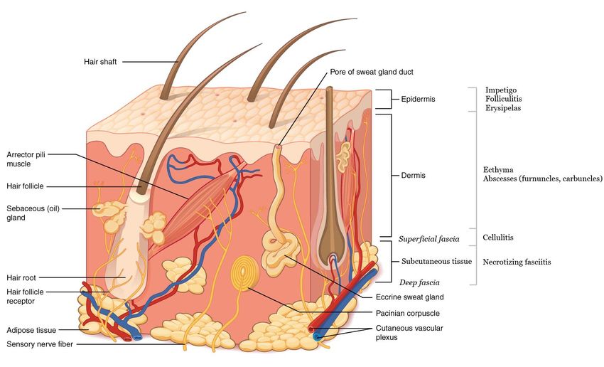
[8,11].by the anatomic tissue layers involved, as seen in
Figure 1 [1,12,13]. Cellulitis is typically
Alternatively, SSTIs can be grouped by in the
the anatomic
dermis and subcutaneous
tissue tissue,
layers involved, and necrotizing
as seen in Figure 1
infections most commonly affect the deep fascia, but can involve the dermis
[1,12,13]. Cellulitis is typically in the dermis and subcutaneous tissue, and necrotizing infections down throughmost to
the muscleaffect
commonly [1,4,9].the deep fascia, but can involve the dermis down through to the muscle [1,4,9].
Figure 1. Infectious processes by depth. Soft tissue structures and layers of the skin are also marked
Figure 1. Infectious
(adapted processes
from Figure by depth.and
5.2 of Anatomy Soft tissue structures
Physiology, andOpenStax
2013, via layers of College;
the skin licensed
are also marked
through
(adapted from Figure 5.2 of Anatomy and Physiology, 2013, via OpenStax College; licensed through
Creative Commons–https://commons.wikimedia.org/wiki/File:501_Structure_of_the_skin.jpg).
Creative Commons – https://commons.wikimedia.org/wiki/File:501_Structure_of_the_skin.jpg).
A third way to classify SSTIs is whether they are suppurative (pus-forming) or not [1,3,14].
Localized purulent
A third way tostaphylococcal
classify SSTIs infections
is whetherresult
they in
arethe formation of
suppurative furuncles (boils),
(pus-forming) or notcarbuncles
[1,3,14].
Localized purulent staphylococcal infections result in the formation of furuncles (boils), carbunclesClin. Pract. 2021, 11 67
(clusters of boils), or larger skin abscesses. Erysipelas, cellulitis, and necrotizing fasciitis are usually
non-purulent, but not always [2].
3.2. History of Present Illness (HPI)
There are often clues in the patient’s history suggesting the depth and severity of the SSTI. Such
indicators include: Was there a traumatic injury? If so, how did it damage the skin’s defenses? Does
the patient have any comorbidities? Diabetes mellitus, a risk factor for both MRSA and necrotizing
fasciitis (NF), can affect a person’s peripheral sensation and wound healing. Venous stasis also affects
the oxygenation of tissue, and edema can stretch and strain the skin itself. Surgery can impair the lymph
flow. Immunocompromise—due to an underlying condition or medical treatment—further weakens
the host’s defenses. Table 1 lists these and other risk factors for SSTIs [1,3–5,10,12].
Table 1. Risk factors for skin and soft tissue infections (SSTIs).
General Risk Factors Risk Factors for MRSA Infections
• Cardiopulmonary disease • Younger age
• Hepatorenal disease • Health care professionals
• Older age • Military personnel
• Debility • Dialysis
• Obesity • Long-term intravascular access
• Asplenia • Prolonged hospitalization
• Immunocompromise (e.g., HIV, chemotherapy)
• Peripheral arteriovenous insufficiency Risk factors for NF
• Peripheral neuropathy • Alcohol abuse
• Lymphedema • Poor nutrition
• Water exposure (salt or freshwater) • Sports participation
• Human/animal bites • Trauma
• Intravenous or subcutaneous drug use • Surgery
3.3. Physical Exam
SSTIs are usually heralded by the signs of inflammatory response—erythema, warmth,
tenderness/pain, and swelling—and other localized and systemic manifestations [5]. (Fluor, or
secretion, has been proposed as a fifth sign of inflammation, but funcio laesa, or loss of function, is
the most commonly added element to this framework. Loss of function results most directly from
pain and swelling). A patient presenting with just skin redness, for example, is more likely to have
a superficial, and therefore mild, infection. Systemic signs such as fever, hypotension, and tachycardiaClin. Pract. 2021, 11 68
suggest a deeper infection. Physical findings such as fluctuance, bullae, crepitus, purpura, and/or
necrosis suggest a more serious infection and/or one needing surgical intervention [10]. The rapid
progression of symptoms, lymphangitic spread, and, in particular, pain out of proportion to exam,
which may suggest tissue ischemia, are also worrisome [3,4]. Staphylococcal toxic shock syndrome
(TTS), for example, which may appear to be more superficial in terms of skin depth, is accompanied
by signs of shock [15]. Therefore, the entirety of the patient’s presentation must be considered when
ascertaining severity.
3.4. Imaging
Plain radiographs have limited utility in the diagnosis or treatment of simple SSTIs in an outpatient
clinic [3]. Ultrasound can be used to evaluate abscesses and fascial inflammation, which may be
suggested by fluctuance or crepitus on physical exam [5]. MRI, which is more sensitive, and CT can
also image fascial planes, but they typically would not change outpatient management. In more serious
cases, surgical intervention—if needed—should not be delayed for imaging [4,16].
3.5. Laboratory Studies
Simple, localized SSTIs typically do not need laboratory evaluation [4]. However, obtaining
a complete blood count, basic metabolic profile, and C-reactive protein may be appropriate if the patient
is likely to be transferred to a higher-level care. Wound cultures have a somewhat low yield, and it can
be challenging sometimes to distinguish between pathogenic bacteria and normal skin colonization.
Blood cultures also have a low yield, unless risk factors are present. Cultures may be more useful in
patients who are hospitalized and febrile, and/or with an underlying disease [4,14,17–21].
3.6. Principles of Treatment
It has been difficult to standardize the classification and, thereby treatment, of SSTIs. Antimicrobials
are typically initiated before a specific etiology is confirmed. Therefore, the selection of an appropriate
regimen is usually empiric, based on the bodily location and perceived severity of the infection. There
is a higher perceived risk of loss of function with infections involving the hands or head, for instance,
so the treatment of infections there should be aggressive. For SSTIs below the waist, pharmacotherapy
should cover Gram-negative and anaerobic bacteria. Pseudomonas aeruginosa is possible with a chronic,
non-healing ulcer [5].
As noted earlier, purulence—or absence thereof—is one fairly easy way to narrow the differential
diagnosis and hone the treatment plan. In each of these groupings, assess whether the infection is
mild, moderate, or severe. Though there is a debate in the literature as to whether IV/IM antibiotics
are truly superior to PO antibiotics in terms of efficacy (parenteral antibiotics at least seem to deliver
higher levels of the drug more rapidly, though it is unclear if outcomes are any better), the general
consensus is to use PO antibiotics for mild infections and IV/IM ones for severe infections [5]. Table 2
summarizes treatment modalities.
Table 2. Simplified management of SSTIs (adapted from [14]).
Purulent Non-Purulent
• Topical antibiotics
Mild • Incision and drainage (I & D)
• Oral antibiotics (cover GAS)
• I&D
• Parenteral antibiotics
Moderate/severe • Antibiotics (cover MRSA)
• Surgical debridement
• Surgical debridementClin. Pract. 2021, 11 69
The treatment for SSTIs attributed to MSSA or MRSA typically lasts 7–14 days, but it should be
individualized based on the patient’s clinical response [5,8,14]. Similarly, reevaluate the diagnosis
and treatment plan if the patient fails to show improvement after five days’ worth of antibiotics [5].
An appropriately-treated SSTI usually shows a reduction in inflammation and no further spreading
within 48–72 h of antibiotic administration [2,4]. Clindamycin is an important therapeutic option
to treat MSSA and MRSA in children [6], but there is increasing resistance to the drug [1].
Trimethoprim-sulfamethoxazole, doxycycline, and minocycline also treat both MSSA and MRSA [1,14].
Local resistance patterns, cost of treatment, ease of adhering to the dosing schedule, and likelihood
and tolerability of side effects are things to consider when selecting one antibiotic over another [3].
3.7. Hospitalization
Indications for hospitalization include intolerance of oral antibiotics or apparent progression
of the infection despite the outpatient treatment. Signs portending a bad outcome—tissue necrosis,
hypotension, severe pain, altered mental status, and organ failure, for example—also warrant in-patient
management with broad-spectrum coverage until specific sensitivities are available. The need for
surgical intervention under anesthesia, as with deep or hard-to-access infections, is yet another reason
for hospitalization [3].
3.8. Adjuvant Therapies
Oral steroids could potentially be used as an adjunct to antibiotics in non-diabetic adults with
severe cellulitis, but more research is needed [2,10,14]. If they are available, negative-pressure wound
therapy (NPWT) devices seem to promote wound healing after a surgical site infection (SSI), diabetic
wound infection, or burn [1,22], though there is some debate on the quality of the available research [23].
Hyperbaric oxygen therapy (HBO) may be useful in some situations (e.g., necrotizing infections),
but not if it delays the current standard of care. More evidence is needed [1,9,10,14].
4. Diagnosis and Treatment
4.1. Impetigo
Impetigo, a superficial, non-purulent SSTI, is characterized by an itchy, vesicular rash on the face or
extremities that evolves into pustules and, subsequently, golden, honey-colored crusts. Since this SSTI
is usually caused by streptococcus species, acute rheumatic fever or glomerulonephritis are possible
complications. For localized, non-bullous lesions, prescribing guidelines in the United Kingdom
advise the hydrogen peroxide cream [24]. Per the Infectious Diseases Society of America (IDSA),
though, a 5-day course of topical mupirocin or retapamulin ointment is the first-line therapy [14]. Oral
antibiotics active against MSSA may be needed if the infection is not improving within 3–5 days of
initial treatment, though some authors suggest it may take up to 7 days to see an improvement [14].
Warm water soaks can help remove the crusts, but take care to avoid further insult to the skin’s
integrity [2].
4.2. Ecthyma
Ecthyma is a deeper form of impetigo also known as ulcerative pyoderma. This should not be
confused with the autoimmune condition pyoderma gangrenosum. Ecthyma features crusted sores
with ulcers beneath them, usually extending into the dermis. If there is pus, the sores usually prevent
easy drainage or successful treatment with topical antibiotics. Therefore, oral antibiotics for 7 or more
days are indicated [14]. A gentle debridement of the crusts may be helpful, but it may be best to defer
this to a Dermatologist or Infectious Disease specialist.Clin. Pract. 2021, 11 70
4.3. Folliculitis
Acute bacterial folliculitis involves the infection of one or more hair follicles. The most common
form of superficial folliculitis has the somewhat confusing eponym “impetigo of Bockhart” and is
caused by S. aureus. Recurrent folliculitis is usually due to community-acquired MRSA. Deeper
folliculitis may be chronic and associated with shaving hair-bearing areas. “Hot tub” folliculitis
is associated with P. aeruginosa. The differential diagnosis also includes viral and fungal etiologies
(e.g., herpes and tinea barbae), as well as keratosis pilaris, skin bumps resulting from the overproduction
of keratin. Since folliculitis is a suppurative infection, warm compresses that allow a natural drainage
of the detritus may be a sufficient treatment. A gentle cleansing of the skin and antibiotics active
against Gram-positive flora may be helpful, as well [2].
4.4. Abscesses
An abscess, by definition, is a collection of pus within the body tissue. Furuncles (boils) or
carbuncles (clusters of boils) are typically deeper than folliculitis, and more painful. If the affected area
is easily accessible—and there is no overlying cellulitis—incision and drainage alone, performed as
an office procedure, should be sufficient treatment [1–3]. Packing of larger abscesses such as infected
pilonidal cysts is done to facilitate drainage of the pus, but not so tightly as to fill the potential space.
Placing just a wick or drain may be preferable for children, as packing the wound is typically rather
painful [14].
Other examples of abscesses frequently encountered in ambulatory facilities include dental
abscesses, hidradenitis suppurativa, and pilonidal abscesses. Generally speaking, dental infections can
involve the gums only (gingivitis and periodontitis) or the gums and teeth. Definitive treatment is
rendered by a Dentist, but in the meanwhile, oral antibiotics such as amoxicillin, amoxicillin-clavulanate,
clindamycin, metronidazole, and azithromycin may be needed. Periodontal infections often involve
anaerobic bacteria.
Topical clindamycin or oral antibiotics such as doxycycline may be used for acute, painful
inflammatory lesions from hidradenitis suppurativa. Despite the suppurative nature of this infection,
incision and drainage should be reserved for severe cases, preferably under the guidance of
a Dermatologist. Similarly, large or otherwise complicated abscesses (e.g., perianal abscesses) are often
polymicrobial and may best be handled by a Colorectal Surgeon, as antibiotics alone may be insufficient.
If it is necessary to drain a large abscess, making multiple counter-incisions is preferable to making
a long, singular incision, which can delay wound healing and is more likely to cause deformity [1].
Recurrent Abscesses
If a patient suffers from recurrent abscesses, consider local causes such as hidradenitis suppurativa
or an infected pilonidal cyst [14]. The treatment includes the use of warm compresses and antibiotics.
Incision and drainage, with culture of the purulent material, may be necessary. If a patient has two
or more infections at different body sites within a 6-month period, consider decolonization with
an intranasal mupirocin ointment, since MRSA often resides in the nose [6]; chlorhexidine body
wash [2]; and decontamination of personal items such as towels and bed linens. Oral medications are
not routinely used for decolonization itself.
4.5. Cellulitis
Cellulitis is a deeper and poorly-demarcated SSTI that can invade lymph tissue and the blood.
The treatment should be directed against typical Gram-positive pathogens—specifically streptococcus
species. A 5-day course of antibiotics may actually be as effective as 10 days’ worth [3,13,14,25].
To avoid recurrence, tissue maceration, edema, eczema, and venous insufficiency should be treated,
though this may be difficult, especially in an ambulatory context [2].Clin. Pract. 2021, 11 71
Recurrent Cellulitis
Similarly, for a patient with recurrent cellulitis, look for predisposing conditions. If the patient still
experiences more than 3–4 episodes annually despite trying to control these factors, oral or parenteral
antibiotic prophylaxis could be considered [2,14].
4.6. Erysipelas
Erysipelas is a more superficial form of cellulitis. It is a distinct entity from erysipeloid, which is
associated with handling fish. It is colloquially known as “Saint Anthony’s fire” due to the intense,
well-demarcated rash and burning sensation associated with it [2]. Erysipelas is more common in
the extremes of age. It is typically caused by group A streptococci (GAS), but groups C and G streptococci
have also been implicated [1]. Treatment for erysipelas and cellulitis includes antibiotics and potentially
surgical intervention such as incision and drainage, surgical debridement, or even fasciotomy.
4.7. Necrotizing Infections
Since necrotizing infections often present a threat to life and limb, recognition of a necrotizing soft
tissue infection (NSTI) is vital. When recognized early and treated promptly, the mortality rate for
necrotizing fasciitis, for instance, drops from 23.5% to 10% [4]. SSTIs that seem to progress rapidly
(i.e., within a couple of days), should be treated as a necrotizing infection until proven otherwise [1].
A history of penetrating or blunt trauma may also suggest a necrotizing infection. NSTIs can involve
any and all layers of soft tissue, from the superficial dermis and subcutaneous tissue to deeper fascia
and muscle, hence terms such as “necrotizing fasciitis” and “necrotizing myositis.” Regardless of
the level of involvement, NSTIs are a medical emergency. They are classified by the bacterial pathogens
present: Type 1 is polymicrobial (aerobic and anaerobic) and more likely in older or sick individuals;
type 2 is monomicrobial and caused by GAS or MRSA. Type 3 is also monomicrobial and synonymous
with gas gangrene (clostridial myonecrosis) [1,2]. Additionally, Panton-Valentine leukocidin (PVL),
a cytotoxin produced by some strains of S. aureus, has been suggested to increase virulence, and should
be considered in severe and/or necrotizing infections [26,27]. Necrotizing infections may also be named
for the body part they affect—for example, Fournier’s gangrene, which occurs in the genital area
and perineum but can easily spread to the abdominal wall, legs, and retroperitoneum.
Immediate surgical exploration is the only definitive means to diagnose a necrotizing infection [4].
En route, blood cultures should be drawn, but other laboratory tests are unlikely to offer much benefit.
Though various imaging modalities may be helpful—plain radiographs excluded—they are often
impractical in this context. Surgical source control should occur within the first 12 h of hospital
admission. Indeed, mortality rates are higher in patients who have waited longer for surgical care.
Fluid resuscitation—there is no “ideal” fluid currently—and broad-spectrum IV antibiotics are given
and the wound is left open. Amputation is a last resort [1].
4.8. Special Situations
SSTIs in injection drug-users (IVDUs) tend to be polymicrobial, so consider combination therapies
(e.g., cephalexin and metronidazole) [5]. Diabetics are particularly susceptible to wounds on their
feet. As such, they should be checking their feet daily and having regular exams with their primary
care provider (PCP), Endocrinologist, and/or Podiatrist. Diabetic foot infections (DFIs) may be more
extensive than they appear and are usually not painful to the patient due to peripheral neuropathy [2,5].
Apparent SSTIs in immunocompromised individuals come with a litany of other possible etiologies
for the signs and symptoms, including bacterial, viral, fungal, parasitic, and autoimmune causes [14].
A biopsy may be helpful.Clin. Pract. 2021, 11 72
4.8.1. Bite Wounds
Animal—and sometimes human—bites are another common presenting concern in walk-in clinics
and facilities. Who (or what) did the biting? Post-exposure prophylaxis (PEP) against rabies is indicated
after mammalian bites or scratches in most countries. Report the bite to animal control and consult
with local health officials [14]. Human bites that have broken the skin and drawn blood—even if
unintentional, as from a fight—can theoretically transmit hepatitis B, C, and HIV [28]. A patient
who has sustained a bite (or any other puncture wound, of course) should confirm that their tetanus
booster is up to date. Unfortunately, signs of infection from a bite may have a delayed presentation,
24–72 h after injury. Overall, between 10–20% of bite wounds become infected. As many as 30–50%
of cat bites will [1]. Human bites often occur in the context of a fight (a so-called “fight bite”):
The pugilist’s fist strikes the other person’s teeth, resulting in a penetrating injury to the extensor
tendon and metacarpophalangeal (MP) joint capsule [29]. By comparison, cat bites are penetrating
and affect the deep tissue and dog bites are more tearing and destroy the tissue. Prophylaxis with
antibiotics is not universally recommended [1]. There is a weak evidence, but experts do recommend
early antibiotics for fresh, deep wounds and wounds in so-called critical body areas—for instance,
the hands, feet, face, genitals, and near the joints [1].
Other risk factors that would suggest the early use of prophylactic antibiotics include
immunocompromise, advanced liver disease, and preexisting or resultant edema of the affected
area. Cover for aerobic and anaerobic bacteria. Amoxicillin-clavulanate, ceftriaxone and metronidazole,
or trimethoprim-sulfamethoxazole and clindamycin are good choices. Gentle irrigation of the bite
wound can help remove foreign debris and pathogens. Primary wound closure is not recommended,
except for wounds on the face [1,2,14].
4.8.2. Other SSTIs and Mimics
Non-infectious etiologies—such as thrombophlebitis, deep venous thrombosis, contact dermatitis,
and gout—can mimic SSTIs, as they may also present with erythema, warmth, and tenderness [4,12,
13,30]. Ingrown nails—most commonly on the great toe—can look red and moist but are not always
infected. Paronychia, a purulent infection in the nail gutter, is more likely in the fingers, potentially
due to seeding with oral flora when someone bites their nails. Infection of the pulp or the pad of
the finger, unceremoniously called a “felon,” is also due to bacteria. A herpetic whitlow, by contrast, is
due to herpes simplex virus, though in milder presentations, it can look a bit as dyshidrotic eczema.
Other viral skin infections elsewhere on the body include shingles, herpes gladiatorum, herpes labialis,
and herpes genitalis. Pyogenic flexor tenosynovitis, a deep hand infection that most likely results
from penetrating trauma, may be secondary to bacterial, viral, or fungal pathogens [29]. Pyogenic
granulomas, on the other hand, which can occur on the digits, the lips or gums, or really anywhere on
the body, are reactive malformations of capillary blood vessels. They are benign but bleed profusely.
Due to their “raw” appearance—just like ingrown toenails or granulation tissue—sometimes pyogenic
granulomas are mistaken for an infection.
5. Conclusions
Since the entities comprising the general concept of SSTIs are diverse, consider the patient’s health
at the baseline and if they appear ill on presentation, where the infection is (and how deep), if it is
suppurative or not, and if there is evidence of tissue necrosis. The diagnosis of SSTIs is clinical, which
is often pattern recognition. Treatment decisions are based on these factors but subject to revision if
the patient does not appear to be on the path towards convalescence [1,3,5].
Funding: This review received no external funding.
Institutional Review Board Statement: Not applicable.
Informed Consent Statement: Not applicable.Clin. Pract. 2021, 11 73
Data Availability Statement: No new data were created or analyzed in this study. Data sharing is not applicable
to this article.
Acknowledgments: This manuscript was adapted from “The Lady is Red: Treatment of Skin and Soft Tissue
Infections (SSTIs)”, presented electronically for the Urgent Care Association’s 2020 annual conference. The author
would like to thank Michael Weinstock, Joshua Russell, Kim Quedado, and Amie Ashcraft for their thoughtful
feedback in the development of this article.
Conflicts of Interest: The author declares no conflict of interest.
References
1. Sartelli, M.; Guirao, X.; Hardcastle, T.; Kluger, Y.; Boermeester, M.A.; Rasa, K.; Ansaloni, L.; Coccolini, F.;
Montravers, P.; Abu-Zidan, F.M.; et al. 2018 WSES/SIS-E consensus conference: Recommendations for
the management of skin and soft tissue infections. World J. Emerg. Surg. 2018, 13, 58. [CrossRef] [PubMed]
2. Chahine, E.B.; Sucher, A.J. Skin and Soft Tissue Infections; Murphy, J.E., Lee, M.W., Eds.; Pharmacotherapy
Self-Assessment Program, Book 1 (Infectious Diseases); ACCP: Lenexa, KS, USA, 2015; pp. 5–27.
3. Ramakrishnan, K.; Salinas, R.C. Skin and soft tissue infections. Am. Fam. Phys. 2015, 92, 474–483.
4. Rajan, S. Skin and soft-tissue infections: Classifying and treating a spectrum. Clevel. Clin. J. Med. 2012,
79, 57–66. [CrossRef] [PubMed]
5. Ki, V.; Rotstein, C. Bacterial skin and soft tissue infections in adults. Can. J. Infect. Dis. Med. Microbiol. 2008,
19, 173–184. [CrossRef]
6. Miller, J.; Leib, A. Reducing morbidity and mortality due to MRSA in the urgent care setting. J. Urgent
Care Med. 2020, 14, 11–15.
7. Jeng, A.; Beheshti, M.; Li, J.; Nathan, R. The role of β-hemolytic streptococci in causing diffuse, nonculturable
cellulitis. Medicine 2010, 89, 217–226. [CrossRef]
8. Lipsky, B.A.; Silverman, M.H.; Joseph, W.S. A proposed new classification of skin and soft tissue infections
modeled on the subset of diabetic foot infection. Open Forum Infect. Dis. 2017, 4, ofw255. [CrossRef]
9. May, A.K.; Stafford, R.E.; Bulger, E.M.; Heffernan, D.; Guillamondegui, O.; Bochicchio, G.; Eachempati, S.R.
Treatment of complicated skin and soft tissue infections. Surg. Infect. 2009, 10, 467–499. [CrossRef]
10. Eron, L.J.; Lipsky, B.A.; Low, D.E.; Nathwani, D.; Tice, A.D.; Volturo, G.A. Managing skin and soft tissue
infections: Expert panel recommendations on key decision points. J. Antimicrob. Chemother. 2003, 52
(Suppl. 1), i3–i17. [CrossRef]
11. Koerner, R.; Johnson, A.P. Changes in the classification and management of skin and soft tissue infections.
J. Antimicrob. Chemother. 2011, 66, 232–234. [CrossRef]
12. Bystritsky, R.; Chambers, H. Cellulitis and soft tissue infections. Ann. Int. Med. 2018, 168, ITC17-32.
[CrossRef] [PubMed]
13. Eron, L.J.; Laine, C.; Goldmann, D.R.; Sox, H.C. Cellulitis and soft-tissue infections. Ann. Int. Med. 2009,
150, ITC1-16.
14. Stevens, D.L.; Bisno, A.L.; Chambers, H.F.; Dellinger, E.P.; Goldstein, E.J.C.; Gorbach, S.L.; Hirschmann, J.;
Kaplan, S.L.; Montoya, J.G.; Wade, J.C. Practice guidelines for the diagnosis and management of skin and soft
tissue infections [IDSA 2014 guidelines]. Clin. Infect. Dis. 2014, 59, e10–e52. [CrossRef] [PubMed]
15. Silversides, J.A.; Lappin, E.; Ferguson, A.J. Staphylococcal toxic shock syndrome: Mechanisms
and management. Curr. Infect. Dis. Rep. 2010, 12, 392–400. [CrossRef]
16. Moffarah, A.S.; Al Mohajer, M.; Hurwitz, B.L.; Armstrong, D.G. Skin and soft tissue infections. Microbiol.
Spectr. 2016, 4, 691–708. [CrossRef]
17. Bailey, E.; Kroshinsky, D. Cellulitis: Diagnosis and management. Dermatol. Ther. 2011, 24, 229–239. [CrossRef]
18. Burnham, J.P.; Kollef, M.H. Treatment of severe skin and soft tissue infections: A review. Curr. Opin. Infect.
Dis. 2018, 31, 113–119. [CrossRef]
19. Leong, H.N.; Kurup, A.; Tan, M.Y.; Kwa, A.L.H.; Liau, K.H.; Wilcox, M.H. Management of complicated
skin and soft tissue infections with special focus on the role of newer antibiotics. Infect. Drug Resist. 2018,
11, 1959–1974. [CrossRef]
20. Prokuski, V.; Strohl, A. Soft tissue coverage for severe infections. Hand Clin. 2020, 36, 369–379. [CrossRef]
21. Yamamoto, L.G. Treatment of skin and soft tissue infections. Pediatr. Emerg. Care 2017, 33, 49–57. [CrossRef]Clin. Pract. 2021, 11 74
22. Diefenbeck, M.; Mennenga, U.; Gückel, P.; Tiemann, A.H.; Mückley, T.; Hofmann, G.O. Vacuum-assisted
closure therapy for the treatment of skin and soft-tissue infections. Are wound specimens of use in planning
secondary wound closure? Z. Orthop. Unf. 2011, 149, 324–329. [CrossRef] [PubMed]
23. Peinemann, F.; Labeit, A. Negative pressure wound therapy: A systematic review of randomized controlled
trials from 2000 to 2017. J. Evid. Based Med. 2019, 12, 125–132. [CrossRef] [PubMed]
24. Impetigo: Antimicrobial Prescribing. Natl. Inst. Health Care Excell. (NICE) 2020. Available online:
https://www.nice.org.uk/guidance/ng153 (accessed on 18 January 2021).
25. Brindle, R.; Williams, O.M.; Barton, E.; Featherstone, P. Assessment of antibiotic treatment of cellulitis
and erysipelas. JAMA Dermatol. 2019, 155, 1033–1040. [CrossRef] [PubMed]
26. Fogo, A.; Kemp, N.; Morris-Jones, R. PVL positive Staphylococcus aureus skin infections. BMJ 2011,
343, d5343. [CrossRef]
27. Tascini, C.; Tagliaferri, E.; Rossolini, G.M.; Mantengoli, E.; Mirarchi, G.; Leonildi, A.; Polidori, M.; Menichetti, F.
Cellulitis caused by a methicillin-sensitive Staphylococcus aureus isolate harboring Panton-Valentine toxin in
an American soldier returning from Iraq. Int. J. Dermatol. 2011, 50, 206–207. [CrossRef]
28. Schürmann, D.; Hoffmann, C.; Stegemann, M.S.; Ruwwe-Glösenkamp, C.; Gürtler, L. HIV transmission by
human bite: A case report and review of the literature—Implications for post-exposure prophylaxis. Infection
2020, 48, 949–954. [CrossRef]
29. Rerucha, C.M.; Ewing, J.T.; Oppenlander, K.E.; Cowan, W.C. Acute hand infections. Am. Fam. Phys. 2019,
99, 228–236.
30. Falagas, M.E.; Vergidis, P.I. Narrative review: Diseases that masquerade as infectious cellulitis. Ann. Int.
Med. 2005, 142, 47–55. [CrossRef]
Publisher’s Note: MDPI stays neutral with regard to jurisdictional claims in published maps and institutional
affiliations.
© 2021 by the author. Licensee MDPI, Basel, Switzerland. This article is an open access
article distributed under the terms and conditions of the Creative Commons Attribution
(CC BY) license (http://creativecommons.org/licenses/by/4.0/).You can also read