Molecular detection and characterisation of Domestic Cat Hepadnavirus (DCH) from blood and liver tissues of cats in Malaysia - BMC ...
←
→
Page content transcription
If your browser does not render page correctly, please read the page content below
Anpuanandam et al. BMC Veterinary Research                 (2021) 17:9
https://doi.org/10.1186/s12917-020-02700-0

 RESEARCH ARTICLE                                                                                                                                    Open Access

Molecular detection and characterisation of
Domestic Cat Hepadnavirus (DCH) from
blood and liver tissues of cats in Malaysia
Khanmani Anpuanandam1, Gayathri Thevi Selvarajah1,2*, Mandy Mun Kei Choy1, Shing Wei Ng1,2, Kiven Kumar3,
Razana Mohd Ali3, Sujey Kumar Rajendran1, Kok Lian Ho3 and Wen Siang Tan2,4

  Abstract
  Background: A new domestic cat hepadnavirus (DCH, family Hepadnaviridae) was first reported from whole blood
  samples of domestic cats in Australia in 2018, and from cat serum samples in Italy in 2019. The pathogenesis of
  DCH is unknown, but it was reported in cats with viraemia (6.5–10.8%), chronic hepatitis (43%) and hepatocellular
  carcinoma (28%). Recent reports suggest that DCH resembles the human hepatitis B virus (HBV) and its related
  hepatopathies. This study aims to detect and characterize DCH among domestic cats in Malaysia. A cross-sectional
  study was performed on 253 cats, of which 87 had paired blood and liver samples, entailing whole-genome
  sequencing and phylogenetic analysis of DCH from a liver tissue sample.
  Results: Among the 253 cats included in this study, 12.3% of the whole blood samples tested positive for DCH. The
  detection rate was significantly higher in pet cats (16.6%, n = 24/145) compared to shelter cats (6.5%, n = 7/108).
  Liver tissues showed higher a DCH detection rate (14.9%, n = 13/87) compared to blood; 5 out of these 13
  cats tested positive for DCH in their paired liver and blood samples. Serum alanine transaminase (ALT) was
  elevated (> 95 units/L) in 12 out of the 23 DCH-positive cats (52.2%, p = 0.012). Whole-genome sequence
  analysis revealed that the Malaysian DCH strain, with a genome size of 3184 bp, had 98.3% and 97.5%
  nucleotide identities to the Australian and Italian strains, respectively. The phylogenetic analysis demonstrated
  that the Malaysian DCH genome was clustered closely to the Australian strain, suggesting that they belong to
  the same geographically-determined genetic pool (Australasia).
  Conclusions: This study provided insights into a Malaysian DCH strain that was detected from a liver tissue.
  Interestingly, pet cats or cats with elevated ALT were significantly more likely to be DCH positive. Cats with
  positive DCH detection from liver tissues may not necessarily have viraemia. The impact of this virus on
  inducing liver diseases in felines warrants further investigation.
  Keywords: Hepadnavirus, Liver, PCR, Prevalence, Risk factors, Clinical pathology, Phylogenetic analysis, Feline, Malaysia

* Correspondence: gayathri@upm.edu.my
1
 Department of Veterinary Clinical Studies, Faculty of Veterinary Medicine,
Universiti Putra Malaysia, Selangor 43400 UPM Serdang, Malaysia
2
 Institute of Bioscience, Universiti Putra Malaysia, 43400 UPM Serdang,
Selangor, Malaysia
Full list of author information is available at the end of the article

                                         © The Author(s). 2021 Open Access This article is licensed under a Creative Commons Attribution 4.0 International License,
                                         which permits use, sharing, adaptation, distribution and reproduction in any medium or format, as long as you give
                                         appropriate credit to the original author(s) and the source, provide a link to the Creative Commons licence, and indicate if
                                         changes were made. The images or other third party material in this article are included in the article's Creative Commons
                                         licence, unless indicated otherwise in a credit line to the material. If material is not included in the article's Creative Commons
                                         licence and your intended use is not permitted by statutory regulation or exceeds the permitted use, you will need to obtain
                                         permission directly from the copyright holder. To view a copy of this licence, visit http://creativecommons.org/licenses/by/4.0/.
                                         The Creative Commons Public Domain Dedication waiver (http://creativecommons.org/publicdomain/zero/1.0/) applies to the
                                         data made available in this article, unless otherwise stated in a credit line to the data.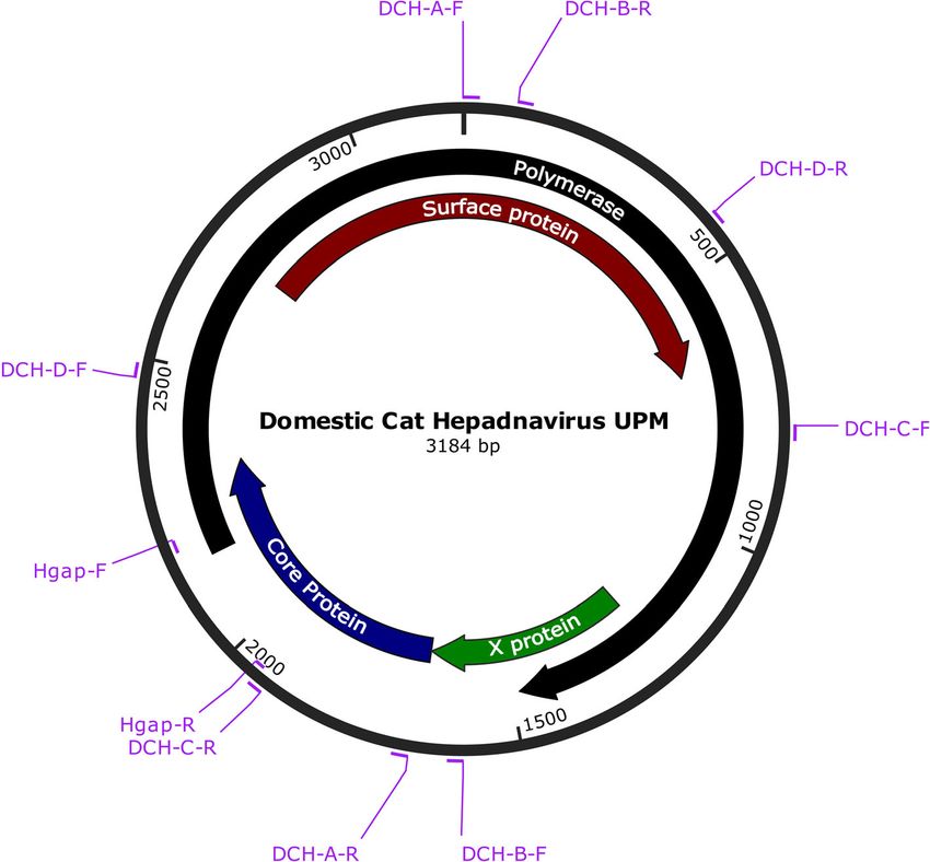
Anpuanandam et al. BMC Veterinary Research   (2021) 17:9                                                      Page 2 of 10

Background                                                     which examined formalin-fixed, paraffin-embedded (FFPE)
Domestic cat hepadnavirus (DCH) is a novel member of           liver tissues from four countries (the United States,
the Hepadnaviridae, detected in domestic cats (Felis catus)    Australia, New Zealand, the UK) demonstrated that up to
[1–3]. Hepadnaviruses are spherical with a diameter of 42–     43% of chronic hepatitis cases and 28% of HCC cases were
50 nm. The viral genome is enclosed in an icosahedral cap-     also DCH-positive.
sid, which is enveloped by a lipid bilayer membrane. The          Cats aged 4 to 7 months had higher odds of being
viral genome comprises a circular, partially double-stranded   DCH-positive, albeit at a p = 0.08061, and with no sex
DNA molecule [4, 5]. Hepadnaviruses can be classified into     predisposition. An in-situ hybridization (ISH) study sug-
five genera according to host: Orthohepadnavirus infects       gested that DCH viral distribution in HCC and chronic
mammals, Avihepadnavirus infects bird species, Herpetohe-      hepatitis cases resembled that of HBV-related liver dis-
padnavirus infects amphibians and reptiles, Metahepadna-       eases in humans [3]. These findings suggest a possible
virus and Parahepadnavirus infect fish [6–8]. The newly        similar pathogenesis for DCH as HBV, whereby the virus
identified DCH has been classified under Orthohepadna-         could be involved in chronic liver inflammation leading
virus [1].                                                     eventually to carcinogenesis. This hypothesis requires
   The orthohepadnavirus has a DNA genome of ap-               further investigations.
proximately 3.2 kb. The genome has four overlapping               Studies on the detection of DCH in paired liver and
open reading frames (ORFs), which encode for the               blood samples from the same cat have not yet been
surface (S), X, core (C) and polymerase (P) proteins [9].      reported. Paired detection results will increase our
Several orthohepadnaviruses have been isolated from            understanding of DCH infection at different clinical
various primates including humans, gorillas, gibbons,          phases as observed in HBV patients, viz. virus carriers
orangutans, woolly monkeys and chimpanzees [10–13].            and immune-tolerant patients, and between active and
Orthohepadnaviruses also infect other mammals includ-          chronic phases. Usually, a higher HBV DNA load in
ing woodchucks, ground squirrels, arctic squirrels and         blood is found in acute infection or in the active chronic
bats [14–17].                                                  phase of infection [21, 22].
   The well-known prototype species of this family, hu-           The discovery of DCH in two different geographical
man HBV, was first discovered in 1966. Approximately           locations revealed two disparate DCH strains: DCH
257 million people worldwide are HBV carriers, and             Australia (3187 bp, AUS/2016/Sydney) and DCH Italy
HBV causes 887,000 deaths annually, mainly through             (3184 bp, ITA/2018/165 − 83) [1, 2]. The Italian and
hepatocellular carcinoma (HCC, 62%) and cirrhosis              Australian strains share 97% nucleotide identity [2].
(29%) [18]. HBV distribution predominates in various           Therefore, determining the complete genome sequence
geographical regions worldwide with high prevalence            of the Malaysian DCH would provide insights into the
reported in Asia, Middle East and Africa. Different HBV        genetic variability of this virus.
genotypes co-circulate in different regions, which in-            Here, we report on the molecular prevalence of DCH
creases the rate of HBV genomic recombination [19].            using conventional PCR assay on both whole-blood and
   HBV has also been reported as a common co-pathogen          liver tissue samples from shelter and pet domestic cats
in human immunodeficiency virus (HIV)-infected individ-        in Malaysia. Possible risk factors for DCH infection were
uals. Chronic HBV infection occurs in up to 10% of HIV         evaluated, including age, sex, type of ownership, eleva-
patients co-infected with HBV. The immune system of            tion in serum alanine transaminase (ALT) levels, and co-
HIV patients has a decreased likelihood of HBV clearance,      infection with FIV and FeLV. In addition, the complete
which leads to higher HBV viral load and the development       genome sequence of the local (Malaysian) strain was de-
of chronic HBV infection [20]. As HBV and HIV co-              termined and compared with two DCH strains from
infection is commonly reported in humans, the initial          Australia and Italy. Phylogenetic analysis was performed
study on DCH detection focused on cats infected with           to relate the evolution of the Malaysian strain of DCH
feline immunodeficiency virus (FIV). DCH prevalence            with those of other hepadnaviruses.
among FIV-infected cats was 10%, which was significantly
higher than that in cats not infected by FIV (3.2%) [1].       Results
   In Italy, cat serum samples sent to diagnostic labora-      Study group
tories under clinical suspicion of infectious diseases re-     A total of 340 samples, comprising 253 blood and 87
vealed a high DCH prevalence of up to 17.8%; of these,         liver samples, were collected. All the liver samples were
33.3% of cats infected with retroviruses (FIV and feline       collected with paired blood samples from shelter cats.
leukemia virus, FeLV) also had DCH co-infection [2].           From pet cats, only blood samples were collected. A
Lanave et al. [2] also found that 7 of 10 suspected            total of 108 (42.7%) cats were from the shelter and the
hepatic disease cases were also DCH-positive. A feline         remaining 145 (57.3%) cats were pet cats. Out of the 253
hepatic lesion-focused study by Pesavento et al. [3],          total cats, 132 (52.2%) were male while the remaining
Anpuanandam et al. BMC Veterinary Research          (2021) 17:9                                                                        Page 3 of 10

121 (47.8%) were female. The mean and median ages of                        (Fig. 2b). Twenty of 31 cats with DCH-positive blood
the cats were 4 and 2 years old with a minimum of one-                      samples were over 2 years of age (64.5%, n = 20/31).
month and a maximum of 19 years. Of the 253 cats,                           DCH was detected in more pet cats (16.6%) than shelter
90.9% were Domestic Short Hair (n = 230). The other                         cats (6.5%, Table 1). Breed and sex were not risk factors
breeds consisted of Persian (n = 10), British Short Hair                    for DCH. Among the different health conditions,
(n = 3), Domestic Long Hair (n = 3), Siamese (n = 2),                       the highest detection (28.6%) of DCH was seen in
Maine Coon (n = 2), American Curl (n = 2), and one                          traumatic injury cases (Fig. 2c). The detection of
Norwegian Forest.                                                           DCH was 12.2% (27/221) in sick cats and 12.5% (4/32) in
  The cats were diagnosed with various health conditions,                   healthy cats.
predominantly respiratory diseases (n = 35), followed by
wounds and skin diseases (n = 27), gastrointestinal diseases                Serum biochemistry
(n = 26), traumatic injuries (n = 14), neoplasia (n = 4), and               Twelve of the 23 DCH-positive blood samples (52.2%) had
urogenital diseases (n = 42). Cats with poor body condition                 elevated ALT levels. Cats with abnormal ALT had a 21%
(n = 73) and with systemic illnesses (anaemia, dehydration,                 chance of having a DCH-positive blood sample (p = 0.012,
general weakness, and anorexia) were also included in this                  CI: 1.239–7.294) (Table 1). When DCH was detected in
study. Samples were also collected from healthy cats (n =                   the blood, the odds of having elevated ALT was 6.3 times
32) undergoing elective procedures such as neutering and                    higher than when DCH was detected solely in the liver
general wellness check-ups. The four neoplasia cases                        tissue. Other serum biochemistry readings, such as
included one each of fibrosarcoma, squamous cell carcin-                    alkaline phosphatase (ALP), urea and creatinine, were not
oma, lymphoma and mammary adenocarcinoma.                                   significantly associated with detecting DCH.

Molecular detection                                                         Co-infection with feline immunodeficiency virus and
Among the 253 cat blood samples, 12.3% (n = 31/253)                         feline leukemia virus
tested positive for DCH. Among the 87 cat liver tissue                      DCH was detected as a co-pathogen of the two common
samples, 14.9% (n = 13/87) tested DCH-positive. Of the                      feline viruses examined in this study. Four out of five
13 cats with DCH-positive liver tissues (Fig. 1), only five                 FeLV-positive cats were DCH-positive, which was sig-
(38.5%) were viraemic.                                                      nificant at p = 0.006 (CI: 1.352–121.212) (Table 1), but
  We analysed the distribution of DCH across different                      the number of FeLV-positive cats (n = 5) was too low to
age groups (Fig. 2a). Cats older than 2 years had signifi-                  estimate the risk of FeLV patients being coinfected with
cantly higher odds of having DCH-positive blood sam-                        DCH. One of the two FIV-positive cats was DCH-
ples than the younger cats (p = 0.042, CI: 1.015–4.848)                     positive with no significant association.

 Fig. 1 Agarose gel electrophoresis of liver samples. PCR of the Hgap gene produced a product of 230 bp. Electrophoresis was performed on 1.5%
 (w/v) agarose gel. Lane L: 1kb DNA ladder, Lane N: negative control. Lanes 1 to 13: positive samples. Liver sample ID: FH4, FH5, FH13, FH14, FH32,
 FH41, FH42, FH43, FH45, FH59, FH62, FH246 and FH248
Anpuanandam et al. BMC Veterinary Research          (2021) 17:9                                                                       Page 4 of 10

 Fig. 2 a Age distribution of cats in relation to detection of DCH included in this study. Highest percentage of DCH detection was seen in age
 group of 4 to 6 years old. b DCH detection in relation to origin and age group. c Health conditions of cats in relation to detection of DCH.
 Among the traumatic injury cases, the highest percentage of detection was seen

                                                                           DCH strain also had four ORFs coding for the P, S,
                                                                           C and X proteins. However, the P and S genes of
Complete genome sequence and phylogenetic analysis of                      the local isolate were each shorter than those of the
Malaysian DCH                                                              Australian strain by one codon. Identical to that of
The complete genome sequence of a local DCH strain                         the Italian DCH, amino acid deletions were also ob-
was 3184 bp; Fig. 3 illustrates its key genomic features.                  served at positions 271 (Pro) and 82 (Ser) in the P
Complete sequencing was performed for the liver tissue                     and S proteins, respectively, in Malaysian strain. The
of a cat which was positive for the Hgap gene. Similar to                  complete genome sequence has been deposited in
Australian and Italian isolates, the genome of the local                   GenBank (accession no. MK902920).
Anpuanandam et al. BMC Veterinary Research (2021) 17:9 Page 5 of 10
Table 1 Chi-square analyses and univariate logistic regression for the risk of DCH in Malaysian cats
Factors Categories Prevalence P value OR 95% CI
Paired Sample Positive 5/13 (38.5%) < 0.001 0.098 0.051–0.188
Negative 0/74 (0%)*
Origin Shelter 7/108 (6.48%)* 0.016 2.862 1.184–6.916
Pet Cats 24/145 (16.55%)
Age Class < 2 years old 11/133 (8.27%)* 0.042 2.218 1.105–4.848
Adult 20/120 (16.7%)
Sex Male 20/132 (15.2%) 0.142 0.56 0.256–1.223
Female 11/121 (9.1%)*
ALT Normal 11/135 (8.15%)* 0.012 3.006 1.239–7.294
Abnormal 12/57 (21.05%)
ALP Normal 22/169 (13.02%) 0.966 0.955 0.112–8.135
Abnormal 1/8 (12.5%)*
Urea Normal 17/132 (12.88%) 0.725 0.846 0.332–2.157
Abnormal 7/63 (11.11%)*
Creatinine Normal 20/168 (11.9%)* 0.563 1.41 0.439–4.527
Abnormal 4/25 (16%)
FIV Positive 1/2 (50%) 0.458 2.783 0.167–46.333
Negative 23/87 (26.44%)*
FeLV Positive 4/5 (80%) 0.006 12.8 1.352–121.212
Negative 20/84 (23.81%)*
OR Odds ratio, OR Odds ratio, CI Confidence interval, *Reference for analysis, numbers in bold indicate pAnpuanandam et al. BMC Veterinary Research (2021) 17:9 Page 6 of 10 Fig. 3 Genomic structure of DCH Malaysia, with the positions of the primers used in this study. The complete genome sequence consists of 3184 bp. The colours indicate the ORFs of polymerase (black), surface protein (red), core protein (blue) and X protein (green) Whether DCH can be transmitted through the saliva, viraemic in the progressive infection phase, when the faeces and urine is still unknown. However, Lanave et al. FeLV antigen is detectable. In the regressive phase, the [2] argued that, since DCH can be detected in the serum virus hides in the bone marrow or other tissues, render- and whole blood samples, it is possible for transmission ing the FeLV antigen undetectable. The FeLV antigen to take place through blood transfusion from one cat to can be reactivated in this phase, and triggers develop- another. ment of clinical signs [25]. The risk of underestimating DCH infection did not appear to be significantly linked FeLV prevalence via antigen detection with ELISA hence with sex. This was in good agreement with that reported warrants closer attention; repeated samplings on FeLV- by Lanave et al. [2] who also detected high DCH preva- negative cats are normally recommended to verify an lence in cats aged 4 to 7 months (20.5%). In contrast, initial ELISA result. The low detection of FIV-positive the present study showed a higher DCH prevalence in samples in the current study was in line with other FIV- cats aged > 2 years compared to younger cats. While related studies on domestic cats in Malaysia [26, 27]. human HBV can cause different phases of infection We found a significant association between elevated prenatally and in young adults and adults [24], the DCH ALT and positive DCH detection in blood samples. infection phases in cats are still unknown. Twelve of the 57 cats with elevated ALT was DCH- The significant association of DCH co-infection with positive, in good agreement with Lanave et al. [2]. For FeLV agrees with Lanave et al. [2], who reported co- chronic HBV infection, at the immune-tolerant phase, infection with both FIV and FeLV. However, separate ALT was within the normal range, but high levels of values for each retrovirus co-infection with DCH were HBV DNA could be detected in the blood, with minimal not mentioned in that study. Although the FeLV or no liver inflammation [24]. In the present study, 4 of antigen-positive sample was small in this study, it is im- 11 cats had no elevated ALT although the PCR analysis portant to note that FeLV detection efficay differs be- showed that they were DCH-positive. These findings tween the phases of FeLV infection. The cats are lead to the possibility of classifying/staging DCH infection
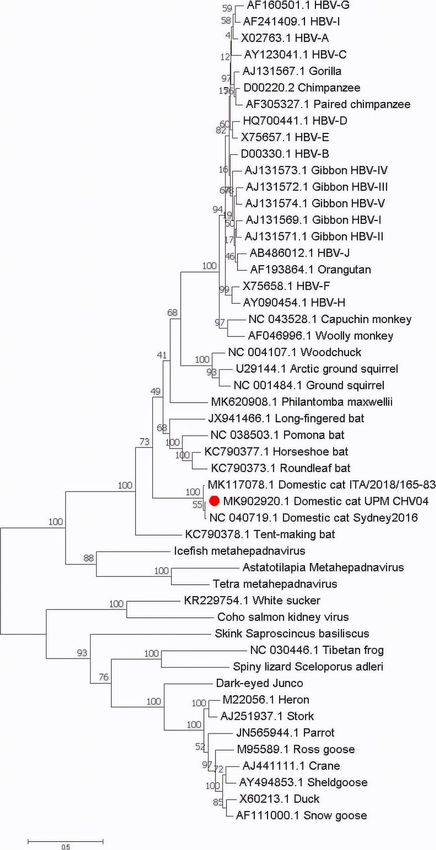
Anpuanandam et al. BMC Veterinary Research          (2021) 17:9                                                                         Page 7 of 10

 Fig. 4 The phylogenetic position of DCH Malaysia within the family Hepadnaviridae. Maximum likelihood phylogeny based on the complete genome
 sequences of a wide range of vertebrate hepadnaviruses retrieved from the Genbank database. The herpetohepadnaviruses, parahepadnaviruses and
 metahepadnaviruses were described in Dill et al. [8] and Lauber et al. [7]. The DCH Malaysia (Domestic cat hepadnavirus UPM MK902920) is indicated
 by a red dot. The tree was drawn to scale, with branch lengths measured in the number of substitutions per site

in cat patients as in HBV infection. In human patients,                     unanswered questions on the disease and provide new
staging HBV infection is important for treatment and                        insights into feline medicine.
prognosis purposes. Future investigations using experi-                       This is the first study in Malaysia to explore the
mental infections of DCH among cats can resolve many                        presence of DCH among local domestic cats. The total
Anpuanandam et al. BMC Veterinary Research   (2021) 17:9                                                      Page 8 of 10

genome sequence of the local strain is similar to that of         Cats were physically restrained and blood (about 1 to
the Italian strain, missing a single codon from the P and       2 mL, depending on the cat’s body weight) was drawn
S genes. However, phylogenetically, the complete gen-           via the jugular vein. All blood samples were transferred
ome sequence of the Malaysian strain clustered together         into a 3 mL plain and EDTA vacutainer tubes (BD Vacu-
with the Australian strain. This could be due to                tainer®, United States). All blood samples in the plain
geographical proximity, since Australia and Malaysia are        tube were left standing for one to three hours at room
close together, while Italy is much further away. The           temperature before centrifugation at 2000 g for 10
Malaysian strain is located between the Italian and             minutes (KUBOTA 2010, Japan). The resultant serum
Australian strains, and more closely related to the             samples were pipetted into 1.5 mL Eppendorf tubes®
Australian strain.                                              (USA) and stored at − 20℃. Whole blood samples in
  All the three DCH strains are grouped together in a           EDTA vacutainer tubes were subjected to DNA isolation.
well-defined branch, which is related to several bat, ro-         Samples were also collected from cats at the shelter
dent and primate hepadnaviruses. The Malaysian DCH              which were euthanized by the shelter veterinarian (not
strain is closely related to the previously published Aus-      solely for this research purpose) for medical conditions
tralian DCH strain, suggesting that different strains of        which was beyond the resources of the shelter to treat
DCH may be discovered across different continents. As           or due to illnesses of a very contagious or zoonosis in
DCH was not discovered until 2018, many of its features         nature. Euthanasia was performed using sodium pento-
such as pathogenesis, cross-species transmission, and           barbital which was administered intravenously at a dose
different stages of infections, have yet to be characterized.   of > 100 mg/kg. Blood was drawn prior to euthanasia
                                                                while liver tissues were collected at post-mortem, snap-
Conclusions                                                     frozen in liquid nitrogen, and stored in cryotubes at -80
This study provided insights into a Malaysian DCH strain        C (SANYO Ultra Low, Japan). Parts of the liver tissue
that was detected from a liver tissue. Interestingly, the       were also placed in 10% (v/v) buffered formalin solution
prevalence of DCH in liver tissue samples was higher than       (Sigma-Aldrich, Germany) for routine histopathology.
that in blood samples from domestic cats in Malaysia, of        Carcasses were disposed based on the shelter's policy.
which adult and pet cats have a higher prevalence. Nearly
50% of the cats tested positive for DCH showed elevated
ALT level. The Malaysian DCH is genetically clustered to-       DNA isolation and PCR assay
gether with Australian and Italian DCH strains. Overall,        Total DNA was extracted from 100 µL whole blood
our study provided valuable knowledge on the occurrence         using the DNeasy Blood & Tissue Kit (Qiagen,
of DCH in Malaysia, of which the true prevalence can be         Germany), following the manufacturer’s protocol. DNA
studied using the liver tissue samples. However, the effect     was extracted from 25 mg liver tissue, after the tissue
of this virus on causing liver disease in felines warrants      was homogenized using a rotor-stator homogenizer and
further study. As the domestic pet cat population in            probe (Qiagen). The total DNA from liver tissue was ex-
Malaysia is growing, veterinarians and pet owners need to       tracted using the DNeasy Blood & Tissue Kit (Qiagen)
be advised sufficiently about new infectious diseases and       according to the manufacturer’s protocol.
possible zoonosis. Investigations on DCH may also offer           PCR primers used for detecting the Hgap gene to
new or more effective ways to study HBV.                        confirm the presence of DCH were synthesized (forward:
                                                                5’-GTGCTCTGATAACCGTATGCTC-3’; reverse: 5’-
Methods                                                         CTAGAATGGCTACATGGGGTTAG-3’) as reported
Sample collection and storage                                   previously [1]. Conventional PCR was performed using
For this study, whole blood samples were collected from         the Bioline MyTaq hot-start polymerase (Bioline,
domestic cats from an animal shelter in Selangor and            Australia) according to the manufacturer’s protocol:
from the University Veterinary Hospital (UVH) of                300 ng DNA template, 5 × MyTaq buffer, 20 µM final
Universiti Putra Malaysia between 2018 and 2019. Blood          primer concentration in a total 25 µL reaction. The PCR
samples were collected from UVH cat patients with               cycling conditions were as follows: Initial denaturation
owners’ consent. Blood was sampled from pet cats                at 95℃ for 1 min, followed by 40 cycles of denaturation
presented to the UVH for annual health examination,             at 95℃ for 15 s, annealing at 55℃ for 15 s, extension at
neutering or health issues. Most often blood was drawn          72℃ for 10 s and final extension at 72℃ for 5 min. The
as part of the routine diagnostics where left over blood        PCR products from both the blood and liver samples
samples were stored for this research. Pet cats were            were analysed with (1.5%) agarose gel electrophoresis,
returned to their respective owners. Blood was sampled          from which specific bands containing the PCR products
from healthy cats at the shelter prior to neutering where       were purified, excised and verified. The amplicons were
they were returned to their enclosures.                         verified using the Sanger DNA sequencing method.
Anpuanandam et al. BMC Veterinary Research (2021) 17:9 Page 9 of 10
Complete genome sequencing and phylogenetic analysis USA). The ALT and ALP results were classified as either
of Malaysian DCH normal (within normal limits) or abnormal (> 105%
The complete DCH viral genome sequence was obtained normal limits); the abnormality thresholds were 95 U/L
from the DCH-positive samples using the four newly de- for ALT and 84 U/L for ALP (cf. normal limits: ALT
signed primers (Table 2). The primers were designed 10–90 U/L; ALP < 80 U/L). The serum samples were
using the National Center for Biotechnology Information tested using the FeLV Ag/FIV Ab rapid test kit (Gen-
(NCBI) Primer-BLAST [28]. Reference was made to the Body, Korea) according to the manufacturer’s protocol.
whole-genome sequence of the Australian strain (Gen-
Bank, accession no. NC040719). PCR was performed as
Data analysis
described above, except that the annealing temperature
Statistical analyses were done using the Statistical Package
was as stated in Table 2. The amplicons were analysed
for the Social Sciences Version 22, (SPSS Inc., USA).
using the Sanger sequencing method (Apical, Malaysia).
Prevalence data are reported as frequencies and percent-
The viral nucleotide sequences were manually edited by
ages. Inferential statistical tests were carried out using χ-
removing the overlapping regions to obtain the complete
squared analysis and by evaluating the odds ratio (OR);
circular genome of the Malaysian DCH. Sequence
95% confidence intervals (CI) were calculated, where ap-
editing, trimming, reverse complement sequencing and
plicable, at a significance level of α = 0.05. The risk factors
assembly of the forward and reverse sequences were
were analysed when the p-value for the likelihood ratio
performed using SnapGene® version 4.3 software [29].
was ≤ 0.05. The χ-squared test was not applicable when
one of the expected cell values was < 5. In these cases,
A total of 50 reference sequences from the family
Fisher’s exact test was used, with α = 0.05, to determine
Hepadnaviridae were included together with the DCH
the association of the risk factors with the DCH-positive
Australia (AUS/2016/Sydney), Italy (ITA/2018/165 − 83)
detection.
and Malaysian (DCH UPM) strains for the phylogenetic
analysis. Evolutionary analyses were conducted using
Abbreviations
Molecular Evolutionary Genetics Analysis 7 (MEGA7) DCH: Domestic cat hepadnavirus; FIV: Feline immunodeficiency virus;
[30] software. Multiple sequence alignments were done FeLV: Feline leukemia virus; HBV: Human hepatitis B virus; ALT: Alanine
transaminase; ALP: Alkaline phosphatase; UPM: Universiti Putra Malaysia;
for the best sequence match alignment. The evolutionary
UVH: University Veterinary Hospital; FFPE: Formalin-fixed, paraffin-embedded;
history was inferred using the Maximum Likelihood HCC: Hepatocellular carcinoma
method. The percentage of trees in which the associated
taxa clustered together is shown next to the branches. Acknowledgements
The tree reliability was evaluated using 1000 bootstrap The authors would like to extend appreciation to the staff of the Clinical
Laboratory, Serology Laboratory and Post-mortem Laboratory of the Faculty
replications. The tree was drawn to scale, with branch of Veterinary Medicine; and the Chemistry Pathology Laboratory of the Fac-
lengths measured in the number of substitutions per ulty of Medicine and Health Sciences for their assistance. The authors thank
site. All positions containing gaps and missing data were Miss Hagilaa Ganesan from the Laboratory of Molecular Virology, Faculty of
Biotechnology and Biomolecular Sciences for technical assistance. We would
eliminated. like to thank the late Mr Maniam for his assistance for blood sampling from
cats.
Serum biochemistry, FIV and FeLV tests
All sera from the shelter and pet cats were measured for Authors' contributions
serum biochemistry (ALT, ALP, urea, creatinine) using a A.K. performed the laboratory tests, collected samples, involved in data
acquisition and interpretation, drafted the work; G.T.S. contributed to the
VetTest Chemistry Analyzer (IDEXX, Washington, conception and designed the work, involved in interpretation of data,
substantively revised the manuscript, principle investigator for the funds;
Table 2 Newly designed primers to amplify the whole genome M.M.K.C. involved in sampling, and processing of samples; S.W.N. involved in
sequence of DCH design of work, interpretation of data; K.K. assisted in laboratory test
Primer Sequence Base Annealing validations, data acquisition and analysis; R.M.A. involved in data analysis;
pair Temperature S.K.R. assisted in processing of samples; K.L.H. provided laboratory
consumables, involved in trouble shooting and interpretation of data; W.S.T.
DCH A F: 5’-TGGGCAACATTACCTCAGGTCC-3’ 1700 56℃ involved in design of work, interpretation of data and substantively revised
R: 5’-GGAACAAAAGAGAACGCACAGG-3’ the manuscript. All authors have approved the submitted version.
DCH B F: 5’-GGTCTGACGCCCAGGTTATG-3’ 1700 56℃
Funding
R: 5’-ACCACGAGTCTGCACTCTGC-3’ This study was partially funded by Universiti Putra Malaysia (grant number:
DCH C F: 5’-CTCAGGTCTTTGCCCACTCA-3’ 1155 55℃ GP/2018/9650300). The Universiti Putra Malaysia funder consented to the
publication of the manuscript but had no role in the study design, data
R: 5’-AGCTGACTCCTCCCAACAGT-3’ collection, analysis, interpretation of data and in writing the manuscript.
DCH D F: 5’-AACTAAGCATGAACTCCGCC-3’ 1173 55℃
Availability of data and materials
R: 5’-TGGGCCAACAGGTGCAATTT-3’
Data will be available upon request from the corresponding author.Anpuanandam et al. BMC Veterinary Research (2021) 17:9 Page 10 of 10
Ethics approval and consent to participate 19. Bonvicino CR, Moreira MA, Soares MA. Hepatitis B virus lineages in
This study was approved by the Institutional Animal Care and Use mammalian hosts: Potential for bidirectional cross-species transmission.
Committee of Universiti Putra Malaysia (AUP - R089/2018). Samples from pet World J Gastroenterol. 2014;20(24):7665–74.
cats were collected with the agreement of the cat owners for clinical and 20. Lisco A, Vanpouille C, Margolis L. Coinfecting viruses as determinants of HIV
laboratory purposes. All efforts were made to minimise the discomfort of the disease. Curr HIV/AIDS Rep. 2009;6(1):5–12.
animals during sampling. 21. Liaw YF, Chu CM. Hepatitis B virus infection. The Lancet. 2009;373(9663):
582–92.
22. Trépo C, Chan HLY, Lok A. Hepatitis B virus infection. The Lancet. 2014;
Consent for publication 384(9959):2053–63.
Not applicable. 23. Seeger C, Mason WS. Molecular biology of hepatitis B virus infection.
Virology. 2015;479:672–86.
24. Lampertico P, Agarwal K, Berg T, Buti M, Janssen HLA, Papatheodoridis G,
Competing interests et al. Clinical Practice Guidelines on the management of hepatitis B virus
The authors declare that they have no competing interests. infection. J Hepatol. 2017;67(2):370–98.
25. Hartmann K. Regressive and progressive feline leukemia virus infections –
Author details clinical relevance and implications for prevention and treatment. Thai J Vet
1
Department of Veterinary Clinical Studies, Faculty of Veterinary Medicine, Med. 2017;Suppl 47:109–12.
Universiti Putra Malaysia, Selangor 43400 UPM Serdang, Malaysia. 2Institute of 26. Sivagurunathan A, Atwa AM, Lobetti R. Prevalence of feline
Bioscience, Universiti Putra Malaysia, 43400 UPM Serdang, Selangor, Malaysia. immunodeficiency virus and feline leukaemia virus infection in Malaysia: a
3
Department of Pathology, Faculty of Medicine and Health Sciences, retrospective study. JFMS Open Rep. 2018;4(1):205511691775258.
Universiti Putra Malaysia, 43400 UPM Serdang, Selangor, Malaysia. 27. Bande F, Arshad SS, Hassan L, Zakaria Z, Sapian NA, Rahman NA, et al.
4
Department of Microbiology, Faculty of Biotechnology and Biomolecular Prevalence and risk factors of feline leukaemia virus and feline
Sciences, Universiti Putra Malaysia, 43400 UPM Serdang, Selangor, Malaysia. immunodeficiency virus in peninsular Malaysia. BMC Vet Res. 2012;8.
28. Primer Designing Tool. https://www.ncbi.nlm.nih.gov/tools/primer-blast/.
Received: 19 May 2020 Accepted: 30 November 2020 Accessed 20 March 2019.
29. Goldberg MF, Roeske EK, Ward LN, Pengo T, Dileepan T, Kotov DI, et al.
Salmonella persist in activated macrophages in T cell-sparse granulomas
but are contained by surrounding CXCR3 ligand-positioned Th1 cells.
References Immunity. 2018;49(6):1090–102.
1. Aghazadeh M, Shi M, Barrs VR, McLuckie AJ, Lindsay SA, Jameson B, et al. A 30. Kumar S, Stecher G, Tamura K. MEGA7: Molecular Evolutionary Genetics
novel hepadnavirus identified in an immunocompromised domestic cat in Analysis version 7.0 for bigger datasets. Mol Biol Evol. 2016;33(7):1870–4.
Australia. Viruses. 2018;10(5):269.
2. Lanave G, Capozza P, Diakoudi G, Catella C, Catucci L, Ghergo P, et al.
Identification of hepadnavirus in the sera of cats. Sci Rep. 2019;9(1):1–6. Publisher’s Note
3. Pesavento PA, Jackson K, Scase T, Tse T, Hampson B, Munday JS, et al. A Springer Nature remains neutral with regard to jurisdictional claims in
novel hepadnavirus is associated with chronic hepatitis and hepatocellular published maps and institutional affiliations.
carcinoma in cats. Viruses. 2019;11(10):1–8.
4. Urban S, Schulze A, Dandri M, Petersen J. The replication cycle of hepatitis B
virus. J Hepatol. 2010;52(2):282–4.
5. Monjardino J. Hepadnaviridae. Virus Taxonomy. 2012;445–455.
6. Schaefer S. Hepatitis B virus taxonomy and hepatitis B virus genotypes.
World J Gastroenterol. 2007;13(1):14–21.
7. Lauber C, Seitz S, Mattei S, Suh A, Beck J, Herstein J, et al. Deciphering the
origin and evolution of Hepatitis B viruses by means of a family of non-
enveloped fish viruses. Cell Host Microbe. 2017;22(3):387–99.
8. Dill JA, Camus AC, Leary JH, Di GF, Holmes EC, Ng TF. Distinct viral lineages
from fish and amphibians reveal the complex evolutionary history of
hepadnaviruses. J Virol. 2016;90:7920–33.
9. Liang TJ. Hepatitis B. The virus and disease. Hepatology. 2009;49(Suppl 5):
513–21.
10. Grethe S, Heckel J, Rietschel W. Molecular epidemiology of hepatitis B virus
variants in nonhuman primates. J Hepatol. 2000;74(11):5377–81.
11. Norder NE, Ebert JW, Fields HA, Mushahwar ISAK, Magnius LO. Complete
sequencing of a gibbon hepatitis B virus genome reveals a unique
genotype distantly related to the chimpanzee hepatitis B virus. Virology.
1996;223(218):214–23.
12. Warren KS, Heeney JL, Swan RA. A New group of hepadnaviruses naturally
infecting orangutans (Pongo pygmaeus). J Virol. 1999;73(9):7860–5.
13. Lanford RE, Chavez D, Brasky KM, Burns IIIRB, Rico-Hesse R. Isolation of a
hepadnavirus from the woolly monkey, a New World primate. Proc Natl
Acad Sci USA. 1998 May;95:5757–61.
14. Tyler GV, Summers JW, Snyder RL. Woodchuck hepatitis virus in natural
woodchuck populations. J Wildl Dis. 1981;17(2):297–301.
15. Helgen KM, Cole FR, Helgen LE, Wilson DE. Generic revision in the holarctic
ground squirrel genus Spermophilus. J Mammal. 2009;90(2):270–305.
16. Testut P, Renard C-A, Terradillos O, Vitvitski-Trepo L, Tekaia F, Degott C, et al.
A new hepadnavirus endemic in arctic ground squirrels in Alaska. J Virol.
1996;70(7):4210–9.
17. He B, Zhang F, Xia L, Hu T. Identification of a novel orthohepadnavirus in
pomona roundleaf bats in China. Arch Virol. 2014;160(1):335–7.
18. Hepatitis B. Key Facts. https://www.who.int/news-room/fact-sheets/detail/
hepatitis-b. Accessed 20 April 2020.You can also read


