Out-of-Distribution Detection for Dermoscopic Image Classification
←
→
Page content transcription
If your browser does not render page correctly, please read the page content below
Out-of-Distribution Detection for Dermoscopic Image Classification

                                         Mohammadreza Mohseni* 1, 2 , Jordan Yap2 , William Yolland2 , Majid Razmara2 , and M Stella Atkins1, 3
                                                                        1
                                                                   School of Computing Science, Simon Fraser University
                                                                                2
                                                                                  MetaOptima Technology Inc
                                                        3
                                                          Department of Skin Science and Dermatology, University of British Columbia
arXiv:2104.07819v2 [cs.CV] 19 Apr 2021

                                                               {mmohseni, stella}@sfu.ca, {jordan, william, majid}@metaoptima.com

                                                                      Abstract                                also be threatening if left untreated [8]. However, differ-
                                                                                                              entiating malignant skin lesions from benign skin lesions
                                             Medical image diagnosis can be achieved by deep neu-             such as nevi and seborrheic keratoses (SK) is often diffi-
                                         ral networks, provided there is enough varied training data          cult even for trained clinicians. Several clinical algorithms
                                         for each disease class. However, a hitherto unknown dis-             have been developed to aid clinicians make a diagnosis of
                                         ease class not encountered during training will inevitably           a skin lesion, such as the visual clues to help to diagnose
                                         be misclassified, even if predicted with low probability. This       malignant melanoma. These clinical algorithms include the
                                         problem is especially important for medical image diagno-            well-known ABCD criteria (Asymmetry, Border irregular-
                                         sis, when an image of a hitherto unknown disease is pre-             ity, Colour irregularity, Diameter) [32, 1], and the 7-point
                                         sented for diagnosis, especially when the images come from           check-list [2, 6]. But even with these clues, Heal et al.
                                         the same image domain, such as dermoscopic skin images.              showed it can be extremely difficult for physicians to make
                                             Current out-of-distribution detection algorithms act un-         a differential diagnosis between the commonly-encountered
                                         fairly when the in-distribution classes are imbalanced, by           skin lesions, such as nevi and non-melanocytic lesions like
                                         favouring the most numerous disease in the training sets.            seborrheic keratoses (SK), and rarely-encountered malig-
                                         This could lead to false diagnoses for rare cases which are          nant melanomas [20].
                                         often medically important.
                                                                                                                 A magnified dermoscopic view taken very close to, or
                                             We developed a novel yet simple method to train neu-
                                                                                                              in contact with, the skin is frequently used to supplement
                                         ral networks, which enables them to classify in-distribution
                                                                                                              the observational clinical view for diagnosis of skin lesions
                                         dermoscopic skin disease images and also detect novel dis-
                                                                                                              [7]. A dermoscopic image, typically taken at magnifica-
                                         eases from dermoscopic images at test time. We show that
                                                                                                              tion 10-15 times and with polarized lighting, can show skin
                                         our BinaryHeads model not only does not hurt classifica-
                                                                                                              details at much higher resolution, including dermoscopic
                                         tion balanced accuracy when the data is imbalanced, but
                                                                                                              structures a few millimeters under the surface of the skin
                                         also consistently improves the balanced accuracy. We also
                                                                                                              including textures such as pigment networks, dots, globules
                                         introduce an important method to investigate the effective-
                                                                                                              and streaks [7, 25].
                                         ness of out-of-distribution detection methods based on pres-
                                         ence of varying amounts of out-of-distribution data, which               In contrast with the previously mentioned clinical algo-
                                         may arise in real-world settings.                                    rithms, a separate set of clues for diagnosis from dermo-
                                                                                                              scopic images has also been developed such as the der-
                                                                                                              moscopy ABCD rule where ”D” stands for dermoscopic
                                         1. Introduction                                                      structures instead of ”diameter” as it does for the clinical
                                                                                                              ABCD observation [16]. These dermoscopy rules are pri-
                                            It is important to diagnose malignant skin lesions early.         marily intended to help identify malignant melanoma, but
                                         In particular, early detection and surgical treatment of ma-         the differential diagnosis remains very challenging, with
                                         lignant melanoma can result in excellent patient outcomes            many possible features to identify and use [44]. Inter-class
                                         [9]. Other malignant skin lesions, including basal cell car-         similarities and intra-class dissimilarities are examples of
                                         cinoma (BCC) and squamous cell carcinoma (SCC), can                  what make differential diagnoses challenging. For example,
                                           * Corresponding   Author                                           benign seborrheic keratoses (SK) mimic SCCs, BCCs, and

                                                                                                          1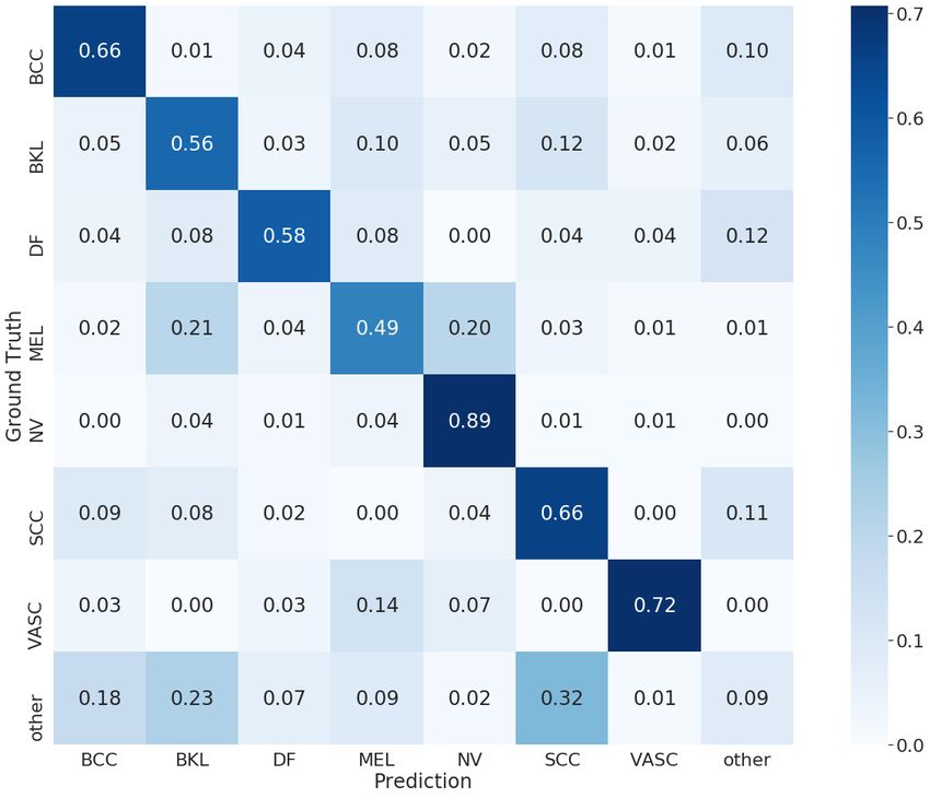
malignant melanomas, especially for patients with many 2. Related Work
atypical (dysplastic) nevi [38]. There are also many intra-
class dissimilarities, where a given disease may have many 2.1. Automated analysis of dermoscopic skin images
subtypes according to colour and texture [25]. Dermoscopic Research on automated analysis of dermoscopic images
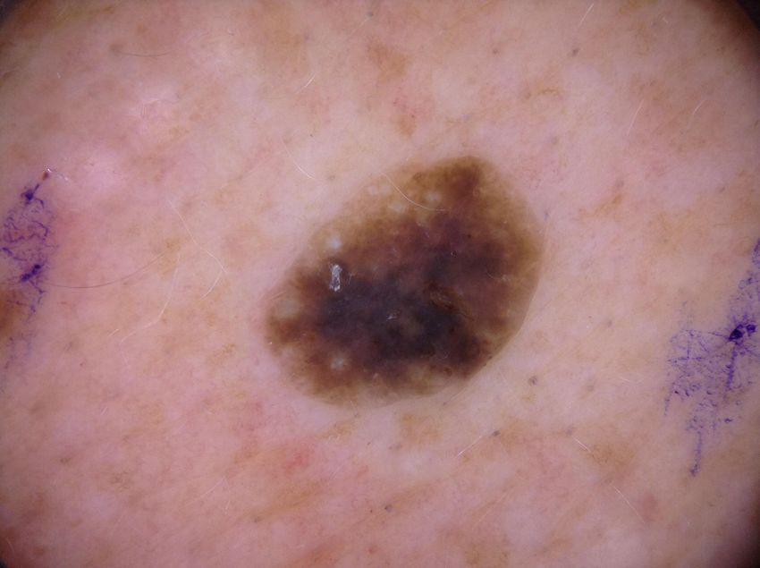
images of two nevi and SKs are shown in Fig 1, illustrat- of skin lesions initially used image processing methods, of-
ing the difficulty in differentiating between these two diag- ten focusing on the ”ABCD” features such as border irreg-
noses. ularity [29], or texture features [36, 35].
Recently deep learning approaches have proven very
successful, despite having been initially hamstrung by a
lack of data [43]. However with much more labelled data,
Esteva et al. showed deep learning models could perform
as well as expert dermatologists [17]. Furthermore, Yap et
al. showed that incorporating metadata provided even better
results [45].
(a) Dermoscopic nevus
(b) Dermoscopic SK The interest in deep learning methods has been catalyzed
by challenges hosted by the International Skin Imaging Col-
Figure 1: Dermoscopic images showing how SK can mimic laboration (ISIC), which has released thousands of high-
a nevus (publicly available skin images from ISIC 2020 quality images to the public.
dataset [34]). In 2018, Tschandl et al. published the HAM10000
dataset [40] with over 10,000 labelled dermoscopic images,
which were used for the 2018 ISIC skin diagnosis chal-
Using dermoscopy images to supplement the clinical lenge. The main task was to perform differential diagnosis
view improves diagnosis [3], [44]. However, it is very im- of 7 classes of skin lesions using the HAM10000 dataset.
portant to provide adequate training in dermoscopy for pri- The possible 7 disease categories were: melanoma (MEL),
mary care physicians to make good diagnoses [27, 14, 26], melanocytic nevus (NV), basal cell carcinoma (BCC), ac-
and even then it is very difficult to make correct differen- tinic keratosis/Bowens disease (AKIEC), benign keratosis
tial diagnoses. There is also a lot of uncertainty in diagno- (BKL) (solar lentigo/ seborrheic keratosis / lichen planus-
sis, which means that many more benign lesions are excised like keratosis), dermatofibroma (DF), and vascular (VASC).
than are strictly necessary [42]. The winning results showed balanced multi-class ac-
We were motivated to develop an automated tool to help curacy of 88.5%, a commendable result given the highly
dermatologists and primary care physicians to perform dif- imbalanced nature of the data in each class. An impor-
ferential diagnosis of dermoscopic skin lesions encountered tant subsequent study by Tschandl et al. showed that the
in the clinic. Since developing an algorithm which is able top three algorithms actually out-performed dermatologists
to detect every skin disease’s type is not possible, we built with respect to most outcome measures [39]. However, as
a tool which is able to express uncertainty when presented Tschandl et al. noted, a limitation of these algorithms is
with a previously unseen disease. Our algorithm can be an their decreased performance for out-of-distribution images,
aid in treatment decisions and detection of lesions which are which should be addressed in future research [39].
not in the commonly encountered categories. The main task in the 2019 ISIC challenge [23] was to
We also show that current out-of-distribution (OOD) classify dermoscopic images among 9 different diagnosis
detection algorithms face many challenges in our evaluation categories: the 7 classes from the 2018 challenge where the
setting. These algorithms act well when the OOD images AK class now specified only actinic keratosis (AK) as op-
are coming from a different domain, but they act poorly posed to (AKIEC), plus Squamous cell carcinoma (SCC),
when the OOD images share many geometric and semantic and a class representing ”none of the others” ie out of dis-
features with the in-distribution images. Also, we show tribution. For the ISIC 2019 challenge, 25,331 images were
that classes with smaller number of images in the training available for training across the 8 known different cate-
set contribute more to false positives in the OOD detection. gories.
The winning balanced multiclass accuracy was 63.6%
Our algorithms are designed to perform multi-class with the addition of external training data, and 60.7% with-
classification of dermoscopic images of skin lesions with out using external data.
high accuracy, and with an important additional out-of- This much lower accuracy reflected the addition of two
distribution (OOD) class by leveraging a separate binary extra classes in the test set (SCC and the ”other” class).
classifier for each in-distribution class. For both of the 2018 and 2019 challenges, the classes had
highly imbalanced distributions, reflecting the distribution
2Figure 2: Range of softmax probabilities predicted for each class. Some classes tend to have higher range of confidence
compared to others.
of lesions biopsied in a real clinical setting. melanoma is an example of one which might accompany
The 2020 ISIC challenge task was to identify melanoma high aleatoric uncertainty. Epistemic uncertainty comes
in images of skin lesions [24]. The leaderboard shows there from a lack of predictive power, due to observing only a
were over 3300 entries, with the winning score of 94.9% subset of the true data distribution at training time. In our
(area under the ROC curve). Of note is the fact that in work, by restricting the domain of novel disease images
2020 there were 33k training images, but only 1.8% were to dermoscopic images, aleatoric uncertainty is increased
malignant (vs 17.8% in 2019, 10 times more) ie the data considerably. We show that even in the scenario when
was much more strongly imbalanced compared to previous there is higher aleatoric uncertainty between in-distribution
years. samples and OOD samples, detecting OOD lesion images
A summary of recent AI approaches, mostly published is still possible.
in 2017-2019, to diagnosing skin lesions is given by Goyal
et al. [19].
Hendrycks and Gimpel proposed an initial baseline
2.2. Out-of-Distribution Detection Algorithms method for OOD detection which works based on thresh-
olding softmax probabilities [21]. Bendale and Boult
Despite all the advances in classification, AI models still proposed a methodology for unknown class detection at
have difficulty in expressing their uncertainty and detecting test time by introducing an OpenMax model layer [5].
OOD samples. Gal et al. estimated uncertainty in deep Liang et al. showed that temperature scaling and adding
neural networks using Monte-Carlo DropOut [18]. They small perturbations to the input image are two effective
proposed that measuring uncertainty can be an effective tools in separating in-distribution samples and OOD
way to detect OOD samples (”special cases”). They pro- samples [30]. Lee et al. calculated a confidence score
vided a mathematically grounded proof on why averaging based on Mahalanobis distance between a given sample
multiple stochastic forward passes can capture uncertainty and class-conditional probability distributions [28]. Vyas et
in dropout neural networks. Der Kiureghian and Ditlevsen al. identified OOD samples by enforcing a margin between
suggested breaking uncertainty into two types: Aleatoric in-distribution and OOD samples softmax probabilities in
and Epistemic [15]. Aleatoric uncertainty comes from the the loss function across an ensemble of classifiers [41].
uncertainty in the data. An image of a lesion which is hard Later, Hendrycks et al. showed that incorporating auxiliary
to tell if it is a benign melanocytic nevus or malignant OOD images during the training stage and enforcing the
3model to produce a uniform response on auxiliary OOD challenge. So images labelled as lichenoid keratosis, so-
samples can boost a model’s uncertainty estimation at test lar lentigo, and seborrheic keratosis were labelled as benign
time [22]. Recently, Liu et al. showed that energy-based keratosis.
models associate higher energy values with OOD samples We treated actinic keratosis as the out-of-distribution
[31]. They identified OOD samples by thresholding a sam- class and trained our model on the rest of the classes. We
ples’ energy calculated over the output of the model. They split the data such that 80% of lesion IDs of in-distribution
also provided a fine-tuning method to improve their results data went to the training set, for a total of 26,400 training
even more in the setting where auxiliary OOD samples are images. The rest of the data (in-distribution and out-of-
available at training time. Shafaei et al. discussed different distribution) was split equally between the validation and
points of views when tackling the OOD detection problem test set. More details can be found in Table 1.
[37]. They also proposed a three-dataset evaluation scheme
for more reliable evaluation of OOD detection algorithms.
Class Name Training Validation Test
Another limitation of current outlier detection algo- BCC 2807 (11%) 299 (12%) 217 (9%)
rithms is that they aim to sacrifice the model’s balanced ac- BKL 2397 (9%) 259 (10%) 147 (6%)
curacy in order to maximize accuracy in outlier detection.
DF 187 (1%) 28 (1%) 24 (1%)
These models look at the OOD detection problem as a bi-
nary classification problem. However, with the existence MEL 4720 (18%) 193 (8%) 193 (8%)
of underrepresented classes in skin diseases, these methods NV 15609 (59%) 1230 (48%) 1229 (53%)
act harshly on underrepresented classes. Fig 2 shows the SCC 496 (2%) 79 (3%) 53 (2%)
variation of predicted probabilities per samples in different VASC 184 (1%) 40 (2%) 29 (1%)
classes. It can be seen that defining a global threshold on
AK 0 (0%) 428 (17%) 439 (19%)
the probabilities only takes the false positive samples from
underrepresented classes. This consequently decreases a
model’s balanced accuracy significantly. Table 1: Number of images per class used and the percent-
age per set.
2.3. Out-of-Distribution Detection in Dermatology
There has also been some research in order to detect
OOD samples in the context of skin disease detection. As 4. Method
mentioned above, ISIC 2019 introduced a challenge [23] One less-travelled path in multi-class classification is us-
where training data contained 8 classes. However, in the ing C one-versus-the-rest classifiers where C is the num-
testing phase there was an ”Other” class which did not be- ber of classes. Although this approach comes with diffi-
long to the training classes. culties in training, it can be shown to effectively identify
Pacheco et al. used the Gram matrix to estimate a given OOD samples. Each of the C classifiers is trained to pre-
sample’s deviation and identify various types of OOD sam- dict whether the given sample belongs to its corresponding
ples from different domains [33]. Their results showed that class. If no classifier accepts the given sample, the sample
OOD detection on domains closer to in-distribution data is will be classified as OOD.
much more challenging than on other domains. Combalia In order to perform this task, we propose a simple yet
et al. [13] used test data augmentation and Monte-Carlo test flexible neural network architecture called BinaryHeads
time dropout ([18]) to measure both aleatoric and epistemic (BH) which transforms training multiple classifiers, into a
uncertainty at the same time. Bagchi et al. utilized a two- multi-task learning framework. A BH network consists of
level ensembling technique to classify skin lesions and de- a shared feature extractor backbone and multiple classifica-
tect novel classes [4]. They also showed that detecting out- tion heads, each corresponding to one known class in the
liers results in a decrease in balanced multi-class accuracy training set. In our experiments we used EfficientNet-B3
measured on the ISIC 2019 test set. as feature extractor backbone and used a simple logistic re-
gression unit for each classification head. An overview of
3. Data our BH model can be seen in Fig 3.
In order to train and evaluate our algorithms, we used
4.1. Training
publicly available dermoscopic images from the ISIC 2019
[40, 11, 12] and ISIC 2020 challenge [34]. When training the BH network, each dermoscopic image
From the ISIC 2020 challenge, we only used the data belongs to exactly one class. We set the ground truth for the
with labels present in the ISIC 2019 classes or where the corresponding classification head to be 1 and the rest should
label could be mapped to one of the classes in ISIC 2019 predict 0. Using this schema we train all classification heads
4Figure 3: BinaryHeads network
together. Each binary head backpropagates through its lo- dent. This means that independent heads can have indepen-
gistic regression unit to the backbone network at the same dent thresholds.
time. In order to find the most suitable per-class thresholds,
we define an initial set of thresholds (can be any arbitrary
4.2. Inference initial value between 0 and 1). Then by local optimization
At inference time, we define C thresholds, each corre- of these thresholds we maximize balanced accuracy on the
sponding to one of the classification classes in the training validation set. In the local optimization process, each time
set. In order to get the network’s prediction on each sample, we randomly choose a disease, and then set its threshold
we first discard the classes where the sample probability is to the value which maximizes the balanced accuracy. We
lower than the class threshold. Among remaining classes, repeat this process until all the diseases have their threshold
we then choose the class with the highest probability, or we set and we converge to a local minimum. We find that this
predicted the sample to be OOD if no class remains. method strikes a good balance between complexity and
effectiveness.
|C|
conf idence = max H(probi − thresholdi ) ∗ probi
i=1
|C|
candidateClass = arg max H(probi − thresholdi ) ∗ probi 5. Experiments
i=1
5.1. Setting
candidateClass if conf idence > 0
prediction = Some additional training details which helped our mod-
OOD otherwise els converge were regimes of weighted sampling and data
(1) augmentation. These were mainly employed to minimize
the effect of class imbalance as much as possible. We aug-
Equation 1 shows the criteria for choosing the predicted mented the data using random rotation, color jitter, and cen-
class. Note that H is heaviside step function. The intuition ter crop.
behind having per-class thresholds is that different classes We trained the models using stochastic gradient descent as
have different amounts of training data and distinctiveness. the optimizer and decreased the learning rate on plateau dur-
This leads to some classes being overconfident and others ing the training process. More information on our training
being underconfident as shown in Fig 2. Hence these thresh- hyperparameters can be found in the projects’ Github repos-
olds are defined to be more fair to underrepresented classes. itory 1 .
It is important to note setting per-class threshold here is pos-
sible since predicted probabilities at each head are indepen- 1 https://github.com/mrm-196/Derm-OOD
5Figure 4: Comparing a BH model with and without OOD detection capability. Note that balanced accuracy is calculated
across all in-distribution and OOD classes, even when no OOD is in the test set, in which case we assume 0% sensitivity for
OOD class)
5.2. Results choosing the class with the maximum probability as the
prediction and does not label any samples as OOD. Fig
In order to effectively evaluate an OOD detection
4 shows the outcome of this experiment across different
algorithm in an imbalanced setting, one needs to show
number of OOD samples in the dataset.
how the presence of differing amounts of OOD data in
the dataset affects accuracy and balanced accuracy of the
system. This is critical, since the number of novel diseases
seen in different clinics can vary greatly and it is difficult We also compared our work against other recent OOD
to impossible to have an accurate estimation of number detection algorithms. Of the algorithms listed in related
of OOD samples across different real world scenarios. works, we chose the baseline OOD detection algorithm [21]
Thus, it is more realistic to evaluate the performance of the and the energy based OOD detection [31]. The energy-
system against various amounts of OOD data present in based method currently holds the state of the art in the
the test set. Also, the class imbalance in the in-distribution OOD detection task. Fig 5 shows a comparison of our
classes suggests the use of a metric which is robust to class method, against the baseline OOD detector, and the energy-
imbalance. To evaluate the performance of our classifier based OOD detector. The baseline method identifies a
at each scenario, we measure both the accuracy of the sample as OOD if the maximum class probability is lower
classifier as well as its balanced accuracy. than a predefined threshold across all classes. The energy
based method initially calibrates the model using tempera-
In practice, a strong consideration for the incorporation ture scaling before calculating the energy for each sample.
of an OOD detection component within a classification Samples with energy higher than a given threshold are then
framework is its overall effect on classification perfor- predicted as OOD.
mance. One needs to show that the inclusion of an OOD
detection algorithm does not hurt general performance of
the system so much as to render it noticeably less useful. Our BH model’s accuracy on the full test set was 65.21%
In order to do such evaluation, we compare a BH model and had a balanced accuracy of 58.10%. The confusion ma-
which has OOD detection capability, against a vanilla trix in Fig 6 shows there is no significant unfairness towards
BH model. The vanilla BH model classifies samples by underrepresented classes.
6Figure 5: Comparing three different systems with three different OOD detection abilities
0.68% balanced accuracy by having per-disease thresholds.
The result shows that when the number of OOD samples
in the test set is relatively high, our per-class thresholding
technique increases both the model’s accuracy as well as
its balanced accuracy. However, when there are not many
OOD samples in the test set, our method hurts accuracy
while increasing balanced accuracy.
Also, an interesting observation is that current OOD
detection approaches seem poorly equipped to detect
OOD data which comes from the same domain as the
in-distribution classes. This result is also supported by
Pacheco et al. [33]. The reason that we limit our focus to
this type of OOD is its importance in the clinical setting.
Consequently, as shown in Fig 5, our method outperforms
the baseline and the energy-based methods in terms
Figure 6: Confusion Matrix of BH model on the full test of balanced accuracy while having very competitive ac-
set. curacy in the presence of various amounts of OOD samples.
Another observation which can be made from Figures
6. Discussion 4 and 5 is the strong performance of the BH model even
without the ability to detect OOD samples compared to the
According to our experiments shown in Fig 4, the baseline model. While conventional knowledge suggests
per-class thresholding technique not only helps with that training binary classifiers to detect each class in a
identifying new diseases, but also decreases the confusion one-vs-rest setting is not the best way to train classifiers,
between in-distribution classes. This can be seen in the we show that such a classifier outperforms its more popular
scenario when there is no OOD available and we gain softmax-based cousin. Additionally, we believe that BH
7model is more flexible and interpretable than a softmax Acknowledgments
classifier since the BH model can propose multiple can-
didate classes for the prediction even if those classes are The reserach is funded by the natural sciences and en-
underrepresented or complex to learn. gineering reserach council (NSERC) OF Canada, grant no.
611106.
Many OOD detection approaches rely on a set of OOD
References
samples in advance in order to find appropriate thresholds.
Another strong point about using BH network in this [1] Naheed R Abbasi, Helen M Shaw, Darrell S Rigel,
setting is that setting thresholds can be done even when no Robert J Friedman, William H McCarthy, Iman Os-
OOD samples are available. We experimented with setting man, Alfred W Kopf, and David Polsky. Early di-
thresholds both with and without using OOD samples. agnosis of cutaneous melanoma: revisiting the abcd
Where no OOD samples were used, performance decreased criteria. JAMA, 292(22):2771–2776, 2004. 1
by only 0.26% accuracy and 0.37% balanced accuracy [2] Giuseppe Argenziano, Gabriella Fabbrocini, Paolo
which is relatively small and provides a good trade-off to Carli, Vincenzo De Giorgi, Elena Sammarco, and
collecting a set of OOD examples, which may not always Mario Delfino. Epiluminescence microscopy for the
be feasible. diagnosis of doubtful melanocytic skin lesions: com-
parison of the abcd rule of dermatoscopy and a new
We should mention that although this approach comes 7-point checklist based on pattern analysis. Archives
with almost no additional costs compared to softmax meth- of Dermatology, 134(12):1563–1570, 1998. 1
ods. Training BH networks can be challenging and in some [3] G Argenziano, S Puig, and I Zalaudek. J Clin Oncol,
cases they may not converge to a good solution. These net- 24:1877–1882, 2006. 2
works are similar to multi-task networks and they might
[4] Subhranil Bagchi, Anurag Banerjee, and Deepti R
benefit from techniques like GradNorm [10] in order to be
Bathula. Learning a meta-ensemble technique for skin
trained properly. Future research is suggested on this matter
lesion classification and novel class detection. In Pro-
to reveal the true potential of BH networks.
ceedings of the IEEE/CVF Conference on Computer
Vision and Pattern Recognition Workshops, pages
7. Conclusion
746–747, 2020. 4
Well-instructed primary care physicians (PCPs) are able [5] Abhijit Bendale and Terrance E Boult. Towards open
to consistently capture dermoscopic images from a similar set deep networks. In Proceedings of the IEEE con-
domain as training images, rendering classification of der- ference on computer vision and pattern recognition,
moscopic lesions feasible. However, training on every pos- pages 1563–1572, 2016. 3
sible class of skin diseases is not possible, because some [6] G. Betta, Giuseppe DI Leo, Gabriella Fabbrocini, Al-
diseases are extremely rare. This suggests a need to iden- fredo Paolillo, and M. Scalvenzi. Automated ap-
tify a usable OOD detection method which is trained on plication of the 7-point checklist diagnosis method
in-domain data (common diseases) and is able to identify for skin lesions: Estimation of chromatic and shape
novel diseases, without largely sacrificing model perfor- parameters. Proceedings of the IEEE Instrumenta-
mance, nor having to be trained/tuned on a large held-out tion and Measurement Technology Conference, page
set of OOD samples. 1818–1822, 2005. 1
We have presented a novel method for detecting OOD
[7] RP Braun, HS Rabinovitz, M Oliviero, AW Kopf, and
classes and have demonstrated how it might be used to
JH Saurat. Dermoscopy of pigmented skin lesions. J
help partition medical images even from within the same
Am Acad Dermatol., 52(1):109–121, 2005. 1
domain, such as dermoscopy skin lesion images, as being
known or unknown diseases (unseen during training). Im- [8] Freddie Bray, Jacques Ferlay, Isabelle Soerjomataram,
portantly, our method does not rely on training with labelled Rebecca L. Siegel, Lindsey A. Torre, and Ahmedin
images of every possible skin disease, nor does it rely on Jemal. Global cancer statistics 2018: Globocan es-
having access to out of distribution samples to tune thresh- timates of incidence and mortality worldwide for 36
olds. cancers in 185 countries. CA: A Cancer journal for
Future work will examine the performance of our Clinicians, 68:394–424, 2018. 1
method when multiple different disease classes are removed [9] Cancer. Types of cancer: skin cancer: Cancer Council
from the training set, and when new disease classes such Australia. 2021. Accessed: 2021-03-27. 1
as some inflammatory conditions are added. Ultimately, [10] Zhao Chen, Vijay Badrinarayanan, Chen-Yu Lee, and
we plan to evaluate the performance of our system through Andrew Rabinovich. Gradnorm: Gradient normal-
comparison with dermatologists. ization for adaptive loss balancing in deep multitask
8networks. In International Conference on Machine [21] D. Hendrycks and K. Gimpel. A baseline for detect-
Learning, pages 794–803. PMLR, 2018. 8 ing misclassified and out-of-distribution examples in
[11] Noel C. F. Codella, David Gutman, M. Emre Celebi, neural networks. In ICLR, 2018. 3, 6
Brian Helba, Michael A. Marchetti, Stephen W. [22] Dan Hendrycks, Mantas Mazeika, and Thomas Diet-
Dusza, Aadi Kalloo, Konstantinos Liopyris, Nabin terich. Deep anomaly detection with outlier exposure.
Mishra, Harald Kittler, and Allan Halpern. Skin lesion arXiv preprint arXiv:1812.04606, 2018. 4
analysis toward melanoma detection 2018: A chal- [23] ISIC. International Skin Imaging Consortium Chal-
lenge hosted by the international skin imaging collab- lenge. 2019. Accessed: 2021-03-27. 2, 4
oration (ISIC). 2018. 4
[24] ISIC. International Skin Imaging Consortium Chal-
[12] Marc Combalia, Noel CF Codella, Veronica Rotem-
lenge. 2020. Accessed: 2021-03-27. 3
berg, Brian Helba, Veronica Vilaplana, Ofer Reiter,
Cristina Carrera, Alicia Barreiro, Allan C Halpern, [25] Natalia Jaimes, Ashfaq A. Marghoob, Harold Ra-
Susana Puig, et al. Bcn20000: Dermoscopic lesions binovitz, Ralph P. Braun, Alan Cameron, Cliff
in the wild. arXiv preprint arXiv:1908.02288, 2019. 4 Rosendahl, Greg Canning, and Jeffrey Keir. Clini-
cal and dermoscopic characteristics of melanomas on
[13] M. Combalia, F. Hueto, S. Puig, J. Malvehy, and
nonfacial chronically sun-damaged skin. Journal of
V. Vilaplana. Uncertainty estimation in deep neu-
the American Academy of Dermatology, 72 (6):1027–
ral networks for dermoscopic image classification.
1035, 2015. 1, 2
IEEE/CVF Conference on Computer Vision and Pat-
tern Recognition Workshops (CVPRW), Seattle, WA, [26] OT Jones, LC Jurascheck, MA van Melle, S Hick-
USA, pages 3211–3220, 2020. 4 man, NP Burrows, PN Hall, J Emeery, and FM Walter.
[14] V De Bedout, NM Williams, AM Muñoz, AM Dermoscopy for melanoma detection and triage in pri-
Londoño, M Munera, N Naranjo, LM Rodriguez, mary care: a systematic review. BMJ Open, 9, 2019.
AM Toro, F Miao, T Koru-Sengul, and N. Jaimes. 2
Skin cancer and dermoscopy training for primary care [27] CJ Koelink, KM Vermeulen, BJ Kollen, de Bock
physicians: A pilot study. Dermatol Pract Concept., G.H., Dekker J.H., Jonkman M.F., and van der Heide
11(1):e2021145, 2021. 2 W.K. Diagnostic accuracy and cost-effectiveness
[15] Armen Der Kiureghian and Ove Ditlevsen. Aleatory of dermoscopy in primary care: a cluster random-
or epistemic? does it matter? Structural safety, ized clinical trial. J Eur Acad Dermatol Venereol.,
31(2):105–112, 2009. 3 28(11):1442–1449, 2014. 2
[16] Dermoscopedia. 2021. Accessed: 2021-03-27. 1 [28] Kimin Lee, Kibok Lee, Honglak Lee, and Jinwoo
Shin. A simple unified framework for detecting
[17] Andre Esteva, Brett Kuprel, Roberto A. Novoa, Justin
out-of-distribution samples and adversarial attacks.
Ko, Susan M. Swetter, Helen M. Blau, and Se-
NEURIPS, 2018. 3
bastian Thrun. Dermatologist-level classification of
skin cancer with deep neural networks. Nature, [29] Tim K. Lee, D McLean, and M. Stella Atkins. Irreg-
542(7639):115–118, Feb. 2017. 2 ularity index: a new border irregularity measure for
cutaneous melanocytic lesions. Medical image analy-
[18] Yarin Gal and Zoubin Ghahramani. Dropout as a
sis, 7 (1):47–64, 2003. 2
bayesian approximation: Representing model uncer-
tainty in deep learning. In Maria Florina Balcan and [30] Shiyu Liang, Yixuan Li, and R. Srikant. Enhancing
Kilian Q. Weinberger, editors, Proceedings of The the reliability of out-of-distribution image detection in
33rd International Conference on Machine Learn- neural networks. 2018. 3
ing, volume 48 of Proceedings of Machine Learning [31] W. Liu, J. Owens, X. Wang, and Y. Li. Energy-based
Research, pages 1050–1059, New York, New York, out-of-distribution detection. In H. Larochelle, M.
USA, 20–22 Jun 2016. PMLR. 3, 4 Ranzato, R. Hadsell, M. F. Balcan, and H. Lin, editors,
[19] Manu Goyal, Thomas Knackstedt, Shaofeng Yan, and Advances in Neural Information Processing Systems,
Saeed Hassanpour. Artificial intelligence-based im- volume 33, pages 21464–21475. Curran Associates,
age classification methods for diagnosis of skin can- Inc., 2020. 4, 6
cer: Challenges and opportunities. Comp. Bio. Med, [32] Franz Nachbar, Wilhelm Stolz, Tanja Merkle, Ar-
2020. 3 mand B. Cognetta, Thomas Vogt, Michael Landthaler,
[20] C.F. Heal, B.A. Raasch, P.G. Buettner, and D. Wee- Peter Bilek, Otto Braun-Falco, and Gerd Plewig. The
don. Accuracy of clinical diagnosis of skin lesions. abcd rule of dermatoscopy: high prospective value in
British Journal of Dermatology, 2008. 1 the diagnosis of doubtful melanocytic skin lesions. J
9of American Academy of Derm., 30(4):551–559, 1994. dermatoscopic images of common pigmented skin le-
1 sions. Sci. Data, 5, 2018. 2, 4
[33] A. G. C. Pacheco, C. S. Sastry, T. Trappenberg, S. [41] Apoorv Vyas, Nataraj Jammalamadaka, Xia Zhu, Di-
Oore, and R. A. Krohling. On out-of-distribution de- pankar Das, Bharat Kaul, and Theodore L Willke.
tection algorithms with deep neural skin cancer clas- Out-of-distribution detection using an ensemble of
sifiers. pages 3152–3161, 2020. 4, 7 self supervised leave-out classifiers. In Proceed-
[34] Veronica Rotemberg, Nicholas Kurtansky, Brigid ings of the European Conference on Computer Vision
Betz-Stablein, Liam Caffery, Emmanouil Chousakos, (ECCV), pages 550–564, 2018. 3
Noel Codella, Marc Combalia, Stephen Dusza, Pas- [42] H Gilbert Welch, Benjamin L Mazer, and Adewole S
cale Guitera, David Gutman, et al. A patient- Adamson. The rapid rise in cutaneous melanoma
centric dataset of images and metadata for identify- diagnoses. The New England journal of medicine,
ing melanomas using clinical context. Scientific data, 384(1):72–79, 2021. 2
8(1):1–8, 2021. 2, 4 [43] P Wighton, Tim K. Lee, H Lui, D.I. McLean, and M.S.
[35] M Sadeghi, Tim K. Lee, D McLean, H Lui, and Atkins. Generalizing common tasks in automated skin
M. Stella Atkins. Detection and analysis of irreg- lesion diagnosis. IEEE Transactions on Information
ular streaks in dermoscopic images of skin lesions. Technology in Biomedicine, 15 (4):622–629, 2011. 2
IEEE Transactions on medical imaging, 32 (5):849– [44] Zachary J. Wolner, Oriol Yelamos, Konstantinos Li-
861, 2013. 2 opyris, Tova Rogers, Michael A. Marchetti, and Ash-
[36] M. Sadeghi, M. Razmara, Tim K. Lee, and M. Stella faq A. Marghoob. Enhancing skin cancer diagnosis
Atkins. A novel method for detection of pigment net- with dermoscopy. Dermatol Clin., 35(4):417–437,
work in dermoscopic images using graphs. Computer- 2017. 1, 2
ized Medical Imaging and Graphics, 35 (2):137–143, [45] J Yap, W Yolland, and P Tschandl. Multimodal skin
2011. 2 lesion classification using deep learning. Exp Derma-
[37] Alireza Shafaei, Mark Schmidt, and James Little. A tol., 27:1261–1267, 2018. 2
Less Biased Evaluation of Out-of-distribution Sample
Detectors. In BMVC, 2019. 4
[38] Luis R. Soenksen, Timothy Kassis, Susan T.
Conover, Berta Marti-Fuster, Judith S. Birkenfeld,
Jason Tucker-Schwartz, Asif Naseem, Robert R.
Stavert, Caroline C. Kim, Maryanne M. Senna, José
Avilés-Izquierdo, James J. Collins, Regina Barzi-
lay, and Martha L. Gray. Using deep learning for
dermatologist-level detection of suspicious pigmented
skin lesions from wide-field images. Science Transla-
tional Medicine, 13(581):eabb3652, Feb. 2021. 2
[39] Philipp Tschandl, Noel Codella, Bengü Nisa Akay,
Giuseppe Argenziano, Ralph P Braun, Horacio Cabo,
David Gutman, Allan Halpern, Brian Helba, Rainer
Hofmann-Wellenhof, Aimilios Lallas, Jan Lapins,
Caterina Longo, Josep Malvehy, Michael A Marchetti,
Ashfaq Marghoob, Scott Menzies, Amanda Oakley,
John Paoli, Susana Puig, Christoph Rinner, Cliff
Rosendahl, Alon Scope, Christoph Sinz, H Peter
Soyer, Luc Thomas, Iris Zalaudek, and Harald Kittler.
Comparison of the accuracy of human readers versus
machine-learning algorithms for pigmented skin le-
sion classification: an open, web-based, international,
diagnostic study. The Lancet Oncology, 20 (7):938–
947, 2019. 2
[40] P. Tschandl, C. Rosendahl, and H. Kittler. The
HAM10000 dataset, a large collection of multi-source
10You can also read