Emerging Parvoviruses in Domestic Cats - Review - MDPI
←
→
Page content transcription
If your browser does not render page correctly, please read the page content below
viruses
Review
Emerging Parvoviruses in Domestic Cats
Paolo Capozza, Vito Martella                 , Canio Buonavoglia            and Nicola Decaro *

                                          Department of Veterinary Medicine, University of Bari Aldo Moro, 70010 Valenzano, Italy;
                                          paolo.capozza@uniba.it (P.C.); vito.martella@uniba.it (V.M.); canio.buonavoglia@uniba.it (C.B.)
                                          * Correspondence: nicola.decaro@uniba.it; Tel.: +39-0804679832

                                          Abstract: Parvovirus infections in cats have been well known for around 100 years. Recently, the
                                          use of molecular assays and metagenomic approaches for virus discovery and characterization has
                                          led to the detection of novel parvovirus lineages and/or species infecting the feline host. However,
                                          the involvement of emerging parvoviruses in the onset of gastroenteritis or other feline diseases is
                                          still uncertain.

                                          Keywords: cat; emerging parvoviruses; protoparvoviruses; bocaparvoviruses; chaphamaparvoviruses

                                          1. Introduction
                                               Parvoviridae is a large and remarkably diverse family of small (22–30 nm in diameter),
                                          non-enveloped, icosahedral viruses. The parvoviral genome is a positive-sense single-
                                          stranded (ss) DNA (4.5–5.5 kb), with complex hairpin-like structures at the 50 and 30
                                          ends [1–3]. The coding region of the genome contains two major expression cassettes,
                                with open reading frames (ORFs) on the left-hand side giving rise to non-structural (NS)
                                   proteins (ORF1), whereas mRNA populations responsible for translating structural proteins
Citation: Capozza, P.; Martella, V.;      (VPs) are transcribed from the right-hand cassette (ORF2) [1,2,4,5].
Buonavoglia, C.; Decaro, N.                    In recent years, using molecular assays and metagenomic approaches for virus dis-
Emerging Parvoviruses in Domestic         covery and characterization, different research groups have detected novel lineages and
Cats. Viruses 2021, 13, 1077.             species of parvoviruses in cats, leading to a change in the classification of the family
https://doi.org/10.3390/v13061077         Parvoviridae [1,2,4,6]. Indeed, according to the classification criteria of the International
                                          Committee on Taxonomy of Viruses (ICTV), the Parvoviridae family is currently divided into
Academic Editor: Julia A. Beatty          three subfamilies: Parvovirinae and Densovirinae, which infect vertebrates and arthropods,
                                          respectively, and the new subfamily Hamaparvovirinae, which infects both [1–4,6] (Table 1).
Received: 17 May 2021                          Parvoviruses have a large host spectrum, spanning from invertebrates to mam-
Accepted: 4 June 2021                     mals [2,7,8]. Since the first identification in 1928 from the fecal samples of cats with
Published: 4 June 2021
                                          gastroenteritis [9,10], feline panleukopenia virus (FPV), currently included in the species
                                          Carnivore Protoparvovirus 1 of the genus Protoparvovirus [1], has been causing the most
Publisher’s Note: MDPI stays neutral
                                          important parvoviral disease in cats. All members of the family Felidae are probably sus-
with regard to jurisdictional claims in
                                          ceptible to infection with FPV, which occurs worldwide. Other carnivores of the families
published maps and institutional affil-
                                          Viverridae, Procyonidae, and Mustelidae also are susceptible to infection, although only a
iations.
                                          smaller number of hosts have been observed to suffer clinical disease, including raccoon
                                          (Procyon lotor), mink (genera Mustela and Neovison), and coatimundi (genus Nasua) [7].
                                          Most wild carnivores are also susceptible to the closely related Carnivore Protoparvovirus 1,
                                          canine parvovirus (CPV) [11]. In cats, FPV infection causes feline panleukopenia (FPL), a
Copyright: © 2021 by the authors.         highly contagious, often fatal disease, characterized by acute severe enteritis, dehydration
Licensee MDPI, Basel, Switzerland.
                                          and sepsis due to lymphoid depletion and pancytopenia [12]. Infection spreads rapidly,
This article is an open access article
                                          especially in cells with high mitotic activity, such as bone marrow, lymphoid tissues, and
distributed under the terms and
                                          intestinal crypt cells.
conditions of the Creative Commons
Attribution (CC BY) license (https://
creativecommons.org/licenses/by/
4.0/).

Viruses 2021, 13, 1077. https://doi.org/10.3390/v13061077                                                      https://www.mdpi.com/journal/viruses
Viruses 2021, 13, 1077 2 of 11
Table 1. Emerging parvoviruses detected in cats and their current classification.
Detection
Subfamily Genus Species Common Names Country Year Reference
Source
Canine parvovirus
Carnivore 2 Stool,
Japan 1993 [13]
protoparvovirus 1 (CPV-2 and its blood
variant 2a, 2b, 2c)
Protoparvovirus
Stool,
Carnivore Feline
Italy 2017 respiratory [14]
protoparvovirus 2 * bufavirus (FeBuV)
samples
Stool, urine,
kidney,
Parvovirinae Carnivore Feline Hong
2012 blood, [15]
bocaparvovirus 3 bocavirus (FBoV-1) Kong
respiratory
samples
Bocaparvovirus
Feline
Carnivore
bocavirus 2 Portugal 212 Stool [16]
bocaparvovirus 4
(FBoV-2)
Feline
Carnivore
bocavirus 3 USA 2014 Stool [17]
bocaparvovirus 5
(FBoV-3)
Feline
Carnivore
chaphamaparvovirus China 2020 Stool [18]
chaphamaparvovirus 1
Hamaparvovirinae Chaphamaparvovirus (FeChPV)
Fechavirus * Fechavirus Canada 2020 Stool [19]
* Tentatively proposed species.
Anorexia, vomiting, diarrhea, neutropenia, and lymphopenia are common in clinically
affected cases. Kittens are most severely affected. In utero or neonatal infection can result
in cerebellar hypoplasia. Depending on the severity of the clinical signs, mortality ranges
from 25% to 100%. FPL is now diagnosed infrequently by veterinarians in several countries,
presumably as a consequence of widespread vaccine use [12,20]. For example, in Australia,
there had been no outbreaks of FPL reported even in shelters for over 30 years [12], but
multiple outbreaks occurred in eastern Australia between 2014 and 2018 [21]. Infection rates
remain high in some unvaccinated cat populations, and the disease occasionally is seen in
vaccinated, pedigreed kittens that have been exposed to a high-titer virus challenge [12,20].
In agreement with current ICTV guidelines, parvoviruses are considered members
of the same species if their NS1 proteins share more than 85% amino acid (aa) sequence
identity. They can be classified into the same taxon if their protein sequences cluster as a
robust monophyletic lineage based on their complete NS1 protein sequence at the subfamily
level and on their SF3 helicase domains at the family level. Additionally, NS1 proteins
of members of the same genus should share at least 35–40% aa sequence identity, with a
coverage of >80% between any two members. Failing the sequence identity-based criteria,
common genus affiliation can also be justified based on a similar genome organization, i.e.,
presence or absence of certain auxiliary-protein-encoding genes, genome length, and/or
transcription strategy [1,2,4,6].
There is limited information on the epidemiology and genetic heterogeneity of these
new parvoviruses, and it is unclear whether these viruses could play a role as enteric
pathogens in cats and what is their impact on feline health. The aim of this review is to
provide an update on emerging feline parvoviruses that have most recently been identified
in association (or not) with enteric signs.Viruses 2021, 13, 1077 3 of 11
2. Protoparvoviruses
The species Carnivore protoparvovirus 1, within genus Protoparvovirus (Table 1), includes
genetically and antigenically related viruses such as FPV, CPV, and parvoviruses of wild an-
imals, all causing clinically important diseases, especially in young animals [1,3,4,6,22–26].
FPV has been known since 1928 [9,10], while CPV emerged as a dog pathogen in the
late 1970s, most likely as a host variant of the feline virus [27] and thanks to an unknown
putative adaptive host [23,28,29]. Currently, the evolutionary studies and host jumps of
protoparvoviruses in carnivores are generating strong scientific interest around the world.
It is known that, among parvoviruses, CPV evolves more rapidly than FPV [30],
showing higher rates of nucleotide changes [31–34]. Indeed, a few years after its onset, the
original strain CPV-2 gave way to two antigenic variants, CPV-2a and CPV-2b [35,36]. In
2000, a third variant, CPV-2c, was identified in Italy [37] and found to spread quickly in
all continents, with the exception of Australia [29,38–50]. Although the main host is the
dog (Canis lupus familiaris) [51], CPV variants are able to infect numerous other carnivores,
including cats (Felis catus) [24,52,53].
Unlike the original CPV-2, which does not infect cats, its antigenic variants have
been widely isolated from the blood [54,55] and feces of cats worldwide, in both natu-
ral [13,52,55–62] and experimental [63] infections. CPV was detected in cats for the first
time in the late 1980s, when CPV-2a-like strains were isolated from non-symptomatic cats
in Japan [13]. Subsequently, CPV was also detected in cats with the identification of 2b
strains in the USA [64] and of CPV-2a/2b strains in Germany [52].
In several cases of natural infection, the CPV variants were reported in the feces of
healthy cats [13,33,65,66], as a probable outcome of prolonged shedding or subclinical
infection [33,55]. A longitudinal study, carried out on cats hosted in two shelters, showed
a high prevalence of CPV, in the total absence of clinical signs and with prolonged fecal
shedding for up to 6 weeks, suggesting a possible role of cats as reservoirs and asymp-
tomatic carriers for CPV infection [65]. This hypothesis was supported by a more recent
study, which detected the DNA of FPV and CPV variants (CPV-2b and 2c) in the white
blood cells (WBCs) of asymptomatic cats, despite the presence of specific antibodies against
parvoviruses [55].
On the other hand, another study, conducted in Australia in 2018, reported the ex-
tremely low prevalence of CPV fecal shedding in asymptomatic shelter-housed cats, con-
cluding that the fecal spread of CPV by healthy cats could not represent a real risk of
infection in mixed cat–dog shelters [67].
Many other studies report CPVs as a cause of clinical signs in cats that are indistin-
guishable from those induced by FPV [24,47,54,59–62,68], although this virus remains the
most common etiological agent of FPL [52,67,68]. CPV variants were also identified in ner-
vous tissues, posing the question of the replicative ability of these viruses in differentiated
and not mitotically active cells [53,69]. One of the first cases of natural CPV infection in
cats occurred in a 1.5-year-old female cat with typical signs of panleukopenia [56]; another
case of a 5-month-old kitten with classic presentation of FPL, characterized by depression,
fever, dehydration, and marked leukopenia, was associated to CPV-2c infection [62].
Subsequently, CPV variants were retrieved from cats with FPL-like disease in Italy,
Germany, USA, Japan, India, Portugal, and Spain [24,47,52,53,55,56,58–61,70,71]
Interestingly, as cats are susceptible to both CPV variants and FPV, superinfection
and co-infection with multiple Carnivore protoparvovirus 1 strains may occur, potentially
facilitating the recombination and high genetic heterogeneity of these viruses. Indeed,
the detection of mixed FPV/CPV infections in cats, associated or not with clinical signs,
emphasizes the possible role of cats as a source of new variants of parvoviruses, with
concrete problems for the implementation of prophylaxis [55,59–61]. Recently, the ability of
FPV to replicate in the dog thymus has been demonstrated in experimental infections [72],
but the virus has not been detected in the canine intestine so far.
In 2017, in Italy, a new protoparvovirus strain was identified from cats with or without
signs of upper respiratory tract disease and from stool samples of diarrheic animals [14]Viruses 2021, 13, 1077 4 of 11
Viruses 2021, 13, 1077 4 of 11
(Table 1). This virus shared more than 99.9% nt sequence identity of VP2 with canine
were protoparvoviruses
bufavirus (CBuV), previously identified
detected in inhuman
a litterand non-human primates,
of five-month-old puppies commonly
during an
termed bufaviruses
outbreak (BuVs) [74–76].
of canine infectious In agreement
respiratory disease with
(CIRD) theinnew ICTV
Italy [73].classification
CBuV displayed crite-
ria, these
low canine(19.3–51.4%)
aa identity and feline protoparvoviruses
in the NS1 protein could be considered
to members members
of the species of a new
Carnivore pro-
species, which
toparvovirus has been
1, while the referred to as Carnivore
closest relatives to CBuV protoparvovirus
(47.2–51.4% aa 2, identity
within the genuswere
in NS1) Pro-
toparvovirus, [14,73].identified in human and non-human primates, commonly termed bu-
protoparvoviruses
In humans
faviruses (BuVs) and,
[74–76]. more In recently,
agreement in with
wildtheanimals
new ICTV(wolves and foxes),
classification BuVs these
criteria, were
identified
canine andalmost exclusively in the could
feline protoparvoviruses entericbetract [77,78]. members
considered However,ofinvestigations
a new species,inwhich
dogs
has beenmonkeys
[73,79], referred to as Carnivore
[76], protoparvovirus
shrews [80], and sea otters2, within
[81] the Protoparvovirus,
genuspossible
suggest [14,73].
extraintestinal
and/orIn systemic
humans and, BuVmore recently,
infections. In ainmore
wildrecent
animals (wolves
study and foxes),
conducted BuVsCBuVs
in China, were identi-
were
fied almost
detected in exclusively
sera from dogs in the enteric
with signs tract [77,78].
of CIRD However,
[79]. Similarly,investigations
feline strains in appear
dogs [73,79],
to be
monkeys
part of the[76], shrews
feline [80], and
respiratory sea otters
virome, [81] suggest
since BuVs have been possible
detectedextraintestinal
more frequentlyand/or in
systemic
respiratoryBuV infections.
samples thanIninarectal
more swabs,
recent study
with aconducted in China, CBuVs
possible age-related were
pattern detected
of infection
in sera from dogs with signs of CIRD [79]. Similarly, feline strains appear to be part of the
[14].
felineAccordingly,
respiratory virome,
in domesticsincecarnivore
BuVs have been detected
species, more frequently
a preferential tropism ofin respiratory
these viruses
samples than in rectal
for the respiratory tractswabs, with
has been a possible age-related
hypothesized, although pattern of infection in
their identification [14].
the enteric
tract Accordingly,
of domestic in domestic and,
carnivores carnivore
morespecies,
recently, a preferential
of wild canids tropism
[77]ofshould
these viruses for
be further
the respiratory tract has been hypothesized, although their identification
investigated in order to rule out their enteropathogenic role, as well as the possibility that in the enteric
tract of domestic
fecal shedding carnivores
of BuVs and, amore
represents recently,
strategy of virusof persistence
wild canidsin[77] should
animal be further
populations.
investigated in order to rule out their enteropathogenic role, as
The multi-host nature of viruses belonging to genus Protoparvirus and the abilitywell as the possibility that
of
fecal shedding of BuVs represents a strategy of virus persistence
some of them to induce severe clinical signs are aspects that must be considered in the in animal populations.
The multi-host
implementation nature of viruses
of individual belonging
and collective prophylaxis Protoparvirus
to genusplans, in orderandnottheonlyability of
to limit
some of them to induce severe clinical signs are aspects that must
the spread of these viruses between individuals of the same species, but also to prevent be considered in the
implementation
the transmissionof ofindividual
other carnivoreand collective
species that prophylaxis
share the plans, in order not only to limit
same environment.
the spread of these viruses between individuals of the same species, but also to prevent the
transmission of other carnivore species that share the same environment.
3. Bocaparvoviruses
Bocaviruses (BoVs) are members of the genus Bocaparvovirus (subfamily Parvoviri-
3. Bocaparvoviruses
nae) that cause disease in various animals and humans, including porcine BoVs [82,83],
Bocaviruses (BoVs) are members of the genus Bocaparvovirus (subfamily Parvovirinae)
bovine parvovirus [84], California sea lion BoV [85], bats BoV [86], rabbit BoV [87], rodent
that cause disease in various animals and humans, including porcine BoVs [82,83], bovine
BoV [88], pine
parvovirus [84],martens BoV
California sea[89],
lionmink BoVbats
BoV [85], [90],BoV
canine
[86],BoVs
rabbit[15,91,92],
BoV [87], feline
rodentBoVs [15–
BoV [88],
17,93], gorilla BoV [94,95], and human BoVs [96,97], thus suggesting a potentially
pine martens BoV [89], mink BoV [90], canine BoVs [15,91,92], feline BoVs [15–17,93], gorilla wide
host range
BoV [94,95],ofand
these parvoviruses.
human BoVs [96,97], thus suggesting a potentially wide host range of
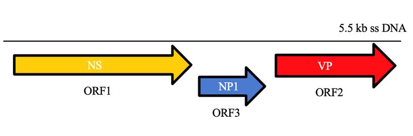
these parvoviruses. among parvoviruses since they contain an additional ORF (ORF3),
BoVS are unique
located
BoVSbetween the non-structural
are unique (ORF1) since
among parvoviruses and structural (ORF2)
they contain coding regions
an additional of their
ORF (ORF3),
genome, a 5.5-kb ssDNA (Figure 1). ORF3 encodes for NP1, a highly phosphorylated
located between the non-structural (ORF1) and structural (ORF2) coding regions of their
protein that
genome, is notssDNA
a 5.5-kb similar(Figure
to proteins of other
1). ORF3 parvoviruses
encodes for NP1, and playsphosphorylated
a highly a role in RNA
protein that is not similar to proteins of other parvoviruses and plays a role in of
processing. NP1 controls the splicing of VP-encoding RNAs and read-through RNAthe
proximal polyadenylation
processing. NP1 controls [3,98–100].
the splicing of VP-encoding RNAs and read-through of the
proximal polyadenylation [3,98–100].
Figure 1. Schematic genome organization of bocaviruses.
Figure 1. Schematic genome organization of bocaviruses.
Following ICTV classification criteria, BoVs are classified into twenty-five officially
recognized species, of which at least five species have been detected in domestic carnivores
Following ICTV classification criteria, BoVs are classified into twenty-five officially
(Carnivore bocaparvovirus 1–5), while a sixth species (Carnivore bocaparvovirus 6) was found
recognized species, of which at least five species have been detected in domestic carni-
in mink [3,4,6,90]. Currently, feline bocaparvoviruses (FBoVs) identified in domestic cats
vores (Carnivore bocaparvovirus 1–5), while a sixth species (Carnivore bocaparvovirus 6) was
found in mink [3,4,6,90]. Currently, feline bocaparvoviruses (FBoVs) identified in do-Viruses 2021, 13, 1077 5 of 11
are classified within the species Carnivore bocaparvovirus 3 to 5 [15–17] (Table 1). FBoV DNA
was detected for the first time in 2012, using molecular tools, in feces, nasal swabs, urine,
kidney, and blood collected from stray cats in Hong Kong [15]. The near-complete genomic
sequences (5179–5331 nt) were obtained from two fecal samples (HK797F and HK875F)
and from a urine sample (HK797U) of two cats. After sequence analysis, the three FBoV
genomes showed an identity of 58.6–59.7% to canine minute virus [15,91].
In agreement with new strict classification criteria, the newly discovered feline par-
voviruses have been classified within a species named Carnivore bocaparvovirus 3 (subspecies
FBoV-1) [1,3,4,6]. Subsequently, using a metagenomics approach on nucleic acids of en-
riched viral particles from the feces of a single healthy cat in Portugal [16], an additional
near-complete bocavirus genome was sequenced (strain POR1). Strain POR1 shares an aa
identity of 58% for NS1 and of 70% for VP1 with the three FBoVs previously identified
in Hong Kong [15,16]. Accordingly, strain POR1 has been classified as a distinct species,
Carnivore bocaparvovirus 4 (FBoV-2) [16]. A third strain of feline bocavirus (FBoV-3) was
detected in 2014 in fecal pools collected from 25 cats from a shelter in California using next-
generation sequencing (NGS). FBoV-3 has been accepted as a prototype of an additional
species, Carnivore bocaparvovirus 5, since it shares an aa sequence identity of 68% for NS1
and of 76% for VP1 with the other feline bocavirus strains previously identified [17].
Since then, different genomes of FBoV have been reported in Europe, China, Japan,
Thailand, and Canada from the feces of cats with and without clinical signs [16,19,93,101–103].
This suggests that different FBoVs circulate in cats in different geographical areas, without
any unambiguous correlation between FBoV genetic diversity and biological properties.
FBoV-1 infection could pose a concrete health risk to cats since it has been associ-
ated with enteritis [104,105]. Several studies have shown that FBoV-1 is more likely to be
detected in cats with diarrhea than in healthy cats [93,102–104]. In a molecular survey per-
formed in China, FBoV-1 DNA was found in 7.7% of the feces from cats with severe enteritis,
while it was not detected in healthy animals [102]. In another investigation conducted in
China, FBoV-1 DNA was evident in 2.8% of cats with severe enteritis, with a statistically
significant association between FBoV-1 infection and the presence of diarrhea [103]. In a
more recent study carried out in Thailand, three independent outbreaks of hemorrhagic
enteritis in household cats associated with FBoV-1 infection were described [93].
Moreover, the detection of FBoV-1 DNA in multiple tissues, including fecal, urine,
blood, respiratory, and kidney samples, collected from stray cats in Hong Kong, suggests a
wide tissue tropism [15].
On the other hand, whether FBoV-2 and 3 can play a role in the occurrence of gas-
troenteritis or other feline diseases remains unclear. Indeed, these viruses were repeatedly
found in fecal samples of healthy cats [16,17]. In a Japanese study, the FBoV-2 genome was
identified in rectal swabs collected from healthy cats (8.4%) and from cats with gastroen-
teritis (11.32%), without a statistically significant association between viral DNA detection
and the presence of clinical signs [101].
In most cases, FBoV DNA was co-detected in cats with other viral pathogens such as
FPV [93], fechaviruses [19], rotaviruses, astroviruses, bocaviruses, sakobuviruses, and/or
picobirnaviruses [16,22,101], suggesting that FBoV could be considered a common com-
ponent of the feline fecal virome. Although the role of systemic infection of cats remains
undetermined, a pathogenic role of FBoV enteric infection is possible. Synergistic effects of
co-infections with other enteric viruses could lead to more severe clinical signs, such as
hemorrhagic enteritis [93,102].
Including systematically FBoVs in the diagnostic algorithm of feline viral enteritis,
using specific molecular tools, could help us to better understand the enteropathogenic
potential of these viruses and the possible correlation between the genetic diversity and
the biological proprieties of each different FBoV species.Viruses 2021, 13, 1077 6 of 11
4. Chaphamaparvoviruses
The genus Chaphamaparvovirus (ChPV) (subfamily Hamaparvovirinae), recently intro-
duced in the family Parvoviridae, includes viruses genetically more related to invertebrate-
infecting parvoviruses than to members of the subfamily Parvovirinae. Future detection and
characterization of new viruses related to currently recognized members of this proposed
taxon might eventually result in splitting the currently recognized single genus into more
genera. Currently, however, clustering of these viruses as a single genus is the only com-
mon node characterized by significant topology support by both Bayesian and maximum
likelihood-based inference [2–4,6].
After the first identification in oropharyngeal swab samples collected from a fruit bat
(Eidolon helvum) in Ghana [106], ChPV-like viruses have been reported in several additional
animal species [2], including dogs and cats [18,19,107–109]. The first description of ChPV
in domestic carnivores occurred in the USA in 2017, where an NGS approach was used
on the feces of two dogs with hemorrhagic diarrhea of unknown etiology [107]. Later,
viruses genetically close to the American canine ChPV strains were found in the feces
of dogs and cats in China and Italy [18,108,109]. In agreement with ICTV classification
criteria, all strains of canine and feline (FeChPV) origin have been segregated in the new
species referred to as Carnivore chaphamaparvovirus 1 (CaChPV-1), sharing an overall aa
identity of 98.6–99.8% in the NS1 protein [4] (Table 1). Recently, a new feline ChPV strain
was recovered from feline feces during an outbreak of vomiting and diarrhea in a multi-
facility feline shelter in Canada using a viral metagenomic approach [19]. NS1 protein
shared 76.0–77.0% aa identity with CaChPV-1 strains previously detected in feline and
canine samples, so that this strain was allocated in another species named Fechavirus [4,19]
(Table 1).
In a recent case–control study carried out in Italy, FeChPV has been identified from
fecal and respiratory samples of cats, displaying a correlation with acute gastroenteri-
tis [110]. Since the Italian strains shared more than 97.7% aa identity in the NS1 protein
with Canadian prototype viruses, they have been clustered into a monophyletic, well-
distinguished species (Fechavirus) with respect to the FeChPV strains found in China and
currently classified as the species Carnivore chaphamaparvovirus 1 [2,18,19,110]. Feciavirus
infection has been correlated with acute gastroenteritis, whereas no correlation has been
found with upper respiratory tract disease [110].
Interestingly, in the first report on the detection of ChPV-related parvovirus in do-
mestic cats in China, there was an attempt to isolate the virus on cell cultures, with the
observation of a cytopathic effect only up to the fifth generation of cultured cells that
were coinfected by FeChPV and FPV [18]. All subsequent studies were mainly focused on
the detection of ChPV DNA in cats using molecular approaches, aiming to highlight the
epidemiology and genetic heterogeneity of these viruses and to find possible correlations
between the presence of viral DNA and clinical signs. To date, however, the information
regarding ChPV in cats is still limited and further studies are needed to investigate im-
portant aspects such as the host range in vitro (the ability of these viruses to adapt to the
in vitro growth is still unclear) and the possibility of detection by immunohistology in the
tissues of infected cats [18,19,110].
The potential clinical impact of ChPVs on feline health and their possible role as
primary enteric/respiratory pathogens remain to be clarified [19,110]. Indeed, ChPVs
DNA was often co-detected in cats with other viral pathogens such as FBoV (Li Y. et al.,
2020), feline coronavirus, kobuvirus, and norovirus [110]. Further epidemiological data
collected from independent studies in other geographical areas are required to confirm the
preliminary findings available so far and assess whether these new parvoviruses can be
considered a stable and common component of the host virome or, on the contrary, they
play a role in the development of disease in infected cats.Viruses 2021, 13, 1077 7 of 11
5. Conclusions
In the last twenty years, using new molecular techniques and metagenomic approaches
for the screening of feline samples, several lineages and species of parvoviruses have been
found in association with enteric and/or respiratory disease in cats. Although several
aspects concerning epidemiology and virus–host interaction remain to be clarified, some
pieces of evidence suggest these emerging feline parvoviruses may act as primary causative
pathogens or synergistic agents in the occurrence of clinical signs in cats. Each emerging
parvovirus should be included systematically in diagnostic algorithms for detection of
feline viral pathogens, chiefly for cats with enteric and/or respiratory disease. Moreover,
large structured epidemiological studies and experimental infections might help clarify
any possible association of emerging parvoviruses with the occurrence of disease and their
distribution in the feline population.
Interestingly, the multi-species circulation of many of these emerging parvoviruses
could represent a concrete problem when devising prophylactic measures in animals living
in the same environment and, in particular, in mixed cat–dog shelters and veterinary clinics.
Vaccines are not available for emerging parvoviruses of cats, so that vaccination
protocols cannot prevent the spread of these viruses, for some of which the cat could
represent the host reservoir. Therefore, in order to limit inter- and intra-species spread
as much as possible, prophylaxis plans should consider strong disinfection protocols and
physical separation, particularly in those facilities housing both dogs and cats.
Author Contributions: Conceptualization, N.D. and V.M.; writing—original draft preparation, P.C.;
supervision, C.B.; writing—review and editing, all authors. All authors have read and agreed to the
published version of the manuscript.
Funding: This research received no external funding.
Conflicts of Interest: The authors declare no conflict of interest.
References
1. Cotmore, S.F.; Agbandje-McKenna, M.; Canuti, M.; Chiorini, J.A.; Eis-Hubinger, A.M.; Hughes, J.; Mietzsch, M.; Modha, S.;
Ogliastro, M.; Penzes, J.J.; et al. ICTV Virus Taxonomy Profile: Parvoviridae. J. Gen. Virol. 2019, 100, 367–368. [CrossRef]
2. Penzes, J.J.; de Souza, W.M.; Agbandje-McKenna, M.; Gifford, R.J. An Ancient Lineage of Highly Divergent Parvoviruses Infects
both Vertebrate and Invertebrate Hosts. Viruses 2019, 11, 525. [CrossRef] [PubMed]
3. ICTV—International Committee on Taxonomy of Viruses. Available online: https://talk.ictvonline.org/ictv-reports/ictv_online_
report/ssdna-viruses/w/parvoviridae (accessed on 4 March 2021).
4. Penzes, J.J.; Soderlund-Venermo, M.; Canuti, M.; Eis-Hubinger, A.M.; Hughes, J.; Cotmore, S.F.; Harrach, B. Reorganizing the
family Parvoviridae: A revised taxonomy independent of the canonical approach based on host association. Arch. Virol. 2020,
165, 2133–2146. [CrossRef] [PubMed]
5. Reed, A.P.; Jones, E.V.; Miller, T.J. Nucleotide sequence and genome organization of canine parvovirus. J. Virol. 1988, 62, 266–276.
[CrossRef]
6. Chung, H.C.; Kim, S.J.; Nguyen, V.G.; Shin, S.; Kim, J.Y.; Lim, S.K.; Park, Y.H.; Park, B. New genotype classification and molecular
characterization of canine and feline parvoviruses. J. Vet. Sci. 2020, 21, e43. [CrossRef] [PubMed]
7. Parrish, C.R. Emergence, natural history, and variation of canine, mink, and feline parvoviruses. Adv. Virus Res. 1990, 38, 403–450.
[CrossRef]
8. MacLachlan, N.J.; Dubovi, E.J. (Eds.) Parvoviridae. In Fenner’s Veterinary Virology, 5th ed.; Academic Press: Cambridge, MA, USA,
2017; pp. 245–257. [CrossRef]
9. Verge, J.; Christoforoni, N. La gastroenterite infectieuse des chats; est-elle due à un virus filtrable? CR Seances Soc. Biol. Fil. 1928,
99, 312.
10. Hindle, E.; Findlay, G.M. Studies on feline distemper. Comp. Pathol. Ther. 1932, 45, 11–26. [CrossRef]
11. Ndiana, L.A.; Lanave, G.; Desario, C.; Berjaoui, S.; Alfano, F.; Puglia, I.; Fusco, G.; Colaianni, M.L.; Vincifori, G.; Camarda, A.; et al.
Circulation of diverse protoparvoviruses in wild carnivores, Italy. Transbound. Emerg. Dis. 2020. [CrossRef]
12. Barrs, V.R. Feline Panleukopenia: A Re-emergent Disease. Vet Clin. North Am. Small Anim. Pract. 2019, 49, 651–670. [CrossRef]
13. Mochizuki, M.; Harasawa, R.; Nakatani, H. Antigenic and genomic variabilities among recently prevalent parvoviruses of canine
and feline origin in Japan. Vet. Microbiol. 1993, 38, 1–10. [CrossRef]
14. Diakoudi, G.; Lanave, G.; Capozza, P.; Di Profio, F.; Melegari, I.; Di Martino, B.; Pennisi, M.G.; Elia, G.; Cavalli, A.;
Tempesta, M.; et al. Identification of a novel parvovirus in domestic cats. Vet. Microbiol. 2019, 228, 246–251. [CrossRef]Viruses 2021, 13, 1077 8 of 11
15. Lau, S.K.P.; Woo, P.C.Y.; Yeung, H.C.; Teng, J.L.L.; Wu, Y.; Bai, R.; Fan, R.Y.Y.; Chan, K.H.; Yuen, K.Y. Identification and
characterization of bocaviruses in cats and dogs reveals a novel feline bocavirus and a novel genetic group of canine bocavirus. J.
Gen. Virol. 2012, 93, 1573–1582. [CrossRef]
16. Ng, T.F.; Mesquita, J.R.; Nascimento, M.S.; Kondov, N.O.; Wong, W.; Reuter, G.; Knowles, N.J.; Vega, E.; Esona, M.D.; Deng, X.; et al.
Feline fecal virome reveals novel and prevalent enteric viruses. Vet. Microbiol. 2014, 171, 102–111. [CrossRef] [PubMed]
17. Zhang, W.; Li, L.; Deng, X.; Kapusinszky, B.; Pesavento, P.A.; Delwart, E. Faecal virome of cats in an animal shelter. J. Gen. Virol.
2014, 95, 2553–2564. [CrossRef]
18. Ji, J.; Hu, W.; Liu, Q.; Zuo, K.; Zhi, G.; Xu, X.; Kan, Y.; Yao, L.; Xie, Q. Genetic Analysis of Cachavirus-Related Parvoviruses
Detected in Pet Cats: The First Report From China. Front Vet. Sci. 2020, 7, 580836. [CrossRef] [PubMed]
19. Li, Y.; Gordon, E.; Idle, A.; Altan, E.; Seguin, M.A.; Estrada, M.; Deng, X.; Delwart, E. Virome of a Feline Outbreak of Diarrhea and
Vomiting Includes Bocaviruses and a Novel Chapparvovirus. Viruses 2020, 12, 506. [CrossRef]
20. Greene, E.C. Feline enteric viral infections. In Infectious Diseases of the Dog and Cat, 4th ed.; Greene, C.E., Ed.; Linda Duncan:
St. Louis, MO, USA, 2012; pp. 80–88.
21. Jenkins, E.; Davis, C.; Carrai, M.; Ward, M.P.; O’Keeffe, S.; van Boeijen, M.; Beveridge, L.; Desario, C.; Buonavoglia, C.;
Beatty, J.A.; et al. Feline Parvovirus Seroprevalence Is High in Domestic Cats from Disease Outbreak and Non-Outbreak Regions
in Australia. Viruses 2020, 12, 320. [CrossRef] [PubMed]
22. Di Martino, B.; Di Profio, F.; Melegari, I.; Marsilio, F. Feline Virome—A Review of Novel Enteric Viruses Detected in Cats. Viruses
2019, 11, 908. [CrossRef]
23. Decaro, N.; Buonavoglia, C. Canine parvovirus—a review of epidemiological and diagnostic aspects, with emphasis on type 2c.
Vet. Microbiol. 2012, 155, 1–12. [CrossRef]
24. Decaro, N.; Buonavoglia, D.; Desario, C.; Amorisco, F.; Colaianni, M.L.; Parisi, A.; Terio, V.; Elia, G.; Lucente, M.S.; Cavalli, A.; et al.
Characterisation of canine parvovirus strains isolated from cats with feline panleukopenia. Res. Vet. Sci. 2010, 89, 275–278.
[CrossRef] [PubMed]
25. Decaro, N.; Buonavoglia, C.; Barrs, V.R. Canine parvovirus vaccination and immunisation failures: Are we far from disease
eradication? Vet. Microbiol. 2020, 247, 108760. [CrossRef] [PubMed]
26. Decaro, N.; Elia, G.; Buonavoglia, C. Challenge studies for registration of canine core vaccines: Is it time to update the European
Pharmacopeia? Vet. Microbiol. 2020, 244, 108659. [CrossRef] [PubMed]
27. Truyen, U. Evolution of canine parvovirus—A need for new vaccines? Vet. Microbiol. 2006, 117, 9–13. [CrossRef]
28. Decaro, N.; Buonavoglia, C. Canine parvovirus post-vaccination shedding: Interference with diagnostic assays and correlation
with host immune status. Vet. J. 2017, 221, 23–24. [CrossRef] [PubMed]
29. Nandi, S.; Chidri, S.; Kumar, M.; Chauhan, R.S. Occurrence of canine parvovirus type 2c in the dogs with haemorrhagic enteritis
in India. Res. Vet. Sci. 2010, 88, 169–171. [CrossRef]
30. Decaro, N.; Desario, C.; Miccolupo, A.; Campolo, M.; Parisi, A.; Martella, V.; Amorisco, F.; Lucente, M.S.; Lavazza, A.;
Buonavoglia, C. Genetic analysis of feline panleukopenia viruses from cats with gastroenteritis. J. Gen. Virol. 2008, 89, 2290–2298.
[CrossRef]
31. Shackelton, L.A.; Parrish, C.R.; Truyen, U.; Holmes, E.C. High rate of viral evolution associated with the emergence of carnivore
parvovirus. Proc. Natl. Acad. Sci. USA 2005, 102, 379–384. [CrossRef]
32. Pereira, C.A.; Leal, E.S.; Durigon, E.L. Selective regimen shift and demographic growth increase associated with the emergence of
high-fitness variants of canine parvovirus. Infect. Genet. Evol. 2007, 7, 399–409. [CrossRef]
33. Hoelzer, K.; Parrish, C.R. The emergence of parvoviruses of carnivores. Vet. Res. 2010, 41, 39. [CrossRef] [PubMed]
34. Decaro, N.; Desario, C.; Parisi, A.; Martella, V.; Lorusso, A.; Miccolupo, A.; Mari, V.; Colaianni, M.L.; Cavalli, A.; Di Trani, L.; et al.
Genetic analysis of canine parvovirus type 2c. Virology 2009, 385, 5–10. [CrossRef]
35. Parrish, C.R.; Aquadro, C.F.; Strassheim, M.L.; Evermann, J.F.; Sgro, J.Y.; Mohammed, H.O. Rapid antigenic-type replacement and
DNA sequence evolution of canine parvovirus. J. Virol. 1991, 65, 6544–6552. [CrossRef] [PubMed]
36. Parrish, C.R.; O’Connell, P.H.; Evermann, J.F.; Carmichael, L.E. Natural variation of canine parvovirus. Science 1985,
230, 1046–1048. [CrossRef] [PubMed]
37. Buonavoglia, C.; Martella, V.; Pratelli, A.; Tempesta, M.; Cavalli, A.; Buonavoglia, D.; Bozzo, G.; Elia, G.; Decaro, N.; Carmichael, L.
Evidence for evolution of canine parvovirus type 2 in Italy. J. Gen. Virol. 2001, 82, 3021–3025. [CrossRef]
38. Martella, V.; Cavalli, A.; Pratelli, A.; Bozzo, G.; Camero, M.; Buonavoglia, D.; Narcisi, D.; Tempesta, M.; Buonavoglia, C. A canine
parvovirus mutant is spreading in Italy. J. Clin. Microbiol. 2004, 42, 1333–1336. [CrossRef] [PubMed]
39. Nakamura, M.; Tohya, Y.; Miyazawa, T.; Mochizuki, M.; Phung, H.T.; Nguyen, N.H.; Huynh, L.M.; Nguyen, L.T.; Nguyen, P.N.;
Nguyen, P.V.; et al. A novel antigenic variant of Canine parvovirus from a Vietnamese dog. Arch. Virol. 2004, 149, 2261–2269.
[CrossRef] [PubMed]
40. Decaro, N.; Desario, C.; Addie, D.D.; Martella, V.; Vieira, M.J.; Elia, G.; Zicola, A.; Davis, C.; Thompson, G.; Thiry, E.; et al. The
study molecular epidemiology of canine parvovirus, Europe. Emerg. Infect. Dis. 2007, 13, 1222–1224. [CrossRef]
41. Decaro, N.; Elia, G.; Martella, V.; Desario, C.; Campolo, M.; Trani, L.D.; Tarsitano, E.; Tempesta, M.; Buonavoglia, C. A real-time
PCR assay for rapid detection and quantitation of canine parvovirus type 2 in the feces of dogs. Vet. Microbiol. 2005, 105, 19–28.
[CrossRef]Viruses 2021, 13, 1077 9 of 11
42. Decaro, N.; Martella, V.; Desario, C.; Bellacicco, A.L.; Camero, M.; Manna, L.; d’Aloja, D.; Buonavoglia, C. First detection of canine
parvovirus type 2c in pups with haemorrhagic enteritis in Spain. J. Vet Med. B Infect. Dis. Vet Public Health 2006, 53, 468–472.
[CrossRef]
43. Perez, R.; Francia, L.; Romero, V.; Maya, L.; Lopez, I.; Hernandez, M. First detection of canine parvovirus type 2c in South
America. Vet. Microbiol. 2007, 124, 147–152. [CrossRef]
44. Hong, C.; Decaro, N.; Desario, C.; Tanner, P.; Pardo, M.C.; Sanchez, S.; Buonavoglia, C.; Saliki, J.T. Occurrence of canine parvovirus
type 2c in the United States. J. Vet. Diagn. Investig. 2007, 19, 535–539. [CrossRef]
45. Calderon, M.G.; Mattion, N.; Bucafusco, D.; Fogel, F.; Remorini, P.; La Torre, J. Molecular characterization of canine parvovirus
strains in Argentina: Detection of the pathogenic variant CPV2c in vaccinated dogs. J. Virol. Methods 2009, 159, 141–145. [CrossRef]
46. Joao Vieira, M.; Silva, E.; Oliveira, J.; Luisa Vieira, A.; Decaro, N.; Desario, C.; Muller, A.; Carvalheira, J.; Buonavoglia, C.;
Thompson, G. Canine parvovirus 2c infection in central Portugal. J. Vet. Diagn. Investig. 2008, 20, 488–491. [CrossRef] [PubMed]
47. Charoenkul, K.; Tangwangvivat, R.; Janetanakit, T.; Boonyapisitsopa, S.; Bunpapong, N.; Chaiyawong, S.; Amonsin, A. Emergence
of canine parvovirus type 2c in domestic dogs and cats from Thailand. Transbound. Emerg. Dis. 2019, 66, 1518–1528. [CrossRef]
[PubMed]
48. Chiang, S.Y.; Wu, H.Y.; Chiou, M.T.; Chang, M.C.; Lin, C.N. Identification of a novel canine parvovirus type 2c in Taiwan. Virol. J.
2016, 13, 160. [CrossRef] [PubMed]
49. Zhao, Z.; Liu, H.; Ding, K.; Peng, C.; Xue, Q.; Yu, Z.; Xue, Y. Occurrence of canine parvovirus in dogs from Henan province of
China in 2009–2014. BMC Vet. Res. 2016, 12, 138. [CrossRef] [PubMed]
50. Kelman, M.; Norris, J.M.; Barrs, V.R.; Ward, M.P. A history of canine parvovirus in Australia: What can we learn? Aust. Vet. J.
2020, 98, 504–510. [CrossRef]
51. Greene, E.C.; Decaro, N. Canine viral enteritis. In Infectious Diseases of the Dog and Cat, 4th ed.; Greene, C.E., Ed.; Linda Duncan:
St. Louis, MO, USA, 2012; pp. 67–79.
52. Truyen, U.; Platzer, G.; Parrish, C.R. Antigenic type distribution among canine parvoviruses in dogs and cats in Germany. Vet.
Rec. 1996, 138, 365–366. [CrossRef]
53. Decaro, N.; Desario, C.; Amorisco, F.; Losurdo, M.; Colaianni, M.L.; Greco, M.F.; Buonavoglia, C. Canine parvovirus type 2c
infection in a kitten associated with intracranial abscess and convulsions. J. Feline Med. Surg. 2011, 13, 231–236. [CrossRef]
54. Ikeda, Y.; Mochizuki, M.; Naito, R.; Nakamura, K.; Miyazawa, T.; Mikami, T.; Takahashi, E. Predominance of canine parvovirus
(CPV) in unvaccinated cat populations and emergence of new antigenic types of CPVs in cats. Virology 2000, 278, 13–19. [CrossRef]
[PubMed]
55. Balboni, A.; Bassi, F.; De Arcangeli, S.; Zobba, R.; Dedola, C.; Alberti, A.; Battilani, M. Molecular analysis of carnivore Protopar-
vovirus detected in white blood cells of naturally infected cats. BMC Vet. Res. 2018, 14, 41. [CrossRef]
56. Mochizuki, M.; Horiuchi, M.; Hiragi, H.; San Gabriel, M.C.; Yasuda, N.; Uno, T. Isolation of canine parvovirus from a cat
manifesting clinical signs of feline panleukopenia. J. Clin. Microbiol. 1996, 34, 2101–2105. [CrossRef]
57. Gamoh, K.; Shimazaki, Y.; Makie, H.; Senda, M.; Itoh, O.; Inoue, Y. The pathogenicity of canine parvovirus type-2b, FP84 strain
isolated from a domestic cat, in domestic cats. J. Vet. Med. Sci. 2003, 65, 1027–1029. [CrossRef]
58. Gamoh, K.; Shimazaki, Y.; Senda, M.; Makie, H.; Itoh, O.; Inoue, Y. Antigenic type distribution of parvovirus isolated from
domestic cats in Japan. Vet. Rec. 2003, 153, 751–752.
59. Battilani, M.; Balboni, A.; Ustulin, M.; Giunti, M.; Scagliarini, A.; Prosperi, S. Genetic complexity and multiple infections with
more Parvovirus species in naturally infected cats. Vet. Res. 2011, 42, 43. [CrossRef]
60. Battilani, M.; Balboni, A.; Giunti, M.; Prosperi, S. Co-infection with feline and canine parvovirus in a cat. Vet. Ital. 2013,
49, 127–129. [PubMed]
61. Battilani, M.; Scagliarini, A.; Ciulli, S.; Morganti, L.; Prosperi, S. High genetic diversity of the VP2 gene of a canine parvovirus
strain detected in a domestic cat. Virology 2006, 352, 22–26. [CrossRef] [PubMed]
62. Miranda, C.; Parrish, C.R.; Thompson, G. Canine parvovirus 2c infection in a cat with severe clinical disease. J. Vet. Diagn. Invest.
2014, 26, 462–464. [CrossRef] [PubMed]
63. Nakamura, K.; Sakamoto, M.; Ikeda, Y.; Sato, E.; Kawakami, K.; Miyazawa, T.; Tohya, Y.; Takahashi, E.; Mikami, T.; Mochizuki, M.
Pathogenic potential of canine parvovirus types 2a and 2c in domestic cats. Clin. Diagn. Lab. Immunol. 2001, 8, 663–668. [CrossRef]
64. Truyen, U.; Evermann, J.F.; Vieler, E.; Parrish, C.R. Evolution of canine parvovirus involved loss and gain of feline host range.
Virology 1996, 215, 186–189. [CrossRef]
65. Clegg, S.R.; Coyne, K.P.; Dawson, S.; Spibey, N.; Gaskell, R.M.; Radford, A.D. Canine parvovirus in asymptomatic feline carriers.
Vet. Microbiol. 2012, 157, 78–85. [CrossRef]
66. Marenzoni, M.L.; Antognoni, M.T.; Baldelli, F.; Miglio, A.; Stefanetti, V.; Desario, C.; Di Summa, A.; Buonavoglia, C.; Decaro, N.
Detection of parvovirus and herpesvirus DNA in the blood of feline and canine blood donors. Vet. Microbiol. 2018, 224, 66–69.
[CrossRef]
67. Byrne, P.; Beatty, J.A.; Slapeta, J.; Corley, S.W.; Lyons, R.E.; McMichael, L.; Kyaw-Tanner, M.T.; Dung, P.T.; Decaro, N.;
Meers, J.; et al. Shelter-housed cats show no evidence of faecal shedding of canine parvovirus DNA. Vet. J. 2018, 239, 54–58.
[CrossRef] [PubMed]
68. Stuetzer, B.; Hartmann, K. Feline parvovirus infection and associated diseases. Vet. J. 2014, 201, 150–155. [CrossRef] [PubMed]Viruses 2021, 13, 1077 10 of 11
69. Url, A.; Truyen, U.; Rebel-Bauder, B.; Weissenbock, H.; Schmidt, P. Evidence of parvovirus replication in cerebral neurons of cats.
J. Clin. Microbiol. 2003, 41, 3801–3805. [CrossRef] [PubMed]
70. Calatayud, O.; Esperon, F.; Velarde, R.; Oleaga, A.; Llaneza, L.; Ribas, A.; Negre, N.; de la Torre, A.; Rodriguez, A.; Millan, J.
Genetic characterization of Carnivore Parvoviruses in Spanish wildlife reveals domestic dog and cat-related sequences. Transbound.
Emerg. Dis. 2020, 67, 626–634. [CrossRef]
71. Mukhopadhyay, H.K.; Nookala, M.; Thangamani, N.R.; Sivaprakasam, A.; Antony, P.X.; Thanislass, J.; Srinivas, M.V.; Pillai, R.M.
Molecular characterisation of parvoviruses from domestic cats reveals emergence of newer variants in India. J. Feline Med. Surg.
2017, 19, 846–852. [CrossRef] [PubMed]
72. Truyen, U.; Parrish, C.R. Canine and feline host ranges of canine parvovirus and feline panleukopenia virus: Distinct host cell
tropisms of each virus in vitro and in vivo. J. Virol. 1992, 66, 5399–5408. [CrossRef]
73. Martella, V.; Lanave, G.; Mihalov-Kovacs, E.; Marton, S.; Varga-Kugler, R.; Kaszab, E.; Di Martino, B.; Camero, M.; Decaro, N.;
Buonavoglia, C.; et al. Novel Parvovirus Related to Primate Bufaviruses in Dogs. Emerg. Infect. Dis. 2018, 24, 1061–1068.
[CrossRef]
74. Phan, T.G.; Vo, N.P.; Bonkoungou, I.J.; Kapoor, A.; Barro, N.; O’Ryan, M.; Kapusinszky, B.; Wang, C.; Delwart, E. Acute diarrhea
in West African children: Diverse enteric viruses and a novel parvovirus genus. J. Virol. 2012, 86, 11024–11030. [CrossRef]
75. Yahiro, T.; Wangchuk, S.; Tshering, K.; Bandhari, P.; Zangmo, S.; Dorji, T.; Tshering, K.; Matsumoto, T.; Nishizono, A.;
Soderlund-Venermo, M.; et al. Novel human bufavirus genotype 3 in children with severe diarrhea, Bhutan. Emerg. Infect.
Dis. 2014, 20, 1037–1039. [CrossRef]
76. Handley, S.A.; Thackray, L.B.; Zhao, G.; Presti, R.; Miller, A.D.; Droit, L.; Abbink, P.; Maxfield, L.F.; Kambal, A.; Duan, E.; et al.
Pathogenic simian immunodeficiency virus infection is associated with expansion of the enteric virome. Cell 2012, 151, 253–266.
[CrossRef]
77. Melegari, I.; Di Profio, F.; Palombieri, A.; Sarchese, V.; Diakoudi, G.; Robetto, S.; Orusa, R.; Marsilio, F.; Banyai, K.; Martella, V.; et al.
Molecular detection of canine bufaviruses in wild canids. Arch. Virol. 2019, 164, 2315–2320. [CrossRef]
78. Vaisanen, E.; Paloniemi, M.; Kuisma, I.; Lithovius, V.; Kumar, A.; Franssila, R.; Ahmed, K.; Delwart, E.; Vesikari, T.;
Hedman, K.; et al. Epidemiology of two human protoparvoviruses, bufavirus and tusavirus. Sci. Rep. 2016, 6, 39267. [CrossRef]
79. Sun, W.; Zhang, S.; Huang, H.; Wang, W.; Cao, L.; Zheng, M.; Yin, Y.; Zhang, H.; Lu, H.; Jin, N. First identification of a novel
parvovirus distantly related to human bufavirus from diarrheal dogs in China. Virus Res. 2019, 265, 127–131. [CrossRef]
80. Sasaki, M.; Orba, Y.; Anindita, P.D.; Ishii, A.; Ueno, K.; Hang’ombe, B.M.; Mweene, A.S.; Ito, K.; Sawa, H. Distinct Lineages of
Bufavirus in Wild Shrews and Nonhuman Primates. Emerg. Infect. Dis. 2015, 21, 1230–1233. [CrossRef]
81. Siqueira, J.D.; Ng, T.F.; Miller, M.; Li, L.; Deng, X.; Dodd, E.; Batac, F.; Delwart, E. Endemic Infection of Stranded Southern Sea
Otters (Enhydra Lutris Nereis) with Novel Parvovirus, Polyomavirus, and Adenovirus. J. Wildl. Dis. 2017, 53, 532–542. [CrossRef]
[PubMed]
82. Cheng, W.X.; Li, J.S.; Huang, C.P.; Yao, D.P.; Liu, N.; Cui, S.X.; Jin, Y.; Duan, Z.J. Identification and nearly full-length genome
characterization of novel porcine bocaviruses. PLoS ONE 2010, 5, e13583. [CrossRef] [PubMed]
83. Shan, T.; Lan, D.; Li, L.; Wang, C.; Cui, L.; Zhang, W.; Hua, X.; Zhu, C.; Zhao, W.; Delwart, E. Genomic characterization and high
prevalence of bocaviruses in swine. PLoS ONE 2011, 6, e17292. [CrossRef]
84. Chen, K.C.; Shull, B.C.; Moses, E.A.; Lederman, M.; Stout, E.R.; Bates, R.C. Complete nucleotide sequence and genome
organization of bovine parvovirus. J. Virol. 1986, 60, 1085–1097. [CrossRef] [PubMed]
85. Li, L.; Shan, T.; Wang, C.; Cote, C.; Kolman, J.; Onions, D.; Gulland, F.M.; Delwart, E. The fecal viral flora of California sea lions. J.
Virol. 2011, 85, 9909–9917. [CrossRef] [PubMed]
86. Wu, Z.; Ren, X.; Yang, L.; Hu, Y.; Yang, J.; He, G.; Zhang, J.; Dong, J.; Sun, L.; Du, J.; et al. Virome analysis for identification of
novel mammalian viruses in bat species from Chinese provinces. J. Virol. 2012, 86, 10999–11012. [CrossRef]
87. Lanave, G.; Martella, V.; Farkas, S.L.; Marton, S.; Feher, E.; Bodnar, L.; Lavazza, A.; Decaro, N.; Buonavoglia, C.; Banyai, K. Novel
bocaparvoviruses in rabbits. Vet. J. 2015, 206, 131–135. [CrossRef]
88. Lau, S.K.; Yeung, H.C.; Li, K.S.; Lam, C.S.; Cai, J.P.; Yuen, M.C.; Wang, M.; Zheng, B.J.; Woo, P.C.; Yuen, K.Y. Identification and
genomic characterization of a novel rat bocavirus from brown rats in China. Infect. Genet. Evol. 2017, 47, 68–76. [CrossRef]
89. Van den Brand, J.M.; van Leeuwen, M.; Schapendonk, C.M.; Simon, J.H.; Haagmans, B.L.; Osterhaus, A.D.; Smits, S.L. Metage-
nomic analysis of the viral flora of pine marten and European badger feces. J. Virol. 2012, 86, 2360–2365. [CrossRef]
90. Yang, S.; Wang, Y.; Li, W.; Fan, Z.; Jiang, L.; Lin, Y.; Fu, X.; Shen, Q.; Sun, Z.; Wang, X.; et al. A novel bocavirus from domestic
mink, China. Virus Genes 2016, 52, 887–890. [CrossRef]
91. Binn, L.N.; Lazar, E.C.; Eddy, G.A.; Kajima, M. Recovery and characterization of a minute virus of canines. Infect. Immun. 1970,
1, 503–508. [CrossRef] [PubMed]
92. Kapoor, A.; Mehta, N.; Dubovi, E.J.; Simmonds, P.; Govindasamy, L.; Medina, J.L.; Street, C.; Shields, S.; Lipkin, W.I. Character-
ization of novel canine bocaviruses and their association with respiratory disease. J. Gen. Virol. 2012, 93, 341–346. [CrossRef]
[PubMed]
93. Piewbang, C.; Kasantikul, T.; Pringproa, K.; Techangamsuwan, S. Feline bocavirus-1 associated with outbreaks of hemorrhagic
enteritis in household cats: Potential first evidence of a pathological role, viral tropism and natural genetic recombination. Sci.
Rep. 2019, 9, 16367. [CrossRef] [PubMed]Viruses 2021, 13, 1077 11 of 11
94. Kapoor, A.; Mehta, N.; Esper, F.; Poljsak-Prijatelj, M.; Quan, P.L.; Qaisar, N.; Delwart, E.; Lipkin, W.I. Identification and
characterization of a new bocavirus species in gorillas. PLoS ONE 2010, 5, e11948. [CrossRef] [PubMed]
95. Sharp, C.P.; LeBreton, M.; Kantola, K.; Nana, A.; Diffo Jle, D.; Djoko, C.F.; Tamoufe, U.; Kiyang, J.A.; Babila, T.G.; Ngole, E.M.; et al.
Widespread infection with homologues of human parvoviruses B19, PARV4, and human bocavirus of chimpanzees and gorillas
in the wild. J. Virol. 2010, 84, 10289–10296. [CrossRef]
96. Allander, T.; Tammi, M.T.; Eriksson, M.; Bjerkner, A.; Tiveljung-Lindell, A.; Andersson, B. Cloning of a human parvovirus by
molecular screening of respiratory tract samples. Proc. Natl. Acad. Sci. USA 2005, 102, 12891–12896. [CrossRef]
97. Vicente, D.; Cilla, G.; Montes, M.; Perez-Yarza, E.G.; Perez-Trallero, E. Human bocavirus, a respiratory and enteric virus. Emerg.
Infect. Dis. 2007, 13, 636–637. [CrossRef]
98. Fasina, O.O.; Dong, Y.; Pintel, D.J. NP1 Protein of the Bocaparvovirus Minute Virus of Canines Controls Access to the Viral
Capsid Genes via Its Role in RNA Processing. J. Virol. 2016, 90, 1718–1728. [CrossRef] [PubMed]
99. Fasina, O.O.; Stupps, S.; Figueroa-Cuilan, W.; Pintel, D.J. Minute Virus of Canines NP1 Protein Governs the Expression of a Subset
of Essential Nonstructural Proteins via Its Role in RNA Processing. J. Virol. 2017, 91, e00260-17. [CrossRef] [PubMed]
100. Zou, W.; Cheng, F.; Shen, W.; Engelhardt, J.F.; Yan, Z.; Qiu, J. Nonstructural Protein NP1 of Human Bocavirus 1 Plays a Critical
Role in the Expression of Viral Capsid Proteins. J. Virol. 2016, 90, 4658–4669. [CrossRef]
101. Takano, T.; Takadate, Y.; Doki, T.; Hohdatsu, T. Genetic characterization of feline bocavirus detected in cats in Japan. Arch. Virol.
2016, 161, 2825–2828. [CrossRef]
102. Liu, C.; Liu, F.; Li, Z.; Qu, L.; Liu, D. First report of feline bocavirus associated with severe enteritis of cat in Northeast China,
2015. J. Vet. Med. Sci. 2018, 80, 731–735. [CrossRef]
103. Yi, S.; Niu, J.; Wang, H.; Dong, G.; Zhao, Y.; Dong, H.; Guo, Y.; Wang, K.; Hu, G. Detection and genetic characterization of feline
bocavirus in Northeast China. Virol. J. 2018, 15, 125. [CrossRef]
104. Wang, Y.; Li, W.; Guo, X.; Zhang, D.; Sun, J.; Fu, Z.; Liu, G.; Li, Y.; Jiang, S. Development of SYBR Green I-based polymerase chain
reaction for feline bocavirus 1 detection. 3 Biotech 2021, 11, 61. [CrossRef] [PubMed]
105. Wang, Y.; Sun, J.; Guo, X.; Zhang, D.; Cui, Y.; Li, W.; Liu, G.; Li, Y.; Jiang, S. TaqMan-based real-time polymerase chain reaction
assay for specific detection of bocavirus-1 in domestic cats. Mol. Cell. Probes 2020, 53, 101647. [CrossRef]
106. Baker, K.S.; Leggett, R.M.; Bexfield, N.H.; Alston, M.; Daly, G.; Todd, S.; Tachedjian, M.; Holmes, C.E.; Crameri, S.; Wang, L.F.; et al.
Metagenomic study of the viruses of African straw-coloured fruit bats: Detection of a chiropteran poxvirus and isolation of a
novel adenovirus. Virology 2013, 441, 95–106. [CrossRef] [PubMed]
107. Fahsbender, E.; Altan, E.; Seguin, M.A.; Young, P.; Estrada, M.; Leutenegger, C.; Delwart, E. Chapparvovirus DNA Found in 4%
of Dogs with Diarrhea. Viruses 2019, 11, 398. [CrossRef] [PubMed]
108. Hu, W.; Liu, Q.; Chen, Q.; Ji, J. Molecular characterization of Cachavirus firstly detected in dogs in China. Infect. Genet. Evol. 2020,
85, 104529. [CrossRef] [PubMed]
109. Palombieri, A.; Di Profio, F.; Lanave, G.; Capozza, P.; Marsilio, F.; Martella, V.; Di Martino, B. Molecular detection and
characterization of Carnivore chaphamaparvovirus 1 in dogs. Vet. Microbiol. 2020, 251, 108878. [CrossRef]
110. Di Profio, F.; Sarchese, V.; Palombieri, A.; Fruci, P.; Massirio, I.; Martella, V.; Fulvio, M.; Di Martino, B. Feline chaphamaparvovirus
in cats with enteritis and upper respiratory tract disease. Transbound. Emerg. Dis. 2021. [CrossRef] [PubMed]You can also read