Bioluminescence of the Largest Luminous Vertebrate, the Kitefin Shark, Dalatias licha: First Insights and Comparative Aspects - Frontiers
←
→
Page content transcription
If your browser does not render page correctly, please read the page content below
ORIGINAL RESEARCH
                                                                                                                                           published: 26 February 2021
                                                                                                                                      doi: 10.3389/fmars.2021.633582

                                            Bioluminescence of the Largest
                                            Luminous Vertebrate, the Kitefin
                                            Shark, Dalatias licha: First Insights
                                            and Comparative Aspects
                                            Jérôme Mallefet 1* † , Darren W. Stevens 2 and Laurent Duchatelet 1†
                                            1
                                             Marine Biology Laboratory, Earth and Life Institute, Université catholique de Louvain – UCLouvain, Louvain-la-Neuve,
                                            Belgium, 2 National Institute of Water and Atmospheric Research (NIWA), Wellington, New Zealand

                                            Bioluminescence has often been seen as a spectacular yet uncommon event at sea but
                                            considering the vastness of the deep sea and the occurrence of luminous organisms
                                            in this zone, it is now more and more obvious that producing light at depth must
                                            play an important role structuring the biggest ecosystem on our planet. Three species
                                            of deepwater sharks (Dalatias licha, Etmopterus lucifer, and Etmopterus granulosus)
                                            were collected from the Chatham Rise, off New Zealand, and for the first time, we
                                            documented their luminescence. Comparison of glowing shark pictures, combined
                                            with histological description of light organs and hormonal control analysis, highlight
                           Edited by:
                       Jacopo Aguzzi,       the evolutive conservation of the bioluminescence process within Dalatiidae and
 Instituto de Ciencias del Mar (CSIC),      Etmopteridae. A special emphasis is placed on the luminescence of D. licha, the largest
                                 Spain
                                            known luminous vertebrate. This first experimental study of three luminous shark species
                       Reviewed by:
                                            from New Zealand provides an insight into the diversity of shark bioluminescence and
                       Alan Jamieson,
Newcastle University, United Kingdom        highlights the need for more research to help understand these unusual deep-sea
                Massimiliano Bottaro,       inhabitants: the glowing sharks.
 University of Naples Federico II, Italy
                *Correspondence:            Keywords: Dalatiidae, Etmopteridae, light emission control, photophore, shark
                     Jérôme Mallefet
        jerome.mallefet@uclouvain.be
     † These   authors have contributed
                                            INTRODUCTION
                    equally to this work
                                            Bioluminescence, defined as the production of visible light by living organisms, is a widespread
                 Specialty section:         phenomenon mainly encountered among various marine taxa (Widder, 1999; Haddock et al.,
       This article was submitted to        2010). This living light, also called cold light, occurs through a biochemical reaction; the
Deep-Sea Environments and Ecology,          oxidation of a substrate, a luciferin, by an enzyme, the luciferase, or through a stabilized complex
             a section of the journal       called photoprotein (Shimomura, 2006). Among Squaliformes, bioluminescence is documented
         Frontiers in Marine Science        for two deep-sea families: Dalatiidae and Etmopteridae (Claes and Mallefet, 2009b; Straube
       Received: 25 November 2020           et al., 2015). A third family, Somniosidae was recently suggested to also contain a luminous
        Accepted: 05 February 2021          species, Zameus squamulosus (Günther, 1877), based on density and upper view of putative
       Published: 26 February 2021
                                            light organs (i.e., photophores) (Straube et al., 2015), new results brought clear evidence
                              Citation:     Z. squamulosus being a luminous species (Duchatelet et al., 2021). The first mentions of shark
           Mallefet J, Stevens DW and       light emission date back to the nineteenth century (Bennett, 1840; Johann, 1899), but it is
Duchatelet L (2021) Bioluminescence
                                            only recently that bioluminescence studies, focusing on physiological control, and photophore
 of the Largest Luminous Vertebrate,
 the Kitefin Shark, Dalatias licha: First
                                            morphology and function, have been developed. These studies investigated bioluminescence
  Insights and Comparative Aspects.         in three etmopterids, Etmopterus spinax (Linnaeus, 1758), Etmopterus molleri (Whitley, 1939),
            Front. Mar. Sci. 8:633582.      Etmopterus splendidus (Yano, 1988), and one dalatiid, Squaliolus aliae (Teng, 1959) (e.g.,
    doi: 10.3389/fmars.2021.633582          Claes and Mallefet, 2009b,c, 2015; Claes et al., 2010a, 2011b, 2012; Renwart et al., 2014, 2015;

Frontiers in Marine Science | www.frontiersin.org                                  1                                         February 2021 | Volume 8 | Article 633582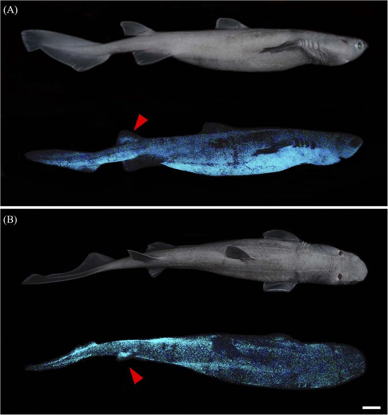
Mallefet et al.                                                                                               Insights on Kitefin Shark Luminescence

Duchatelet et al., 2019b, 2020b). Luminous sharks appear to                scales at the ventral side of the body like the related cookie
produce blue-green light (between 455 and 486 nm; Claes et al.,            cutter shark, Isistius brasiliensis (Quoy and Gaimard, 1824) (Reif,
2014a) for multiple purposes, such as counterillumination (Claes           1985; Widder, 1998; Delroisse et al., 2021). Nevertheless, no clear
et al., 2010a), aposematism (Claes et al., 2013; Duchatelet et al.,        evidence has been put forward to confirm its luminescence status.
2019b), and conspecific recognition (Claes et al., 2014a, 2015).               The diet of the kitefin shark is mainly composed of
Luminescence is achieved via thousands of photophores located              small demersal sharks such as lanternsharks (Etmopteridae),
within the epidermis. Each photophore is composed of a cup-                gulpersharks (Centrophoridae), and catsharks (Scyliorhinidae),
shaped layer of pigmented cells encapsulating one to more than             followed by demersal fishes, crustaceans, and cephalopods
twelve photogenic cells (i.e., photocytes) and topped by one or            (Macpherson, 1980; Matallanas, 1982; Dunn et al., 2010;
more lens cells. In E. spinax, a guanine crystal reflector structure       Navarro et al., 2014). Chunks of large fast swimming epipelagic
is located between the cup-shaped pigmented layer and the                  fishes have been also reported in the stomach contents of
photocyte (Renwart et al., 2014, 2015). Photophores also display           kitefin sharks (Matallanas, 1982), similar to what is observed
an iris-like structure (ILS), composed mainly of chromatophores,           for I. brasiliensis (Jones, 1971; Muñoz-Chápuli et al., 1988;
between the photocytes and the lens cells (Renwart et al., 2014;           Papastamatiou et al., 2010).
Duchatelet et al., 2020b). Recently, studies of the luminous                   Along the coast of New Zealand, D. licha inhabit waters
system of E. spinax failed to identify the reactive compounds              where at least six lanternshark species have been reported:
underlying the emission of light (i.e., luciferin/luciferase or            E. lucifer (Jordan and Snyder, 1902), E. granulosus (Günther,
photoprotein) (Renwart and Mallefet, 2013). Moreover, it has               1880), Etmopterus molleri, Etmopterus pusillus (Lowe, 1839),
been demonstrated that shark luminescence is not due to                    Etmopterus unicolor (Engelhardt, 1912), and Etmopterus viator
symbiotic luminous bacteria (Duchatelet et al., 2019a). Therefore,         (Straube, 2011) (Roberts et al., 2015). Photophores have been
the nature of the shark luminous system remains enigmatic.                 observed for these species (Ohshima, 1911; Last and Stevens,
    In Metazoans, sharks are the only known bioluminescent                 1994; Tracey and Shearer, 2002; Straube et al., 2011), but
organisms to hormonally control light emission. For the studied            bioluminescence has only been confirmed for Etmopterus molleri
species, researchers have demonstrated the involvement of                  (Claes and Mallefet, 2015). The blackbelly lanternshark
several hormones in the control of light emission: melatonin               (E. lucifer) and the southern lanternshark (E. granulosus) are
(MT) triggers light production, while alpha-melanocyte-                    the most common shark by-catch species in New Zealand
stimulating (α-MSH) and adrenocorticotropic hormones                       deep-sea trawl fisheries (Blackwell, 2010). Studying light
(ACTH) inhibit it (Claes and Mallefet, 2009c; Duchatelet et al.,           emission of the kitefin shark, the blackbelly lanternshark, and
2020b). Prolactin triggers brighter and faster light emission than         the southern lanternshark, might increase our understanding of
MT in Etmopteridae (Claes and Mallefet, 2009c; Claes et al.,               their bioluminescence functions, and possible prey-predation
2011b), while this hormone inhibits light production in S. aliae           relationships between these species.
(Claes et al., 2012). More recently, in silico mRNA sequences                  Here, organization, morphology, density, and physiological
and expression sites of MT and α-MSH/ACTH receptors                        control of kitefin shark photophores were investigated. To
were highlighted within the photophores, but neither mRNA                  determine if this species displays the same photophore
sequences nor protein presence was found for the prolactin                 structure and hormonal control, a comparative analysis
receptor (Duchatelet et al., 2020a). Other molecules, such as              was performed on the two most abundant New Zealand
nitric oxide or γ-aminobutyric acid, also exhibited modulatory             lanternshark species, E. lucifer and E. granulosus. Results are
effects on light emission in some Etmopteridae (Claes et al.,              compared to previously studied dalatiids and etmopterids.
2010b, 2011a). Finally, an extraocular opsin (Es-Opn3) has been            Homogeneity of light emission control among luminous
demonstrated to be involved in a secondary control targeting               elasmobranch and photophore structures among each
the ILS and modulating the aperture of this pigmented structure            shark families are observed, strengthening a conservative
acting as a light organ shutter (Duchatelet et al., 2020c). To             evolution of light emission capabilities among sharks.
establish the conservation of photophore morphology and the                These observations and results raise questions on the
control of hormonal light emission in the evolution of luminous            luminescence role for the largest luminous vertebrate. The
Squaliformes, increasing the knowledge on bioluminescent                   use of counterillumination for this giant luminous shark is here
sharks is crucial.                                                         suggested to be co-opted for a camouflage-type approach as
    While the majority of Squaliformes never reach more than               a predatory tool.
60 cm in adulthood, the kitefin shark (also named seal shark
or black shark), Dalatias licha (Bonnaterre, 1788), can grow
to 180 cm (Compagno, 1984; Roberts et al., 2015). This giant               MATERIALS AND METHODS
holobenthic dalatiid has a worldwide distribution at depths
ranging from 50 to 1800 m but it is usually found in depths                Specimen Sampling
below 300 m (Compagno, 1984; Roberts et al., 2015). Recently,              Shark specimens were captured during the Chatham Rise Trawl
through baited-remote video and muscle enzymatic activity                  survey by the R.V. Tangaroa in January 2020 off the coast of
analysis, D. licha was suggested to be one of the slowest moving           eastern New Zealand. The survey used the same eight-seam hoki
elasmobranch species (Pinte et al., 2020). Reif (1985) assumed             bottom trawl and survey methodology that was used on previous
that this shark is luminous as it presents pavement-like placoid           surveys (Hurst et al., 1992; Stevens et al., 2018). The net has 100 m

Frontiers in Marine Science | www.frontiersin.org                      2                                    February 2021 | Volume 8 | Article 633582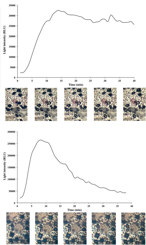
Mallefet et al.                                                                                                 Insights on Kitefin Shark Luminescence

sweeps, 50 m bridles, 12 m backstrops, 58.8 m groundrope, 45 m                Pharmacological Studies
headline, and 60 mm codend mesh. The trawl doors were Super                   In addition to the skin patches used for histology, round
Vee type with an area of 6.1 m2 .                                             skin patches were dissected from the ventral luminous area
    The following depth range information are available: D. licha –           of each shark using a metal cap driller (6 mm diameter) as
mean maximal depth 678 ± 26 m (min-max 443–997 m);                            described in Duchatelet et al. (2020b). Freshly dissected patches
E. lucifer – mean maximal depth 542 ± 8 m [min-max 235–                       were rinsed and kept in shark saline [292 mmol L−1 NaCl,
1078 m]; E. granulosus – mean maximal depth 903 ± 13 m                        3.2 mmol L−1 KCl, 5 mmol L−1 CaCl2 , 0.6 mmol L−1 MgSO4 ,
(min-max 498–1269 m).                                                         1.6 mmol L−1 Na2 SO4 , 300 mmol L−1 urea, 150 mmol L−1
    A total of 37 D. licha [40.9–138.0 cm total length (TL)],                 trimethylamine N-oxide, 10 mmol L−1 glucose, 6 mmol L−1
304 E. lucifer (16.2–53.2 cm TL), and 281 E. granulosus                       NaHCO3 ; total osmolarity: 1.080 mOsmol; pH 7.7 (Bernal
(19.3–75.6 cm TL) were captured on the survey, of which                       et al., 2005)] at 4◦ C in dark conditions before being used for
13 D. licha, 7 E. lucifer, and 4 E. granulosus were used                      pharmacological tests.
for bioluminescence studies. Each specimen was maintained                         Hormones known to trigger or inhibit light emission in
in a tank with fresh cold sea water in a dark cold room                       luminous elasmobranchs were applied (Claes and Mallefet, 2009c;
until manipulation. Each shark was sexed, measured, weighed                   Duchatelet et al., 2020b). Here, evaluations of the effect of MT,
(Supplementary Table 1) and photographed in dim daylight                      α-MSH and ACTH were conducted for the first time on the
and in dark conditions using Sony α7SII camera before having                  dalatiid species, D. licha, and the etmopterid species, E. lucifer,
a full incision of the spinal cord at the level of the first                  and E. granulosus.
vertebrae, according to the European regulation for animal                        Experiments were first conducted on 10 D. licha specimens.
research handling. Ventral skin of a specimen of S. aliae and                 To obtain a dose response curve for MT application, three
I. brasiliensis, collected, respectively, as in Delroisse et al. (2021)       different concentrations of MT (i.e., 10−6 , 10−7 , 10−8 mol
and Duchatelet et al. (2020b), were used for dalatiid comparative             L−1 ) were used. Skin patches were immersed in 200 µL of MT
photophore histology.                                                         solution (either 10−6 , 10−7 , 10−8 mol L−1 ). To analyze the
                                                                              effect of α-MSH and ACTH on the light emission of D. licha,
Photophore Histology and Density                                              another set of skin patches were subjected to an immersion
Skin patches of 3 cm2 were dissected from different locations                 in 100 µL of MT 10−6 mol L−1 followed after 5 min by an
along the body of D. licha specimens (i.e., rostral, mandibular,              application of 100 µL of either α-MSH 10−6 mol L−1 or
pecto-ventral, pectoral, ventral, dorsal, dorsal fin, pelvic, flank,          ACTH 10−5 mol L−1 . Luminescence of ventral skin patches
infra-caudal, precaudal, and caudal zones; Figure 1A) to                      subjected to the various treatments was measured using a FB12
assess photophore presence, size and densities. Skin patches                  tube-luminometer (Titertek-Berthold, Pforzheim, Germany)
were fixed in 4% formalin at least overnight before being                     calibrated as in Duchatelet et al., 2020b. Lights emissions were
transferred to phosphate buffer saline (PBS). Skin patches                    recorded through FB12- Sirius, multiple kinetics software
were observed and photographed under a transmitted light                      (Titertek-Berthold) for at least 30 min with a measurement
microscope (Leitz Diaplan, Germany) coupled with a ToupCam                    every 58 s. For comparative purposes, similar treatments
camera (UCMOS Series C-mount USB2.0 CMOS camera,                              were performed on seven specimens of E. lucifer (same
ToupTek, Zhejiang, China). Photophore densities (per mm2 )                    experiments) and four E. granulosus specimens (MT dose
and mean diameter (n = 30 or 50 zones per species) were also                  response and α-MSH treatments). In parallel, for D. licha and
measured on the two etmopterid species using the same protocol                E. lucifer, photophore aperture and closure were observed
(Supplementary Figure 1).                                                     after drug application by taking a time-lapse series of pictures
   In parallel, skin patches of D. licha, E. lucifer, E. granulosus,          (every 10 min) with a Sony α7SII camera mounted on a
S. aliae, and I. brasiliensis were used to perform histological               binocular microscope.
sections across the photogenic organ. Skin tissues were bathed                    Luminescence measurements were characterized as follows
for 7 days in decalcifying solution (OsteoRAL, Fast decalcifier               (Duchatelet et al., 2020b): the maximum intensity of light
for Large Anatomical Specimens, RAL Diagnostics, France)                      emission [Lmax, in megaquanta per second (Mq s−1 )], the
with constant agitation and renewal of the solution every                     total amount of light emitted during experimentation [Ltot, in
2 days, rinsed in PBS, and placed in PBS with increasing                      Gigaquanta per hour (Gq h−1 )] and the time to reach maximum
concentrations of sucrose (10% for 1 h, 20% for 1 h, and                      light intensity [TLmax, in seconds (s)]. Inhibitory actions of
30% overnight). Tissues were then embedded in optimal                         α-MSH and ACTH were measured as the total amount of light
cutting temperature compound (O.C.T. compound, Tissue-                        emitted after the second drug application [Ltotapp , in Gq h−1 ].
Tek, Netherlands) and rapidly frozen at −80◦ C. Sections of                   All light parameters were standardized according to the surface
10 µm were obtained with a cryostat microtome (CM3050S,                       area of each skin patch (in cm2 ). A second treatment (α-MSH or
Leica, Solms, Germany). Sections were placed on coated                        ACTH) was added to the first one when the light intensity plateau
Superfrost slides (Thermo Scientific) and left overnight to                   was reached with the MT application, each timing being species-
dry. All sections were observed under a transmitted light                     specific. Results of the luminescence decrease were expressed as a
microscope (Leitz Diaplan) equipped with a ToupCam                            percentage of the maximal luminescence value (i.e., plateau MT)
camera (ToupTek).                                                             measured before the second application.

Frontiers in Marine Science | www.frontiersin.org                         3                                   February 2021 | Volume 8 | Article 633582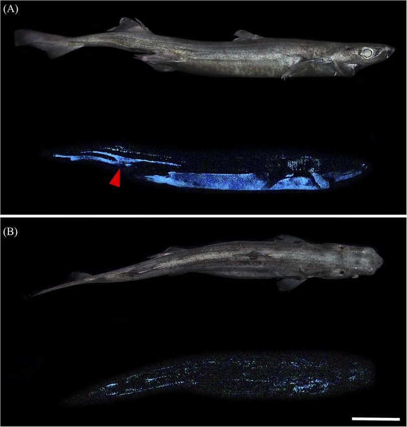
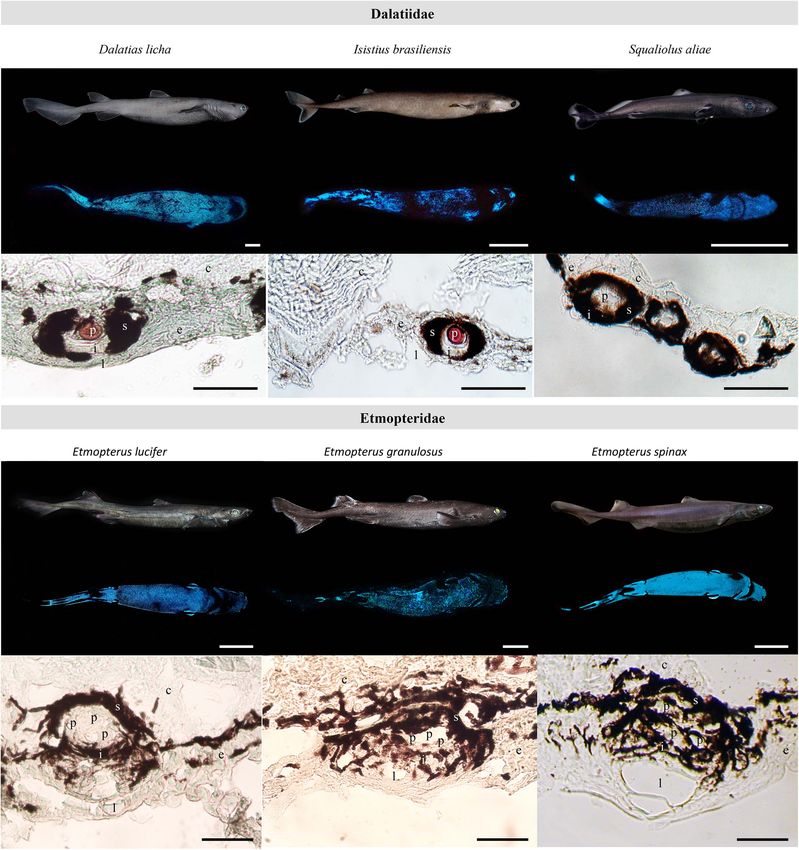
Mallefet et al.                                                                                                                    Insights on Kitefin Shark Luminescence

  FIGURE 1 | Dalatias licha photophore visualization and density measurements. (A) Photophore densities for each studied zone along the shark body.
  (B) Representation of the dorso-ventral photophore density gradient. Black-dotted photophores (red arrowhead) observed between the placoid scales (delimited
  areas) at the (C) rostral and (D) ventral areas. Rostral area presents specific leaf-shaped placoid scales, while ventral area harbors typical pavement type placoid
  scales. (E) Close-up of the black circular-shaped photophores within the integument surrounding the ventral placoid scales. d, placoid scale; e, epidermis; m,
  melanophore; p, photophore. (F) Photophore density variation across the studied zones. Different lettering indicates statistical differences. All density values are
  expressed as mean ± SEM.

   To evaluate the putative evolutive conservation of the                                assumptions were not met, a non-parametric Kruskal-Wallis
hormonal control of light emission in dalatiids and etmopterids,                         ANOVA was used. Post hoc Tukey’s tests or Wilcoxon tests
pharmacological data on shark luminescence were extracted                                allowed pair-wised comparison of means, attributing different
from literature.                                                                         letters to significantly different values (P-value < 0.05).

Statistical Analyses
All analyses were performed with the software R studio (version                          RESULTS
1.1.383, 2009, R Studio Inc., United States). Variance normality
and homoscedasticity assumptions were tested by Shapiro-                                 Luminous Pattern and Photophore
Wilk and Levene’s test, respectively, before running ANOVA                               Morphology
which reveals significant differences between skin photophore                            A blue glow was observed on the ventral surface of D. licha,
densities or pharmacological treatments. When these parametric                           E. lucifer, and E. granulosus specimens kept in a fully dark

Frontiers in Marine Science | www.frontiersin.org                                    4                                           February 2021 | Volume 8 | Article 633582
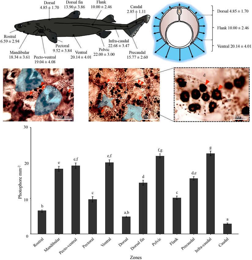
Mallefet et al.                                                                                                                        Insights on Kitefin Shark Luminescence

environment (Figures 2A,D,E). D. licha also emit a faint                                   photophores distributed between placoid scales for all the
blue glow from the lateral and dorsal areas and at the two                                 observed sharks (Figures 1C–E and Supplementary Figure 1).
dorsal fins (Figure 3 – Mallefet personal observation). Both                               The mean photophore diameters are 83.9 ± 9.5, 122.4 ± 10.8,
etmopterids present a more complex pattern of light emission                               and 132.3 ± 14.5 µm, for D. licha, E. lucifer, and E. granulosus,
with flank marks, and lateral, dorsal, and rostral patterns                                respectively. No statistical differences in photophore diameter
(Figure 4; E. granulosus – Mallefet personal observation).                                 were observed between the zones presenting large amount of
Skin patches observed in toto present black round-shaped                                   photophores (ventral, pecto-ventral and infra-caudal) (D. licha

  FIGURE 2 | Dalatiidae and Etmopteridae ventral luminous pattern and photophore histology. Picture of the lateral side in daylight, ventral luminescent pattern and
  section across ventral integument photophore of (A) Dalatias licha, (B) Isistius brasiliensis, (C) Squaliolus aliae, (D) Etmopterus lucifer, (E) Etmopterus granulosus,
  and (F) Etmopterus spinax. Ventral luminescence in dalatiid shows a homogenous pattern, while etmopterids show a heterogenous pattern with different zones.
  Photophores histology highlights a single photocyte within small photophores in dalatiids, while etmopterids harbor bigger and more complex photophores. c,
  connective tissue; e, epidermis; i, iris-like structure cells; l, lens cell; p, photocyte; s, pigmented sheath. In toto shark picture scale bar: 10 cm; photophore section
  scale bar: 100 µm.

Frontiers in Marine Science | www.frontiersin.org                                      5                                            February 2021 | Volume 8 | Article 633582
Mallefet et al.                                                                                                                Insights on Kitefin Shark Luminescence

  FIGURE 3 | Lateral and dorsal luminescent pattern of Dalatias licha. (A) Lateral daylight view and luminescent pattern highlighting the dorso-ventral luminous
  pattern. (B) Dorsal daylight view and luminescent pattern. Luminescence of the second dorsal fin is observable on this specimen (red arrowhead). Scale bar: 10 cm.

ANOVA: F(2,183) = 1,1928, P-value = 0,3057; E. lucifer ANOVA:                         Conversely, for both species, only a few photophores were spread
F(2,183) = 0,1014, P-value = 0,9036; E. granulosus ANOVA:                             within the dorsal epidermis. Both species have a well-defined
F(2,183) = 0.1376, P-value = 0.8716).                                                 flank mark with photophores. All the remaining photophore
   Analyses of photophore density along the D. licha body                             densities and their respective statistical differences [E. lucifer
show an increasing dorso-ventral repartition of photophores                           ANOVA: F(9,490) = 263.39, P-value < 2.2 10−16 ; E. granulosus
reaching up to 20.14 ± 4.01 photophores per mm2 at the ventral                        ANOVA: F(10,298) = 175.16, P-value < 2.2 10−16 ] are reported
side of the shark (Figures 1A,B,F). The lowest densities were                         in Supplementary Figure 1 and Supplementary Tables 2B,C.
observed for the caudal and dorsal areas with a mean density of                       Both etmopterids present needle-shaped placoid scales in all the
2.85 ± 1.11 and 4.85 ± 1.70 photophores per mm2 , respectively                        studied zones (Supplementary Figure 1).
(Figure 1A). Statistical differences [ANOVA: F(11,514) = 128.64,                          Histological sections across photogenic skin highlight the
P-value < 2.2 × 10⁻¹⁶ ] in photophore densities are illustrated                       structure of D. licha photophores. Each light organ is embedded
Figure 1F and Supplementary Table 2A. The scales of the rostral                       in the stratified squamous epidermis and is composed of a cup-
area were leaf-like in shape while the scales of the remaining body                   shaped pigmented sheath containing a unique photocyte, topped
parts were pavement-like in shape (Figures 1C,D).                                     by a lens cell with a few diffuse pigmented cells between the
   For both studied etmopterids, a high density of photophores                        photocyte and lens cell (Figure 2A). This structural organization
was observed at the pectoral zone with 34.00 ± 6.20, and                              is similar to that found in S. aliae and I. brasiliensis photophores
15.63 ± 2.50 photophores per mm2 for E. lucifer and                                   (Figures 2B,C).
E. granulosus, respectively. E. granulosus also have a high                               Photophore morphologies of E. lucifer and E. granulosus are
density of photophores at the infra-caudal and caudal zones.                          consistent with those already described for other etmopterids

Frontiers in Marine Science | www.frontiersin.org                                 6                                         February 2021 | Volume 8 | Article 633582
Mallefet et al.                                                                                                                  Insights on Kitefin Shark Luminescence

  FIGURE 4 | Lateral and dorsal luminescent pattern of Etmopterus lucifer. (A) Lateral daylight view and luminescent pattern. The species-specific flank mark is
  indicated by a red arrowhead (B) dorsal daylight view and luminescent pattern with specific luminous lines. Scale bar: 10 cm.

(i.e., E. spinax, Etmopterus molleri, and Etmopterus splendidus)                       MT 10−6 mol L−1 treatment was significantly different [Kruskal-
(Figures 2D–F). They are composed of a cup-shaped pigmented                            Wallis χ2 (2) = 10.14, P-value = 0.0063], E. lucifer produced a
sheath embedded with luminous cells and topped a with lens.                            mean total amount of light during the experiment 2.5 and 5
They are similar to dalatiid photophores, but they harbor a higher                     times higher than D. licha and E. granulosus, respectively. Similar
number of photocytes, a larger iris-like structure area, and more                      patterns of bioluminescence were observed for the three species
lens cells (up to 3) (Figures 2D–F).                                                   (Figures 2, 5).
                                                                                          The effect of α-MSH was evaluated for the three species after
Light Emission Control                                                                 reaching the Lmax triggered through MT 10−6 mol L−1
The effect of MT on D. licha, E. lucifer, and E. granulosus                            application. Application of α-MSH 10−6 mol L−1
was tested through a dose-dependent response. For the studied                          induced a rapid decrease of light emission for the studied
species, MT 10−6 mol L−1 triggered a long-lasting light emission,                      luminous sharks (Figures 6A–C and Supplementary
significantly different from the MT 10−8 mol L−1 application                           Table 3B). After MT-induced bioluminescence, Ltotapp
(P-value < 0.05; Figures 5A–C and Supplementary Tables 3A,                             values of α-MSH were statistically significant compared
4), while MT 10−7 mol L−1 triggered an intermediate light                              with the MT 10−6 mol L−1 control (Figures 6A–C and
emission and Ltot value (Figures 5A–C and Supplementary                                Supplementary Tables 3B, 5).
Tables 3A, 4). All treatments were significantly different from                           The effect of ACTH was evaluated on D. licha and E. lucifer
the shark saline control, except for the MT 10−7 and 10−8 mol                          bioluminescence. Similar to the results obtained for α-MSH,
L−1 treatments of E. granulosus (P-value < 0.05; Supplementary                         ACTH 10−5 mol L−1 applications rapidly induced a decrease
Tables 3A, 4). Although the total amount of light emitted under                        in light emission (Figures 6A,B and Supplementary Table 3B).

Frontiers in Marine Science | www.frontiersin.org                                  7                                          February 2021 | Volume 8 | Article 633582
Mallefet et al. Insights on Kitefin Shark Luminescence FIGURE 5 | Effect of MT on the studied species luminescence. Time course of the mean light emissions (Mq s−1 cm−2 ) and total amount of light produced (Gq h−1 cm−2 ) from ventral skin patches under hormonal treatments (MT 10−8 to 10−6 mol L−1 ) for (A) Dalatias licha (n = 10), (B) Etmopterus lucifer (n = 7), and (C) Etmopterus granulosus (n = 4). Different lettering indicates statistical differences [Kruskal-Wallis ANOVA: D. licha χ2 (3) = 23.95, P-value = 2.56 × 10⁻⁵ ; E. lucifer χ2 (3) = 23.823, P-value = 2.72 × 10⁻⁵ ; E. granulosus χ2 (3) = 8.5368, P-value = 0.0361]. Error bars correspond to SEM. FIGURE 6 | Effect of ACTH and α-MSH on luminescence induced by MT in the studied species. Time course of the light produced (expressed as percentage of maximal melatonin control value), and total amount of light produced (Gq h−1 cm−2 ) after melatonin pretreatment from ventral skin patches under melanocortin treatments (ACTH 10−5 mol L−1 /α-MSH 10−6 mol L−1 ) for (A) Dalatias licha (n = 10), (B) Etmopterus lucifer (n = 7), and (C) Etmopterus granulosus (n = 4 – no ACTH treatment). Hormonal treatments are expressed in mol L−1 . Different lettering indicates statistical differences [ANOVA: D. licha F(2,33) = 2.585, P-value = 0.0437; E. lucifer F(2,18) = 14.482, P-value = 0.0002; Kruskal-Wallis ANOVA: E. granulosus χ2 (1) = 3.857, P-value = 0.0495]. Error bars correspond to SEM. Each Ltotapp value of ACTH 10−5 mol L−1 was not significantly control (Figures 6A,B and Supplementary Tables 3B, 5). different from those of α-MSH 10−6 mol L−1 , respectively, Mean values of Lmax, TLmax, Ltot, Ltotapp are presented in but were statistically different from the MT 10−6 mol L−1 Supplementary Table 3. Frontiers in Marine Science | www.frontiersin.org 8 February 2021 | Volume 8 | Article 633582
Mallefet et al.                                                                                                    Insights on Kitefin Shark Luminescence

   The time-course of light emission in D. licha under MT                       no place to hide, hence the need for glowing camouflage
10−6 mol L−1 stimulation revealed a concomitant opening of                      or counterillumination, first proposed by Clarke, 1963. The
the photophore ILS within 15 min of luminescence, in which                      mesopelagic zone, often called the twilight zone, ranges from
the ILS stayed open for the next 30 min while the light level                   200 to 1000 m depth (maximal depth of solar light penetration)
remained high (Figure 7A). In the case of E. lucifer MT-                        and is the realm of bioluminescence (Martini and Haddock,
induced luminescence, a rapid opening of the photophore ILS                     2017; Martini et al., 2019). At 200 m the residual solar light is
was observed within 8 min followed by a slow decrease during                    considered too weak to initiate photosynthesis but organisms
which a closure of the ILS was visible (Figure 7B). Aperture                    living there are well adapted to see in low light conditions
and closure of photophores showed pigment movements                             (Nicol, 1978). Mesopelagic cephalopods, sharks and bony fishes
concomitant to light emission.                                                  have large eyes with specialized structures such as a large iris, a
                                                                                tapetum, huge rod density, high content of opsins (rhodopsin and
                                                                                chrysopsin), and an elevated integration rate at the optical nerve
DISCUSSION                                                                      which allows them to perceive very low light levels down to 800 m
                                                                                depth (Douglas et al., 1998; Warrant, 2004; Warrant and Locket,
The three studied shark species inhabit the mesopelagic zone                    2004; Claes et al., 2014a,b).
(Roberts et al., 2015), therefore they face an environment with
                                                                                Luminescent Pattern
                                                                                The light emission pattern observed in D. licha is similar
                                                                                to that observed in previously studied dalatiids i.e., S. aliae
                                                                                and I. brasiliensis (Claes et al., 2012; Delroisse et al., 2021).
                                                                                The dorso-ventral gradient and the relative homogeneity in
                                                                                ventral photophore densities suggest the luminescence is used
                                                                                for counterillumination. The luminous pelvic zone of D. licha
                                                                                reveals a sexual dimorphism but, contrary to E. spinax and
                                                                                Etmopterus molleri (Claes and Mallefet, 2010b; Duchatelet et al.,
                                                                                2020c), it is not brighter than the rest of the ventral body,
                                                                                suggesting it is less important for sexual signaling. The kitefin
                                                                                shark D. licha, like other dalatiids, does not have flank marking
                                                                                or specific dorsal patterns. The lack of these luminescent patterns,
                                                                                previously suggested to be used as conspecific signaling for group
                                                                                aggregation, swimming, or hunting in etmopterids (Claes et al.,
                                                                                2015; Duchatelet et al., 2019b), rules out this function in D. licha.
                                                                                The aposematic function described for etmopterids (Claes et al.,
                                                                                2013; Duchatelet et al., 2019b) is also ruled out for D. licha
                                                                                luminescence due to the absence of dorsal fin defensive spines.
                                                                                Nevertheless, D. licha is the first shark with fully luminous
                                                                                dorsal fins (Figures 1A, 3), which raises questions about its
                                                                                luminescence function.
                                                                                   The light emission patterns of E. lucifer and E. granulosus,
                                                                                are similar to that of previously studied etmopterids. The
                                                                                dorsal photophores, flank markings, and brighter pectoral
                                                                                fin and claspers are likely to be used for intraspecific
                                                                                communications while the ventrally emitted light is likely to
                                                                                be used for counterillumination. These functions have been
                                                                                documented for E. spinax (Claes and Mallefet, 2009a; Claes
                                                                                and Mallefet, 2010a), Etmopterus molleri (Claes and Mallefet,
                                                                                2015), and Etmopterus splendidus (Claes et al., 2011b). However,
                                                                                a bioluminescence aposematic function through specific spine-
                                                                                associated photophores (Claes et al., 2013; Duchatelet et al.,
                                                                                2019b) was not documented for E. lucifer and E. granulosus.
                                                                                   Reif (1985) postulated that a trade-off exists between the
  FIGURE 7 | Time-course of MT-induced luminescence, and time-lapse of          space occupied by placoid scales and luminous organs, and that
  photophore pigment movements. Luminescence in relative light unit (RLU)       four different types of placoid scales have evolved to allow this
  recorded during a 40 min MT 10−6 mol L−1 application on ventral skin          trade-off: pavement, cross-, bristle/ needle-, and hook-shaped
  patches and time-lapse pictures (times: 0, 10, 20, 30, and 40 min,
                                                                                placoid scales. A new type of squamation with overlapping leaf-
  respectively) of photophore pigment movements of (A) Dalatias licha,
  (B) Etmopterus lucifer.
                                                                                shaped placoid scales is present in the luminous rostral area
                                                                                of D. licha. This new bioluminescent-associated squamation

Frontiers in Marine Science | www.frontiersin.org                           9                                    February 2021 | Volume 8 | Article 633582
Mallefet et al. Insights on Kitefin Shark Luminescence
was observed in the somniosid, Zameus squamulosus, which is in the two studied etmopterids, as well as in D. licha,
assumed to be luminous (Straube et al., 2015). This new type might share common features. Moreover, the involvement of
of bioluminescence-associated placoid scale needs to be highly extraocular photoreception events in the light emission control
translucent or possess specific physical characteristics to allow of photophores (Duchatelet et al., 2020d), remains to be
efficient light transmission. The use of Reif placoid scale types to deciphered for these sharks. Further research are necessary to
assess the bioluminescent status of a shark species is not a decisive fully demonstrate the evolutive conservation of luminescence
character as shown by a recent study of Ferrón et al. (2018); control within etmopterids and dalatiids.
highlighting the presence of bioluminescent-like squamation
in a galeomorph shark, Apristurus ampliceps, a species not Luminescence of Dalatias licha
known to be luminous. The question remains concerning bioluminescence in the largest
luminous vertebrate; why does D. licha emit light ventrally to
Photophore Morphology Conservation counterilluminate when it has few or no predators? Pinte et al.
Histology revealed an evolutive conservation of photophore (2020), analyzed the swimming speed of several New Zealand
morphology across each family. Kitefin shark photophores are deep-sea sharks, and found that D. licha possesses one of
larger (mean diameter 83.9 µm) than those observed in S. aliae the slowest cruise swimming speeds ever measured in sharks.
and I. brasiliensis [i.e., 50 and 56 µm, respectively (Claes et al., Conversely, this species is assumed to possess a high burst
2012; Delroisse et al., 2021)] while the internal structure of typical capability (Pinte et al., 2020). Stomach content analyses have
dalatiid photophores is conserved. Here, D. licha photophores revealed that this shark species hunts and eats etmopterids, which
are depicted as morphologically similar to those of S. aliae, S. have a higher cruise swimming speed. Therefore, there are two
laticaudus and I. brasiliensis (Seigel, 1978; Delroisse et al., 2021). hypotheses which might explain the ventral luminescence of this
E. lucifer and E. granulosus showed typical etmopterid holobenthic species: luminescence might be used (i) to illuminate
photophore histology (Claes and Mallefet, 2009b, 2015; Claes the ocean floor while searching and hunting for prey; or (ii)
et al., 2011b; Renwart et al., 2014; Duchatelet et al., 2020b). to stealthily approach toward prey, using counterillumination
These observations provide further insights on the evolutive camouflage, before striking fast when close enough (Zintzen
conservation of light organ morphology across luminous et al., 2011), allowing them to predate etmopterids. In both
squaliform radiation (Straube et al., 2015). cases, the principle of counterillumination would have been
distorted to serve as a predation tool instead of an avoidance
mechanism, a hypothesis already proposed for the cookie cutter
Luminescence Control Evolutive shark, I. brasiliensis (Widder, 1998). However, to validate such
Conservation hypotheses for these dalatiid species, in vivo observations and
The effect of hormones on light emission in D. licha, E. lucifer behavioral studies are essential.
and E. granulosus are consistent with increasing literature on light
emission control in sharks (Claes and Mallefet, 2009c; Claes et al.,
2012; Duchatelet et al., 2020b,d): MT, and α-MSH/ACTH, have CONCLUSION
been demonstrated as the main triggering and inhibiting agents
of shark luminescence, respectively. Similar to observations Through a histological and pharmacological approach, the
of E. spinax and Etmopterus molleri photophores (Claes and bioluminescence of three different shark species was investigated.
Mallefet, 2010a; Duchatelet et al., 2020b), aperture and closure Our results support evolutive conservation of light organ
of D. licha and E. lucifer photophores involved pigment motion morphology and luminescence control. For the first time,
within the ILS cells. Simultaneities of curves kinetics and pigment luminescence was recorded and analyzed for the largest luminous
motions highlight the evolutive conservation of hormonally vertebrate, D. licha and two lanternsharks, E. lucifer and
controlled pigment motion regulating luminescence. These data E. granulosus. Dalatiid photophores are similar between species
strongly suggest that luminous etmopterids and dalatiids share and are structurally composed of a single photocyte embedded
a common luminescence control mechanism, involving at least in a cup-shaped pigmented cell and surmounted by lens
MT, and α-MSH/ACTH hormones. This control is assumed to cells. The same observation was made for etmopterids, which
have been successfully and evolutionary co-opted from shark showed a conservation of photophore structure between species.
melanophore pigment motion control by a common ancestor of Etmopterid photophores are slightly more complex than those
these two squaliform families. For both families, luminescence of dalatiids, with several photocytes and a well-developed ILS
appears to be dually controlled at the level of (i) the photocyte, between the lens cells and the photocytes. Through this study, the
site of luminescent reaction, and (ii) the ILS cells, acting action of MT and α-MSH/ACTH in the bioluminescence control
as a diaphragm capable of occluding light produced by the in these two families was shown to be identical and seem to
photocytes, via melanophore-associated pigment movements have been co-opted during evolution from the regulation of skin
(Duchatelet et al., 2020b,d). This was recently demonstrated pigment movements. With these data, we can assume that the
within ILS cells of the lanternshark, E. spinax (Duchatelet et al., common luminous ancestor of etmopterids and dalatiids likely
2020d) i.e., transduction pathways that activate cellular motors had hormonal control of its luminescence and had luminous
such as dynein and kinesin, leading pigment movements within organs similar to those of the dalatiids (i.e., the simplest structure)
ILS melanophores. The bioluminescence control mechanisms for counterillumination.
Frontiers in Marine Science | www.frontiersin.org 10 February 2021 | Volume 8 | Article 633582Mallefet et al. Insights on Kitefin Shark Luminescence
DATA AVAILABILITY STATEMENT This study is the contribution BRC #276 of the Biodiversity
Research Center (UCLouvain) from the Earth and Life Institute
The original contributions presented in the study are included Biodiversity (ELIB) and the “Centre Interuniversitaire de Biologie
in the article/Supplementary Material, further inquiries can be Marine” (CIBIM).
directed to the corresponding author/s.
ETHICS STATEMENT SUPPLEMENTARY MATERIAL
Ethical review and approval was not required for this The Supplementary Material for this article can be found
study. The shark specimens were captured as bycatch of a online at: https://www.frontiersin.org/articles/10.3389/fmars.
fisheries assessment survey for the New Zealand Ministry for 2021.633582/full#supplementary-material
Primary Industries.
Supplementary Figure 1 | External features and densities of photophores in
Etmopterus lucifer and Etmopterus granulosus. Black-dotted photophores
observed at the ventral side (A), flank mark (B), and pectoral (C) specific area of
AUTHOR CONTRIBUTIONS E. lucifer. Dotted line corresponds to the flank mark boundaries. (D) Measured
photophore densities for the studied zones of E. lucifer (n = 50 for each zones).
JM and DS collected the samples. JM collected the Black-dotted photophore observed at the ventral (E), infra-caudal (F) and rostral
bioluminescence pictures and performed pharmacological (G) areas of E. granulosus. (H) Measured photophore densities for the studied
studies and fixations on the survey. LD performed the classical zones of E. granulosus (n = 30 for each zones). Different lettering indicates
histology, pattern, and pharmacological analyses. LD and statistical differences. Values are expressed as mean ± SEM. Scale bars: 750 µm.
JM were major contributors to the initial manuscript that Supplementary Table 1 | Experimental specimens. Morphometrics
was improved by DS revisions. All authors approved the measurements of Dalatias licha, Etmopterus lucifer, E. granulosus, Squaliolus aliae
final manuscript. and I. brasiliensis studied specimens. ♀, female; ♂, male.
Supplementary Table 2 | Photophore density, statistical analyses. Results of
Tukey’s test for the photophore density of (A) D. licha, (B) E. lucifer, and (C)
FUNDING E. granulosus different skin zones. Gray-shaded cases represent not
significant differences.
This work was supported by an F.R.S.– FNRS Grant (T.0169.20) Supplementary Table 3 | Hormone-induced luminescence parameters (mean
awarded to the Université Catholique de Louvain Marine Biology maximal light intensity: Lmax; time to reach the Lmax: TLmax; total amount of
Laboratory and the Université de Mons Biology of Marine emitted light: Ltot; total amount of emitted light after second drug application:
Organisms and Biomimetics Laboratory. JM received a travel Ltotapp ). (A) luminescence recorded parameters for the melatonin (MT) dose
response treatments for Dalatias licha (n = 12), Etmopterus lucifer (n = 7) and
grant (35401759) from F.R.S.– FNRS Belgium. E. granulosus (n = 4). ∗ indicate significant differences (P-value < 0.05) from the
shark saline control experiment. (B) Ltotapp for each treatment and each shark
species. ∗ indicate differences (P-value < 0.05) from the melatonin 10−6 mol L−1
ACKNOWLEDGMENTS control experiment. All data are means ± SEM.
Supplementary Table 4 | MT dose response, statistical analyses. Kruskal-Wallis
The authors acknowledge R. O’Driscoll, Program ANOVA and pairwise Wilcoxon test results for the MT dose response of the three
Leader – Fisheries Monitoring NIWA, the scientific staff, and studied sharks. Gray-shaded cases represent not significant differences.
the skillfull crew of R.V. Tangaroa on voyage TAN2001
Supplementary Table 5 | α-MSH and ACTH effects, statistical analyses. ANOVA
(Chatham Rise fish survey, NIWA). The authors thank Dr. and Tukey’s test results for the decrease of light triggered by α-MSH and ACTH
Nicolas Pinte and Constance Coubris for the help during treatments (except for E. granulosus non-parametric test). Gray-shaded cases
statistical analyses. JM is Research Associate F.R.S.– FNRS. represent not significant differences.
REFERENCES Claes, J. M., Dean, M. N., Nilsson, D. E., Hart, N. S., and Mallefet, J. (2013). A
deepwater fish with ‘lightsabers’ – dorsal spine-associated luminescence in a
Bennett, F. D. (1840). Narrative of a Whaling Voyage Round the Globe, from the counterilluminating lanternshark. Sci. Rep. 3:1308. doi: 10.1038/srep01308
Year 1833 To 1836, Vol. 2. Moscow: Рипол Классик. Claes, J. M., Ho, H.-C., and Mallefet, J. (2012). Control of luminescence from
Bernal, D., Donley, J. M., Shadwick, R. E., and Syme, D. A. (2005). Mammal- pygmy shark (Squaliolus aliae) photophores. J. Exp. Biol. 215, 1691–1699. doi:
like muscles power swimming in a cold-water shark. Nature 437, 1349–1352. 10.1242/jeb.066704
doi: 10.1038/nature04007 Claes, J. M., Krönström, J., Holmgren, S., and Mallefet, J. (2010b). Nitric
Blackwell, R. G. (2010). Distribution and Abundance of Deepwater Sharks in oxide in the control of luminescence from lantern shark (Etmopterus spinax)
New Zealand Waters, 2000–01 to 2005–06. New Zealand Aquatic Environment photophores. J. Exp. Biol. 213, 3005–3011. doi: 10.1242/jeb.040410
and Biodiversity Report No. 57. Wellington: Ministry of Fisheries. Claes, J. M., Krönström, J., Holmgren, S., and Mallefet, J. (2011a). GABA inhibition
Claes, J. M., Aksnes, D. L., and Mallefet, J. (2010a). Phantom hunter of the fjords: of luminescence from lantern shark (Etmopterus spinax) photophores. Comp.
camouflage by counterillumination in a shark (Etmopterus spinax). J. Exp. Mar. Biochem. Physiol. C Toxicol. Pharmacol. 153, 231–236. doi: 10.1016/j.cbpc.2010.
Biol. Ecol. 388, 28–32. doi: 10.1016/j.jembe.2010.03.009 11.002
Frontiers in Marine Science | www.frontiersin.org 11 February 2021 | Volume 8 | Article 633582Mallefet et al. Insights on Kitefin Shark Luminescence Claes, J. M., and Mallefet, J. (2009a). Ontogeny of photophore pattern in the velvet Dunn, M. R., Szabo, A., McVeagh, M. S., and Smith, P. J. (2010). The diet of belly lantern shark. Etmopterus spinax. Zoology 112, 433–441. doi: 10.1016/j. deepwater sharks and the benefits of using DNA identification of prey. Deep zool.2009.02.003 Sea Res. 57(Pt I), 923–930. doi: 10.1016/j.dsr.2010.02.006 Claes, J. M., and Mallefet, J. (2009b). “Bioluminescence of sharks: first synthesis,” Ferrón, H. G., Paredes-Aliaga, M. V., Martínez-Pérez, C., and Botella, H. (2018). in Bioluminescence in Focus - A Collection of Illuminating Essays, ed. V. Meyer Bioluminescent-like squamation in the galeomorph shark Apristurus ampliceps Rochow (Thiruvananthapuram: Research Signpost), 51–65. (Chondrichthyes: Elasmobranchii). Contrib. Zool. 87, 187–196. doi: 10.1163/ Claes, J. M., and Mallefet, J. (2009c). Hormonal control of luminescence from 18759866-08703004 lantern shark (Etmopterus spinax) photophores. J. Exp. Biol. 212, 3684–3692. Haddock, S. H. D., Moline, M. A., and Case, J. F. (2010). Bioluminescence in the sea. doi: 10.1242/jeb.034363 Annu. Rev. Mar. Sci. 2, 443–493. doi: 10.1146/annurev-marine-120308-081028 Claes, J. M., and Mallefet, J. (2010a). The lantern shark’s light switch: turning Hurst, R. J., Bagley, N., Chatterton, T., Hanchet, S., Schofield, K., and Vignaux, M. shallow water crypsis into midwater camouflage. Biol. Lett. 6, 685–687. doi: (1992). Standardisation of Hoki/Middle Depth Time Series Trawl Surveys. MAF 10.1098/rsbl.2010.0167 Fisheries Greta Point Internal Report No. 194. Wellington: Draft Report Held in Claes, J. M., and Mallefet, J. (2010b). Functional physiology of lantern shark MAF Fisheries Great point library, 89. (Etmopterus spinax) luminescent pattern: differential hormonal regulation of Johann, L. (1899). Über eigentümliche epitheliale Gebilde (Leuchtorgane) bei luminous zones. J. Exp. Biol. 213, 1852–1858. doi: 10.1242/jeb.041947 Spinax niger Aus dem zoologischen Institut der Universität Rostock. Von. Claes, J. M., and Mallefet, J. (2015). Comparative control of luminescence in sharks: Zeitschr. Wissenschaf. Zool. 66, 136–160. new insights from the slendertail lanternshark (Etmopterus molleri). J. Exp. Mar. Jones, E. C. (1971). Isistius brasiliensis, a squaloid shark, the probable cause of crater Biol. Ecol. 467, 87–94. doi: 10.1016/j.jembe.2015.03.008 wounds on fishes and cetaceans. Fish. Bull. U.S.A. 69, 791–798. Claes, J. M., Nilsson, D. E., Mallefet, J., and Straube, N. (2015). The presence Last, P. R., and Stevens, J. D. (1994). Sharks and Rays of Australia. Canberra, ACT: of lateral photophores correlates with increased speciation in deep-sea CSIRO, 513. bioluminescent sharks. Royal Soc. Open Sci. 2:150219. doi: 10.1098/rsos.150219 Macpherson, E. (1980). Régime alimentaire de Galeus melastomus Rafinesque, Claes, J. M., Nilsson, D. E., Straube, N., Collin, S. P., and Mallefet, J. (2014a). Iso- 1810, Etmopterus spinax (L., 1758), et Scymnorhinus licha (Bonnaterre, 1788) luminance counterillumination drove bioluminescent shark radiation. Sci. Rep. en Méditerranée occidentale. Vie Milieu 30, 139–148. 4:4328. doi: 10.1038/srep04328 Martini, S., and Haddock, S. H. D. (2017). Quantification of bioluminescence from Claes, J. M., Partridge, J. C., Hart, N. S., Garza-Gisholt, E., Ho, H. C., Mallefet, the surface to the deep sea demonstrates its predominance as an ecological trait. J., et al. (2014b). Photon hunting in the twilight zone: visual features of Sci. Rep. 7, 1–11. doi: 10.1038/srep45750 mesopelagic bioluminescent sharks. PLoS One 9:e104213. doi: 10.1371/journal. Martini, S., Kuhnz, L., Mallefet, J., and Haddock, S. H. D. (2019). Distribution and pone.0104213 quantification of bioluminescence as an ecological trait in the deep sea benthos. Claes, J. M., Sato, K., and Mallefet, J. (2011b). Morphology and control of Sci. Rep. 9, 1–11. doi: 10.1038/s41598-019-50961-z photogenic structures in a rare dwarf pelagic lantern shark (Etmopterus Matallanas, J. (1982). Feeding habits of Scymnorhinus licha in Catalan waters. J. Fish splendidus). J. Exp. Mar. Biol. Ecol. 406, 1–5. doi: 10.1016/j.jembe.2011.05.033 Biol. 20, 155–163. doi: 10.1111/j.1095-8649.1982.tb03916.x Clarke, W. D. (1963). Function of bioluminescence in mesopelagic organisms. Muñoz-Chápuli, R., Rel Salgado, J. C., and De La Serna, J. M. (1988). Biogeography Nature 198, 1244–1246. doi: 10.1038/1981244a0 of Isistius brasiliensis in the North-Eastern Atlantic, inferred from crater Compagno, L. J. V. (1984). Sharks of the world. an annoted and illustrated wounds on Swordfish (Xiphias gladius). J. Mar. Biol. Assoc. U. K. 68, 315–321. catalogue of shark species known to date. FAO Fisher. Sympos. 125:249. doi: 10.1017/S0025315400052218 Delroisse, J., Duchatelet, L., Flammang, P., and Mallefet, J. (2021). Photophore Navarro, J., López, L., Coll, M., Barría, C., and Sáez-Liante, R. (2014). Short- and distribution and enzymatic diversity within the photogenic integument of the long-term importance of small sharks in the diet of the rare deep-sea shark cookie cutter shark Isistius brasiliensis (Chondrichthyes: Dalatiidae). Front. Dalatias licha. Mar. Biol. 161, 1697–1707. doi: 10.1007/s00227-014-2454-2 Mar. Sci. Nicol, J. A. (1978). “Bioluminescence and vision,” in Bioluminescence in Action, ed. Douglas, R. H., Partridge, J. C., and Marshall, N. J. (1998). The eyes of deep-sea P. J. Herring (London: Academic Press), 367–408. fish I: lens pigmentation, tapeta and visual pigments. Prog. Retin. Eye Res. 17, Ohshima, H. (1911). Some observations on the luminous organs of fishes. J. Coll. 597–636. doi: 10.1016/S1350-9462(98)00002-0 Sci. Imp. Univ. Tokyo 27, 1–25. doi: 10.1002/jez.1402590102 Duchatelet, L., Delroisse, J., Flammang, P., Mahillon, J., and Mallefet, J. (2019a). Papastamatiou, Y. P., Wetherbee, B. M., O’Sullivan, J., Goodmanlowe, G. D., and Etmopterus spinax, the velvet belly lanternshark, does not use bacterial Lowe, C. G. (2010). Foraging ecology of cookiecutter sharks (Isistius brasiliensis) luminescence. Acta Histochem. 121, 516–521. doi: 10.1016/j.acthis.2019.04.010 on pelagic fishes in Hawaii, inferred from prey bite wounds. Environ. Biol. Fish. Duchatelet, L., Delroisse, J., and Mallefet, J. (2020a). Bioluminescence in 88, 361–368. doi: 10.1007/s10641-010-9649-2 lanternsharks: Insight from hormone receptor localization. Gen. Comp. Pinte, N., Parisot, P., Martin, U., Zintzen, V., De Vleeschouwer, C., Roberts, C. D., Endocrinol. 294:113488. doi: 10.1016/j.ygcen.2020.113488 et al. (2020). Ecological features and swimming capabilities of deep-sea sharks Duchatelet, L., Delroisse, J., Pinte, N., Sato, K., Ho, H. C., and Mallefet, J. from New Zealand. Deep Sea Res. 156:103187. doi: 10.1016/j.dsr.2019.103187 (2020b). Adrenocorticotropic hormone and cyclic adenosine monophosphate Reif, W.-E. (1985). Functions of scales and photophores in mesopelagic are involved in the control of shark bioluminescence. Photochem. Photobiol. 96, luminescent sharks. Acta Zool. 66, 111–118. doi: 10.1111/j.1463-6395.1985. 37–45. doi: 10.1111/php.13154 tb00829.x Duchatelet, L., Marion, R., and Mallefet, J. (2021). A third luminous shark family: Renwart, M., Delroisse, J., Claes, J. M., and Mallefet, J. (2014). Ultrastructural confirmations of luminescence ability for Zameus squamulosus (Squaliformes; organization of lantern shark (Etmopterus spinax Linnaeus, 1758) photophores. Somniodidae). Photochem. Photobiol. doi: 10.1111/php13393 [Epub ahead of Zoomorphology 133, 405–416. doi: 10.1007/s00435-014-0230-y print]. Renwart, M., Delroisse, J., Flammang, P., Claes, J. M., and Mallefet, J. Duchatelet, L., Oury, N., Mallefet, J., and Magalon, H. (2020c). In the intimacy (2015). Cytological changes during luminescence production in lanternshark of the darkness: genetic polyandry in deep-sea luminescent lanternsharks (Etmopterus spinax Linnaeus, 1758) photophores. Zoomorphology 134, 107– Etmopterus spinax and Etmopterus molleri (Squaliformes, Etmopteridae). 116. doi: 10.1007/s00435-014-0235-6 J. Fish. Biol. 96, 1523–1529. doi: 10.1111/jfb.14336 Renwart, M., and Mallefet, J. (2013). First study of the chemistry of the luminous Duchatelet, L., Pinte, N., Tomita, T., Sato, K., and Mallefet, J. (2019b). system in a deep-sea shark, Etmopterus spinax Linnaeus, 1758 (Chondrichthyes: Etmopteridae bioluminescence: dorsal pattern specificity and aposematic use. Etmopteridae). J. Exp. Mar. Biol. Ecol. 448, 214–219. doi: 10.1016/j.jembe.2013. Zool. Lett. 5:9. doi: 10.1186/s40851-019-0126-2 07.010 Duchatelet, L., Sugihara, T., Delroisse, J., Koyanagi, M., Rezsohazy, R., Terakita, Roberts, C. D., Stewart, A. L., and Struthers, C. D. (2015). The Fishes of A., et al. (2020d). From extraocular photoreception to pigment movement New Zealand, Vol. 2. Wellington: Te Papa Press, 1–576. regulation: a new control mechanism of the lanternshark luminescence. Sci. Seigel, J. A. (1978). Revision of the dalatiid shark genus Squaliolus: anatomy, Rep. 10:10195. doi: 10.1038/s41598-020-67287-w systematics, ecology. Copeia 1978, 602–614. doi: 10.2307/1443686 Frontiers in Marine Science | www.frontiersin.org 12 February 2021 | Volume 8 | Article 633582
Mallefet et al.                                                                                                                       Insights on Kitefin Shark Luminescence

Shimomura, O. (2006). Bioluminescence: Chemical Principles and Methods.                  Warrant, E. J., and Locket, N. A. (2004). Vision in the deep sea. Biol. Rev. 79,
   Singapore: World Scientific.                                                             671–712. doi: 10.1017/S1464793103006420
Stevens, D. W., O’Driscoll, R. L., Ballara, S. L., and Schimel, A. C. G.                 Widder, E. A. (1998). A predatory use of counterillumination by the squaloid
   (2018). Trawl Survey Of Hoki and Middle-Depth Species on the Chatham                     shark, Isistius brasiliensis. Environ. Biol. Fishes 53, 267–273. doi: 10.1023/A:
   Rise, January 2018 (TAN1801). New Zealand Fisheries Assessment Report                    1007498915860
   2018/41. Available online at: https://fs.fish.govt.nz/Doc/24639/FAR-2018-41-          Widder, E. A. (1999). “Bioluminescence,” in Adaptative Mechanisms in the Ecology
   Trawl-Survey-TAN1801.pdf.ashx (accessed January 15, 2021).                               of Vision, eds S. N. Archer, M. B. A. Djamgoz, E. R. Loew, J. C. Partridge., and S.
Straube, N., Duhamel, G., GaSco, N., Kriwet, J., and Schliewen, U. K.                       Vallerga (Dordrecht: Springer). doi: 10.1007/978-94-017-0619-3-19
   (2011). “Description of a new deep-sea lantern shark Etmopterus viator                Zintzen, V., Roberts, C. D., Anderson, M. J., Stewart, A. L., Struthers, C. D., and
   sp. nov. (Squaliformes: Etmopteridae) from the Southern hemisphere,”                     Harvey, E. S. (2011). Hagfish predatory behavior and slime defence mechanism.
   in The kerguelen Plateau, Marine Ecosystem and Fisheries, eds G.                         Sci. Rep. 1:131.
   Duhamel., and D. Welsford (Paris: Société française d’ichtyologie),
   135–148.                                                                              Conflict of Interest: The authors declare that the research was conducted in the
Straube, N., Li, C., Claes, J. M., Corrigan, S., and Naylor, G. J. P.                    absence of any commercial or financial relationships that could be construed as a
   (2015). Molecular phylogeny of squaliformes and first occurrence of                   potential conflict of interest.
   bioluminescence in sharks. BMC Evol. Biol. 15:162. doi: 10.1186/s12862-015-
   0446-6                                                                                Copyright © 2021 Mallefet, Stevens and Duchatelet. This is an open-access article
Tracey, D., and Shearer, P. (2002). An Identification Guide for Deepwater Shark          distributed under the terms of the Creative Commons Attribution License (CC BY).
   Species. Wellington: NIWA, 16.                                                        The use, distribution or reproduction in other forums is permitted, provided the
Warrant, E. J. (2004). Vision in the dimmest habitats on earth. J. Comp. Physiol.        original author(s) and the copyright owner(s) are credited and that the original
   A Neuroethol. Sens. Neural Behav. Physiol. 190, 765–789. doi: 10.1007/s00359-         publication in this journal is cited, in accordance with accepted academic practice. No
   004-0546-z                                                                            use, distribution or reproduction is permitted which does not comply with these terms.

Frontiers in Marine Science | www.frontiersin.org                                   13                                             February 2021 | Volume 8 | Article 633582
You can also read