Screening of Fungi for Antimycobacterial Activity Using a Medium-Throughput Bioluminescence-Based Assay
←
→
Page content transcription
If your browser does not render page correctly, please read the page content below
ORIGINAL RESEARCH
                                                                                                                                       published: 06 September 2021
                                                                                                                                     doi: 10.3389/fmicb.2021.739995

                                              Screening of Fungi for
                                              Antimycobacterial Activity Using a
                                              Medium-Throughput
                                              Bioluminescence-Based Assay
                                              Alexander B. J. Grey 1 , Melissa M. Cadelis 1,2 , Yiwei Diao 1 , Duckchul Park 3 ,
                                              Thomas Lumley 4 , Bevan S. Weir 3 , Brent R. Copp 2† and Siouxsie Wiles 1* †
                                              1
                                               Bioluminescent Superbugs Lab, Department of Molecular Medicine and Pathology, School of Medical Sciences, The
                                              University of Auckland – Waipapa Taumata Rau, Auckland, New Zealand, 2 School of Chemical Sciences, The University of
                                              Auckland – Waipapa Taumata Rau, Auckland, New Zealand, 3 Manaaki Whenua – Landcare Research, Auckland, New
                                              Zealand, 4 Department of Statistics, The University of Auckland – Waipapa Taumata Rau, Auckland, New Zealand

                                              There is a real and urgent need for new antibiotics able to kill Mycobacteria, acid-
                                              fast bacilli capable of causing multiple deadly diseases. These include members of the
                                              Mycobacterium tuberculosis complex, which causes the lung disease tuberculosis (TB)
                                              as well as non-tuberculous Mycobacteria (NTM) a growing cause of lung, skin, soft
                                              tissue, and other infections. Here we describe a medium-throughput bioluminescence-
                              Edited by:      based pipeline to screen fungi for activity against Mycobacteria using the NTM species
                           Paola Angelini,    Mycobacterium abscessus and Mycobacterium marinum. We used this pipeline to
               University of Perugia, Italy
                                              screen 36 diverse fungal isolates from the International Collection of Microorganisms
                      Reviewed by:
                                              from Plants (ICMP) grown on a wide variety of nutrient-rich and nutrient-poor media and
            Joseph Oliver Falkinham,
         Virginia Tech, United States         discovered that almost all the tested isolates produced considerable anti-mycobacterial
                     Renuka Kapoor,           activity. Our data also provides strong statistical evidence for the impact of growth
      Emory University, United States
                                              media on antibacterial activity. Chemical extraction and fractionation of a subset of
                    *Correspondence:
                          Siouxsie Wiles      the ICMP isolates revealed that much of the activity we observed may be due to
                 s.wiles@auckland.ac.nz       the production of the known anti-mycobacterial compound linoleic acid. However,
     † These   authors have contributed       we have identified several ICMP isolates that retained their anti-mycobacterial activity
                    equally to this work
                                              in non-linoleic acid containing fractions. These include isolates of Lophodermium
                   Specialty section:
                                              culmigenum, Pseudaegerita viridis, and Trametes coccinea, as well as an unknown
         This article was submitted to        species of Boeremia and an isolate of an unknown genus and species in the family
                   Microbiotechnology,
                                              Phanerochaetaceae. Investigations are ongoing to identify the sources of their anti-
               a section of the journal
             Frontiers in Microbiology        mycobacterial activity and to determine whether any may be due to the production
            Received: 12 July 2021            of novel bioactive compounds.
        Accepted: 10 August 2021
     Published: 06 September 2021             Keywords: mycobacteria, Mycobacterium marinum, Mycobacterium abscessus, bioluminescence, luciferase,
                                              minimum inhibitory concentration, screening, antibacterial
                             Citation:
      Grey ABJ, Cadelis MM, Diao Y,
 Park D, Lumley T, Weir BS, Copp BR
     and Wiles S (2021) Screening of
                                              INTRODUCTION
  Fungi for Antimycobacterial Activity
         Using a Medium-Throughput
                                              There is a real and urgent need for new antibiotics able to kill Mycobacteria, acid-fast bacilli
      Bioluminescence-Based Assay.            capable of causing multiple deadly diseases. Because of their slow growth and hydrophobic, lipid-
         Front. Microbiol. 12:739995.         rich outer membrane, treatment of mycobacterial infections can take months to years and require
    doi: 10.3389/fmicb.2021.739995            multiple antibiotics (Seaworth and Griffith, 2017; Pontali et al., 2019). The major mycobacterial

Frontiers in Microbiology | www.frontiersin.org                                    1                                     September 2021 | Volume 12 | Article 739995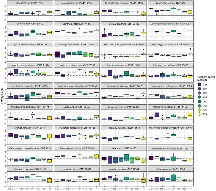
Grey et al.                                                                                              Screening Fungi for Anti-mycobacterial Activity

human pathogens are members of the Mycobacterium                            et al., 2020; Jain et al., 2020) or in vivo using sensitive imaging
tuberculosis complex, which causes the lung disease tuberculosis            equipment (Andreu et al., 2013).
(TB) (Gagneux, 2018), described by the World Health                            Here we describe a medium-throughput bioluminescence-
Organization as a global epidemic. Also of concern are the                  based pipeline to screen fungi for activity against Mycobacteria
non-tuberculous Mycobacteria (NTM), free living opportunistic               using the NTM species Mycobacterium abscessus and
pathogens that are ubiquitous in the environment and able to                Mycobacterium marinum. Our results indicate that many
cause lung, skin, and soft tissue infections (Mirsaeidi et al., 2014;       of the ICMP fungal isolates are anti-mycobacterial and have
Gonzalez-Santiago and Drage, 2015; Koh and Schlossberg, 2017).              identified isolates of Lophodermium culmigenum, Pseudaegerita
Almost two hundred NTM species have been identified to date,                viridis, and Trametes coccinea, as well as an unknown species of
which were recently found to divide into five clades based on               Boeremia and an isolate of an unknown genus and species in the
phylogenetic characteristics (Gupta et al., 2018). Rates of NTM             family Phanerochaetaceae as suitable for further study.
infections are increasing globally, including in hospital settings
(Al-Mahruqi et al., 2009; Roux et al., 2009; Moore et al., 2010;
Morimoto et al., 2014; Donohue and Wymer, 2016; Donohue,                    MATERIALS AND METHODS
2018; Ratnatunga et al., 2020). NTM are natural inhabitants
of water and their inclusion in implanted devices such as                   Bacterial Strains and Growth Conditions
catheters, prosthetics, and pacemakers, have resulted in cases of           In this study, we used M. abscessus BSG301 (Cadelis et al., 2021)
bacteremia and disseminated infection, while NTM outbreaks                  and M. marinum BSG101 (Dalton et al., 2017) which are stable
have been associated with invasive procedures such as cosmetic              bioluminescent derivatives transformed with the integrating
surgeries, intramuscular injections, and tattooing (Griffin et al.,         plasmid pMV306G13ABCDE (Andreu et al., 2010). We grew
2019; Jabbour et al., 2020). Recently, some cases of pulmonary              mycobacterial cultures with shaking (200 rpm) in Middlebrook
infections with Mycobacterium chimaera were traced back to site             7H9 broth (Fort Richard, New Zealand) supplemented with
of manufacture of heater-cooler units routinely used during open            10% Middlebrook ADC enrichment media (Fort Richard,
heart surgery (Williamson et al., 2017).                                    New Zealand), 0.4% glycerol (Sigma-Aldrich, New Zealand)
   Aotearoa New Zealand is an archipelago which split from                  and 0.05% tyloxapol (Sigma-Aldrich, New Zealand). We grew
the Gondwanan supercontinent approximately 85 million years                 M. abscessus at 37◦ C and M. marinum at 28◦ C.
ago and has since gradually become more isolated from other
land masses (Wallis and Trewick, 2009). This geographical
separation has led to the evolution of iconic native flora,
                                                                            Fungal Material
                                                                            Fungal isolates (Table 1) were provided by Manaaki Whenua –
fauna, and fungi. The Crown Research Institute Manaaki
                                                                            Landcare Research, a New Zealand Crown Research Institute
Whenua is the custodian of the International Collection of
                                                                            responsible for the curation of the International Collection of
Microorganisms from Plants (ICMP) (Johnston et al., 2017).
                                                                            Microorganisms from Plants (ICMP). We stored fungal isolates
The ICMP contains over 10,000 fungal cultures derived
                                                                            individually in cryotubes at −80◦ C. We made freezer stocks
from plants and soil from Aotearoa New Zealand and the
                                                                            by growing each fungus on 1.5% Potato Dextrose Agar (PDA)
South Pacific. The collection has a great diversity of fungal
                                                                            and excising small cubes of agar (5–6 mm in length) from the
species, host substrates, and collection localities, with the
                                                                            fungus’ growing edge. We placed these cubes within a cryovial
earliest cultures dating from the 1960s. While the collection
                                                                            containing 1 mL of 10% glycerol and rested them for 1 h after
contains some of the fungal genera traditionally used for
                                                                            which we removed the remaining liquid glycerol and stored the
antibiotic production it has not been rigorously tested for
                                                                            tubes at -80◦ C.
antimicrobial activity against mycobacterial species. In our view,
this makes the ICMP an excellent and untapped resource for
antibiotic discovery.                                                       Fungal DNA Extraction and ITS
   The search for new antibiotics with activity against                     Sequencing
Mycobacteria is complicated by their slow growth, with species              We used a small portion of mycelium from growing fungi and
like M. tuberculosis having a doubling time of approximately                extracted DNA using the REDExtract-N-AmpTM Plant PCR
24 h. Mycobacteria also tend to clump in liquid culture due to              Kit (Sigma-Aldrich) according to the manufacturer’s protocol.
their hydrophobic cell envelope. These properties make the two              We diluted DNA samples five-fold and amplified using the
most common methods of measuring antibacterial activity, the                ITS1F (50 CTTGGTCATTTAGAGGAAGTAA 30 ) and ITS4 (50
production of zones of inhibition when grown on agar, or degree             TCCTCCGCTTATTGATATGC 30 ) primer set in a 10 µl reaction
of turbidity when grown in liquid culture, slow and unreliable.             volume using the REDExtract-N-Amp Plant PCR Kit (Sigma-
Tagging bacteria with the genes that encode for luciferase-based            Aldrich) according to the manufacturer’s instructions. We used
reporters allows light to be used as a rapid surrogate marker               the following PCR conditions: initial denaturation at 94◦ C
for bacterial viability (Andreu et al., 2012). We and others have           for 3 min, followed by 40 cycles of denaturation at 94◦ C for
shown that bioluminescence is an excellent non-destructive                  30 s, annealing at 52◦ C for 30 s and extending at 72◦ C for
real-time reporter to assay for anti-mycobacterial activity in              30 s. The final extension was performed at 72◦ C for 7 min.
microtiter plate formats using a luminometer (Andreu et al.,                We checked the amplified DNA by gel electrophoresis before
2012; Dalton et al., 2016; Early et al., 2019; Chengalroyen                 sequencing using an Applied BiosystemsTM 3500xL Genetic

Frontiers in Microbiology | www.frontiersin.org                         2                                September 2021 | Volume 12 | Article 739995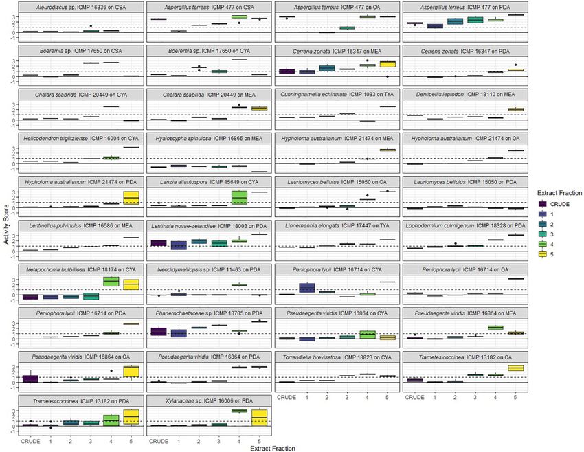
Grey et al.                                                                                                         Screening Fungi for Anti-mycobacterial Activity

TABLE 1 | Fungal isolates used in this study.

Fungus                           ICMP             GenBank     Description
                                 number           Accession

Agaricales sp.                   17554            MT107903    An undescribed crust fungus in the cyphellaceae – a family closely related to mushroom species but
                                                              forming crust or simple hood-like fruitbodies. It was isolated in Kerikeri, New Zealand in August
                                                              2007
Aleurodiscus sp.                 16336            MZ325955    Aleurodiscus sp. is a pinkish crust fungus. This culture was isolated from dead wood near Lake
                                                              Waikaremoana, New Zealand in May 1985
Amylostereum sacratum            10158            MZ325952    Amylostereum sacratum is a plant pathogen causing root rot. This culture was isolated from an
                                                              apple tree in Nelson, New Zealand in May 1977
Aspergillus terreus              477              MW862777    Aspergillus terreus is a common cosmopolitan saprotrophic soil-inhabiting fungus. This culture was
                                                              isolated in September 1961 in Auckland, New Zealand from sheep’s wool incubated at 30◦ C
Boeremia sp.                     17650            MW862790    This isolate is an unknown species of Boeremia which are often plant pathogens. It was isolated
                                                              from the surface of a mushroom in the Mamaku Plateau, New Zealand in May 1991
Cerrena zonata                   16347            MW862786    Cerrena zonata is a white rot decay fungus of dead wood. This culture was isolated from
                                                              Ngāruawāhia, New Zealand in April 1995
Chalara scabrida                 20449            MK432752    Chalara scabrida is an endemic saprobic fungus. The culture was isolated from a living Phormium
                                                              cookianum leaf in Mt Hutt, New Zealand in February 2014
Cunninghamella echinulata        1083             MZ325951    Cunninghamella echinulata is a common soil saprotroph. This culture was isolated from Auckland,
                                                              New Zealand in December 1978
Cylindrobasidium sp.             16397            MZ325956    This isolate is a crust fungus in the family physlacriaceae and related to the Armillaria mushroom.
                                                              This culture was isolated from apple wood in Auckland, New Zealand in June 1973
Dentipellis leptodon             18110            MZ325966    Dentipellis leptodon grows on the underside of dead wood and has dangling spines and is related
                                                              to the Lion’s Mane Hericium fungus. This culture was isolated from Metrosideros robusta wood in
                                                              Mamaku, New Zealand in March 1984
Helicodendron triglitziense      16004            MK432688    Helicodendron triglitziense is an aero-aquatic species isolated from dead alder leaves in Horseshoe
                                                              Lake Reserve wetland, Christchurch, New Zealand in June 2005
Hyaloscypha spinulosa            16865            MK432695    Hyaloscypha spinulosa is an aero-aquatic species isolated from a dead rimu twig in Pigeon Bay,
                                                              New Zealand in September 2006
Hypholoma australianum           21474            MZ325972    Hypholoma australianum is an orange mushroom with a white stem. This culture was isolated from
                                                              wood buried in soil in Otago Lakes, New Zealand in May 2016
Laetiporus portentosus           15555            MZ325953    Laetiporus portentosus is a soft bracket fungus, traditionally used as a tinder and wound packing
                                                              material by Mâori, the indigenous people of New Zealand. This culture was isolated from a beech
                                                              tree in Rimutaka Forest Park, New Zealand in May 1999
Lanzia allantospora              15649            AY755334    Lanzia allantospora is an endemic cup fungus found on kauri wood in Northland, New Zealand in
                                                              April 1992
Lauriomyces bellulus             15050            EF029218    Lauriomyces bellulus is a saprophytic fungus. This culture was isolated from a dead leaf of
                                                              Weinmannia racemosa in Katikati, New Zealand in May 2003
Lentinellus pulvinulus           16586            MW862787    Lentinellus pulvinulus is a white rot wood decay mushroom. This culture was isolated from a dead
                                                              wood in Pehitawa Kahikatea Forest Reserve, New Zealand in May 2006.
Lentinula novae-zelandiae        18003            MZ325965    Lentinula novae-zelandiae is a native edible “shitake” mushroom. This culture was isolated from
                                                              dead wood in Dunedin, New Zealand in September 1991
Linnemannia elongate             17447            MZ325962    Linnemannia elongate is a Mucorales fungus. The culture was isolated from a kauri tree in Rotorua,
                                                              New Zealand in January 2008
Lophodermium culmigenum          18328            MZ325968    Lophodermium culmigenum is a plant decay fungus. This culture was isolated from Trounson Kauri
                                                              Park, Chatham Islands, New Zealand in November 1992
Metapochonia bulbillosa          18174            MZ325967    Metapochonia bulbillosa is an insect pathogen fungus. This culture was isolated from dead leaves
                                                              of Marram grass in Lake Tennant, New Zealand in 1985
Mortierella sp.                  20597            MZ325970    This isolate is an unknown species of Mortierella, a common Mucorales soil fungus. This culture
                                                              was isolated from rotting wood from Farewell Spit, New Zealand in May 2014
Mucor laxorrhizus                20877            MZ325971    Mucor laxorrhizus is a Mucorales saprobe. This culture was isolated from rotten wood from a
                                                              stream in St Arnaud, New Zealand in January 2015
Neodidymelliopsis sp.            11463            MW862783    This isolate is an unknown species of Neodidymelliopsis which are typically plant pathogens. This
                                                              culture was isolated from Pittosporum leaves in Albany, Auckland, New Zealand in October 1991
Peniophora lycii                 16714            MZ325959    Peniophora lycii is a crust fungus. This culture was isolated from decaying wood in Te Waiiti,
                                                              New Zealand in May 2001
Phanerochaetaceae sp.            18785            MZ325969    This isolate is an unknown genus and species of crust fungi in the family Phanerochaetaceae. The
                                                              culture was isolated from beech leaves from Matakitaki, New Zealand in December 2010
Pleurotus australis              18149            MH395972    Pleurotus australis is an edible wood decay mushroom. This culture was isolated from the
                                                              Waitakere Ranges near Auckland, New Zealand in February 1987

                                                                                                                                                        (Continued)

Frontiers in Microbiology | www.frontiersin.org                              3                                       September 2021 | Volume 12 | Article 739995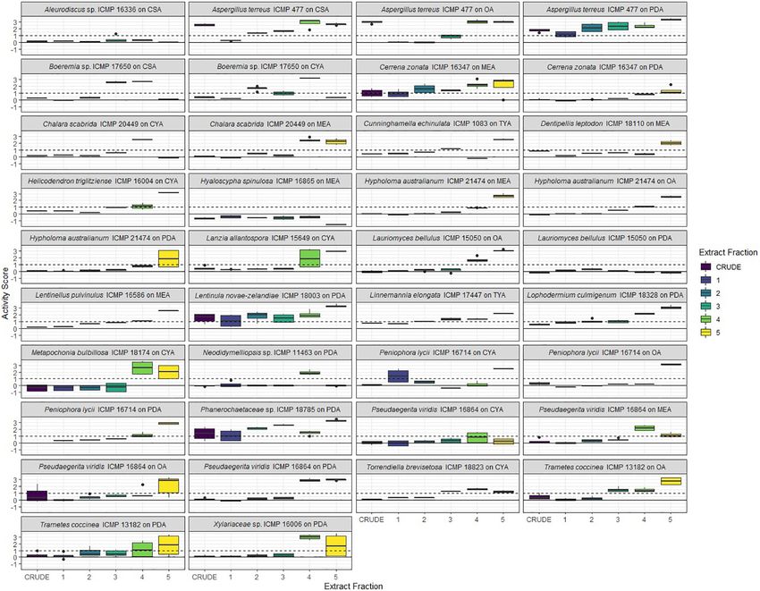
Grey et al.                                                                                                               Screening Fungi for Anti-mycobacterial Activity

TABLE 1 | (Continued)

Fungus                            ICMP            GenBank           Description
                                  number          Accession

Pleurotus purpureo-olivaceus      9630            MH395959          Pleurotus purpureo-olivaceus is an edible wood decay mushroom. This culture was isolated from
                                                                    Manapouri, New Zealand in May 1990
Pleurotus purpureo-olivaceus      17077           GQ411512          Pleurotus purpureo-olivaceus is an edible wood decay mushroom. This culture was isolated from
                                                                    the Craigieburn Range, New Zealand in May 2006
Pseudaegerita viridis             16864           MZ325960          Pseudaegerita viridis is an aero-aquatic species. This culture was isolated from a dead rimu twig in
                                                                    Pigeon Bay, New Zealand in September 2006
Stereum sp.                       16953           MZ325961          This isolate is an unknown species of Stereum, a wood decay bracket fungus, and was isolated
                                                                    from Rangitoto Station, New Zealand in November 2006
Torrendiella brevisetosa          18823           JN225946          Torrendiella brevisetosa is a cup fungus. This culture was isolated from beech leaves in Matakitaki,
                                                                    New Zealand in December 2010
Trametes coccinea                 13182           MW862784          Trametes coccinea is a wood decay bracket fungus. This culture was isolated from a dead radiata
                                                                    pine in Northland, New Zealand in September 1985
Umbelopsis sp.                    17492           EU770239          This isolate is an unknown species of Umbelopsis, a Mucorales saprobe, isolated from grapevines
                                                                    in Whenuapai, New Zealand in April 2007
Vararia fusispora                 17544           MZ325963          Vararia fusispora is a crust fungus. This culture was isolated from a decaying rimu branch in
                                                                    Owhango, New Zealand in October 2007
Xylariaceae sp.                   16006           MZ325954          This isolate is an unknown genus and species of the Xylariaceae family that was isolated from
                                                                    Ahuriri Reserve, Christchurch, New Zealand in May 2005

Analyzer using both ITS1F and ITS4 primers. We trimmed                                 triplicate wells of a black 24 well plate (4titude, Millennium
and combined the sequence data using Geneious (Geneious                                Science, New Zealand) and allowed them to set. We obtained
Biologics), removed any low-quality reads and used BLAST                               all media from Fort Richard (New Zealand). In addition to
to check fungal identification. Optimized sequence data were                           PDA, these comprised: Czapek Solution Agar (CSA), Czapek
aligned using MEGA7 (Kumar et al., 2016).                                              Yeast Extract Agar (CYA), Malt Extract Agar (MEA), Malt
                                                                                       Yeast Extract Agar (MYA), Oatmeal Agar (OA), Rice Extract
Primary Fungal Screening                                                               Agar (REA), and Tryptone Yeast Extract Agar (TYA). With
We grew fungal isolates on PDA (Fort Richard, New Zealand)                             the aid of a sterile scalpel blade, we sectioned fungal isolates
prior to screening for antibacterial activity using a 24 well                          grown on PDA into cubes ≤5 mm in diameter, and then
plate assay. Briefly, we added 0.5 mL aliquots of agar to                              transferred the cubes to the agar-filled wells of the 24-well
                                                                                       plates ensuring that each cube was placed fungus-side down
                                                                                       and touching the agar. We covered the inoculated 24-well
                                                                                       screening plates, sealed them with parafilm, and incubated them
                                                                                       at room temperature.
                                                                                           We monitored fungal growth visually at regular intervals and
                                                                                       recorded the time taken for them to either cover the entire well
                                                                                       or to stop visibly growing. At twice this time, we removed a 6 mm
                                                                                       plug of agar from each well using a biopsy punch. To screen
                                                                                       for antibacterial activity, we resuspended M. abscessus BSG301
                                                                                       and M. marinum BSG101 in 0.8% Middlebrook 7H9 broth (Fort
                                                                                       Richard, New Zealand) supplemented with 10% Middlebrook
                                                                                       ADC enrichment media (Fort Richard, New Zealand) to a
                                                                                       final concentration of 107 colony forming units (CFU)/mL for
                                                                                       M. abscessus and 108 CFU/mL for M. marinum. With the aid of a
                                                                                       pipette, we pipetted 50 µL of the bacterial-agar mixture into the
                                                                                       cylindrical holes left after removal of the fungal-agar plugs and
                                                                                       allowed the mixture to set. We measured bacterial luminescence
                                                                                       at regular intervals using a Victor X-3 luminescence plate
                                                                                       reader (PerkinElmer) with an integration time of 1 s. Between
                                                                                       measurements, plates were covered, placed in a plastic box
                                                                                       lined with damp paper towels, and incubated static at 37◦ C for
                                                                                       M. abscessus and 28◦ C for M. marinum. We performed these
  FIGURE 1 | Geographical spread of the isolation locations for the fungal             assays three times. We have published a more detailed description
  isolated used in this study within the archipelago of Aotearoa New Zealand.
                                                                                       of our methods on the protocol repository website protocols.io
  Black dots are individual ICMP isolates.
                                                                                       (Wiles and Grey, 2021a,b).

Frontiers in Microbiology | www.frontiersin.org                                    4                                      September 2021 | Volume 12 | Article 739995
Grey et al. Screening Fungi for Anti-mycobacterial Activity
TABLE 2 | ICMP isolates belonging to novel fungal taxa likely endemic to Aotearoa New Zealand.
Phylum Fungus ICMP number GenBank Accession Isolation substrate and location Isolation year
Ascomycota Boeremia sp. 17650 MW862790 Isolated from the surface of a mushroom in the Mamaku 1991
Plateau
Neodidymelliopsis sp. 11463 MW862783 Isolated from Pittosporum leaves in Albany, Auckland 1991
Xylariaceae sp. 16006 MZ325954 Isolated in Ahuriri Reserve, Christchurch 2005
Basidiomycota Agaricales sp. 17554 MT107903 Isolated in Kerikeri, Northland 2007
Aleurodiscus sp. 16336 MZ325955 Isolated from dead wood near Lake Waikaremoana 1985
Cylindrobasidium sp. 16397 MZ325956 Isolated from apple wood in Auckland 1973
Phanerochaetaceae sp. 18785 MZ325969 Isolated from beech leaves from Matakitaki 2010
Stereum sp. 16953 MZ325961 Isolated at Rangitoto Station 2006
Mucoromycota Mortierella sp. 20597 MZ325970 Isolated from rotting wood from Farewell Spit 2014
Umbelopsis sp. 17492 EU770239 Isolated from grapevines in Whenuapai 2007
FIGURE 2 | Phylogeny and activity of ICMP isolates grown in different media against M. abscessus BSG301 and M. marinum BSG101. Active media for each isolate
are shown in gray. CSA, Czapek Solution Agar; CYA, Czapek Yeast extract Agar; MEA, Malt Extract Agar; MYA, Malt Yeast extract Agar; OA, Oatmeal Agar; PDA,
Potato Dextrose Agar; REA, Rice Extract Agar; TYA, Tryptone Yeast extract Agar. ICMP isolates are described as active if they caused a minimum 1-log (90%)
reduction in the bioluminescence of the mycobacterial strains. The phylogenetic tree was constructed by comparing ITS sequences to those in GenBank and the
phylogenetic tree was constructed using the Molecular Evolutionary Genetics Analysis version 7.0 software (MEGA7) using the neighbor-joining method and
p-distance as the substitution model. Each phylogeny was tested using the bootstrap method with 500 replications.
Fungal Fermentation and Extraction Extract Screening
We grew fungal cultures either in liquid media or on We grew mycobacterial cultures until they reached stationary
solid media at room temperature and then freeze-dried phase (approximately 3–5 days for M. abscessus BSG301 and
them. We extracted the dry cultures with MeOH (Sigma- 7–10 days for M. marinum BSG101) and then diluted these in
Aldrich, New Zealand) for 4 h followed by CH2 Cl2 Mueller Hinton broth II (MHB) (Fort Richard) supplemented
(Sigma-Aldrich, New Zealand) overnight. We concentrated with 10% Middlebrook ADC enrichment media and 0.05%
the combined organic extracts under reduced pressure tyloxapol to give an optical density at 600 nm (OD600 ) of 0.001
and subjected the crude extracts to C8 reversed-phase which is the equivalent of ∼106 bacteria per mL. We dissolved
column chromatography eluting with a gradient of the fungal fractions in DMSO (Sigma-Aldrich, New Zealand) and
H2 O/MeOH (Sigma-Aldrich, New Zealand) to afford added these in duplicate to the wells of a black 96-well plate
five fractions (F1–F5). Full details are provided in (Nunc, Thermo Scientific) at doubling dilutions with a maximum
Supplementary Material. concentration of 50 mg/mL. Then we added 50 µL of diluted
Frontiers in Microbiology | www.frontiersin.org 5 September 2021 | Volume 12 | Article 739995Grey et al. Screening Fungi for Anti-mycobacterial Activity FIGURE 3 | Antibacterial activity of ICMP fungal isolates against Mycobacterium abscessus BSG301 when grown on different media. Data is presented as box and whisker plots of activity scores. The solid line shown at 0 is the median control value while the dotted line at 1 is the activity threshold. Scores above 1 correspond to a >90% reduction in bacterial bioluminescence compared to the corresponding no-fungi control. Similarly, an activity score above 2 means corresponds to a >99% reduction. CSA, Czapek Solution Agar; CYA, Czapek Yeast Extract Agar; MEA, Malt Extract Agar; MYA, Malt Yeast Extract Agar; OA, Oatmeal Agar; PDA, Potato Dextrose Agar; REA, Rice Extract Agar; TYA, Tryptone Yeast Extract Agar. Boxes are upper and lower quartiles with median shown. The whiskers extend up to 1.5× the inter-quartile range and any dots beyond those bounds are outliers. bacterial culture to each well of the fraction containing plates description of our methods on the protocol repository website giving final extract concentrations of 0–1000 µg/mL and a cell protocols.io (Wiles and Grey, 2021c,d). density of ∼5 × 10⁵ CFU/mL. We used the antibiotic rifampicin (Sigma-Aldrich, General Chemistry Conditions New Zealand) as a positive control at 1000 µg/mL for We recorded NMR spectra using a Bruker Avance DRX- M. abscessus and 10 µg/mL for M. marinum. Between 400 spectrometer or an Avance III-HD 500 spectrometer measurements, plates were covered, placed in a plastic box operating at 400 MHz or 500 MHz for 1 H nuclei and lined with damp paper towels, and incubated with shaking at 100 MHz or 125 MHz for 13 C nuclei utilizing standard 100 rpm at 37◦ C for M. abscessus and 28◦ C for M. marinum. pulse sequences at 298 K. We recorded high resolution We measured bacterial luminescence at regular intervals using mass spectra on a Bruker micrOTOF QII (Bruker Daltonics, a Victor X-3 luminescence plate reader (PerkinElmer) with an Bremen, Germany). We carried out analytical thin layer integration time of 1 s. We have defined the MIC as causing chromatography (TLC) on 0.2 mm thick plates of DC- a 1 log reduction in light production, as previously described plastikfolien Kieselgel 60 F254 (Merck). We carried out reversed- (Dalton et al., 2016, 2017). We have published a more detailed phase column chromatography on C8 support with a pore Frontiers in Microbiology | www.frontiersin.org 6 September 2021 | Volume 12 | Article 739995
Grey et al. Screening Fungi for Anti-mycobacterial Activity
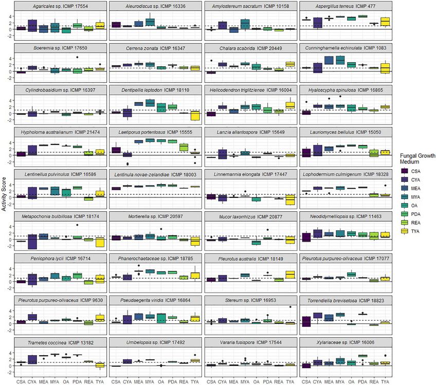
FIGURE 4 | Antibacterial activity of ICMP fungal isolates against Mycobacterium marinum BSG101 when grown on different media. Data is presented as box and
whisker plots of activity scores. The solid line shown at 0 is the median control value while the dotted line at 1 is the activity threshold. Scores above 1 correspond to
a >90% reduction in bacterial bioluminescence compared to the corresponding no-fungi control. Similarly, an activity score above 2 means corresponds to a >99%
reduction. CSA, Czapek Solution Agar; CYA, Czapek Yeast Extract Agar; MEA, Malt Extract Agar; MYA, Malt Yeast Extract Agar; OA, Oatmeal Agar; PDA, Potato
Dextrose Agar; REA, Rice Extract Agar; TYA, Tryptone Yeast Extract Agar. Boxes are upper and lower quartiles with median shown. The whiskers extend up to 1.5×
the inter-quartile range and any dots beyond those bounds are outliers.
size of 40–63 µm (Merck). We carried out gel filtration RESULTS
chromatography on Sephadex LH-20 (Pharmacia). We carried
out flash chromatography on Diol-bonded silica with a pore Identification of Novel Fungal Taxa
size of 40–63 micron (Merck). We used solvents that were
Endemic to Aotearoa New Zealand
of analytical grade or better and/or purified according to
The 36 ICMP fungal isolates used in this study were collected
standard procedures.
between 1961 and 2016 and from locations across Aotearoa
New Zealand, including the North, South, and Chatham Islands
Statistical Analysis (Figure 1). Of the 36 isolates, nine were not able to be identified
We fitted a logistic mixed model for activity with a random as a known species and one is from both an unknown genus
effect for biologic replicates. We tested the main effects and and species (Table 2). These isolates likely represent novel
second-order interactions of the variables, using the lme4 and car taxa endemic to Aotearoa New Zealand. As with the broader
packages in R (Bates et al., 2015; Fox and Weisberg, 2019; R Core collection, they cover a range of isolation dates, the earliest
Team, 2020). being isolated in 1973 (Cylindrobasidium sp. ICMP 16397)
Frontiers in Microbiology | www.frontiersin.org 7 September 2021 | Volume 12 | Article 739995Grey et al. Screening Fungi for Anti-mycobacterial Activity
and the most recent in 2014 (Mortierella sp. ICMP 20597). The fungal isolates we tested belong to three Phyla: the
They also cover a broad range of isolation locations within Basidiomycota (18 ICMP isolates), the Ascomycota (13 ICMP
New Zealand, from Kerikeri in the North Island (Agaricales sp. isolates), and the Mucoromycota (5 ICMP isolates) (Figure 2).
ICMP 17554) to Christchurch in the South Island (Xylariaceae We observed that the group with the most active fungi-
sp. ICMP 16006). medium combinations was the Basidiomycota (55%), followed
by the Mucoromycota (52%), and then the Ascomycota (46%%)
(Figure 2). More Basidiomycota-medium combinations were
Whole Cell Screening Identified Many active against M. marinum [92/144 (64%)] (Figures 2, 4)
ICMP Fungal Isolates as Having than M. abscessus [67/144 (47%)] (Figures 2, 3). In contrast,
Anti-mycobacterial Activity more Ascomycota-medium combinations were active against
We screened 36 ICMP fungal isolates for antibacterial activity M. abscessus [53/104 (51%)] (Figures 2, 3) than M. marinum
against M. abscessus BSG301 and M. marinum BSG101. The [43/104 (41%)] (Figures 2, 4).
isolates belong to three different fungal Phyla, and we grew them
on eight different media giving us a total of 288 fungi-media
Differential Impact of Growth Medium on
combinations tested for each bacterium (Figures 2–4). Anti-mycobacterial Activity
We measured antibacterial activity as reductions in light We observed that many of the ICMP fungi displayed differential
output of our bioluminescent mycobacterial strains over a 72-h activity depending on their growth medium with the majority
period. We calculated activity scores by first converting the being active on more than one medium. An isolate of the native
luminescence measurement at each time-point into an area New Zealand “shiitake” mushroom Lentinula novae-zelandiae
under the curve (AUC) value for each well. We then divided (ICMP 18003) and an isolate of unknown genus and species in
this number by the median AUC of a sterile control plate the family Phanerochaetaceae (ICMP 18785) were active against
inoculated and incubated at the same time as the fungus- both M. abscessus and M. marinum when grown on an all 8
containing plates. The negative log of this value corresponds media (Figures 2–4). An isolate of Aspergillus terreus (ICMP
to the activity score. We define a fungus-media combination as 477) was also active against M. abscessus regardless of growth
active/antibacterial if the median activity score is above 1 which media (Figures 2, 4), while Amylostereum sacratum ICMP 10158,
corresponds to a > 90% reduction in light compared to the Cerrena zonata ICMP 16347, Hypholoma australianum ICMP
control. Similarly, an activity score above 2 means corresponds 21474, Laetiporus portentosus ICMP 15555, and Pseudaegerita
to a > 99% reduction. viridis ICMP 16864 were active against M. marinum when grown
We observed no consistent difference in activity by on all media (Figures 2, 4).
mycobacterial strain, but there is strong statistical evidence When assessing for activity against M. abscessus, 2/36 fungal
of differences in activity between media and fungal Phyla, and isolates were only active when grown on one of the eight media,
that these vary by mycobacterial strain (Table 3). an unknown species of Mortierella (ICMP 20597) when grown
on PDA and an unknown species of Stereum (ICMP 16953) on
MYA (Figures 2, 3). ICMP 20597 was also only active against
More ICMP Fungal Isolates Are Active Against M. marinum when grown on one of the eight media, though in
M. marinum Than M. abscessus this case it was TYA (Figures 2, 4). Three other fungal isolates
We observed that 28/36 (77%) fungal isolates were active were only active against M. marinum when grown on one of
against M. abscessus when grown in at least one medium, the eight media, Helicodendron triglitziense ICMP 16004 on
with 130/288 (45%) fungi-medium combinations being anti- TYA, and Metapochonia bulbillosa ICMP 18174 and an unknown
mycobacterial against this bacterium (Figures 2, 3). In contrast, species of Umbelopsis (ICMP 17492) on OA (Figures 2, 4).
34/36 (94%) fungal isolates were active against M. marinum,
with 146/288 (51%) fungi-medium combinations being anti- Potato Dextrose Agar (PDA) Is the Most Active
mycobacterial against this bacterium (Figures 2, 4). Of the two Culture Medium for Screening for Anti-mycobacterial
fungal isolates that were not active against M. marinum when Activity
grown in any of the media tested, only the Mucoromycota We observed that PDA was the most active culture medium
fungus Mucor laxorrhizus ICMP 20877 displayed no activity with 29/36 (80%) fungal isolates active against either bacterium
against M. abscessus. The second isolate, Pleurotus purpureo- (Figure 2). For M. marinum, the most active culture medium
olivaceus ICMP 9630 was active against M. abscessus when was OA, followed by PDA (24 and 23 active fungi, respectively)
grown on CYA, MYA, and TYA. However, a second isolate of while for M. abscessus it was MYA followed by PDA (24 and
P. purpureo-olivaceus we tested, ICMP 17077, was active against 22 active fungi, respectively). We observed that isolates grown
both mycobacterial strains. We could discern no obvious pattern on REA and CSA were the least active, with only 14/36 isolates
between fungal species or genus for those isolates that were (39%) being active against either mycobacterium species when
only active against M. marinum, namely Agaricales sp. ICMP cultured on these media (Figure 2). MYA and MEA were the two
17554, Boeremia sp. ICMP 17650, Cylindrobasidium sp. ICMP media that favored M. abscessus activity, with 24 fungal isolates
16397, Lanzia allantospora ICMP 15649, Linnemannia elongata active when grown on MYA and 20 isolates active when grown on
ICMP 17447, Metapochonia bulbillosa ICMP 18174, and Vararia MEA, compared to 17 and 16 being active against M. marinum,
fusispora ICMP 17544. respectively (Figure 2).
Frontiers in Microbiology | www.frontiersin.org 8 September 2021 | Volume 12 | Article 739995Grey et al. Screening Fungi for Anti-mycobacterial Activity
TABLE 3 | Analysis of Deviance Table (Type II Wald Chi2 tests). acids and sterols. Fractions F2, F3, and F4 typically contain the
chemical compounds we are most interested in pursuing, with
Chi2 Degrees of Significance
freedom
the potential to be bioactive.
We tested, at a single concentration of 1000 µg/mL,
Fungal phyla 2.57 2 p = 0.28 the crude extracts and fractions F1–F5 from all 41 fungus-
Mycobacterial strain 3.48 1 p = 0.06 medium combinations for activity against M. marinum BSG101
Fungal growth medium 273.30 7 p < 0.0001 (Figures 5, 6) and for 38 of the combinations for activity against
Fungal Phyla × Mycobacterial strain 99.50 2 p < 0.0001 M. abscessus BSG301 (Figures 5, 7). As described previously,
Fungal Phyla × fungal growth 117.96 14 p < 0.0001 we measured antibacterial activity as reductions in light output
medium
of our bioluminescent mycobacterial strains over a 72-h period
Mycobacterial strain × fungal 124.95 7 p < 0.0001
and calculated activity scores as the negative log of the ratio
growth medium
of the AUC values of the fungus-containing measurements and
the control measurements. We define an extract/fraction as
active/antibacterial if the median activity score is above 1, which
Screening of Extracts and Fractions corresponds to a >90% reduction in light compared to the
control, as previously described. Similarly, an activity score above
From ICMP Fungal Isolates for 2 means corresponds to a > 99% reduction.
Anti-mycobacterial Activity
We prepared extracts from 41 fungus-medium combinations ICMP Fungal Extracts and Fractions Retain
which were further separated into 5 fractions, designated F1– Anti-mycobacterial Activity
F5. Fraction F1 (100% water) is generally comprised of sugars We observed that only 3/38 of the fungus-medium combinations
while fraction F5 (100% methanol) contains predominantly fatty we tested for activity against M. abscessus BSG301 did not
FIGURE 5 | Phylogeny and activity of crude extracts and fractions from ICMP isolates grown in different media against M. abscessus BSG301 and M. marinum
BSG101. Active extracts and fractions for each isolate are shown in gray. CSA, Czapek Solution Agar; CYA, Czapek Yeast extract Agar; MEA, Malt Extract Agar;
MYA, Malt Yeast extract Agar; OA, Oatmeal Agar; PDA, Potato Dextrose Agar; REA, Rice Extract Agar; TYA, Tryptone Yeast extract Agar. ICMP isolates are
described as active if they caused a minimum 1-log (90%) reduction in the bioluminescence of the mycobacterial strains. The phylogenetic tree was constructed by
comparing ITS sequences to those in GenBank and the phylogenetic tree was constructed using the Molecular Evolutionary Genetics Analysis version 7.0 software
(MEGA7) using the neighbor-joining method and p-distance as the substitution model. Each phylogeny was tested using the bootstrap method with 500 replications.
Frontiers in Microbiology | www.frontiersin.org 9 September 2021 | Volume 12 | Article 739995Grey et al. Screening Fungi for Anti-mycobacterial Activity FIGURE 6 | Antibacterial activity of crude extracts and fractions 1–5 from ICMP fungal isolates against Mycobacterium marinum BSG101. Data is presented as box and whisker plots of activity scores. The solid line shown at 0 is the median control value while the dotted line at 1 is the activity threshold. Scores above 1 correspond to a >90% reduction in bacterial bioluminescence compared to the corresponding no-fungi control. Similarly, an activity score above 2 means corresponds to a >99% reduction. CSA, Czapek Solution Agar; CYA, Czapek Yeast Extract Agar; MEA, Malt Extract Agar; OA, Oatmeal Agar; PDA, Potato Dextrose Agar; TYA, Tryptone Yeast Extract Agar. Boxes are upper and lower quartiles with median shown. The whiskers extend up to 1.5× the inter-quartile range and any dots beyond those bounds are outliers. retain any activity in either the crude extract or any of the 5 F4 [11/17 (65%)]. Seven Basidiomycota-medium combinations fractions. These belonged to Aleurodiscus sp. ICMP 16336 grown also displayed some activity from fractions F2 and/or F3. on CSA, Pseudaegerita viridis ICMP 16864 grown on CYA, and Like the Basidiomycota-medium combinations, of the 21 Lauriomyces bellulus ICMP 15050 grown on PDA (Figures 5, 7). Ascomycota-medium combinations we observed to be active, Of the remaining 14 Basidiomycota-medium combinations the most active fraction was F5 [21/21 (100%)], followed by tested, the most active fraction was F5 [14/14 (100%)], followed F4 [16/21 (76%)]. Eight Ascomycota-medium combinations by F4 [7/14 (50%)]. Four Basidiomycota-medium combinations also displayed some activity from fractions F2 and/or F3. also displayed some activity from fractions F2 and/or F3. Of Both Mucoromycota-medium combinations tested had multiple the 17 Ascomycota-medium combinations we observed to be active fractions. active, the most active fraction was F4 [17/17 (100%)], followed by F5 [13/17 (76%)]. Six Ascomycota-medium combinations Linoleic Acid Is Likely the Anti-mycobacterial also displayed some activity from fractions F2 and/or F3. Compound Present in Fraction F5 Both Mucoromycota-medium combinations tested had multiple Given the anti-mycobacterial activity we observed from the active fractions. F5 fractions of so many of the ICMP isolates, we analyzed We observed that all 41 of the fungus-medium combinations this fraction in more detail from four phylogenetically diverse we tested for activity against M. marinum BSG101 retained some ICMP isolates: the Basidiomycota Aleurodiscus sp. ICMP activity in either the crude extract or at least one of the 5 fractions 16336, the Ascomycota Hyaloscypha spinulosa ICMP 16865 (Figures 5, 6). Of the 18 Basidiomycota-medium combinations and Lanzia allantospora ICMP 15649, and the Mucoromycota tested, the most active fraction was F5 [17/18 (94%)], followed by Cunninghamella echinulata ICMP 1083. NMR spectroscopic and Frontiers in Microbiology | www.frontiersin.org 10 September 2021 | Volume 12 | Article 739995
Grey et al.                                                                                                              Screening Fungi for Anti-mycobacterial Activity

  FIGURE 7 | Antibacterial activity of crude extracts and fractions 1–5 from ICMP fungal isolates against Mycobacterium abscessus BSG301. Data is presented as
  box and whisker plots of activity scores. The solid line shown at 0 is the median control value while the dotted line at 1 is the activity threshold. Scores above 1
  correspond to a >90% reduction in bacterial bioluminescence compared to the corresponding no-fungi control. Similarly, an activity score above 2 means
  corresponds to a >99% reduction. CSA, Czapek Solution Agar; CYA, Czapek Yeast Extract Agar; MEA, Malt Extract Agar; OA, Oatmeal Agar; PDA, Potato Dextrose
  Agar; TYA, Tryptone Yeast Extract Agar. Boxes are upper and lower quartiles with median shown. The whiskers extend up to 1.5× the inter-quartile range and any
  dots beyond those bounds are outliers.

Mass spectrometric analysis confirmed the presence of linoleic                            In contrast to M. abscessus, of the 31 fungus-medium
acid in fraction F5 of these fungi.                                                    combinations tested against M. marinum BSG101, the most
                                                                                       active fractions were F4 rather than F3, with 17/31 (55%) F4
Identification of Active ICMP Fungal Fractions for                                     fractions having an MIC ≤ 500 µg/mL. Of these, the most active
Further Analysis                                                                       fungus-medium combinations were Pseudaegerita viridis ICMP
To prioritize the most active anti-mycobacterial fungus-                               16864 grown on PDA and MEA, with an MIC of 31.25 and
medium combinations for further NMR spectroscopic and Mass                             62.5 µg/mL, respectively, and Lophodermium culmigenum ICMP
spectrometric analysis, we tested those fractions F2, F3, and                          18328, with an MIC of 62.5 µg/mL (Table 4). Three F2 fractions
F4 that were active at 1000 mg/mL to obtain their minimum                              and six F3 fractions had an MIC ≤ 500 µg/mL.
inhibitory concentration (MIC) (Table 4).
   Of the 31 fungus-medium combinations tested against
M. abscessus BSG301, 11/31 (35%) F3 fractions had an                                   DISCUSSION
MIC ≤ 500 µg/mL. Of these, the most active fungus-medium
combinations were Pseudaegerita viridis ICMP 16864 grown on                            In this study, we describe a medium-throughput
PDA with an MIC of 31.25 µg/mL, and Aspergillus terreus ICMP                           bioluminescence-based pipeline to screen fungi for activity
477 grown on CSA, with an MIC of 125 µg/mL (Table 4). In                               against Mycobacteria using bioluminescent derivates of
contrast, only three F4 fractions and one F2 fraction had an                           M. abscessus and M. marinum as the testing strains. We included
MIC ≤ 500 µg/mL.                                                                       M. abscessus as it is a relatively fast-growing non-tuberculous

Frontiers in Microbiology | www.frontiersin.org                                   11                                      September 2021 | Volume 12 | Article 739995
Grey et al. Screening Fungi for Anti-mycobacterial Activity
TABLE 4 | Minimum inhibitory concentrations (MIC) of ICMP fungal fractions (F2, F3, and F4) against M. abscessus and M. marinum.
Species ICMP Media1 M. abscessus MIC (µg/mL) M. marinum MIC (µg/mL)
F2 F3 F4 F2 F3 F4
Aspergillus terreus 477 CSA 1000 125 1000 250 500 125
OA >1000 250 >1000 >1000 1000 250
PDA 500 500 500 500 500 500
Boeremia sp. 17650 CSA 1000 500 >1000 >1000 >1000 1000
CYA >1000 250 1000 >1000 >1000 500
Cerrena zonata 16347 MEA 1000 1000 1000 1000 1000 500
PDA >1000 >1000 >1000 >1000 >1000 1000
Chalara scabrida 20449 CYA >1000 1000 >1000 >1000 1000 >1000
MEA >1000 1000 >1000 >1000 >1000 500
Cunninghamella echinulata 1083 TYA 1000 >1000 >1000 1000 500 >1000
Hypholoma australianum 21474 MEA >1000 >1000 >1000 >1000 >1000 500
OA >1000 1000 >1000 >1000 >1000 1000
PDA >1000 >1000 >1000 >1000 >1000 1000
Lauriomyces bellulus 15050 OA >1000 1000 >1000 >1000 >1000 1000
PDA >1000 >1000 >1000 >1000 >1000 >1000
Lentinellus pulvinulus 16586 MEA >1000 1000 >1000 >1000 >1000 1000
Lentinula novae-zelandiae 18003 PDA 1000 1000 1000 1000 500 500
Linnemannia elongate 17447 TYA 1000 1000 1000 1000 >1000 1000
Lophodermium culmigenum 18328 PDA 1000 500 >1000 1000 1000 62.5
Metapochonia bulbillosa 18174 CYA >1000 250 1000 >1000 500 500
Neodidymelliopsis sp. 11463 PDA >1000 1000 >1000 >1000 >1000 500
Peniophora lycii 16714 PDA >1000 >1000 >1000 >1000 1000 1000
Phanerochaetaceae sp. 18785 PDA 500 1000 1000 500 250 1000
Pseudaegerita viridis 16864 CYA >1000 >1000 >1000 >1000 >1000 250
MEA >1000 500 >1000 1000 >1000 62.5
OA >1000 >1000 >1000 >1000 >1000 250
PDA >1000 31.25 >1000 1000 >1000 31.25
Torrendiella brevisetosa 18823 CYA 500 500 >1000 >1000 1000 >1000
Trametes coccinea 13182 OA 1000 1000 >1000 >1000 1000 500
PDA >1000 1000 >1000 1000 1000 250
Xylariaceae sp. 16006 PDA >1000 500 >1000 >1000 >1000 1000
1 CSA, Czapek Solution Agar; CYA, Czapek Yeast Extract Agar; MEA, Malt Extract Agar; OA, Oatmeal Agar; PDA, Potato Dextrose Agar; TYA, Tryptone Yeast Extract Agar.
MIC values ≤500 µg/mL are shown in bold.
mycobacterial species and is a cause of opportunistic infections active against M. marinum than M. abscessus. This is not
in patients with cystic fibrosis or chronic pulmonary disease, unsurprising given their different ecological niches and divergent
and of skin and soft tissue infections, for which treatment genomes (Malhotra et al., 2017). Identification of the chemical
options are limited (To et al., 2020; Victoria et al., 2021). We compounds responsible may shed some further light on
also included M. marinum as, despite being a pathogen of these differences.
fish, amphibians, and reptiles, it shares conserved virulence Our screening pipeline involves growing fungi in 24 well
determinants with M. tuberculosis (Bouz and Al Hasawi, 2018; plates on multiple growth media. It has previously been
Ramakrishnan, 2020) and is a Biosafety Level (BSL) 2 rather than shown that different culture conditions can alter the expression
an airborne BSL 3 organism. of biosynthetic gene clusters and therefore the structural
We screened 36 ICMP fungal isolates using our assay diversity and quantity of secondary metabolites produced by
and discovered that almost all produced considerable anti- microorganisms, including fungi (Bills et al., 2008). For example,
mycobacterial activity. This is in contrast with our experience growing Fusarium tricinctum on Rice medium supplemented
screening ICMP isolates for activity against other human with fruit and vegetable juices led to the discovery of Fusarielin
pathogens such as Escherichia coli, Pseudomonas aeruginosa, J (Hemphill et al., 2017) while growing Asteromyces cruciatus
and Staphylococcus aureus, where we find just 5–20% of on Czapek-Dox medium with an altered nitrogen source led to
fungal isolates have some antibacterial activity (unpublished the discovery of Lajollamide A (Gulder et al., 2012). We selected
data). We also observed some differences between the two media that cover a broad range of carbon and nitrogen sources, as
mycobacterial species, with more ICMP isolates being well as different pH and metal ions. We have also included media
Frontiers in Microbiology | www.frontiersin.org 12 September 2021 | Volume 12 | Article 739995Grey et al. Screening Fungi for Anti-mycobacterial Activity
with less chemically defined elements, including from potatoes, efforts on Polyporales fungi could prove fruitful for the discovery
rice, and oatmeal. Our data provides strong statistical evidence of new antibacterial compounds.
for the impact of growth media on antibacterial activity of the
ICMP fungi we tested, with some fungal isolates only active when
grown on one medium and others active when grown on several, DATA AVAILABILITY STATEMENT
or even all. The highest proportion of ICMP isolates were active
when grown on the nutritionally rich Oatmeal Agar (OA), Potato The datasets presented in this study can be found in
Dextrose Agar (PDA), and (MYA) while the lowest proportion online repositories. The names of the repository/repositories
were active when grown on the more nutritionally poor Rice and accession number(s) can be found below: https://
Extract Agar (REA) and Czapek Solution Agar (CSA). www.ncbi.nlm.nih.gov/genbank/, MT107903, MZ325955,
The ICMP fungi we screened in this project included MZ325952, MW862777, MW862790, MW862786, MK432752,
isolates of several species well known to produce antimicrobial MZ325951, MZ325956, MZ325966, MK432688, MK432695,
compounds. For example, Aspergillus terreus produces terrein MZ325972, MZ325953, AY755334, EF029218, MW862787,
(Goutam et al., 2017), maunakeanolic acid A and B (Zaman MZ325965, MZ325962, MZ325968, MZ325967, MZ325970,
et al., 2020), and helvolic acid (Zaman et al., 2020), amongst MZ325971, MW862783, MZ325959, MZ325969, MH395972,
other compounds. Chemical extraction and fractionation of a MH395959, GQ411512, MZ325960, MZ325961, JN225946,
subset of the ICMP isolates revealed that much of the activity MW862784, EU770239, MZ325963, and MZ325954
we observed may be due to the production of the known anti- and https://figshare.com/, https://auckland.figshare.com/
mycobacterial compound linoleic acid (Kanetsuna, 1985; Choi, articles/dataset/Antibacterial_activity_of_fungal_isolates_
2016). However, we have identified several ICMP isolates that from_the_International_Collection_of_Microorganisms_
retained their anti-mycobacterial activity in non-linoleic acid from_Plants_ICMP_against_Mycobacterium_abscessus_and_
containing fractions. These include isolates of Lophodermium Mycobacterium_marinum_/14937894.
culmigenum, Pseudaegerita viridis, and Trametes coccinea, as
well as an unknown species of Boeremia and an isolate of an
unknown genus and species in the family Phanerochaetaceae. AUTHOR CONTRIBUTIONS
Investigations are ongoing to identify the sources of their anti-
mycobacterial activity and to determine whether any may be BW, BC, and SW contributed to conception and design of the
due to the production of novel bioactive compounds. Species study. AG, MC, YD, and DP performed the experiments. AG,
of Lophodermium and Pseudaegerita have previously been found MC, TL, BW, and SW were involved in data analysis. AG and
to produce several antifungal compounds (Hosoya et al., 2007; SW wrote the manuscript. All authors contributed to manuscript
Sumarah et al., 2011; McMullin et al., 2015), while T. coccinea revision, read, and approved the submitted version.
is predicted to have secondary metabolite pathways though
genomic analysis (Zhang et al., 2020).
An interesting observation we have made, is of the abundant FUNDING
anti-mycobacterial activity of fungi we tested in the order
Polyporales. The isolates Cerrena zonata ICMP 16347, Laetiporus This work was supported by funds from Cure Kids (9102
portentosus ICMP 15555, Phanerochaetaceae sp. ICMP 18785, 3715810), NZ Carbon Farming (9102 3718092), Maurice
and T. coccinea ICMP 13182, were active against both Wilkins Centre for Molecular Biodiscovery (9159 3715235), and
mycobacterial species when grown in almost all media. These donations from the New Zealand public. BW, DP, and the
fungi are bracket-like fungi with pores on the under surface. To ICMP culture collection were funded by the SSIF infrastructure
fulfill their ecological niche of digesting moist wood, these fungi investment fund of the New Zealand Ministry of Business,
first need to colonize the wood. To do this they need to compete Innovation and Employment.
with other microorganisms, including bacteria, and producing
antimicrobial compounds would be beneficial on this process.
An alternative hypothesis could be that their antibacterial activity SUPPLEMENTARY MATERIAL
is a by-product of these fungi producing the peroxidases and
oxidases they need to digest wood (Sulej et al., 2019). Should The Supplementary Material for this article can be found
the activity prove not to be the result of peroxidase/oxidase online at: https://www.frontiersin.org/articles/10.3389/fmicb.
production, this would suggest that focusing future screening 2021.739995/full#supplementary-material
REFERENCES Andreu, N., Fletcher, T., Krishnan, N., Wiles, S., and Robertson, B. D. (2012). Rapid
measurement of antituberculosis drug activity in vitro and in macrophages
Al-Mahruqi, S. H., van Ingen, J., Al-Busaidy, S., Boeree, M. J., Al- using bioluminescence. J. Antimicrob. Chemother. 67, 404–414. doi: 10.1093/
Zadjali, S., Patel, A., et al. (2009). Clinical relevance of nontuberculous jac/dkr472
mycobacteria, Oman. Emerg. Infect. Dis. 15, 292–294. doi: 10.3201/eid1502.0 Andreu, N., Zelmer, A., Fletcher, T., Elkington, P. T., Ward, T. H., Ripoll,
80977 J., et al. (2010). Optimisation of bioluminescent reporters for use
Frontiers in Microbiology | www.frontiersin.org 13 September 2021 | Volume 12 | Article 739995Grey et al. Screening Fungi for Anti-mycobacterial Activity
with mycobacteria. PLoS One 5:e10777. doi: 10.1371/journal.pone.001 Hemphill, C. F. P., Sureechatchaiyan, P., Kassack, M. U., Orfali, R. S., Lin, W.,
0777 Daletos, G., et al. (2017). OSMAC approach leads to new fusarielin metabolites
Andreu, N., Zelmer, A., Sampson, S. L., Ikeh, M., Bancroft, G. J., Schaible, U. E., from Fusarium tricinctum. J. Antibiot. (Tokyo) 70, 726–732. doi: 10.1038/ja.
et al. (2013). Rapid in vivo assessment of drug efficacy against Mycobacterium 2017.21
tuberculosis using an improved firefly luciferase. J. Antimicrob. Chemother. 68, Hosoya, T., Ohsumi, J., Hamano, K., Ono, Y., and Miura, M. (2007). Method for
2118–2127. doi: 10.1093/jac/dkt155 Producing Cercosporamide. Worldwide Patent No WO2007018194A1.
Bates, D., Mächler, M., Bolker, B., and Walker, S. (2015). Fitting Linear mixed- Jabbour, S. F., Malek, A. E., Kechichian, E. G., Tomb, R. R., and Nasr, M. W.
effects models using lme4. J. Stat. Softw. 67, 1–48. (2020). Nontuberculous mycobacterial infections after cosmetic procedures:
Bills, G. F., Platas, G., Fillola, A., Jiménez, M. R., Collado, J., Vicente, F., et al. a systematic review and management algorithm. Dermatol. Surg. 46,
(2008). Enhancement of antibiotic and secondary metabolite detection from 116–121.
filamentous fungi by growth on nutritional arrays. J. Appl. Microbiol. 104, Jain, P., Garing, S., Verma, D., Saranathan, R., Clute-Reinig, N., Gadwa, J.,
1644–1658. doi: 10.1111/j.1365-2672.2008.03735.x et al. (2020). Nanoluciferase reporter mycobacteriophage for sensitive and
Bouz, G., and Al Hasawi, N. (2018). The zebrafish model of tuberculosis – no lungs rapid detection of Mycobacterium tuberculosis drug susceptibility. J. Bacteriol.
needed. Crit. Rev. Microbiol. 44, 779–792. doi: 10.1080/1040841X.2018.1523132 202:e00411-20. doi: 10.1128/JB.00411-20
Cadelis, M. M., Gordon, H., Grey, A., Geese, S., Mulholland, D. R., Weir, B. S., et al. Johnston, P. R., Weir, B. S., and Cooper, J. A. (2017). Open data on fungi and
(2021). Isolation of a novel polyketide from Neodidymelliopsis sp. Molecules bacterial plant pathogens in New Zealand. Mycology 8, 59–66. doi: 10.1080/
26:3235. doi: 10.3390/molecules26113235 21501203.2016.1278409
Chengalroyen, M. D., Jordaan, A., Seldon, R., Ioerger, T., Franzblau, S. G., Kanetsuna, F. (1985). Bactericidal effect of fatty acids on mycobacteria, with
Nasr, M., et al. (2020). Biological profiling enables rapid mechanistic particular reference to the suggested mechanism of intracellular killing.
classification of phenotypic screening hits and identification of KatG activation- Microbiol. Immunol. 29, 127–141. doi: 10.1111/j.1348-0421.1985.tb00811.x
dependent pyridine carboxamide prodrugs with activity against Mycobacterium Koh, W.-J., and Schlossberg, D. (2017). Nontuberculous mycobacteria-overview.
tuberculosis. Front. Cell. Infect. Microbiol. 10:699. doi: 10.3389/fcimb.2020. Microbiol. Spectr. 5:5.1.11. doi: 10.1128/microbiolspec.TNMI7-0024-2016
582416 Kumar, S., Stecher, G., and Tamura, K. (2016). MEGA7: molecular evolutionary
Choi, W. H. (2016). Evaluation of anti-tubercular activity of linolenic genetics analysis version 7.0 for bigger datasets. Mol. Biol. Evol. 33, 1870–1874.
acid and conjugated-linoleic acid as effective inhibitors against Malhotra, S., Vedithi, S. C., and Blundell, T. L. (2017). Decoding the similarities and
Mycobacterium tuberculosis. Asian Pac. J. Trop. Med. 9, 125–129. doi: differences among mycobacterial species. PLoS Negl. Trop. Dis. 11:e0005883.
10.1016/j.apjtm.2016.01.021 doi: 10.1371/journal.pntd.0005883
Dalton, J. P., Uy, B., Okuda, K. S., Hall, C. J., Denny, W. A., Crosier, P. S., et al. McMullin, D. R., Green, B. D., and Miller, J. D. (2015). Antifungal sesquiterpenoids
(2017). Screening of anti-mycobacterial compounds in a naturally infected and macrolides from an endophytic Lophodermium species of Pinus strobus.
zebrafish larvae model. J. Antimicrob. Chemother. 72, 421–427. doi: 10.1093/ Phytochem. Lett. 14, 148–152. doi: 10.1016/j.phytol.2015.10.006
jac/dkw421 Mirsaeidi, M., Farshidpour, M., Allen, M. B., Ebrahimi, G., and Falkinham, J. O.
Dalton, J. P., Uy, B., Phummarin, N., Copp, B. R., Denny, W. A., Swift, S., et al. (2014). Highlight on advances in nontuberculous mycobacterial disease in
(2016). Effect of common and experimental anti-tuberculosis treatments on North America. BioMed Res. Int. 2014:919474. doi: 10.1155/2014/919474
Mycobacterium tuberculosis growing as biofilms. PeerJ 4:e2717. doi: 10.7717/ Moore, J. E., Kruijshaar, M. E., Ormerod, L. P., Drobniewski, F., and Abubakar, I.
peerj.2717 (2010). Increasing reports of non-tuberculous mycobacteria in England, Wales
Donohue, M. J. (2018). Increasing nontuberculous mycobacteria reporting rates and Northern Ireland, 1995-2006. BMC Public Health 10:612. doi: 10.1186/
and species diversity identified in clinical laboratory reports. BMC Infect. Dis. 1471-2458-10-612
18:163. doi: 10.1186/s12879-018-3043-7 Morimoto, K., Iwai, K., Uchimura, K., Okumura, M., Yoshiyama, T., Yoshimori,
Donohue, M. J., and Wymer, L. (2016). Increasing prevalence rate of K., et al. (2014). A steady increase in nontuberculous mycobacteriosis mortality
nontuberculous mycobacteria infections in five states, 2008–2013. Ann. Am. and estimated prevalence in Japan. Ann. Am. Thorac. Soc. 11, 1–8. doi: 10.1513/
Thorac. Soc. 13, 2143–2150. doi: 10.1513/AnnalsATS.201605-353OC AnnalsATS.201303-067OC
Early, J. V., Mullen, S., and Parish, T. (2019). A rapid, low pH, nutrient stress, assay Pontali, E., Raviglione, M. C., and Migliori, G. B. (2019). Regimens to treat
to determine the bactericidal activity of compounds against non-replicating multidrug-resistant tuberculosis: past, present and future perspectives. Eur.
Mycobacterium tuberculosis. PLoS One 14:e0222970. doi: 10.1371/journal.pone. Respir. Rev. 28:190035. doi: 10.1183/16000617.0035-2019
0222970 R Core Team. (2020). R: A Language And Environment for Statistical Computing.
Fox, J., and Weisberg, S. (2019). An R Companion to Applied Regression, 3rd Edn. Available online at: https://www.R-project.org/ (accessed July 12, 2021)
Thousand Oaks, CA: Sage. Ramakrishnan, L. (2020). Mycobacterium tuberculosis pathogenicity viewed
Gagneux, S. (2018). Ecology and evolution of Mycobacterium tuberculosis. Nat. Rev. through the lens of molecular Koch’s postulates. Curr. Opin. Microbiol. 54,
Microbiol. 16, 202–213. doi: 10.1038/nrmicro.2018.8 103–110. doi: 10.1016/j.mib.2020.01.011
Gonzalez-Santiago, T. M., and Drage, L. A. (2015). Nontuberculous mycobacteria: Ratnatunga, C. N., Lutzky, V. P., Kupz, A., Doolan, D. L., Reid, D. W., Field, M.,
skin and soft tissue infections. Granulomat. Disord. Adult Skin 33, 563–577. et al. (2020). The rise of non-tuberculosis mycobacterial lung disease. Front.
doi: 10.1016/j.det.2015.03.017 Immunol. 11:303. doi: 10.3389/fimmu.2020.00303
Goutam, J., Sharma, G., Tiwari, V. K., Mishra, A., Kharwar, R. N., Ramaraj, Roux, A.-L., Catherinot, E., Ripoll, F., Soismier, N., Macheras, E., Ravilly, S., et al.
V., et al. (2017). Isolation and characterization of “Terrein” an antimicrobial (2009). Multicenter study of prevalence of nontuberculous mycobacteria in
and antitumor compound from endophytic fungus Aspergillus terreus (JAS-2) patients with cystic fibrosis in France. J. Clin. Microbiol. 47, 4124–4128. doi:
associated from Achyranthus aspera Varanasi, India. Front. Microbiol. 8:1334. 10.1128/JCM.01257-09
doi: 10.3389/fmicb.2017.01334 Seaworth, B. J., and Griffith, D. E. (2017). Therapy of multidrug-resistant and
Griffin, I., Schmitz, A., Oliver, C., Pritchard, S., Zhang, G., Rico, E., et al. (2019). extensively drug-resistant Tuberculosis. Microbiol. Spectr. 5, doi: 10.1128/
Outbreak of tattoo-associated nontuberculous mycobacterial skin infections. microbiolspec.TNMI7-0042-2017
Clin. Infect. Dis. 69, 949–955. doi: 10.1093/cid/ciy979 Sulej, J., Osińska-Jaroszuk, M., Jaszek, M., Gra̧z, M., Kutkowska, J., Pawlik, A.,
Gulder, T. A. M., Hong, H., Correa, J., Egereva, E., Wiese, J., Imhoff, J. F., et al. et al. (2019). Antimicrobial and antioxidative potential of free and immobilised
(2012). Isolation, structure elucidation and total synthesis of lajollamide A cellobiose dehydrogenase isolated from wood degrading fungi. Fungal Biol. 123,
from the marine fungus Asteromyces cruciatus. Mar. Drugs 10, 2912–2935. 875–886. doi: 10.1016/j.funbio.2019.09.007
doi: 10.3390/md10122912 Sumarah, M. W., Kesting, J. R., Sørensen, D., and Miller, J. D. (2011). Antifungal
Gupta, R. S., Lo, B., and Son, J. (2018). Phylogenomics and comparative genomic metabolites from fungal endophytes of Pinus strobus. Phytochemistry 72, 1833–
studies robustly support division of the Genus Mycobacterium into an emended 1837. doi: 10.1016/j.phytochem.2011.05.003
Genus Mycobacterium and four novel genera. Front. Microbiol. 9:67. doi: 10. To, K., Cao, R., Yegiazaryan, A., Owens, J., and Venketaraman, V. (2020).
3389/fmicb.2018.00067 General overview of nontuberculous mycobacteria opportunistic pathogens:
Frontiers in Microbiology | www.frontiersin.org 14 September 2021 | Volume 12 | Article 739995You can also read