Ding's herbal enema treats dextran sulfate sodium induced colitis in mice by regulating the gut microbiota and maintaining the Treg/Th17 cell balance
←
→
Page content transcription
If your browser does not render page correctly, please read the page content below
EXPERIMENTAL AND THERAPEUTIC MEDICINE 22: 1368, 2021

          Ding's herbal enema treats dextran sulfate sodium‑induced
                colitis in mice by regulating the gut microbiota
                  and maintaining the Treg/Th17 cell balance
          YAN‑YAN TAN1*, YANG DING2*, XUEPING ZHENG1, GONG‑JIAN DAI1, SU‑MIN ZHANG1, XU YANG1,
             DA‑CHAO XU1, PENG CHEN2, JIA‑MIN ZHANG2, JIA‑ZE MA2, MENG LI2, SHI‑CAI HUANG2,
                   YAN LIU2, YU‑TING ZHANG2, HAN XING2, KANG DING1 and YI‑JIANG DING1

      1
   Department of National Center of Colorectal Surgery, Jiangsu Integrate Colorectal Oncology Center, Nanjing Hospital of
             Chinese Medicine Affiliated to Nanjing University of Chinese Medicine, Nanjing, Jiangsu 210001;
2
  Department of First Clinical Medical College, Nanjing University of Chinese Medicine, Nanjing, Jiangsu 210029, P.R. China

                                            Received July 27, 2020; Accepted August 18, 2021

                                                        DOI: 10.3892/etm.2021.10802

Abstract. Ding's herbal enema (DHEP) is a traditional                  DHEP in UC that may be associated with the regulation of the
Chinese medicinal therapy that has been used to treat ulcer‑           gut microbiota composition, and maintenance of the balance
ative colitis (UC) in China. The present study determined the          between Treg and Th17 cells. Furthermore, β‑sitosterol exhib‑
molecular mechanism of the effect of DHEP in UC treatment.             ited the same effects with DHEP and it could be a possible
C57BL/6J mice were treated with 3.5% (w/v) dextran sulfate             substitute for DHEP in UC treatment.
sodium (DSS) for 7 days to establish an animal model of colitis.
The mice were divided into five groups (n=5): Control, vehicle,        Introduction
DHEP, mesalazine and β‑sitosterol. After oral administration
for 7 days, the body weight, disease activity index, histopa‑          Inflammatory bowel disease (IBD), including ulcerative
thology and inflammatory factors were analyzed. The fractions          colitis (UC) and Crohn's disease, is an autoimmune disease
of CD4+Foxp3+ regulatory T (Treg) cells and CD4+IL‑17A+                of the intestine (1). IBD is influenced by a number of factors,
T helper (Th) cells were determined by flow cytometry. Gut             such as eating habits, disorders of the intestinal flora (2) and
microbiota composition was analyzed by next‑generation                 the immune system (3). There is a lack of specific therapeutic
sequencing. The results revealed that DHEP and β‑sitosterol            target in UC; therefore, the strategy of UC treatment involves
could significantly alleviate the symptoms of DSS‑induced              immune regulation and inhibition of inflammation (4,5).
UC. Furthermore, the levels of IL‑6, cyclooxygenase‑2,                 There have been previous precedents of successful prevention
TNF‑α and p65 were reduced after administration of DHEP.               and cure of inflammatory diseases using traditional Chinese
Additionally, the data indicated that DHEP could increase the          herbal medicines (6,7). Ding's herbal enema (DHEP) was
abundance of seven operational taxonomic units (OTUs) and              created by Ding Zemin, an eighth‑generation descendant of
decrease the abundance of 12 OTUs in the gut microbiota. The           the Ding family. DHEP has been used in The Third Affiliated
content of short‑chain fatty acids in the colon remodeled the          Hospital of Nanjing University of Chinese Medicine (Nanjing,
balance of Treg/Th17 cells in DSS‑induced UC in mice. The              China) for >50 years and has an excellent curative effect
present study preliminarily defined the mechanism of action of         in IBD treatment (8). DHEP contains Lonicerae japonicae
                                                                       flos (Jinyinhua), Sanguisorba officinalis L. (Diyu), Bletilla
                                                                       striata (Thunb.) Rchb.F. (Baiji), Phellodendron chinense
                                                                       Schneid. (Huangbo), Coptis chinensis Franch (Huanglian)
Correspondence to: Professor Kang Ding or Professor Yi‑Jiang Ding,     and Portulaca oleracea L. (Machixian). All of the constitu‑
Department of National Center of Colorectal Surgery, Jiangsu           ents of the DHEP solution that are from traditional Chinese
Integrate Colorectal Oncology Center, Nanjing Hospital of Chinese      medicine.
Medicine Affiliated to Nanjing University of Chinese Medicine,             Studies have demonstrated that Jinyinhua, Diyu and Baiji
1 Jinling Road, Nanjing, Jiangsu 210001, P.R. China
                                                                       contain β‑sitosterol (9,10). β‑sitosterol has immunomodulatory
E‑mail: dingkang1123@163.com
                                                                       and anti‑inflammatory activity, and is present in numerous
E‑mail: 2543775235@qq.com
                                                                       plants (11). A number of studies have revealed that β‑sitosterol
*
    Contributed equally                                                can inhibit inflammation through the NF‑κ B pathway (12‑16).
                                                                       Animal experiments suggest that β‑sitosterol can significantly
Key words: Ding's herbal enema, β‑sitosterol, ulcerative colitis,      reduce colonic shortening, disease activity index (DAI) and
short‑chain fatty acids, T cell differentiation, gut microbiota        fetal hemoglobin content in mice (17). Additionally, β‑sitosterol
                                                                       specifically increases the activity of T helper (Th) cells, and
                                                                       increases the activity of T cells and natural killer cells (18).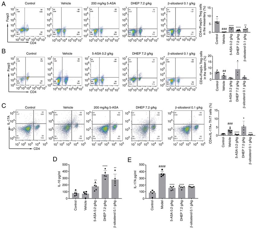
2                 TAN et al: DING'S ULCER ENEMA TREATS MURINE COLITIS BY REGULATING GUT MICROBIOTA

    The present study investigated the mechanism of action of     The oven temperature was gradually increased from 100˚C to
DHEP and its possible active ingredient, β‑sitosterol, in the     250˚C at a rate of 50˚C/min and from 250˚C to 280˚C at a rate
treatment of UC. C57BL/6J mice were treated with 3.5% (w/v)       of 10˚C/min and was held for 4 min.
dextran sulfate sodium (DSS) for 7 days to generate an animal
model of colitis. DHEP and β‑sitosterol were administered for     Animal models of DSS‑induced acute colitis and treatments.
7 days, and the results indicated that DHEP and β‑sitosterol      Mice were treated with 3.5% (w/v) DSS in their drinking water
inhibited the inflammatory response and restored the              for 7 days, followed by switching to regular drinking water
Th17/regulatory T cell (Treg) balance by regulating intestinal    for 7 days. Test compounds were administered via an enema
microbiota and short‑chain fatty acids (SCFAs).                   per day for 7 days following DSS treatment. To investigate the
                                                                  effect of DHEP, all mice were randomly divided into 5 groups:
Materials and methods                                             Control (no DSS treatment, orally administered with sterile
                                                                  water; n=12), vehicle (DSS treated for 7 days, orally adminis‑
Animals. A total of 50 adult female C57BL/6 mice (6‑8 weeks       tered with sterile water; n=12), DHEP (administered by enema
old), weighing 20‑24 g, were obtained from Yangzhou               at a dose of 7.2 g/kg; n=12), 5‑ASA (orally administered at a
University Comparative Medical Center (Yangzhou, China).          dose of 0.2 g/kg; N = 12) and β ‑sitosterol (administered by
Mice were fed with free access to food and drinking water and     enema at dose of 0.1 g/kg; n=12). At 24 h after the last enema,
housed in a temperature‑controlled room (22±4˚C) under a          mice that had been fasting for 12 h were sacrificed.
12‑h dark‑light cycle with a relative humidity of 50%. Animal
welfare and experimental procedures were carried out in           Evaluation of DAI. Body weight, stool consistency and rectal
accordance with the guidelines and the associated ethical regu‑   bleeding of the mice were recorded daily and scored according
lations of The Experimental Animal Ethics Committee of the        to Cooper's scoring criteria (19). The mean of the three scores
Nanjing Hospital of Chinese Medicine Affiliated to Nanjing        was calculated and recorded as the DAI. The body weight
University of Chinese Medicine (Nanjing, China), which            loss was scored as 0 when there was no loss in weight; 1 for
approved the animal experiments (approval no. 2020‑10‑002).       a loss of 1‑5%; 2 for a loss of 5‑10%; 3 for a loss of 10‑15%;
                                                                  or 4 for a loss of >15%. Regarding stool consistency, 0 points
Reagents. DHEP was obtained from The Third Affiliated             indicated normal pellets, 2 points indicated loose stools that
Hospital of Nanjing University of Chinese Medicine (Nanjing,      did not stick to the anus and 4 points indicated diarrhea. For
China). β ‑sitosterol was purchased from Beijing Solarbio         rectal bleeding, 0 points were given for negative (‑) results in
Science & Technology Co., Ltd. (cat. no. IS0690). Mesalazine      the Hemoccult test (20); 1 point for positive (+) results; 2 points
(5‑ASA) was purchased from Ethypharm SAS (cat. no. 160306).       for positive (++) results; 3 points for positive (+++) results; and
DSS (36‑50 kDa) was purchased from MP Biomedicals, LLC            4 points for gross rectal bleeding (++++) results.
(cat. no. 160110). IL‑6 (cat. no. 12912), TNF‑α (cat. no. 3707)
and p65 (cat. no. 6956) antibodies were obtained from Cell        Histopathology. A representative sample from the middle
Signaling Technology, Inc. Cyclooxygenase (COX)‑2 (cat.           region of the colon was fixed in 4% paraformaldehyde at 4˚C
no. BM4419) antibodies were obtained from Wuhan Boster            for 24 h, embedded in paraffin, sectioned (5‑µm), stained with
Biological Technology, Ltd. IL‑17A (cat. no. ml037864) and        hematoxylin (at room temperature for 5 min) and eosin (at
IL‑10 (cat. no. ml037873) enzyme‑linked immunosorbent assay       room temperature for 2 min) and then observed using an IX 51
(ELISA) kits were purchased from Shanghai Enzyme‑Linked           epifluorescence Olympus microscope (Olympus Corporation)
Biotechnology Co., Ltd.                                           equipped with a DP‑26 digital camera at x40 magnification.

Determination of β ‑sitosterol in DHEP. DHEP powder               16S DNA high‑throughput sequencing. The construction of a
(20 g) was extracted using methanol (100 ml x 3) at room          high‑throughput sequencing library and sequencing based on
temperature. After removal of methanol under reduced pres‑        the Illumina MiSeq platform was performed by Genewiz, Inc.
sure (~‑0.1 Mpa), the aqueous brownish syrup (120 ml) was         A Qubit 2.0 Fluorometer (Invitrogen; Thermo Fisher Scientific,
suspended in H2O (100 ml) and then partitioned with petroleum     Inc.) was used to detect the DNA concentration of the samples
ether (50 ml x 3) to obtain 500 mg petroleum ether‑soluble        and to construct a sequencing library using the MetaVxTM
fraction. The fraction was further fractionated on a silica gel   Library Construction kit (Genewiz, Inc.). Using 10 nmol of DNA
column eluted with petroleum ether‑acetone (from 1:0 to 0:1)      as a template, a series of PCR primers designed by Genewiz,
to obtain eleven fractions (1‑10) according to thin‑layer chro‑   Inc. were used to amplify the prokaryotic 16S ribosomal (r)
matography analysis. Fraction three (65 mg) was loaded onto       DNA, including two highly variable regions of V3 and V4. The
a silica gel column and eluted with methanol‑CHCl3 (20:80,        V3 and V4 regions were amplified using an upstream primer
30:70 and 40:60; 50 ml of each mixture) to yield four subfrac‑    containing the 5'‑CCTACGGRRBGCASCAGKVRVGAAT‑3'
tions (fractions 3‑1 to 3‑4). Fraction 3‑2 was analyzed by gas    sequence and a downstream primer containing the 5'‑GGA
chromatography. The analytes (1 µl) were separated using          CTACNVGGGTWTCTAATCC‑3' sequence. In addition, a
an Agilent 6890N‑5975B GC system (Agilent Technologies,           primer with an index was added to the end of the PCR product
Inc.) equipped with a flame ionization detector and an HP‑5       of the 16S rDNA by PCR for next‑generation sequencing.
column (30 m x 250 µm x 0.25 µm) using 99.99% nitrogen as a       The library quality was determined using an Agilent 2100
carrier gas and 99.99% hydrogen as an auxiliary gas at a flow     Bioanalyzer (Agilent Technologies, Inc.) and library concen‑
rate of 1.2 ml/min with a split ratio of 10:1. The temperatures   trations were determined using a Qubit 2.0 fluorometer
of injector and detector were 250˚C and 280˚C, respectively.      (Invitrogen; Thermo Fisher Scientific, Inc.). After the DNA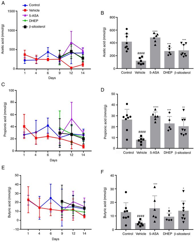
EXPERIMENTAL AND THERAPEUTIC MEDICINE 22: 1368, 2021                                             3

library was mixed, the Illumina MiSeq (Illumina, Inc.)            Bioengineering Institute) according to the to the manufac‑
instrument was used in accordance with the manufacturer's         turer's protocols.
instructions. The instrument performed 2x300 bp double‑end
sequencing (paired end) reactions, and the sequence informa‑      Western blotting. Mouse colon tissue was collected and lysed
tion was read by the MiSeq Control Software (v4.0) that comes     in RIPA lysis buffer (cat. no. P0013B; Beyotime Institute of
with MiSeq.                                                       Biotechnology). The protein content was determined by the
                                                                  BCA method. Equal quantities (50 µg) of protein lysate were
SCFAs measurements. Fresh feces (0.2 g) were suspended in         loaded into each lane of a 10% SDS‑polyacrylamide gel and
1 ml of deionized water and homogenized by vortexing for          wet‑transferred onto a polyvinylidene difluoride membrane
1 min before centrifugation (138,00 x g at 4˚C for 30 min). The   (Bio‑Rad Laboratories, Inc.) using a Bio‑Rad electro transfer
supernatant from the colon and cecum (630 and 720 µl, respec‑     apparatus. After blocking with 5% skimmed milk powder in
tively) was mixed with 25% metaphosphoric acid at a ratio         Tris‑buffered saline and 0.1% Tween‑20 at room temperature
of 9:1 (v:v) and was shaken at 37˚C for 4 h. The supernatant      for 2 h, the membranes were incubated with the primary
was collected and stored at ‑20˚C before gas chromatog‑           antibodies. The following antibodies were used: p65 (1:500),
raphy analysis. Standard curves were constructed according        COX‑2 (1:500), IL‑6 (1:1,000), TNF‑ α (1:100) and actin
to a previously described method (15). Specifically, 1 µl of      (cat. no. ab6276, 1:5,000; Epitomics; Abcam). Horseradish
the analytes was separated using an Agilent 6890N‑5975B           peroxidase (HRP)‑conjugated goat anti‑rabbit IgG antibody
GC system equipped with an flame ionization detector and          (cat. no. D110058, 1:10,000; Sangon Biotech Co., Ltd.) was
a DB‑FFAP column (30 m x 250 µm x 0.25 µm; Agilent                used as the secondary antibody. Immunoblotted bands were
Technologies Inc.) (21) using 99.99% nitrogen as a carrier        visualized with the Immobilon Western Chemilum HRP
gas and 99.99% hydrogen as an auxiliary gas at a flow rate        Substrate (cat. no. WBKLS0100; MilliporeSigma) using a
of 0.8 ml/min with a split ratio of 50:1. The temperatures of     JS‑680 B Gel Documentation and Analysis System (Bio‑Rad
injector and detector were 250˚C and 280˚C, respectively. The     Laboratories, Inc.) and quantified using Image Lab v3.0
oven temperature was gradually increased from 60˚C to 220˚C       (Bio‑Rad Laboratories, Inc.) and Quantity One v4.62 (Bio‑Rad
at a rate of 20˚C/min and held for 1 min (22).                    Laboratories, Inc.) software. All the proteins were normalized
                                                                  to the corresponding actin level.
Flow cytometry. The Treg and Th17 cells in the mouse
spleen and mesenteric lymph nodes (MLNs) were isolated            Statistical analysis. Statistical analysis was performed using
using a Mouse Lymphocyte Separation Solution kit (cat.            one‑way ANOVA, followed by Tukey's post hoc test. Ordinal
no. DKW33‑R0100; Dakewe Biotech Co., Ltd.) according to           data were analyzed using Kruskal‑Wallis with Dunn's post
the manufacturer's instructions. The differentiation of Treg      hoc test. Quantitative data are presented as mean ± standard
and Th17 cells in the spleen and MLNs were analyzed with          deviation (unless otherwise shown). P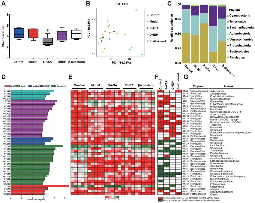
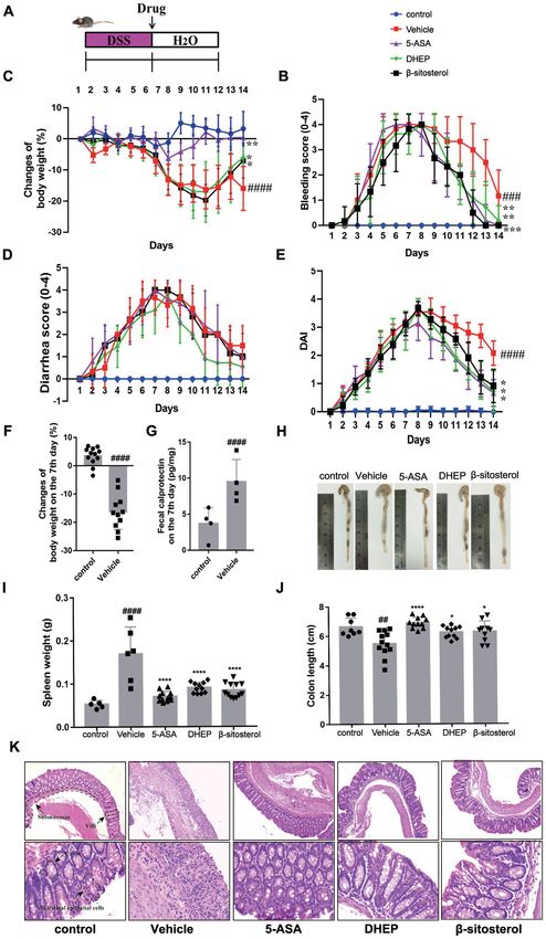
4                      TAN et al: DING'S ULCER ENEMA TREATS MURINE COLITIS BY REGULATING GUT MICROBIOTA

Figure 1. DHEP and β‑sitosterol treatment of acute colitis in vivo. (A) Schematic overview of the experimental design. Changes in (B) body weights, (C) clinical
bleeding scores, (D) clinical diarrhea scores and (E) DAI are presented, and all statistical values are compared on day 14. (F) Weight loss and (G) fecal calpro‑
tectin content in feces after colitis induction. (H) Representative images of whole colons and colon length. (I) Spleen mass of mice after the end of the experiment.
(J) Quantification of whole colons and colon length. (K) Colon sections were counterstained with hematoxylin and eosin, and high‑magnification images are
presented in the bottom row (top row magnification, x100; bottom row, x400). n=4‑12. ##P
EXPERIMENTAL AND THERAPEUTIC MEDICINE 22: 1368, 2021                                            5

model was successfully established (27,28). DHEP, compared          were significantly altered in the control, 5‑ASA, DHEP and
with the vehicle group, was administered by enema for 7 days        β ‑sitosterol groups (Fig. 2F). Comparison with the vehicle
and attenuated the symptoms of colitis (loss of body weight,        group indicated that 5‑ASA altered 21 OTUs. The 12 reduced
diarrhea, rectal bleeding and DAI) (Fig. 1B‑E). In addition, for    OTUs in the 5‑ASA group (OTU 42, 321, 8, 7, 43, 182,
β‑sitosterol treatment, the loss of body weight, rectal bleeding    100, 203, 183, 48, 125 and 286) are highlighted in red, and
and DAI score were relieved remarkably compared with the            9 increased OTUs (OTU 73, 16, 13, 60, 77, 39, 41, 136 and
vehicle group (Fig. 1B‑E). These results suggested that DHEP        71) are highlighted in green in Fig. 2F. In the DHEP‑treated
and β‑sitosterol might be a positive effect on the treatment of     group, 19 OTUs were altered compared with those in the
DSS‑induced UC mice model.                                          vehicle group. As presented in Fig. 2F, 11 OTUs (OTU 321,
    As presented in Fig. 1H and J, the colon was edematous and      12, 120, 182, 239, 203, 83, 233, 183, 93 and 196) were reduced
significantly shortened in the vehicle group of mice compared       and are highlighted in red, which belonged to the genera of
with that in the control group. A reduction in colon length in      Erysipelatoclostridium, Bifidobacterium, Anaerotruncus,
DSS‑treated mice was significantly attenuated by treatment          Defluviitaleaceae UCG‑011, Family XIII AD3011 group,
with 5‑ASA, DHEP and β ‑sitosterol. Additionally, 5‑ASA,            Tyzzerella, Eubacterium coprostanoligenes, Proteus and three
DHEP and β ‑sitosterol significantly reduced the weight of          uncultured genera; and a total of seven OTUs (OTU 73, 3, 13,
the spleen compared with the vehicle, which was the largest         29, 41, 136 and 1) were increased, which belonged to the genera
immune organ (Fig. 1I). Comparison with the control group           of Bacteroides, Lactobacillus, Turicibacter, Parabacteroides,
mice identified certain pathological changes in the DSS‑treated     Eubacterium xylanophilum group and one uncultured genus.
mice (Fig. 1K), including damaged intestinal epithelial cells,      In the β ‑sitosterol‑treated group, six OTUs (OTU 22, 182,
submucosal edema, disappearance of villi and crypts and a           100, 203, 93 and 286) were reduced (Fig. 2F). The six reduced
large number of infiltrating inflammatory cells (Fig. 1K).          OTU belonged to Akkermansia, Defluviitaleaceae UCG‑011,
However, these pathological changes were relieved after             Ruminococcaceae UCG‑005, Tyzzerella and Oligella Only
administration of 5‑ASA, DHEP and β‑sitosterol (Fig. 1K).           four OTUs were increased (OTU 270, 13, 29 and 41), which
                                                                    belonged to Lactobacillus, Parabacteroides and two uncul‑
DHEP and β‑sitosterol remodels the distribution of intestinal       tured genera. Thus, 34 OTUs were altered by 5‑ASA, DHEP
microbiota in mice. To determine whether intestinal micro‑          and β‑sitosterol, and only four OTUs (OTU 182, 203, 13 and 41)
biota were affected by treatment with 5‑ASA, DHEP and               were modulated in the same direction as in the control group
β‑sitosterol, 16S rDNA sequencing was used to evaluate the          for all three treatment groups. These four OTUs belonged to
abundance of intestinal flora in mice. After the sequencing         the genera Anaerotruncus, Ruminococcaceae UCG‑005 and
data were analyzed by QIIME, the α diversity for all groups         Turicibacter and uncultured bacterium, which belonged to the
represented by the Shannon indexes is presented in Fig. 2A.         Firmicutes, Firmicutes, Bacteroidetes and Firmicutes phyla,
Notably, the diversity in the DSS‑treated groups were similar       respectively (Fig. 2G).
to that in the control group. By contrast, the diversity in the
5‑ASA group was significantly decreased compared with that in       DHEP and β ‑sitosterol treatment increases the profiles of
the vehicle group. However, these differences were not signifi‑     SCFAs in the colon of mice with DSS‑induced UC. SCFAs
cant in the DHEP‑ and β‑sitosterol‑treated groups (Fig. 2A).        are the end‑products generated from food with a high dietary
β diversity represented by PCA is presented in Fig. 2B. The         content by gut microbiota and can alleviate IBD in animal
distributions in the DHEP and 5‑ASA groups were relatively          models (29). To evaluate whether DHEP and β ‑sitosterol
concentrated, and the distribution between the groups was           increased the release of SCFAs, the SCFA content in the colon
relatively dispersed. There was no overlap between the DHEP         of mice was evaluated (Fig. 3). The levels of SCFAs, including
and 5‑ASA groups. There was a partial overlap between the           acetic acid (Fig. 3A and B), propionic acid (Fig. 3C and D) and
vehicle and β‑sitosterol groups and the control group; however,     butyric acid (Fig. 3E and F) were significantly reduced in the
the overall distribution was relatively dispersed, which may        colon of DSS‑treated mice compared with the control group,
be due to individual differences and intervention effects           and the levels of SCFAs were increased in the 5‑ASA, DHEP
in mice. The bacterial composition at the phylum level is           and β ‑sitosterol groups compared with the vehicle group.
presented in Fig. 2C. The abundance at this level was vari‑         These results indicated that the beneficial effects of DHEP in
able, including Firmicutes, Bacteroidetes and Proteobacteria.       colitis involved an increase in the SCFA content in the colon.
The abundance of Bacteroidetes and Proteobacteria were
increased in DHEP group compared with vehicle group, but            DHEP and β ‑sitosterol regulates the Th17/Treg balance in
the abundance of Firmicutes was decreased. These results            mice with DSS‑induced UC. The Th17/Treg transformation
indicated that DHEP might be able to regulate the ratios of         balance is important for the maintenance of intestinal homeo‑
Bacteroidetes/Firmicutes and Proteobacteria/Firmicutes. In          stasis (30). To investigate whether DHEP and β‑sitosterol had a
addition, in the β‑sitosterol group, the ratios of Bacteroidetes/   positive effect on the balance of Th17/Treg cells, the expression
Firmicutes and Proteobacteria/ Firmicutes were upregulated,         levels of IL‑17A and IL‑10, which are specifically expressed
but not to the same level as the DHEP group.                        in Th17 cells and Treg cells, respectively, were determined.
    To identify the bacteria possibly regulated by DHEP and         The results demonstrated that the numbers of Treg cells in the
β‑sitosterol, LEfSe analysis was used to obtain the OTUs for        MLNs (Fig. 4A) and spleen (Fig. 4B) of mice with DSS‑induced
each group. As a result, 42 significant OTUs were identified,       UC were significantly lower compared with those in the
along with their linear discriminant analysis score (Fig. 2D),      control mice. After administration with 5‑ASA or β‑sitosterol
and a heatmap was generated (Fig. 2E). Notably, 39 OTUs             for 7 days, the number of Treg cells was significantly increased
6 TAN et al: DING'S ULCER ENEMA TREATS MURINE COLITIS BY REGULATING GUT MICROBIOTA Figure 2. DHEP and β‑sitosterol treatments affects the distribution of flora in vivo. (A) Comparison of α diversity assessed by Shannon index‑based genus level. (B) PC analysis of gut microbiota communities‑based genus level. (C) Column diagram of microbial composition at the phylum level. (D) Linear discriminant analysis at the OTU level. (E) Heatmap showing the abundance of 42 OTUs in different groups based on linear discriminant analysis. (F) Changing direction of OTUs induced by DSS, 5‑ASA, DHEP or β‑sitosterol intervention. (G) OTUs represent bacterial taxonomic information (n=5). #P
EXPERIMENTAL AND THERAPEUTIC MEDICINE 22: 1368, 2021                                                                       7

Figure 3. DHEP and β‑sitosterol can increase short chain fatty acid content in vivo. (A) Acetic acid content across the 14‑day period. (B) Acetic acid content at day 14.
(C) Propionic acid content across the 14‑day period. (D) Propionic acid content at day 14. (E) Butyric acid content across the 14‑day period. (F) Butyric acid content
at day 14. n=5‑8. ####P
8                     TAN et al: DING'S ULCER ENEMA TREATS MURINE COLITIS BY REGULATING GUT MICROBIOTA

Figure 4. DHEP and β‑sitosterol treatments promote T cell differentiation in mice. Representative flow cytometric profiles and quantitative analysis of the
(A) spleen and (B) mesenteric lymph nodes Treg cells, and (C) spleen Th17 cells (n= 3‑9). ELISA showing (D) IL‑10 and (E) IL‑17A protein expression
levels (NN=5). ##P
EXPERIMENTAL AND THERAPEUTIC MEDICINE 22: 1368, 2021                                                                 9

Figure 5. Expression levels of various inflammatory factors in mice. (A) Fecal calprotectin content in feces after sacrifice of the mice. MPO content in (B) colon
tissue and (C) serum of mice. Quantification of western blotting showing (D) p65, (E) COX‑2, (F) IL‑6 and (G) TNF‑α. (H) Western blot analysis of actin,
p65, COX‑2, IL‑6 and TNF‑α. N=4‑6. ###P
10                 TAN et al: DING'S ULCER ENEMA TREATS MURINE COLITIS BY REGULATING GUT MICROBIOTA

suggested that β‑sitosterol can significantly reduce colonic         the 16s rDNA sequencing data. JMZ, JZM and ML obtained
shortening, DAI and fecal hemoglobin content in mice. In addi‑       and analyzed the SCFAs measurement data. SCH and YL
tion, β‑sitosterol specifically increases the activity of Th cells   performed the flow cytometry experiment and data analysis.
and increases the activity of T cells (18). In the present study,    YTZ and HX performed the western blotting experiment
β‑sitosterol significantly reduced body weight loss, diarrhea        and data analysis. KD and YJD designed the study, provided
score, bleeding score, colon index and spleen index. In addi‑        final approval of the version to be published and agreed to be
tion, both DHEP and β‑sitosterol significantly increased the         accountable for all aspects of the work in ensuring that ques‑
proportion of Tregs in the MLNs, and there was no significant        tions related to the accuracy or integrity of any part of the
difference in the ratio of Tregs between DHEP and β‑sitosterol       work are appropriately investigated and resolved. YJD and KD
treatments. β‑sitosterol could significantly reduce Th17 cells in    confirm the authenticity of all the raw data. All authors have
the spleen, but DHEP had no significant effect. These results        read and approved the final manuscript.
suggested that β‑sitosterol could be used as a potential drug for
the treatment of UC.                                                 Ethics approval and consent to participate
     Thus, the current study demonstrated that β ‑sitosterol
and the clinically effective DHEP enema solution protected           The Experimental Animal Ethics Committee of the Nanjing
mice from DSS‑induced colitis. The present study identified          Hospital of Chinese Medicine Affiliated to Nanjing University
four therapeutic mechanisms of both treatments, including            of Chinese Medicine approved the animal experiments
effective inhibition of inflammatory responses, and regulation       (approval no. 2020‑10‑002).
of intestinal microbiota and SCFAs to restore the Th17/Treg
balance. However, additional comprehensive studies may be            Patient consent for publication
focused on the following three points: i) Investigation of the
notable role of the gut microbiota in the mechanism of action;       Not applicable.
ii) identification of other active substances in DHEP that are
effective in the treatment of UC in addition to β‑sitosterol; and    Competing interests
iii) identification of DHEP‑induced changes in other intestinal
flora metabolites in addition to SFCAs, which are important          The authors declare that they have no competing interests.
for the treatment of UC. Furthermore, the efficacy of the active
ingredient β‑sitosterol is clinically validated, thus simplifying    References
the convenient administration of the preparation.
                                                                      1. Guan Q: A Comprehensive review and update on the patho‑
                                                                         genesis of inflammatory bowel disease. J Immunol Res: Dec 1,
Acknowledgements                                                         2019 (Epub ahead of print). doi: 10.1155/2019/7247238.
                                                                      2. Desreumaux P and Colombel JF: Modifications and roles of
Not applicable.                                                          intestinal flora in inflammatory bowel diseases. Gastroenterol
                                                                         Clin Biol 25: C89‑C93, 2001.
                                                                      3. Huang Y and Chen Z: Inflammatory bowel disease related innate
Funding                                                                  immunity and adaptive immunity. Am J Transl Res 8: 2490‑2497,
                                                                         2016.
                                                                      4. Coskun M, Vermeire S and Nielsen OH: Novel targeted therapies
This work was supported by Nanjing Medical Science and                   for inflammatory bowel disease. Trends Pharmacol Sci 38:
Technique Development Foundation (grant no. QRX17090),                   127‑142, 2017.
Nanjing Famous Traditional Chinese Medicine Studio (grant             5. Rogler G: Where are we heading to in pharmacological IBD
                                                                         therapy? Pharmacol Res 100: 220‑227, 2015.
no. ZSM‑2017‑NJ), Multi‑Disciplinary Integrated Diagnosis             6. Zheng K, Shen H, Jia J, Lu Y, Zhu L, Zhang L and Shen Z:
and Treatment Platform for Inflammatory Bowel Disease (grant             Traditional Chinese medicine combination therapy for patients
                                                                         with steroid‑dependent ulcerative colitis: Study protocol for a
no. 00302010524), Natural Science Foundation of Jiangsu                  randomized controlled trial. Trials 18: 8, 2017.
Province (grant no. SBK20180140), Young Talent Cultivation            7. Sałaga M, Zatorski H, Sobczak M, Chen C and Fichna J: Chinese
Program of the Key Subject of ‘Chinese Medicine Anorectal                herbal medicines in the treatment of IBD and colorectal cancer:
                                                                         A review. Curr Treat Options Oncol 15: 405‑420, 2014.
Diseases’ of the State Administration of Chinese Medicine             8. Shicai H, Kang D and Xu Y: Clinical efficacy of Kuijie enema
(grant nos. GCPY201701 and GCPY201902) and The Third                     liquid by air‑ ressured herb enema combined with Zhuling
Chinese Medicine Experts' Academic Experience Succession                 Xianglian decoction in the treatment of ulcerative colitis and its
                                                                         effect on the level of inflammatory factors of patients. Hebei J
Work Project of Jiangsu Provincial (grant no. 2019‑SSPSC‑DK).            Tradit Chin Med 41: 367‑371, 2019 (In Chinese).
                                                                      9. Shen T, He YL, Sun GP, Liu WX and Zheng SZ: Studies on
Availability of data and materials                                       chemical constituents of Sanguisorba longifolia Bertol. Indian J
                                                                         Chem B 47: 1600‑1604, 2008.
                                                                     10. He X, Wang X, Fang J, Zhao Z, Huang L, Guo H and Zheng X:
The datasets generated and/or analyzed during the current                Bletilla striata: Medicinal uses, phytochemistry and pharmaco‑
                                                                         logical activities. J Ethnopharmacol 195: 20‑38, 2017.
study are available in the BioProject repository (https://www.       11. Feng S, Dai Z, Liu A, Wang H, Chen J, Luo Z and Yang CS:
ncbi.nlm.nih.gov/bioproject/PRJNA753609).                                β ‑Sitosterol and stigmasterol ameliorate dextran sulfate
                                                                         sodium‑induced colitis in mice fed a high fat Western‑style diet.
                                                                         Food Funct 8: 4179‑4186, 2017.
Authors' contributions                                               12. Paniagua‑Pérez R, Flores‑Mondragón G, Reyes‑Legorreta C,
                                                                         Herrera‑López B, Cervantes‑Hernández I, Madrigal‑Santillán O,
YYT and YD acquired and analyzed the data, and drafted                   Morales-González JA, Álvarez‑González I and Madrigal-
                                                                         Bujaidar E: Evaluation of the anti‑inflammatory capacity of
and revised the manuscript. XZ, SMZ and GJD performed the                beta‑sitosterol in rodent assays. Afr J Tradit Complement Altern
animal experiments. XY, DCX and PC obtained and analyzed                 Med 14: 123‑130, 2016.
EXPERIMENTAL AND THERAPEUTIC MEDICINE 22: 1368, 2021 11
13. Liz R, Zanatta L, dos Reis GO, Horst H, Pizzolatti MG, 29. Chang PV, Hao L, Offermanns S and Medzhitov R: The
Silva FR and Fröde TS: Acute effect of β‑sitosterol on calcium microbial metabolite butyrate regulates intestinal macrophage
uptake mediates anti‑inflammatory effect in murine activated function via histone deacetylase inhibition. Proc Natl Acad Sci
neutrophils. J Pharm Pharmacol 65: 115‑122, 2013. USA 111: 2247‑2252, 2014.
14. Kim KA, Lee IA, Gu W, Hyam SR and Kim DH: β ‑Sitosterol 30. Yan JB, Luo MM, Chen ZY and He BH: The function and
attenuates high‑fat diet‑induced intestinal inflammation in mice role of the Th17/Treg cell balance in inflammatory bowel
by inhibiting the binding of lipopolysaccharide to toll‑like disease. J Immunol Res: Dec 15, 2020 (Epub ahead of print).
receptor 4 in the NF‑ κ B pathway. Mol Nutr Food Res 58: doi: 10.1155/2020/8813558.
963‑972, 2014. 31. Hansberry DR, Shah K, Agarwal P and Agarwal N: Fecal
15. Bin Sayeed MS, Karim SMR, Sharmin T and Morshed MM: myeloperoxidase as a biomarker for inflammatory bowel disease.
Critical analysis on characterization, systemic effect, and Cureus 9: e1004‑e1004, 2017.
therapeutic potential of beta‑sitosterol: A plant‑derived orphan 32. Huang JH, Huang XH, Chen ZY and Zheng QS: Dose conversion
phytosterol. Medicines (Basel) 3: 3, 2016. among different animals and healthy volunteers in pharmaco‑
16. Yin Y, Liu X, Liu J, Cai E, Zhu H, Li H, Zhang L, Li P and Zhao Y: logical study. Chinese J Pharmacol Toxicol 9: 1069-1072, 2004.
Beta‑sitosterol and its derivatives repress lipopolysaccharide/d‑galac‑ 33. Wlodarska M, Kostic AD and Xavier RJ: An integrative view of
tosamine‑induced acute hepatic injury by inhibiting the oxidation microbiome‑host interactions in inflammatory bowel diseases.
and inflammation in mice. Bioorg Med Chem Lett 28: 1525‑1533, Cell Host Microbe 17: 577‑591, 2015.
2018. 34. Zhu ZH, Wang QY and Wu Q: On the examination of the Darcy
17. Simin F, Ke N, Ping S, Guoping R, Peilong S and Zisheng L: permeability of soft fibrous porous media; new correlations.
Research on the β‑sitosterol and stigmasterol therapeutic effect of Chem Eng Sci 173: 525‑536, 2017.
acute colitis in mice. J Chin Cereals Oils Assoc 33: 80‑86, 94, 2018. 35. Bamias G, Pizarro TT and Cominelli F: Pathway‑based
18. Fraile L, Crisci E, Córdoba L, Navarro MA, Osada J and approaches to the treatment of inflammatory bowel disease.
Montoya M: Immunomodulatory properties of beta‑sitosterol in Transl Res 167: 104‑115, 2016.
pig immune responses. Int Immunopharmacol 13: 316‑321, 2012. 36. Eck A, Zintgraf LM, de Groot EF, de Meij TG, Cohen TS,
19. Wirtz S, Neufert C, Weigmann B and Neurath MF: Chemically Savelkoul PH, Welling M and Budding AE: Interpretation of
induced mouse models of intestinal inflammation. Nat Protoc 2: microbiota‑based diagnostics by explaining individual classifier
541‑546, 2007. decisions. BMC Bioinformatics 18: 441, 2017.
20. Ramadass SK, Jabaris SL, Perumal RK, HairulIslam VI, 37. Feng Y, Wang Y, Wang P, Huang Y and Wang F: short‑chain fatty
Gopinath A and Madhan B: Type I collagen and its daughter acids manifest stimulative and protective effects on intestinal
peptides for targeting mucosal healing in ulcerative colitis: A barrier function through the inhibition of NLRP3 inflammasome
new treatment strategy. Eur J Pharm Sci 91: 216‑224, 2016. and autophagy. Cell Physiol Biochem 49: 190‑205, 2018.
21. Xia Z, Han Y, Wang K, Guo S, Wu D, Huang X, Li Z and Zhu L: 38. Tian Y, Xu Q, Sun L, Ye Y and Ji G: Short‑chain fatty acids
Oral administration of propionic acid during lactation enhances administration is protective in colitis‑associated colorectal cancer
the colonic barrier function. Lipids Health Dis 16: 62, 2017. development. J Nutr Biochem 57: 103‑109, 2018.
22. Tao JH, Duan JA, Jiang S, Guo JM, Qian YY and Qian DW: 39. Levy M, Thaiss CA, Zeevi D, Dohnalová L, Zilberman‑Schapira G,
Simultaneous determination of six short‑chain fatty acids in Mahdi JA, David E, Savidor A, Korem T, Herzig Y, et al:
colonic contents of colitis mice after oral administration of Microbiota‑modulated metabolites shape the intestinal micro‑
polysaccharides from Chrysanthemum morifolium Ramat by gas environment by regulating nlrp6 inflammasome signaling.
chromatography with flame ionization detector. J Chromatogr B Cell 163: 1428‑1443, 2015.
Analyt Technol Biomed Life Sci 1029‑1030: 88‑94, 2016. 40. Zhang SL, Wang SN and Miao CY: Influence of microbiota
23. Patel A, Panchal H and Dubinsky MC: Fecal calprotectin levels on intestinal immune system in ulcerative colitis and its inter‑
predict histological healing in ulcerative colitis. Inflamm Bowel vention. Front Immunol 8: 1674, 2017.
Dis 23: 1600‑1604, 2017. 41. Ahmadi M, Yousefi M, Abbaspour‑Aghdam S, Dolati S,
24. Okayasu I, Hatakeyama S, Yamada M, Ohkusa T, Inagaki Y Aghebati-Maleki L, Eghbal‑Fard S, Khabbazi A, Rostamzadeh D,
and Nakaya R: A novel method in the induction of reliable Alipour S, Shabani M, et al: Disturbed Th17/Treg balance,
experimental acute and chronic ulcerative colitis in mice. cytokines, and miRNAs in peripheral blood of patients with
Gastroenterology 98: 694‑702, 1990. Behcet's disease. J Cell Physiol 234: 3985‑3994, 2019.
25. Gisbert JP and McNicholl AG: Questions and answers on the 42. Gálvez J: Role of Th17 cells in the pathogenesis of human IBD.
role of faecal calprotectin as a biological marker in inflammatory ISRN Inflamm 2014: 928461, 2014.
bowel disease. Dig Liver Dis 41: 56‑66, 2009. 43. Miossec P and Kolls JK: Targeting IL‑17 and TH17 cells in
26. Erbayrak M, Turkay C, Eraslan E, Cetinkaya H, Kasapoglu B and chronic inflammation. Nat Rev Drug Discov 11: 763‑776, 2012.
Bektas M: The role of fecal calprotectin in investigating inflam‑ 44. Hus I, Maciag E and Roliński J: The role of Th17 cells in anti‑cancer
matory bowel diseases. Clinics (São Paulo) 64: 421‑425, 2009. immunity. Postepy Hig Med Dosw 64: 244‑250, 2010 (In Polish).
27. Zha Z, Lv Y, Tang H, Li T, Miao Y, Cheng J, Wang G, Tan Y, 45. Bettelli E, Carrier Y, Gao W, Korn T, Strom TB, Oukka M,
Zhu Y, Xing X, et al: An orally administered butyrate‑releasing Weiner HL and Kuchroo VK: Reciprocal developmental
xylan derivative reduces inflammation in dextran sulphate pathways for the generation of pathogenic effector TH17 and
sodium‑induced murine colitis. Int J Biol Macromol 156: regulatory T cells. Nature 441: 235‑238, 2006.
1217‑1233, 2020.
28. Gao X, Cao Q, Cheng Y, Zhao D, Wang Z, Yang H, Wu Q, This work is licensed under a Creative Commons
You L, Wang Y, Lin Y, et al: Chronic stress promotes colitis by Attribution-NonCommercial-NoDerivatives 4.0
disturbing the gut microbiota and triggering immune system International (CC BY-NC-ND 4.0) License.
response. Proc Natl Acad Sci USA 115: E2960‑E2969, 2018.You can also read