Viral control of biomass and diversity of bacterioplankton in the deep sea - Nature
←
→
Page content transcription
If your browser does not render page correctly, please read the page content below
ARTICLE
                   https://doi.org/10.1038/s42003-020-0974-5               OPEN

                  Viral control of biomass and diversity
                  of bacterioplankton in the deep sea
                  Rui Zhang 1 ✉, Yanxia Li1, Wei Yan1, Yu Wang                          1,   Lanlan Cai2,3, Tingwei Luo1, Huifang Li1,
1234567890():,;

                  Markus G. Weinbauer4 & Nianzhi Jiao1

                  Viral abundance in deep-sea environments is high. However, the biological, ecological and
                  biogeochemical roles of viruses in the deep sea are under debate. In the present study,
                  microcosm incubations of deep-sea bacterioplankton (2,000 m deep) with normal and
                  reduced pressure of viral lysis were conducted in the western Pacific Ocean. We observed a
                  negative effect of viruses on prokaryotic abundance, indicating the top-down control of
                  bacterioplankton by virioplankton in the deep-sea. The decreased bacterial diversity and a
                  different bacterial community structure with diluted viruses indicate that viruses are sus-
                  taining a diverse microbial community in deep-sea environments. Network analysis showed
                  that relieving viral pressure decreased the complexity and clustering coefficients but
                  increased the proportion of positive correlations for the potentially active bacterial com-
                  munity, which suggests that viruses impact deep-sea bacterioplankton interactions. Our
                  study provides experimental evidences of the crucial role of viruses in microbial ecology and
                  biogeochemistry in deep-sea ecosystems.

                  1 StateKey Laboratory of Marine Environmental Science, Fujian Key Laboratory of Marine Carbon Sequestration, College of Ocean and Earth Sciences,
                  Xiamen University (Xiang’an), 361102 Xiamen, Fujian, China. 2 Department of Ocean Science, The Hong Kong University of Science and Technology, Clear
                  Water Bay, Hong Kong, China. 3 Southern Marine Science and Engineering Guangdong Laboratory (Zhuhai), 519080 Zhuhai, China. 4 Laboratoire
                  d’Océanographie de Villefranche (LOV), UPMC, Université Paris 06, CNRS, Sorbonne Universités, 181 Chemin du Lazaret, 06230 Villefranche-sur-
                  Mer, France. ✉email: ruizhang@xmu.edu.cn

                  COMMUNICATIONS BIOLOGY | (2020)3:256 | https://doi.org/10.1038/s42003-020-0974-5 | www.nature.com/commsbio                                        1
ARTICLE COMMUNICATIONS BIOLOGY | https://doi.org/10.1038/s42003-020-0974-5
V
irioplankton are the most abundant biological entities not oligotrophic gyre (Fig. 1 and Supplementary Fig. 1). The in situ-
only in the surface ocean but also in the deep sea (the part prokaryotic and viral abundances decreased from 0.70 and 6.17 ×
of the water column below 200 m in depth)1. Compared 106 particles mL−1 in surface waters to 0.49 and 1.70 × 10⁶
with their hosts (mainly prokaryotes), the number of viral particles particles mL−1 at 2000 m. With a series of filtrations and dilutions
decreases much more slowly with depth1. For example, in the of water from 2000 m, microcosms were set up with the prokar-
North Atlantic, the average abundances of viruses decreased less yotic community either exposed to viruses (+virus) or with a
than 50% from the surface to 3000 m in depth while those of reduced viral abundance (−virus) (Fig. 1). In order to investigate
picoplankton decreased more than 90%2. However, the biological, the development of deep sea bacterioplankton and avoid the
ecological, and biogeochemical roles of virioplankton in the deep possible limitation for bacterial growth which usually occurred in
sea are still largely unknown. Based on the observation of low host in situ deep sea environments (e.g., see refs. 1,14), prokaryotes
abundance and activity, previous studies argued that virioplankton were diluted at the beginning of the incubation. The initial pro-
in the deep sea are in a state of “maintenance” and thus unlikely to karyotic abundances were 1.5 × 10⁴ particles mL−1 and 2.1 × 10⁴
infect their hosts1. In addition, a decoupling of viral abundance particles mL−1 for −virus and +virus treatments, respectively
between prokaryotic abundance and other microbial parameters (Fig. 2a, Supplementary Data 1). Although the filtration and
(e.g., prokaryotic production) has been usually recorded in deep dilution steps may produce slight differences between treatments
sea3–5. The high abundances of viral particles in the deep sea were (e.g., the amount of organic matter), reducing the bacter-
therefore explained by allochthonous input of viral particles from ioplankton population size in our experimental setup should have
the upper ocean (e.g., via sedimenting particles) and a low viral alleviated this influence, and organic matter was likely not a
decay rate in cold, dark deep-sea environments1,2. However, there limiting factor in our incubation system15–17. Resource competi-
is also an indication that viral abundance in the deep sea is high in tion among the bacteria would also have been relieved by dilution,
areas with low particle export3,6. as fewer bacteria would have been competing for the same amount
One of the ecologically important processes mediated by vir- of nutrients. For the −virus treatments, viral abundance was
ioplankton in an environment is the infection and lysis of their reduced to 3.55 × 10⁵ particles mL−1 at the beginning of incu-
hosts (e.g., bacterioplankton, phytoplankton, and zooplankton). bation and showed small variations during incubation (Fig. 2b).
Lysis eliminates infected hosts while stimulating the growth of After 12 days of incubation, viral abundance in the +virus
noninfected hosts, thus altering the general diversity and com- treatments were higher and showed clear fluctuations, increasing
munity composition of microbial populations7. A number of from 5.83 × 10⁵ particles mL−1 to 1.44 × 10⁶ particles mL−1
studies have demonstrated the influence of viruses on bacterial (Fig. 2b). The virus-to-bacteria ratios in both treatments displayed
communities and therefore on the function of the microbial loop similar temporal patterns and decreased after 5–7 days of incu-
in the surface ocean8–10. Recently, the measurements of viral bation (Supplementary Fig. 2), which was caused by bacter-
production in the Atlantic Ocean, Pacific Ocean, and Mediterra- ioplankton growth in the −virus treatments and viral decay in the
nean Sea3,4,6,11,12 indicate that virioplankton are active players in +virus treatments, respectively. Higher virus-to-bacteria ratios
the deep-sea ecosystem and in biogeochemical cycling. At depths were always observed in the +virus treatments throughout the
below 1000 m in the North Atlantic Ocean, lytic viral production incubation, demonstrating the successful experimental setup with
varied from 0.50 to 9.55 × 10⁸ L−1 d−13,6. A similar viral pro- relatively strong top-down control by viruses of deep-sea bacter-
duction rate was observed in a study performed in the western ioplankton. In addition, compared with similar experiments for
Pacific Ocean4. On average, 13.4% of deep-sea prokaryotic mor- surface bacterioplankton and virioplankton10, a longer incubation
tality was contributed by viruses in the bathypelagic waters of the time (12 days) was required for the deep-sea bacterioplankton and
Mediterranean Sea12. In deep-sea sediments, viruses can be the virioplankton in our study. This is consistent with the relatively
main cause of prokaryotic mortality and can greatly impact the low bacterial and viral activity, and then longer bacterial and viral
ecology and biogeochemistry of benthic deep-sea ecosystems13. In turnover time, recorded in deep sea1. For example, the viral
surface waters, it has been demonstrated that viruses can shape turnover times in the deep North Atlantic Ocean were 11–39 days
host biomass and diversity; however, direct experimental evidence and the turnover times of the prokaryotic community were about
is lacking to show the viral impacts on prokaryotic biomass, 34–54 days2–4,6.
diversity, and community structure in the deep sea.
If viruses in the deep sea exert a similar top-down control,
mirroring the surface oligotrophic gyre environment, the bacter-
ioplankton are expected to be dominated by defense specialists
against viral infection. In parallel, one may expect that the fast
growing opportunistic competition specialists will bloom once the
top-down control by viruses is relieved. To verify this hypothesis,
we used filtration and dilution techniques in microcosms to reduce
the pressure of viral lysis to study the impact of virioplankton on
bacterioplankton in the western Pacific deep sea (depth 2000 m).
Our results showed a negative effect of viruses on prokaryotic
abundance. Decreased bacterial diversity and a differing bacterial
community structures in treatments with diluted viruses showed the
importance of viruses for shaping bacterial diversity and sustaining
a highly diverse bacterial community in the deep sea. Our study
provides the experimental demonstration of the impacts of active
virioplankton on host communities in deep-sea ecosystems.
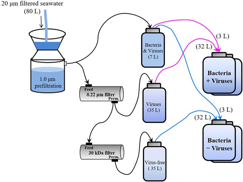
Results Fig. 1 Schematic of experimental setup used in this study. Schematic
The microcosm experiments were conducted with 2000 m deep diagram of the experimental setup for deep sea bacterioplankton with
water samples from the western Pacific Ocean, a typical normal and reduced pressure of viral lysis.
2 COMMUNICATIONS BIOLOGY | (2020)3:256 | https://doi.org/10.1038/s42003-020-0974-5 | www.nature.com/commsbio
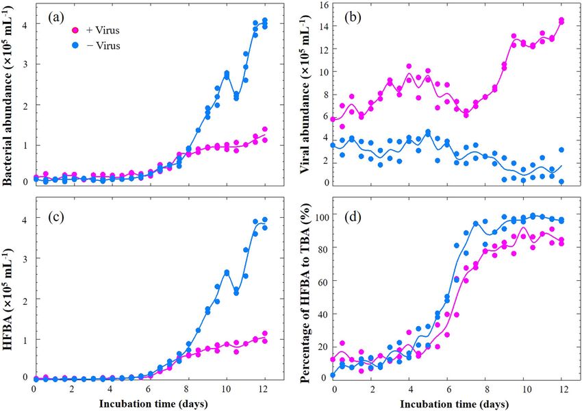
COMMUNICATIONS BIOLOGY | https://doi.org/10.1038/s42003-020-0974-5                                                                  ARTICLE

Fig. 2 Dynamics of microbial abundance during microcosm incubation. a Bacterial abundance, b Viral abundance, c High-fluorescence bacterial
abundance, d Percentages of high-fluorescence bacterial abundance to total bacterial abundance. HFBA high-fluorescence bacterial abundance, TBA total
bacterial abundance. Dots show two replicates at each time point and lines represent the average values.

Effects of viruses on deep-sea bacterioplankton abundance. The                high-fluorescence cells were observed in the +virus than in the
bacterial abundances in the +virus and −virus treatments                      −virus treatments, especially in the latter period (Fig. 2d). The
showed a rapid growth between Days 5 and 7, raising to 0.48 ×                 repression of active cells caused by the presence of viruses
105 particles mL−1 and 0.51 × 10⁵ particles mL−1, respectively                accounted for most (70%) of the difference between the
(Fig. 2a). Afterwards, the −virus treatments were characterized by            abundance of total bacterioplankton in the two treatments. Our
more rapid growth of bacterioplankton, reaching a higher                      finding suggests that viruses infect and lyse active bacterioplank-
abundance, than the +virus treatments (Fig. 2a). After                        ton cells in the deep sea, which is consistent with observations of
10–12 days of incubation, the bacterioplankton abundance in the               surface water18. In contrast, potentially less active bacterioplank-
–virus treatments was 3.2 times higher than that in the +virus                ton as indicated by low nucleic acid content might be less
treatments (Fig. 2a). Concurrently, the abundance of viruses in               susceptible to viral infection or the infection processes are much
the two treatments showed distinct trends: viral abundance                    slower21. Therefore, in the deep-sea environments, the majority
increased in the +virus incubations but decreased slightly in the             of bacterioplankton might be slow growing defense strategists,
−virus incubations (Fig. 2b). The repressing effects on prokar-               while the potential winners of competition for nutrients would be
yotic abundance in the +virus treatments demonstrated the                     controlled by viruses.
presence of top-down control by viruses of deep-sea bacter-
ioplankton. The reduction of viral abundance in the −virus                    Effects of viruses on deep-sea bacterial diversity and commu-
treatment suggests decay of viral particles due to reduced                    nity structure. Bacterial diversity and community composition
encounter rates with hosts, hence resulting in lower viral                    were investigated with 454 sequencing of PCR-amplified 16S
infection rates.                                                              rRNA genes at the DNA and RNA levels, respectively. After
   Since it has been proposed that viruses mainly infect active               quality control and normalization, 64,427 sequences were
hosts, e.g., cells with high RNA content18, we investigated the               obtained, and 975 OTUs were assigned based on 97% similarity of
dynamics of high-fluorescence signal bacterioplankton, contain-                sequences. At the beginning of the incubation, the bacterial
ing relatively high nucleic acids19,20, using flow cytometry (Fig. 3).         diversity (Shannon index) at the DNA level was 5.7. After 12 days
For both treatments, the abundances of high-fluorescence                       of incubation, bacterial diversity decreased to 4.7 in the +virus
bacterioplankton remained relatively stable until Day 6. But in               treatments and to 2.0 in the −virus treatments (Fig. 4a). Similarly,
the latter period (i.e., after Day 6), high-fluorescence bacterial             the bacterial diversity revealed by RNA analysis decreased after
abundance (HFBA) increased more rapidly in the −virus                         incubation and was higher in the +virus treatments (Shannon:
treatments than in the +virus treatments were observed in the                 3.9, Phylogenetic Diversity: 13.1) than in the −virus treatments
latter period (Fig. 2c). For both treatments, the percentages of              (Shannon: 3.1, Phylogenetic Diversity: 10.3). Although rRNA is
high-fluorescence bacterial to total bacterial abundance (TBA)                 not simply related to bacterial activity, it has been used as ten-
ranged from 3.0 to 49.0% for the first half of the incubation and              tative indicator for the active bacterial community or for an
then exceeded more than 70% during the second half. The                       anticipatory life strategy, i.e., being able to respond quickly to
increasing trends of HFBA were similar to those of TBA,                       environmental changes (e.g., see ref. 22). For the deep-sea, such
suggesting that the growth and development of potentially active              environmental changes could include the mixing of water mas-
bacterial assemblages in our incubation were mainly due to the                ses23, sinking particles, or the in situ formation of particles (e.g.,
cells containing high nucleic acids content. Lower percentages of             see ref. 14).

COMMUNICATIONS BIOLOGY | (2020)3:256 | https://doi.org/10.1038/s42003-020-0974-5 | www.nature.com/commsbio                                        3
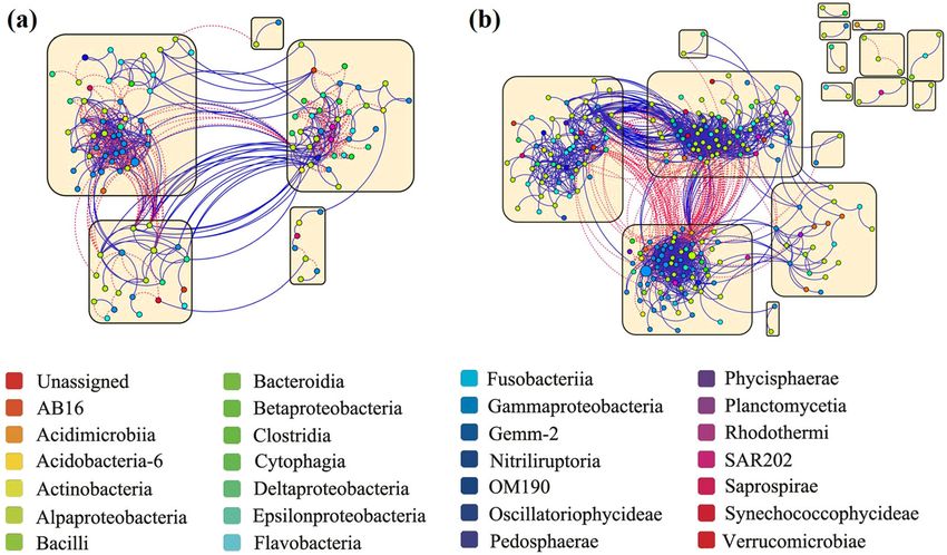
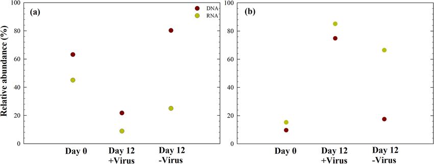
ARTICLE                                                                 COMMUNICATIONS BIOLOGY | https://doi.org/10.1038/s42003-020-0974-5

Fig. 3 Distinct deep sea bacterioplankton populations developed with different pressure of virus lysis. a Flow cytograms showing development of
bacterioplankton with normal viral lysis pressure. b Flow cytograms showing development of bacterioplankton with reduced viral lysis pressure. Red: high-
DNA bacteria; Blue: low-DNA bacteria.

   For the major phyla present at the beginning of the experiment             Effects of viruses on the relative contribution of deep-sea
(e.g., Alphaproteobacteria and Gammaproteobacteria), diversity                bacterial lineages. The relative abundances of specific bacterial
generally decreased after incubation; moreover, higher diversity              lineages (DNA level) and potentially active bacterial lineages
was always observed for the +virus treatment than for the –virus              (RNA level) were assessed. Before incubation, Alphaproteo-
treatment at both the DNA and RNA levels (Fig. 4b, c). Previous               bacteria dominated the bacterial population at both the DNA
studies have suggested that the presence of viruses supports                  level and RNA level (Fig. 6). After 12 days of incubation with
higher bacterial diversity in surface oceans8,10. The mechanism               viruses, Gammaproteobacteria dominated the potentially active
behind this phenomenon could be explained by the “kill the                    bacterial community (rRNA level). Detailed examination of the
winner” hypothesis, which states that viruses infect and lyse the             bacterial taxa in our experiments showed that nine of the top ten
bacterial winner of resource competition, prevent their blooming,             abundant OTUs showed a difference between the +virus and
and create niches for other bacteria24–26. This would allow for               −virus DNA libraries, including Sulfitobacter, Pseudoalter-
more bacterial species to be exit in a specific environment, leading           omonas, Marinobacter, Alteromonas, Pseudomonas, Oleispira,
to higher diversity. Furthermore, the release of organic matter               Oceanobacter, and Oceaniserpentilla (Supplementary Data 2).
during cell lysis alters the composition and bioavailability of               This indicates that viruses have potentially strong effects on the
organic nutrients, thus changing the composition of the bacterial             majority of deep-sea bacterioplankton. Alphaproteobacteria (e.g.,
community in surface waters27. Our study suggests that viruses                SAR11 Clade Ia, Thalassococcus, Paracoccus) were the dominant
play a similar ecological role in structuring the microbial                   groups in the in situ bacterioplankton community. In the +virus
community in deep-sea ecosystems.                                             treatment, the abundance and activity of Alphaproteobacteria
   At the beginning of the incubation, the RNA-based bacterial                were repressed compared with the −virus treatment, resulting in
diversity was lower than the DNA-based, indicating that some                  a population shift to Gammaproteobacteria accounting for
bacterial lineages were inactive (or not anticipatory) in the original        approximately 80% of total and active bacterioplankton. When
bacterioplankton community. In the Day 0 samples, Alphapro-                   viral pressure was relieved in the −virus treatment, Alphapro-
teobacteria showed relatively higher diversity at the RNA level               teobacteria dominating the original environments retained their
than at the DNA level (Fig. 4b), suggesting that they are highly              dominant status. These bacteria may be comparatively inactive
active in situ. The loss of Gammaproteobacteria diversity was                 because of the differing conditions between the in situ and the
greater than that of Alphaproteobacteria after incubation with                microcosm environments. In the −virus treatment, Gammapro-
viruses (Fig. 4b, c), contributing to the majority of diversity loss of       teobacteria and some Alphaproteobacteria (e.g., Sulfitobacter)
total bacterioplankton, especially at the DNA level. Similar to the           could have outcompeted other bacterioplankton in terms of
total bacterial population, higher diversity loss was observed for            nutrient utilization or adaption to environmental changes, thus
both Alphaproteobacteria and Gammaproteobacteria in the                       dominating the active bacterioplankton. Such a pattern, i.e., that
−virus treatments than in the +virus treatments. In addition,                 numerically not dominating but potentially highly active bacteria
higher diversity of active bacteria compared with that of total               are controlled by viruses, has been demonstrated for surface
bacteria was observed in the −virus treatments, which was due to              water29. In addition, the relative abundances of Bacteroidetes and
the same trend occurring in Alphaproteobacteria (Fig. 4a, b).                 Actinobacteria decreased after incubation, and the treatments
   To represent the major bacterial lineages across all samples, the          with and without the pressure of virus lysis did not differ sig-
58 most abundant OTUs (accounting for 90% of all sequences                    nificantly, which suggest that these bacterial groups may be less
after normalization) were selected. These major OTUs were                     susceptible to viral infection (Supplementary Fig. 3).
affiliated with Proteobacteria, Bacteroidetes, and Cyanobacteria
(Fig. 5). Cluster analysis showed that the +virus treatments
displayed more similarity to bacterial communities in the −virus              Effects of viruses on the interactions among deep-sea bacteria.
treatments than those in situ (Day 0 samples) (Fig. 5). However,              To evaluate whether the presence or absence of viruses may have
at both the DNA and RNA levels, the similarities between the                  affected the active interactions among deep-sea bacterioplankton
+virus treatments and in situ environment were higher than                    during the 12-day incubation, we constructed co-occurrence
those between the −virus treatments and in situ environment                   networks for RNA-inferred bacterial communities (Fig. 7 and
(Fig. 5). This indicates that in addition to general influences on             Supplementary Fig. 4; Table 1 and Supplementary Data 3),
diversity, viruses also sustain the community structure of deep-              assuming that RNA rather than DNA reflects the active com-
sea bacterioplankton. Similar trends have been recorded in                    munity. In network analysis, the topological indices mathemati-
studies on surface water (e.g., see refs. 10,28).                             cally describe the microbial interactions, including degree,

4                           COMMUNICATIONS BIOLOGY | (2020)3:256 | https://doi.org/10.1038/s42003-020-0974-5 | www.nature.com/commsbio
COMMUNICATIONS BIOLOGY | https://doi.org/10.1038/s42003-020-0974-5                                                                   ARTICLE

                                                                               +virus treatment than −virus treatment reveals the closer con-
                                                                               nection among the bacteria with the occurrence of virus (Table 1).
                                                                               The more complexity of microbial community shown by network
                                                                               density and clustering coefficient is consistent with the higher
                                                                               bacterial diversity found in the +virus treatments (Fig. 4). This
                                                                               could be explained by the concept of “kill the winner”24 that
                                                                               viruses control competitive dominates for nutrients and allow for
                                                                               the co-existence of more diverse bacterial community. Previous
                                                                               studies have shown that abiotic and biotic pressures might
                                                                               increase the network complexity of microbial communities31. Our
                                                                               data demonstrate that top-down pressure from viral lysis could
                                                                               have similar ecological influences. A positive correlation between
                                                                               two nodes in the microbial network is usually explained as a
                                                                               mutualism or cooperation relationship, while theoretically, this
                                                                               can also be produced by co-infection of different hosts by the
                                                                               same viruses. In our incubation, a lower proportion of positive
                                                                               correlations was observed in the +virus bacterial community
                                                                               compared with the −virus bacterial community (ca. 56% vs. 85%)
                                                                               (Table 1), suggesting that the positive correlation of bacteria may
                                                                               not be the direct result of bacterial mortality induced by viral
                                                                               lysis. This is reasonable as viral infection is always host-specific
                                                                               and their host range is narrow. Also, viral lysis does not only
                                                                               cause mortality of hosts (hence influence competition among
                                                                               bacteria), but also causes a release of the cell content and cell wall
                                                                               content (lysis products) which can be used by bacterioplankton
                                                                               (“viral shunt”). This supposedly “lubricates” the microbial food
                                                                               web and could result in positive correlations of the network
                                                                               analysis.
                                                                                  Four large modules contained more than five nodes were
                                                                               observed in each of +virus and −virus treatments, showing an
                                                                               evident modular architecture for both networks (Table 1). Since
                                                                               the OTUs within a module might occupy a similar ecological
                                                                               niche, a higher modularity value and module number observed in
                                                                               the −virus treatment (Fig. 7) indicated that number of niches
                                                                               increased without virus. In addition, the presence of virus
                                                                               stimulated the connections among bacteria within the module,
                                                                               which was evidenced by the higher clustering coefficients and
                                                                               increased proportion of links within modules in the +virus
                                                                               treatment (Table 1 and Fig. 7). Thus, the presence of virus might
                                                                               facilitate the stability of the niches by increasing the interactions
                                                                               among the bacteria in each niche. We also measured the
                                                                               proportions of Alphaproteobacteria and Gammaproteobacteria
                                                                               (the two major bacterial groups in the present study) in the
                                                                               networks (Supplementary Data 3). The Alphaproteobacteria to
                                                                               Gammaproteobacteria ratio was higher in the +virus treatments
                                                                               than in the −virus treatments, and the proportion of positive
                                                                               correlations for Gammaproteobacteria was greater than that for
                                                                               Alphaproteobacteria. Our results suggest that the removal of
                                                                               viruses might change the connection type (positive or negative)
                                                                               among bacteria, especially the Gammaproteobacteria. Thus,
                                                                               viruses could play a fundamental role in sustaining the deep-sea
                                                                               ecosystem by shaping the interactions among bacteria.

Fig. 4 Effects of viruses on diversity of deep sea bacterioplankton. a Total   Methodological considerations. In our study, we used onboard
and active bacterial diversity, b Total and active Alphaproteobacterial        microcosm incubation (20 L) in the dark at the in situ tempera-
diversity, c Total and active Gammaproteobacterial diversity. Diversity is     ture to represent deep-sea environments. A main difference
shown as Shannon index and phylogenetic diversity. Data show the mean of       between our incubation and the in situ deep-sea environment was
two independent incubations.                                                   pressure. The inactive deep-sea microbial community (e.g.,
                                                                               allochthonous and piezosensitive) may become dominant while
number of nodes, density, and clustering coefficient30. The den-                piezophilic and hyperpiezophilic microorganisms may disappear
sity, the proportion of actual connections to the potential con-               when incubated under atmospheric pressure32. However, there
nections in a network, is higher in the +virus treatment than in               are evidences that pressure did not significantly affect viral
the −virus treatment. This indicates that more bacteria are                    activities and, subsequently, the impact of viruses on their host
connected and a more complex network when viruses were                         population13,33. In addition, the enclosure of microbial plankton
present (Table 1). Similarly, the higher clustering coefficient of              communities may be biased by “bottle effects”, which may either

COMMUNICATIONS BIOLOGY | (2020)3:256 | https://doi.org/10.1038/s42003-020-0974-5 | www.nature.com/commsbio                                         5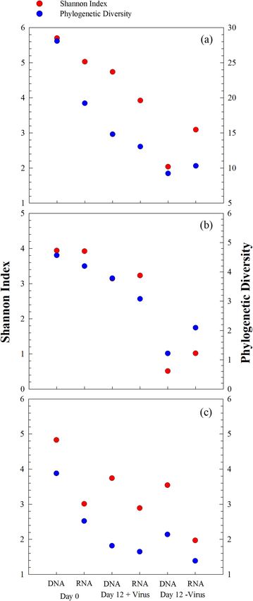
ARTICLE                                                                COMMUNICATIONS BIOLOGY | https://doi.org/10.1038/s42003-020-0974-5

Fig. 5 Heatmap showing the relative abundances and activity of the top 58 OTUs across the samples. The cluster analysis of bacterial community
structure was generated based on Bray-Curtis distances between samples. Heatmap shows the relative abundance (log [relative abundance + 1]) for each
OTU. Phylogenetic tree (on left) was reconstructed by representative sequence from each OTU with Maximum-Likelihood method using FastTree. Filled
circle on the nodes show bootstrap values higher than 50% (1000 resamplings). One archaeal sequence from Picrophilus torridus (AE017261) was used as
outgroup. The two heatmaps on the bottom show the similarity (%) of Bray–Curtis distances between samples. Day 0 samples were collected before
aliquoting into microcosms and considered as control for both +virus and −virus treatments.

Fig. 6 Effects of viruses on the relative abundance of two major classes of bacteria. a Total and active Alphaproteobacteria, b Total and active
Gammaproteobacteria. Data show the mean of two independent incubations.

stimulate the growth or enhance the loss of different planktonic             library, while Gammaproteobacteria predominated in the RNA
groups34,35. However, if we assume that the “bottle effects” and             library. This difference clearly indicates that viruses can also play
effects of pressure changes in the +virus and −virus treatments              a major role in shaping the bacterial community structure in the
were similar (as all incubation conditions were the same), dif-              deep sea. In addition, the PCR-based method was applied to
ferences in the ecological characteristics of bacterioplankton               estimate bacterial diversity and relative abundance. Although we
(Figs. 2–7) in our study should be due to the presence or absence            controlled the PCR cycle numbers and performed triplicate
of the top-down pressure of viral lysis. For example, the repla-             amplification for each sample to minimize any possible bias
cement of Alphaproteobacteria by Gammaproteobacteria has                     introduced by PCR, the relative abundance estimation may be
been frequently observed in microcosm incubations of bacter-                 biased by the variation of 16S rRNA gene copy number in the
ioplankton, and this was usually explained as resulting from the             deep-sea bacterial genome (e.g., see ref. 38).
fast growth rate and opportunistic life strategy of Gammapro-
teobacteria in bottle incubations36,37. In our study, however, the           Discussion
simultaneous incubation in the −virus treatment showed a con-                The deep ocean contains approximately 75% of the total pelagic
trasting pattern: Alphaproteobacteria predominated in the DNA                prokaryotic biomass1. The diversity of bacterioplankton

6                           COMMUNICATIONS BIOLOGY | (2020)3:256 | https://doi.org/10.1038/s42003-020-0974-5 | www.nature.com/commsbio
COMMUNICATIONS BIOLOGY | https://doi.org/10.1038/s42003-020-0974-5                                                                              ARTICLE

Fig. 7 Connections among active bacteria with different pressure of virus lysis. a Co-occurrence network graph of bacterioplankton with normal viral
lysis pressure. b Co-occurrence network graph of bacterioplankton with reduced viral lysis pressure. The nodes, representing OTUs, are colored according
to phylogenetic taxa. Node size is proportional to the relative abundance of OTU. Blue and red lines indicate positive and negative connections,
respectively, between nodes. Yellow squares show modules in the network.

(including Bacteria and Archaea) in the deep ocean is almost as               to solve the paradox that organic carbon supply is not sufficient
high as that in the upper ocean1,39. Also, the variability of                 for sustaining the observed microbial metabolic activity14.
microbial and viral communities on a seasonal scale is almost as                 Here we present, to the best of our knowledge, the first
high in deep water as in surface water40, thus supporting the idea            experimental evidence that deep-sea pelagic viruses actively
that deep-sea environments are more dynamic than previously                   control prokaryotic population size while sustaining bacterial
thought. However, the mechanisms controlling population size                  diversity and community structure. As a consequence, viral lysis
and diversity of deep sea bacterioplankton remain unclear. The                could be influencing microbial ecology and biogeochemical
relatively low concentration and bioavailability of dissolved                 cycling in the deep ocean.
organic matter in deep-sea environments lead to the assumption
of bottom-up control of the population size of bacterioplankton1.             Methods
The measured viral production rates are relatively low compared               Deep-sea water collection and experimental setup. Microcosm experiments
with those obtained in the surface and upper ocean3,4,6,11,12.                were performed onboard the R/V Kexue-1 during the NSFC western Pacific Ocean
                                                                              cruise in December 2010. Samples were collected at a 2000 m depth using 12 L
Deep-sea virioplankton have thus been regarded as a compara-                  Niskin bottles mounted on a CTD-carousel sampler. Approximately 80 L of deep-
tively inactive component. However, the few vertical investiga-               sea water was filtered through a 20 μm mesh and 1.0 μm polycarbonate membranes
tions of lysis and grazing suggest that in deep waters, viral lysis           to remove large particles and obtain the fraction containing bacteria and viruses
might be more important than grazing in terms of relative con-                (Fig. 1). Seventy liters of filtrate was filtered through a 0.22 m polypropylene car-
tribution to the mortality of prokaryotes (e.g., see ref. 16). Addi-          tridge (Prep-Scale/TFF, Millipore) to obtain the fraction containing viruses. Then,
                                                                              35 L of filtrate was filtered through a 30 kDa cut-off polysulfone cartridge (Prep-
tionally, an inverse relationship between grazing and lysis usually           Scale/TFF, Millipore) to obtain virus-free seawater. The bacteria plus virus fractions
appeared with variations in community structure and system                    were diluted with virus-containing and virus-free water, respectively, to ca. 10% of
productivity (e.g., see ref. 16). These observations suggest that in          the initial volume to obtain two treatments for deep-sea bacterioplankton: with
the deep sea, where grazing is potentially limited, viral lysis could         viruses at an ambient concentration and with diluted viruses (Fig. 1). Two repli-
                                                                              cates of each treatment were set up in 20 L polycarbonate bottles (Nalgene) and
be a major top-down control of prokaryotes. Such a viral impact               incubated in the dark at the ambient seawater temperature (ca. 2 °C). All bottles
has also been demonstrated for deep-sea sediments13. Our                      and containers were acid-rinsed before use.
experiment showed that viral communities likely exert a strong
control on the population size and community structure of pro-                Time series sampling and measurements. Subsamples for determining bacterial
karyotes in the deep sea and may play more active roles in the                and viral abundance were collected twice daily during the incubation and analyzed
dark ocean than previously thought.                                           using an onboard FACSAria flow cytometer (Becton, Dickinson and Company,
                                                                              USA) with SYBR Green I staining following procedures described previously42,43.
   Almost half of the heterotrophic prokaryotic production in the             On flow cytometry, particles with high nucleic acids (DNA or RNA) content always
ocean occurs in mesopelagic and bathypelagic waters1. However,                show high-fluorescence signal19,20. Therefore, the present study considered bac-
there is an obvious imbalance between the metabolic activity and              terioplankton with high-fluorescence signal as potentially active bacterial popula-
the supply of organic carbon (including sinking particular organic            tions. For molecular ecology analysis, 2 L of samples were filtered onto 0.22 μm
                                                                              pore size, 47 mm-diameter polycarbonate filters (Millipore, Bedford, MA, USA) to
carbon and chemolithoautotrophic production) in the deep                      retrieve the total bacterioplankton population. To start the incubation as soon as
ocean14,41. Based on our study and recent evidence of deep-sea                possible, we collected bacterial diversity samples before splitting them into +virus
viral activity3,4,11,12, we propose that viral cycling may account for        and −virus treatments, assuming the bacterial community composition in the
at least some of the imbalance. Viral lysis shunts organic carbon             treatments at the beginning of the incubation were the same. The filters for DNA
back to the dissolved organic carbon pool, which can be used by               analysis were stored at −80 °C, and the samples for RNA analysis were immersed
                                                                              in 600 μL of an RLT solution (Qiagen, Chatsworth, CA, USA) and stored at
prokaryotes, increase the supply of “bottom-up” resources, and                −80 °C. Ribosomal RNA (rRNA) has been widely employed to indicate potentially
thus, support prokaryote activity (e.g., see ref. 13). This may help          active microbial assemblage in various environments (e.g., see refs. 22,38).

COMMUNICATIONS BIOLOGY | (2020)3:256 | https://doi.org/10.1038/s42003-020-0974-5 | www.nature.com/commsbio                                                        7
ARTICLE COMMUNICATIONS BIOLOGY | https://doi.org/10.1038/s42003-020-0974-5
Modularity
Therefore, we apply bacterial 16S rRNA gene amplicon from environmental DNA
describes the proportion of closed triplets to the total number of triplets. A clustering coefficient close to 0 means that there are hardly any connections with neighbors, while a value of 1 means a node is fully connected to its neighbors. P link # and N link # represent the
aNode # and link # represent the node number and the connection number in each network, respectively. Density refers to the proportion of actual connections to the potential connections in a network. Higher density means a more complex network. The clustering coefficient
and RNA to characterize the whole and potentially active bacterioplankton,
0.584
0.674
respectively, in our study.
Pyrosequencing and analysis of bacterial diversity. For DNA-based and RNA-
based high throughput sequencing of microbial communities, DNA and RNA were
between module
extracted using a MoBio PowerWater DNA Isolation Kit (MoBio, San Diego, CA,
USA) and an RNeasy Mini Kit (Qiagen, Hilden, Germany), respectively, according
to the manufacturers’ protocols. During RNA purification, gDNA contamination
N link #
was removed using an RNase-Free DNase Set (Qiagen, Hilden, Germany), which
was verified by PCR amplification of bacterial 16S rRNA gene. The SuperScript III
127
35
First-Strand Synthesis System with random hexamers (Invitrogen, Carlsbad, CA,
USA) was used to synthesize first-strand cDNA for the RT-PCR. Please note that
one RNA extraction of Day 12 sample of −virus treatments was not successful.
Partial 16S rRNA gene fragments were amplified using the universal primers
within module
F515 (GTGNCAGCMGCCGCGGTAA) and R926 (CCGYCAATTYMTTTRAG
TTT), which cover the V4 and V5 regions of the gene44. PCR was carried out in
25 μL reaction volumes with 2× Premix Ex Taq (Takara, Dalian, China) under the
N link #
following conditions: initial denaturation at 95 °C for 3 min, followed by 30 cycles
104
201
of 95 °C for 30 s, 55 °C for 45 s, and 72 °C for 45 s, with a final extension at 72 °C
Table 1 Properties of bacterial communities’ phylogenetic network at the RNA level with normal and reduced viral lysis pressure.
for 10 min. Triplicate PCR products for each sample were pooled together and
purified using a MiniBEST Agarose Gel DNA Purification kit Ver. 3.0 (Takara).
Finally, 454 library preparation and sequencing were conducted at the Shanghai
N link #
Hanyu Biotechnology Co. (Shanghai, China).
Sequence analysis was carried out using QIIME 1.8.045. The raw sequences were
Total
236
231
quality filtered for length (300 bp < length < 460 bp), quality score (>25), number
of homopolymer runs (0.9 andCOMMUNICATIONS BIOLOGY | https://doi.org/10.1038/s42003-020-0974-5 ARTICLE
Data availability 21. Weinbauer, M. G. Ecology of prokaryotic viruses. FEMS Microbiol. Rev. 28,
The entire sequencing data set has been deposited in the NCBI SRA database under 127–181 (2004).
project number SRP139068. The data of microbial abundance during microcosm 22. Blazewicz, S. J., Barnard, R. L., Daly, R. A. & Firestone, M. K. Evaluating rRNA
incubation have been provided in Supplementary Data 1. The distribution of major as an indicator of microbial activity in environmental communities:
OTUs in each sample is shown Supplementary Data 2. Supplementary Data 3 shows the limitations and uses. ISME J. 7, 2061–2068 (2013).
proportion of nodes belonging to α-Proteobacteria and γ-Proteobacteria in the networks 23. Winter, C. et al. Mixing alters the lytic activity of viruses in the dark ocean.
and the proportion of these two classes in the +virus and −virus treatments at Ecology 99, 700–713 (2018).
RNA level. 24. Thingstad, T. F. & Lignell, R. Theoretical models for the control of bacterial
growth rate, abundance, diversity and carbon demand. Aquat. Microb. Ecol.
13, 19–27 (1997).
Received: 1 November 2019; Accepted: 29 April 2020; 25. Thingstad, T. F. Elements of a theory for the mechanisms controlling
abundance, diversity, and biogeochemical role of lytic bacterial viruses in
aquatic systems. Limnol. Oceanogr. 45, 1320–1328 (2000).
26. Winter, C., Bouvier, T., Weinbauer, M. G. & Thingstad, T. F. Trade-offs
between competition and defense specialists among unicellular planktonic
organisms: the “killing the winner” hypothesis revisited. Microbiol Mol. Biol.
References Rev. 74, 42–57 (2010).
1. Arístegui, J., Gasol, J. M., Duarte, C. M. & Herndl, G. J. Microbial 27. Middelboe, M. Bacterial growth rate and marine virus-host dynamics. Micro.
oceanography of the dark ocean’s pelagic realm. Limnol. Oceanogr. 54, Ecol. 40, 114–124 (2000).
1501–1529 (2009). 28. Bonilla-Findji, O. et al. Viral effects on bacterial respiration, production and
2. Parada, V., Sintes, E., van Aken, H. M., Weinbauer, M. G. & Herndl, G. J. Viral growth efficiency: Consistent trends in the Southern Ocean and the
abundance, decay, and diversity in the meso- and bathypelagic waters of the Mediterranean Sea. Deep-Sea Res. Pt II 55, 790–800 (2008).
North Atlantic. Appl. Environ. Microbiol. 73, 4429–4438 (2007). 29. Bouvier, T. & del Giorgio, P. A. Key role of selective viral-induced mortality in
3. De Corte, D., Sintes, E., Yokokawa, T., Reinthaler, T. & Herndl, G. J. Links determining marine bacterial community composition. Environ. Microbiol. 9,
between viruses and prokaryotes throughout the water column along a North 287–297 (2007).
Atlantic latitudinal transect. ISME J. 6, 1566–1577 (2012). 30. Deng, Y. et al. Molecular ecological network analyses. BMC Bioinform. 13, 113
4. Li, Y. et al. Lytic viral infection of bacterioplankton in deep waters of the (2012).
western Pacific Ocean. Biogeosciences 11, 2531–2542 (2014). 31. Deng, Y. et al. Network succession reveals the importance of competition in
5. Yang, Y. H., Yokokawa, T., Motegi, C. & Nagata, T. Large-scale distribution of response to emulsified vegetable oil amendment for uranium bioremediation.
viruses in deep waters of the Pacific and Southern Oceans. Aquat. Microb. Environ. Microbiol. 18, 205–218 (2016).
Ecol. 71, 193–202 (2014). 32. Tamburini, C., Boutrif, M., Garel, M., Colwell, R. R. & Deming, J. W.
6. De Corte, D. et al. Links between viral and prokaryotic communities Prokaryotic responses to hydrostatic pressure in the ocean—a review. Environ.
throughout the water column in the (sub)tropical Atlantic Ocean. ISME J. 4, Microbiol. 15, 1262–1274 (2013).
1431–1442 (2010). 33. Dell’Anno, A., Corinaldesi, C. & Danovaro, R. Virus decomposition provides
7. Weinbauer, M. G. & Rassoulzadegan, F. Are viruses driving microbial an important contribution to benthic deep-sea ecosystem functioning. Proc.
diversification and diversity? Environ. Microbiol 6, 1–11 (2004). Natl Acad. Sci. USA 112, E2014–2019 (2015).
8. Weinbauer, M. G. et al. Synergistic and antagonistic effects of viral lysis and 34. Eilers, H., Pernthaler, J. & Amann, R. Succession of pelagic marine bacteria
protistan grazing on bacterial biomass, production and diversity. Environ. during enrichment: a close look at cultivation-induced shifts. Appl. Environ.
Microbiol 9, 777–788 (2007). Microbiol. 66, 4634–4640 (2000).
9. Winter, C., Herndl, G. J. & Weinbauer, M. G. Diel cycles in viral 35. Calvo-Diaz, A. et al. Decrease in the autotrophic-to-heterotrophic biomass
infection of bacterioplankton in the North Sea. Aquat. Microb. Ecol. 35, ratio of picoplankton in oligotrophic marine waters due to bottle enclosure.
207–216 (2004). Appl. Environ. Microbiol. 77, 5739–5746 (2011).
10. Zhang, R., Weinbauer, M. G. & Qian, P. Y. Viruses and flagellates 36. Comte, J., Lindstrom, E. S., Eiler, A. & Langenheder, S. Can marine bacteria
sustain apparent richness and reduce biomass accumulation of be recruited from freshwater sources and the air? ISME J. 8, 2423–2430
bacterioplankton in coastal marine waters. Environ. Microbiol. 9, 3008–3018 (2014).
(2007). 37. Pinhassi, J. & Berman, T. Differential growth response of colony-forming α-
11. Lara, E. et al. Unveiling the role and life strategies of viruses from the surface and γ-proteobacteria in dilution culture and nutrient addition experiments
to the dark ocean. Sci. Adv. 3, e1602565 (2017). from Lake Kinneret (Israel), the eastern Mediterranean Sea, and the Gulf of
12. Umani, S. F. et al. Disentangling the effect of viruses and nanoflagellates on Eilat. Appl. Environ. Microbiol. 69, 199–211 (2003).
prokaryotes in bathypelagic waters of the Mediterranean Sea. Mar. Ecol. Prog. 38. Engelbrektson, A. et al. Experimental factors affecting PCR-based estimates of
Ser. 418, 73–85 (2010). microbial species richness and evenness. ISME J. 4, 642–647 (2010).
13. Danovaro, R. et al. Major viral impact on the functioning of benthic deep-sea 39. Nagata, T. et al. Emerging concepts on microbial processes in the bathypelagic
ecosystems. Nature 454, 1084–1087 (2008). ocean - ecology, biogeochemistry, and genomics. Deep-Sea Res. Pt II 57,
14. Herndl, G. J. & Reinthaler, T. Microbial control of the dark end of the 1519–1536 (2010).
biological pump. Nat. Geosci. 6, 718–724 (2013). 40. Winter, C., Kerros, M.-E. & Weinbauer, M. G. Seasonal and depth-related
15. Weinbauer, M. G., Winter, C. & Höfle, M. G. Reconsidering transmission dynamics of prokaryotes and viruses in surface and deep waters of the
electron microscopy based estimates of viral infection of bacterio-plankton northwestern Mediterranean Sea. Deep-Sea Res. Pt I 56, 1972–1982 (2009).
using conversion factors derived from natural communities. Aquat. Microb. 41. Burd, A. B. et al. Assessing the apparent imbalance between geochemical and
Ecol. 27, 103–110 (2002). biochemical indicators of meso- and bathypelagic biological activity: What the
16. Tsai, A. Y., Gong, G. C. & Chao, C. F. Contribution of viral lysis and @$#! is wrong with present calculations of carbon budgets? Deep-Sea Res. Pt II
Nanoflagellate grazing to bacterial mortality at surface waters and deeper 57, 1557–1571 (2010).
depths in the coastal ecosystem of subtropical western Pacific. Estuar. Coast 42. Brussaard, C. P. Optimization of procedures for counting viruses by flow
39, 1357–1366 (2016). cytometry. Appl. Environ. Microbiol. 70, 1506–1513 (2004).
17. Tsai, A.-Y., Gong, G.-C., Sanders, R. W. & Huang, J.-K. Contribution of viral 43. Marie, D., Brussaard, C. P. D., Thyrhaug, R., Bratbak, G. & Vaulot, D.
lysis and nanoflagellate grazing to bacterial mortality in the inner and outer Enumeration of marine viruses in culture and natural samples by flow
regions of the Changjiang River plume during summer. J. Plankton Res. 35, cytometry. Appl. Environ. Microbiol. 65, 45–52 (1999).
1283–1293 (2013). 44. Quince, C., Lanzen, A., Davenport, R. J. & Turnbaugh, P. J. Removing noise
18. Bonilla-Findji, O., Herndl, G. J., Gattuso, J. P. & Weinbauer, M. G. Viral and from pyrosequenced amplicons. BMC Bioinform. 12, 38 (2011).
flagellate control of prokaryotic production and community structure in 45. Caporaso, J. G. et al. QIIME allows analysis of high-throughput community
offshore Mediterranean Waters. Appl. Environ. Microbiol. 75, 4801–4812 sequencing data. Nat. Methods 7, 335–336 (2010).
(2009). 46. Haas, B. J. et al. Chimeric 16S rRNA sequence formation and detection in
19. Lebaron, P., Servais, P., Agogue, H., Courties, C. & Joux, F. Does the high Sanger and 454-pyrosequenced PCR amplicons. Genome Res. 21, 494–504
nucleic acid content of individual bacterial cells allow us to discriminate (2011).
between active cells and inactive cells in aquatic systems? Appl. Environ. 47. Edgar, R. C. Search and clustering orders of magnitude faster than BLAST.
Microbiol. 67, 1775–1782 (2001). Bioinformatics 26, 2460–2461 (2010).
20. Gasol, J. M. & Del Giorgio, P. A. Using flow cytometry for counting natural 48. DeSantis, T. Z. et al. Greengenes, a chimera-checked 16S rRNA gene database
planktonic bacteria and understanding the structure of planktonic bacterial and workbench compatible with ARB. Appl. Environ. Microbiol. 72,
communities. Sci. Mar. 64, 197–224 (2000). 5069–5072 (2006).
COMMUNICATIONS BIOLOGY | (2020)3:256 | https://doi.org/10.1038/s42003-020-0974-5 | www.nature.com/commsbio 9ARTICLE COMMUNICATIONS BIOLOGY | https://doi.org/10.1038/s42003-020-0974-5
49. Wang, Q., Garrity, G. M., Tiedje, J. M. & Cole, J. R. Naive Bayesian classifier Competing interests
for rapid assignment of rRNA sequences into the new bacterial taxonomy. The authors declare no competing interests.
Appl. Environ. Microbiol. 73, 5261–5267 (2007).
50. Caporaso, J. G. et al. PyNAST: a flexible tool for aligning sequences to a
template alignment. Bioinformatics 26, 266–267 (2010).
Additional information
Supplementary information is available for this paper at https://doi.org/10.1038/s42003-
51. Price, M. N., Dehal, P. S. & Arkin, A. P. FastTree: computing large minimum
020-0974-5.
evolution trees with profiles instead of a distance matrix. Mol. Biol. Evol. 26,
1641–1650 (2009).
Correspondence and requests for materials should be addressed to R.Z.
52. Ludwig, W. et al. ARB: a software environment for sequence data. Nucleic
Acids Res. 32, 1363–1371 (2004).
Reprints and permission information is available at http://www.nature.com/reprints
53. Friedman, J. & Alm, E. J. Inferring correlation networks from genomic survey
data. PLoS Comput. Biol. 8, e1002687 (2012).
Publisher’s note Springer Nature remains neutral with regard to jurisdictional claims in
54. Shannon, P. et al. Cytoscape: a software environment for integrated models of
published maps and institutional affiliations.
biomolecular interaction networks. Genome Res. 13, 2498–2504 (2003).
55. Morris, J. H. et al. clusterMaker: a multi-algorithm clustering plugin for
cytoscape. BMC Bioinform. 12, 436 (2011).
56. Girvan, M. & Newman, M. E. Community structure in social and biological
networks. Proc. Natl Acad. Sci. USA 99, 7821–7826 (2002).
Open Access This article is licensed under a Creative Commons
Attribution 4.0 International License, which permits use, sharing,
Acknowledgements adaptation, distribution and reproduction in any medium or format, as long as you give
This work was supported by the National Natural Science Foundation of China appropriate credit to the original author(s) and the source, provide a link to the Creative
(91951209, 41706154) and China Ocean Mineral Resources R&D Association (DY135- Commons license, and indicate if changes were made. The images or other third party
E2-1-04). R.Z. was partially supported by Qingdao National Laboratory for Marine material in this article are included in the article’s Creative Commons license, unless
Science and Technology (QNLM2016ORP0303) and the Fundamental Research Funds indicated otherwise in a credit line to the material. If material is not included in the
for the Central Universities (20720190141, 20720170107). article’s Creative Commons license and your intended use is not permitted by statutory
regulation or exceeds the permitted use, you will need to obtain permission directly from
Author contributions the copyright holder. To view a copy of this license, visit http://creativecommons.org/
R.Z. developed ideas and organized the study; Y.L. and R.Z. performed the onboard licenses/by/4.0/.
experiments and collected the samples; R.Z., Y.L., W.Y., Y.W., L.C., T.L., and H.L.
analyzed the data; R.Z., W.Y., Y.W., L.C., M.W., and N.J. wrote the manuscript. All
© The Author(s) 2020
authors participated in early stages of data interpretation, provided comments on the
manuscript and approved the final manuscript.
10 COMMUNICATIONS BIOLOGY | (2020)3:256 | https://doi.org/10.1038/s42003-020-0974-5 | www.nature.com/commsbioYou can also read