Phylogeography, Transmission, and Viral Proteins of Nipah Virus
←
→
Page content transcription
If your browser does not render page correctly, please read the page content below
Virologica Sinica                                                                                              www.virosin.org
https://doi.org/10.1007/s12250-018-0050-1                                                              www.springer.com/12250(0123456789().,-volV)(0123456789().,-volV)

    REVIEW

Phylogeography, Transmission, and Viral Proteins of Nipah Virus
Bangyao Sun1,2 • Lijia Jia1,2 • Bilin Liang1,2 • Quanjiao Chen1 • Di Liu1,2

Received: 14 June 2018 / Accepted: 5 September 2018
Ó The Author(s) 2018

Abstract
Nipah virus (NiV), a zoonotic paramyxovirus belonging to the genus Henipavirus, is classified as a Biosafety Level-4
pathogen based on its high pathogenicity in humans and the lack of available vaccines or therapeutics. Since its initial
emergence in 1998 in Malaysia, this virus has become a great threat to domestic animals and humans. Sporadic outbreaks
and person-to-person transmission over the past two decades have resulted in hundreds of human fatalities. Epidemio-
logical surveys have shown that NiV is distributed in Asia, Africa, and the South Pacific Ocean, and is transmitted by its
natural reservoir, Pteropid bats. Numerous efforts have been made to analyze viral protein function and structure to
develop feasible strategies for drug design. Increasing surveillance and preventative measures for the viral infectious
disease are urgently needed.

Keywords Nipah virus  Viral transmission  Geographical distribution  Protein structure

Introduction                                                      serious illness most probably caused by NiV was reported
                                                                  in several people after contact with infected horses or
In February 2018, Nipah Virus (NiV) infection was listed          patients in the Philippines (Ching et al. 2015). NiV infec-
as a priority disease posing a public health risk by the          tion can cause fever and encephalitis in humans and a
World Health Organization (http://www.who.int/blueprint/          neurological and respiratory syndrome in pigs or horses
en/). NiV was named after Kampung Sungai Nipah (Nipah             (Lee et al. 1999; CDC 1999a, b). To date, over 600 human
River Village) in Malaysia, where it was first isolated in        cases of NiV infection have been reported in South Asia
1998, before its subsequent spread into Singapore via             and South-East Asia, with fatality ranging from 40% to
exported pigs in 1999, leading to the abattoir worker             70%, accordingly it poses a major threat to human health
infections (CDC 1999a, b; Paton et al. 1999; Epstein et al.       (Clayton 2017).
2006). In 2001, human cases of NiV infection were dis-                Belonging to the genus Henipavirus [the other patho-
covered independently in India and Bangladesh, and since          genic member of the genus is Hendra virus (HeV),
then, infections have been observed annually in Bangla-           reviewed in (Middleton 2014; Enchéry and Horvat 2017)]
desh, and human-to-human transmission through direct              of the family Paramyxoviridae (Chua et al. 2000), NiV is
contact with infected individuals is common (Hsu et al.           classified as a Biosafety Level-4 (BSL-4) pathogen due to
2004; Chadha et al. 2006a, b). In 2007, an NiV outbreak           its high pathogenicity and the lack of any effective treat-
occurred in India, killing five people (Arankalle et al.          ments or vaccines (Wit and Munster 2015; Angeletti et al.
2011). In 2018, NiV infection was ongoing in Kerala,              2016). The NiV genome consists of a negative-sense, sin-
India, with 16 cases succumbed (Paul 2018). In 2014, a            gle-stranded RNA of approximately 18.2 kb, encoding six
                                                                  structural proteins, nucleoprotein (N), phosphoprotein (P),
                                                                  matrix protein (M), fusion protein (F), attachment glyco-
& Di Liu                                                          protein (G), and the large protein or RNA polymerase
  liud@wh.iov.cn                                                  protein (L). In addition, the P gene encodes three non-
1
     Wuhan Institute of Virology, Chinese Academy of Sciences,    structural proteins by RNA editing (V and W proteins) or
     Wuhan 430071, China                                          an alternative open reading frame (C protein) (Wang et al.
2
     University of Chinese Academy of Sciences, Beijing 100049,   2001) (Fig. 1).
     China

                                                                                                                   123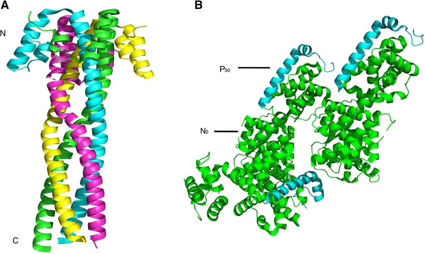
Virologica Sinica

                                                                       no NiV outbreaks have been reported is ongoing. Extensive
                                                                       studies and sample collections (swabs, sera, saliva, and
                                                               N
                                                                       urine) and analyses from bats have indicated that in addi-
                                                               P       tion to the countries where NiV outbreaks have occurred,
                                                               M       NiV is also distributed in China (Yan et al. 2008), Vietnam
                                                                       (Hasebe et al. 2012), Thailand (Supaporn et al. 2005),
                                                               F
                                                                       Cambodia (Reynes et al. 2005), Indonesia (Sendow et al.
                                                               G       2010), East Timor (Breed et al. 2013), Madagascar (Iehlé
                                                                       et al. 2007), New Caledonia (Enchéry and Horvat 2017)
                                                               L       and Papua New Guinea (Breed et al. 2010; Field et al.
                                                                       2013) (Fig. 2). In addition, anti-NiV neutralization anti-
                                                             ssRNA
                                                                       body test of the serum from people involved in hunting bats
                                                                       as bushmeat revealed evidence of NiV spillover (Pernet
                                                                       et al. 2013), which emphasizes the significance of further
       N      P/W/V/C     M       F       G          L                 NiV monitoring.
 3’                                                            5’

Fig. 1 Schematic representation of the viral structure (upper panel)
and genome organization (lower panel). Different genes or proteins     Hosts and Transmission of Nipah Virus
are indicated in different colors.
                                                                       Understanding susceptible hosts and routes for the spread
   NiV has a wide range of hosts, from its natural reservoir           of viral disease raises knowledge to curb epidemics. Bats
Pteropid bats to humans, horses, dogs, cats, cows, and pigs            are the second largest order of mammals after rodents
(Calisher et al. 2006; Halpin et al. 2011; Weatherman et al.           (Moratelli and Calisher 2015; Ming and Dong 2016), and
2018). Close contact with infected patients (Tan and Tan               harbor in excess of 200 types of viruses, including many
2001) or domestic animals (e.g., pigs and horses) plays an             highly pathogenic to humans (e.g., rabies, Ebola, severe
important role in the spread of NiV (Clayton 2017). Fur-               acute respiratory syndrome (SARS), NiV, HeV) (Li et al.
thermore, palm sap is also currently regarded as a crucial             2005; Calisher et al. 2006). NiV circulates within bat
NiV transmission medium in Bangladesh (Luby et al.                     populations via close mutual contact when bats crowd
2006; Nahar et al. 2010; Rahman et al. 2012). Intraspecific            together (Middleton et al. 2007). NiV transmission from
transmission (in bats, pigs, and horses) is also possible via          bats to humans is through two main pathways, i.e., inter-
saliva, urine or secretions upon high density populations of           mediate hosts (pigs and horses) and food-borne transmis-
animals (Middleton et al. 2007; Weatherman et al. 2018).               sion via date palm sap contaminated with the saliva or
In this review, an overview of recent studies on the geo-              urine of fruit bats (Enchéry and Horvat 2017) [reviewed in
graphical and phylogenetic properties, transmission, and               (Clayton 2017)]. A retrospective study in Malaysia found
protein structure and function of NiV is provided.                     that workers show severe influenza-like symptoms after
                                                                       slaughtering NiV-infected swine (Hsu et al. 2004). In the
                                                                       Philippines, people were infected by butchering horses or
Geographical Distribution of Nipah Virus                               consuming horsemeat (Ching et al. 2015). No cases of
                                                                       person-to-person spread have been found in Malaysia or
Since it emerged in Malaysia in 1998, NiV caused a series              Singapore, but in the Philippines, direct human-to-human
of outbreaks in Singapore, India, and Bangladesh (Clayton              virus transmission has been reported (Ching et al. 2015). In
2017; Enchéry and Horvat 2017) (Fig. 2), which has                    India, human-to-human transmission of NiV was discov-
caused hundreds of human deaths and thereby represents a               ered in 2001. In a case of NiV infection in a human in
great challenge to public health. Numerous efforts have                2007, date palm sap contaminated by bats was considered
been made to trace the origin, distribution, and probable              to mediate NiV spillover from bats to humans (Chadha
transmission route of NiV in nature. For example, in                   et al. 2006a, 2006b; Arankalle et al. 2011). In Bangladesh,
Bangladesh, where humans are frequently infected by NiV,               where people consume palm sap, frequent infection by NiV
retrospective investigations combined with the collect of              and person-to-person NiV transmission has occurred.
biological samples from patients or contaminated envi-                 Recently, it was demonstrated that Syrian hamster infection
ronment have been conducted to evaluate potential risk                 could occur after drinking artificial palm sap mixed with
factors and to develop a feasible strategy for prevention              NiV (Wit et al. 2014) and infrared camera monitoring
and control (Hsu et al. 2004; Gurley et al. 2007; Rahman               showed that bats frequently fly around or directly contact
et al. 2012). Additionally, NiV surveillance in areas where            palm sap trees to urinate or defecate (Khan et al. 2010;

123
B. Sun et al.: Characterization of Nipah virus

Fig. 2 Summary of the known
geographical distribution of
Nipah virus in the world.
Yellow stars represent reported
Nipah virus outbreaks and green
star shows the likely presence of
Nipah-like virus.

Rahman et al. 2012). These findings provide further sup-           Furthermore, the nucleotide sequence identity of N, P, L, and
port for palm sap-mediated bat-to-person transmission,             C genes were 94.3%, 92.0%, 93.4%, and 97.6%, respec-
despite the lack of Nipah virus detection in natural date          tively, with corresponding amino acid sequence identity of
palm sap until now (Fig. 3). Measures have been taken to           98.3%, 92%, 98.2%, and 95.2%, respectively (Harcourt et al.
prevent bat access to sap using bamboo skirts or lime              2005) (Fig. 4A). These findings provided a basis for further
smudged on date palm trees. Further steps to prevent the           investigations of the biological characteristics of NiV.
transmission of NiV infection are necessary.                          Based on the time-scaled tree constructed using the
                                                                   currently available viral genomes, the time to the most
                                                                   recent common ancestor (tMRCA) of NiV could be dated
Viral Genomics and Phylogenetics                                   to 1356 (95% highest posterior density, 95% HPD: 482—
                                                                   1884). The strains were divided into two lineages (NiV-
Genomic and amino acid differences may explain differ-             Bangladesh, n = 4, and NiV-Malaysia, n = 11), with dif-
ences in viral pathogenicity and virulence between isolates in     ferent clinical features and transmission routes in Bangla-
Bangladesh and Malaysia, particularly given the contribu-          desh and Malaysia (Fig. 4B). Due to the limited viral
tions of the N, P and L proteins to viral replication and          genomes available, the detailed divergent time of Bangla-
transcription and the role of the nonstructural protein C in the   desh and Malaysia lineages needs further investigation.
virulence of NiV (see ‘‘Viral Protein Function and Structure’’     More frequent person-to-person contact and more severe
section). In sequence analyses (Harcourt et al. 2005),             respiratory disease have been observed in Bangladesh than
nucleotide sequence identity between the genomes of NiV-           in Malaysia (Goh et al. 2000; Chong et al. 2008; Hossain
Bangladesh and NiV-Malaysia was only 91.8%, with an                et al. 2008), consistent with the higher level of viral
uneven distribution of differences throughout the genome.          replication in ferrets for NiV-Bangladesh than NiV-
The amino acid sequence identity between NiV-Bangladesh            Malaysia (Clayton et al. 2012). However, a recent study
and NiV-Malaysia proteins were all greater than 92%.               suggested that social and environmental factors impact the

                                                                                                                     123
Virologica Sinica
Malaysia
India/Bangladesh
Philippines
Fig. 3 Schematic representation of transmission routes for Nipah persons infected by NiV after consuming the date palm sap
virus. In Malaysia, the fruit trees where fruit bats reside are in contaminated by bat saliva or urine, followed by person-to-person
proximity to pig farms and domestic pigs infected by NiV via contact transmission by close contact. In the Philippines, people were infected
with materials contaminated by bats, and NiV is subsequently by consuming horsemeat or contact with infected horses, and then
transmitted to humans by direct contact. In India or Bangladesh, healthy individuals were infected after contact with patients.
spread of NiV (Clayton and Marsh 2014; Clayton et al. envelope consists of its genome and the N protein, which is
2016). In Malaysia, no person-to-person transmission was essential for the viral life cycle as a template for RNA-
reported when NiV emerged in 1998 and NiV strains dependent RNA-polymerase (RdRp), composed of poly-
derived from humans were isolated in 1999–2000 merase L and a polymerase cofactor P (Diederich and
(Fig. 4B), whereas, in Bangladesh, NiV has appeared Maisner 2007; Cox and Plemper 2017). Within the RNP, N
nearly annually since 2001 along with significant human- is responsible for viral genome wrapping and facilitates
to-human transmission. Thus, comparative genomics and viral replication and transcription (Lee et al. 2012). The
reverse genetics approaches are required to uncover the synthesis of viral mRNA is catalyzed by L and P (Morin
different features between NiV-Bangladesh and NiV- et al. 2013), and the latter also inhibits interferon signaling
Malaysia. via host STAT-1 (Lo et al. 2012) and acts as a chaperone of
N0 (the unassembled form of N) to prevent it nonspecific
binding to host RNA (Habchi and Longhi 2012). The M
Viral Protein Function and Structure protein contributes to viral assembly and release (Dietzel
et al. 2015). G and F are two important surface glycopro-
Nipah virus has an approximately 18.2 kb genome encod- teins of NiV; the former induces viral attachment to two
ing six structural proteins and three nonstructural proteins. cellular receptors, ephrin-B2 and ephrin-B3, despite a lack
The viral ribonucleocapsid (RNP) surrounded by the viral of hemagglutination or neuraminidase activity (Bonaparte
123B. Sun et al.: Characterization of Nipah virus
AF212302.2_Malaysia_1999_Homosapiens
A
KY425655.1_Malaysia_1999_Homosapiens
Gene N P L C AJ564622.1_Malaysia_1999_Swine
% nucleotide 94.3 92 93.4 97.6 KY425646.1_Malaysia_1999_Homosapiens
identity
AY029767.1_Malaysia_2000_Homosapiens
% amino acid
98.3 92 98.2 95.2
identity
AY029768.1_Malaysia_2000_Homosapiens
AJ564621.1_Malaysia_1999_Swine
AJ564623.1_Malaysia_1999_Homosapiens
B
1905 1775-1987 1 AF376747.1_Malaysia_1999_Pteropus
AJ627196.1_Malaysia_1999_Swine
FN869553.1_Malaysia_2008_Pteropus
1356 482 -1884 1
JN808857.1_Bangladesh_2008_Homosapiens
JN808863.1_Bangladesh_2008_Homosapiens
1951 1877-1998 1 FJ513078.1_India_2007_Homosapiens
AY988601.1_Bangladesh_2004_Homosapiens
JN808864.1_Bangladesh_2010_Homosapiens
70.0
500 1000 1500 2000
Fig. 4 Genomic and phylogenetic analyses of Nipah virus. A Com- model, under an uncorrelated relaxed clock, and the exponential
parisons of both nucleotide sequence and amino acid sequence growth demographic model. NiV-Bangladesh is represented in pink
identity between NiV-Bangladesh and NiV-Malaysia. B Maximum and NiV-Malaysia is in brown. The purple bars represent the 95%
clade credibility (MCC) tree of Nipah virus genomes. The tree was highest posterior density intervals of the estimation of the dates.
performed by using BEAST package 2.4.8 with the HKY SRD06
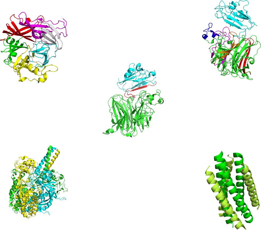
Fig. 5 Crystal structures of P
and the N0-P complex.
A Cartoon representation of
NiV-P (Protein Data Bank
accession number 4N5B). Four
chains are indicated in four
different colors. N and C
termini are shown. B Structure
of three heterodimers of the N0-
P complex (PDB: 4CO6). P50 is
shown in cyan and N0 is in
green.
123Virologica Sinica
A B Ephrin-B3
N
C
N C
B1
B6
B5
B2 B4
Ephrin-B2 C
C
NiV-G
B3
N
Residues 107–125
NiV-G
C HR1
D E
HR2
C
N
N
Fig. 6 Crystal structures of G and F protein. A Cartoon representation B4S4–B5S1 in magenta, and B5S2–B5S3 in orange. C An overview
of NiV-G (PDB: 3D11) in the receptor-unbound state. Six blades are of the G/Ephrin-B2 complex structure (PDB: 2VSM). NiV-G and
represented as follows: B1 in red, B2 in green, B3 in yellow, B4 in Ephrin-B2 are shown in green and cyan, respectively, and residues
cyan, B5 in gray and B6 in magenta. N and C termini are shown in the 107–125 is in green. D The structure of NiV-F pre-fusion (PDB:
figure. The disulfide bond (C181–C601) is not shown. B Structure of 5EVM). Three chains of F trimers are indicated in green, yellow, and
the G/Ephrin-B3 complex (PDB: 3D12). The upper Ephrin-B3 is cyan, and the hexamer form of F trimers is not represented. E Cartoon
labeled in cyan and the lower G is in green. Four G loops are representation of the fusion core (PDB: 1WP7). HR1 is indicated in
indicated as following, B1S2–B1S3 in red, B3H2–B3H3 in blue, green and HR2 is in lemon with N and C termini.
et al. 2005; Negrete et al. 2005, 2006; Bishop et al. 2007). conformation in the complex due to P-mediated inhibition
And this subsequently triggers F-mediated membrane of the polymerization of N (Bruhn et al. 2014; Yabukarski
fusion between virus and host cells (Bossart et al. 2002; et al. 2014) (Fig. 5B).
Tamin et al. 2002). The nonstructural protein C participates The extracellular region of the attachment glycoprotein
in the host immune response and serves as a virulence G, which is a tetrameric type II membrane protein, is a
factor (Mathieu et al. 2012). disk-like b-propeller with six blades (B1–B6) encircling
Elucidating the viral structure is regarded as a promising the center in its receptor-unbound status and changes its
approach for antiviral drug design, and several crystal form in the G/Ephrin-B3 complex. Each blade module in
structures of NiV proteins have been reported. The crystal the b-propeller enhanced by a disulfide bond (C181–C601)
structure of the P protein is a tetramer with a parallel coiled contains a four-stranded (strands S1–S4) antiparallel b-
coil (Fig. 5A), while the N0-P complex, whose binding site sheet (Fig. 6A). The structure of the G/Ephrin-B3 complex
is located in residues 1–50 (P50) of the N-terminal domain is elongated with a heterodimeric assembly compared to G
of P, is characterized by an asymmetric pea-like form (Xu et al. 2008). Ephrin-B3 attaches to the upper face of
composed of three heterodimers. N0 remains an open the G b-propeller and their interaction is mediated by
123B. Sun et al.: Characterization of Nipah virus
several G loops, including B1S2–B1S3, B3H2–B3H3, although no human infection was found. More exposure to
B4S4–B5S1, and B5S2–B5S3 (Fig. 6B). The crystal areas of bat movement augments extremely risk of infect-
structure of the G/Ephrin-B2 complex is characterized by a ing bat-derived viral diseases. Unfortunately, human
capacious protein–protein interface containing crucial activities are altering the frequency of contact with bats.
residues 107–125 on Ephrin-B2 (Bowden et al. 2008) For instance, deforestation in tropical zones forces bats to
(Fig. 6C). F, a typical trimeric class I membrane protein, is migrate from their habitats to human areas (Daszak et al.
synthesized as an immature precursor (called F0) and is 2001) and many bats are hunted for consumption or so-
then cleaved by cellular protease into F1 and F2 subunits called harmfulness (Enchéry and Horvat 2017). Therefore,
linked by a disulfide bond. Two heptad-repeat regions continuous surveillance, reducing human activities that
(HR1 and HR2) in F1 contribute to the membrane merger promote contact with bats, and enhancing scientific
(Michalski et al. 2000; Tamin et al. 2002). In the pre- research will help the prevention and control of bat-derived
fusion form of the ectodomain of F, six copies of F trimers viral infectious disease. Given limited viral genomes
assemble a hexamer around a central axis (Xu et al. 2015), available, restrictions on investigating the origin and evo-
which confers the stability of pre-fusion F (Fig. 6D). lution of Nipah virus have been imposed; therefore, con-
During subsequent membrane fusion, the conformation of tinuous epidemiological surveillance must be strengthened
two HR domains changes and a six-helix bundle, called a in the future. In addition, several structures of viral proteins
fusion core, takes shape to facilitate viral penetration after remain unknown; accordingly, it is necessary to increase
the merger, characterized by three parallel HR1 domains basic research efforts on protein structures and functional
surrounded by three anti-parallel HR2 domains (Xu et al. analyses of viral proteins to provide data for antiviral drug
2004; Lou et al. 2006) (Fig. 6E). Given that only sup- and vaccine development.
portive treatments are available in hospital setting for NiV
[detailed information about viral tropism, vaccines and Acknowledgements This work was supported by National Key
Research and Development Program (2016YFC1200800) and
antiviral strategies of NiV was reviewed in references Advanced Customer Cultivation Project of Wuhan National Biosafety
(Broder et al. 2016; Ang et al. 2018)], these results provide Laboratory, Chinese Academy of Sciences.
potential targets for drug or vaccine design.
Compliance with Ethical Standards
Conclusion Conflict of interest The authors declare that they have no conflict of
interest.
Knowing the geographic distribution and transmission of a Animal and Human Rights Statement This article does not contain
virus is the priority for the control of infection and any studies with human or animal subjects performed by any of the
resolving the structure and function of viral protein is the authors.
basis for anti-viral drug development. In this review, we
Open Access This article is distributed under the terms of the Creative
are focusing on these aspects of the NiV. As a huge natural Commons Attribution 4.0 International License (http://creative
reservoir of viruses, including NiV, bats have been under commons.org/licenses/by/4.0/), which permits unrestricted use, dis-
renewed interest. Bats appear asymptomatic when infected tribution, and reproduction in any medium, provided you give
by many viruses and play a pivotal role in viral spillover. appropriate credit to the original author(s) and the source, provide a
link to the Creative Commons license, and indicate if changes were
Continually emerging and reemerging viruses from bats made.
have been reported. In 2012, a novel rubula-like
paramyxovirus from fruit bats was found to be responsible
for a series of severe clinical symptoms appearing on a References
female wildlife biologist who performed a 6-week field
exploration in South Sudan and Uganda (Albariño et al. Albariño CG, Foltzer M, Towner JS, Rowe LA, Campbell S,
2014). In 2017, a huge gene pool of SARS-like coron- Jaramillo CM, Bird BH, Reeder DM, Vodzak ME, Rota P,
Metcalfe MG, Spiropoulou CF, Knust B, Vincent JP, Frace MA,
aviruses was found in horseshoe bats in a cave in Yunnan Nichol ST, Rollin PE, Ströher U (2014) Novel paramyxovirus
province, China, which indicated the close relationship associated with severe acute febrile disease, South Sudan and
between those isolates and SARS coronavirus (Hu et al. Uganda, 2012. Emerg Infect Dis 20:211–216
2017). Recently, a novel bat-originated coronavirus, swine Ang BSP, Lim TCC, Wang L (2018) Nipah virus infection. J Clin
Microbiol 56:e01875-17
acute diarrhea syndrome coronavirus (SADS-CoV), which Angeletti S, Presti AL, Cella E, Ciccozzi M (2016) Molecular
led to more than 24,000 piglet deaths at four pig farms in epidemiology and phylogeny of Nipah virus infection: a mini
Guangdong province, China was reported (Zhou et al. review. Asian Pac J Trop Med 9:630–634
2018). In particular, a similar transmission pathway Arankalle VA, Bandyopadhyay BT, Ramdasi AY, Jadi R, Patil DR,
Rahman M, Majumdar M, Banerjee PS, Hati AK, Goswami RP,
between SADS-CoV and NiV (bats to pigs) occurs,
123Virologica Sinica
Neogi DK, Mishra AC (2011) Genomic characterization of Clayton BA, Middleton D, Bergfeld J, Haining J, Arkinstall R, Wang
Nipah virus, West Bengal, India. Emerg Infect Dis 17:907–909 L, Marsh GA (2012) Transmission routes for Nipah virus from
Bishop KA, Stantchev TS, Hickey AC, Khetawat D, Bossart KN, Malaysia and Bangladesh. Emerg Infect Dis 18:1983–1993
Krasnoperov V, Gill P, Feng YR, Wang L, Eaton BT (2007) Clayton BA, Middleton D, Arkinstall R, Frazer L, Wang LF, Marsh
Identification of Hendra virus G glycoprotein residues that are GA (2016) The nature of exposure drives transmission of Nipah
critical for receptor binding. J Virol 81:5893–5901 viruses from Malaysia and Bangladesh in ferrets. Plos Negl Trop
Bonaparte MI, Dimitrov AS, Bossart KN, Crameri G, Mungall BA, Dis 10:e0004775
Bishop KA, Choudhry V, Dimitrov DS, Wang LF, Eaton BT Cox RM, Plemper RK (2017) Structure and organization of
(2005) Ephrin-B2 ligand is a functional receptor for Hendra virus paramyxovirus particles. Curr Opin Virol 24:105–114
and Nipah virus. Proc Natl Acad Sci U S A 102:10652–10657 Daszak P, Cunningham AA, Hyatt AD (2001) Anthropogenic
Bossart KN, Wang LF, Flora MN, Chua KB, Lam SK, Eaton BT, environmental change and the emergence of infectious diseases
Broder CC (2002) Membrane fusion tropism and heterotypic in wildlife. Acta Trop 78:103–116
functional activities of the Nipah virus and Hendra virus Diederich S, Maisner A (2007) Molecular characteristics of the Nipah
envelope glycoproteins. J Virol 76:11186–11198 virus glycoproteins. Ann N Y Acad Sci 1102:39–50
Bowden TA, Aricescu AR, Gilbert RJ, Grimes JM, Jones EY, Stuart Dietzel E, Kolesnikova L, Sawatsky B, Heiner A, Weis M, Kobinger
DI (2008) Structural basis of Nipah and Hendra virus attachment GP, Becker S, Von MV, Maisner A (2015) Nipah virus matrix
to their cell-surface receptor ephrin-B2. Nat Struct Mol Biol protein influences fusogenicity and is essential for particle
15:567–572 infectivity and stability. J Virol 90:2514–2522
Breed AC, Meers J, Sendow I, Bossart KN, Barr JA, Smith I, Enchéry F, Horvat B (2017) Understanding the interaction between
Wacharapluesadee S, Wang L, Field HE (2013) The distribution henipaviruses and their natural host, fruit bats: paving the way
of henipaviruses in Southeast Asia and Australasia: Is Wallace’s toward control of highly lethal infection in humans. Int Rev
line a barrier to Nipah virus? PLoS ONE 8:e61316 Immunol 36:108–121
Breed AC, Yu M, Barr JA, Crameri G, Thalmann CM, Wang LF Epstein JH, Field HE, Luby S, Pulliam JR, Daszak P (2006) Nipah
(2010) Prevalence of henipavirus and rubulavirus antibodies in virus: impact, origins, and causes of emergence. Curr Infect Dis
pteropid bats, Papua New Guinea. Emerg Infect Dis Rep 8:59–65
16:1997–1999 Field H, de Jong CE, Halpin K, Smith CS (2013) Henipaviruses and
Broder CC, Weir DL, Reid PA (2016) Hendra virus and Nipah virus fruit bats, Papua New Guinea. Emerg Infect Dis 19:670–671
animal vaccines. Vaccine 34:3525–3534 Goh KJ, Tan CT, Chew NK, Tan PSK, Kamarulzaman A, Sarji SA,
Bruhn JF, Barnett KC, Bibby J, Thomas JM, Keegan RM, Rigden DJ, Wong KT, Abdullah BJJ, Chua KB, Lam SK (2000) Clinical
Bornholdt ZA, Saphire EO (2014) Crystal structure of the Nipah features of Nipah virus encephalitis among pig farmers in
virus phosphoprotein tetramerization domain. J Virol Malaysia. N Engl J Med 342:1229–1235
88:758–762 Gurley ES, Montgomery JM, Hossain MJ, Bell M, Azad AK, Islam
Calisher CH, Childs JE, Field HE, Holmes KV, Schountz T (2006) MR, Molla MA, Carroll DS, Ksiazek TG, Rota PA, Lowe L,
Bats: important reservoir hosts of emerging viruses. Clin Comer JA, Rollin P, Czub M, Grolla A, Feldmann H, Luby SP,
Microbiol Rev 19:531–545 Woodward JL, Breiman RF (2007) Person-to-person transmis-
Centers for Disease Control and Prevention (CDC) (1999a) Update: sion of Nipah virus in a Bangladeshi community. Emerg Infect
outbreak of Nipah virus–Malaysia and Singapore, 1999. MMWR Dis 13:1031–1037
Morb Mortal Wkly Rep 48:335–337 Habchi J, Longhi S (2012) Structural disorder within paramyxovirus
Centers for Disease Control and Prevention (CDC) (1999b) Outbreak nucleoproteins and phosphoproteins. Mol Biosyst 8:69–81
of Hendra-like virus–Malaysia and Singapore, 1998–1999. Halpin K, Hyatt AD, Fogarty R, Middleton D, Bingham J, Epstein JH,
MMWR Morb Mortal Wkly Rep 48:265–269 Rahman SA, Hughes T, Smith C, Field HE, Daszak P,
Chadha MS, Comer JA, Luis L, Rota PA, Rollin PE, Bellini WJ, Henipavirus Ecology Research Group (2011) Pteropid bats are
Ksiazek TG, Mishra AC (2006a) Nipah virus-associated confirmed as the reservoir hosts of henipaviruses: a comprehen-
encephalitis outbreak, Siliguri, India. Emerg Infect Dis sive experimental study of virus transmission. Am J Trop Med
12:235–240 Hyg 85:946–951
Chadha MS, Comer JA, Lowe L, Rota PA, Rollin PE, Bellini WJ, Harcourt BH, Lowe L, Tamin A, Liu X, Bankamp B, Bowden N,
Ksiazek TG, Mishra A (2006b) Nipah virus-associated Rollin PE, Comer JA, Ksiazek TG, Hossain MJ (2005) Genetic
encephalitis outbreak, Siliguri, India. Emerg Infect Dis characterization of Nipah virus, Bangladesh, 2004. Emerg Infect
12:235–240 Dis 11:1594–1597
Ching PK, de los Reyes VC, Sucaldito MN, Tayag E, Columna- Hasebe F, Thuy NTT, Inoue S, Yu F, Kaku Y, Watanabe S, Akashi H,
Vingno AB, Malbas FF Jr, Bolo GC Jr, Sejvar JJ, Eagles D, Dat DT, Mai LTQ, Morita K (2012) Serologic evidence of Nipah
Playford G, Dueger E, Kaku Y, Morikawa S, Kuroda M, Marsh virus infection in bats, Vietnam. Emerg Infect Dis 18:536–537
GA, McCullough S, Foxwell AR (2015) Outbreak of henipavirus Hossain MJ, Gurley ES, Montgomery JM, Bell M, Carroll DS, Hsu
infection, Philippines, 2014. Emerg Infect Dis 21:328–331 VP, Formenty P, Croisier A, Bertherat E, Faiz MA (2008)
Chong HT, Hossain MJ, Tan CT (2008) Differences in epidemiologic Clinical presentation of nipah virus infection in Bangladesh. Clin
and clinical features of Nipah virus encephalitis between the Infect Dis 46:977–984
Malaysian and Bangladesh outbreaks. Neurology Asia 13:23–26 Hsu VP, Hossain MJ, Parashar UD, Ali MM, Ksiazek TG, Kuzmin I,
Chua KB, Bellini WJ, Rota PA, Harcourt BH, Tamin A, Lam SK, Niezgoda M, Rupprecht C, Bresee J, Breiman RF (2004) Nipah
Ksiazek TG, Rollin PE, Zaki SR, Goldsmith CS (2000) Nipah virus encephalitis reemergence, Bangladesh. Emerg Infect Dis
virus: a recently emergent deadly paramyxovirus. Science 10:2082–2087
288:1432 Hu B, Zeng LP, Yang XL, Ge XY, Zhang W, Li B, Xie JZ, Shen XR,
Clayton BA (2017) Nipah virus: transmission of a zoonotic Zhang YZ, Wang N (2017) Discovery of a rich gene pool of bat
paramyxovirus. Curr Opin Virol 22:97–104 SARS-related coronaviruses provides new insights into the
Clayton BA, Marsh GA (2014) Nipah viruses from Malaysia and origin of SARS coronavirus. PLoS Pathog 13:e1006698
Bangladesh: Two of a kind? Future Virol 9:935–946 Iehlé C, Razafitrimo G, Razainirina J, Andriaholinirina N, Goodman
SM, Faure C, Georges-Courbot MC, Rousset D, Reynes JM
123B. Sun et al.: Characterization of Nipah virus
(2007) Henipavirus and Tioman virus antibodies in pteropodid Pernet O, Schneider BS, Beaty SM, Lebreton M, Yun TE, Park A,
bats, Madagascar. Emerg Infect Dis 13:159–161 Zachariah TT, Bowden TA, Hitchens P, Ramirez CM (2013)
Khan MSU, Hossain J, Gurley ES, Nahar N, Sultana R, Luby SP Evidence for henipavirus spillover into human populations in
(2010) Use of infrared camera to understand bats’ access to date Africa. Nat Commun 5:5342
palm sap: implications for preventing Nipah virus transmission. Rahman MA, Hossain MJ, Sultana S, Homaira N, Khan SU, Rahman
EcoHealth 7:517 M, Gurley ES, Rollin PE, Lo MK, Comer JA (2012) Date palm
Lee KE, Umapathi T, Mmed CBT, Chua TS, Oh ML, Frcp KMF, sap linked to Nipah virus outbreak in Bangladesh, 2008. Vector
Kurup A, Asha Das MD, Tan KY (1999) The neurological Borne Zoonotic Dis 12:65–72
manifestations of Nipah virus encephalitis, a novel paramyx- Reynes JM, Counor D, Ong S, Faure C, Seng V, Molia S, Walston J,
ovirus. Ann Neurol 46:428–432 Georgescourbot MC, Deubel V, Sarthou JL (2005) Nipah virus
Lee B, Rota PA, Lee B, Rota PA (2012) Henipavirus: ecology, in Lyle’s flying foxes, Cambodia. Emerg Infect Dis
molecular virology, and pathogenesis. Curr Top Microbiol 11:1042–1047
Immunol 359:V–VI Sendow I, Field HE, Adjid A, Ratnawati A, Breed AC, Darminto
Li W, Shi Z, Yu M, Ren W, Smith C, Epstein JH, Wang H, Crameri Morrissy C, Daniels P (2010) Screening for Nipah virus infection
G, Hu Z, Zhang H (2005) Bats are natural reservoirs of SARS- in West Kalimantan province, Indonesia. Zoonoses Public
like coronaviruses. Science 310:676–679 Health 57:499–503
Lo MK, Peeples ME, Bellini WJ, Nichol ST, Rota PA, Spiropoulou Supaporn W, Boonlert L, Kalyanee B, Sawai W, Lawan C, Pierre R,
CF (2012) Distinct and overlapping roles of Nipah virus P gene Patrick S, Rupprecht CE, Ksiazek TG, Thiravat H (2005) Bat
products in modulating the human endothelial cell antiviral Nipah virus, Thailand. Emerg Infect Dis 11:1949–1951
response. PLoS ONE 7:e47790 Tan CT, Tan KS (2001) Nosocomial transmissibility of Nipah virus.
Lou Z, Xu Y, Xiang K, Su N, Qin L, Li X, Gao GF, Bartlam M, Rao Z J Infect Dis 184:1367
(2006) Crystal structures of Nipah and Hendra virus fusion core Tamin A, Harcourt BH, Ksiazek TG, Rollin PE, Bellini WJ, Rota PA
proteins. FEBS J 273:4538–4547 (2002) Functional properties of the fusion and attachment
Luby SP, Rahman M, Hossain MJ, Blum LS, Husain MM, Gurley E, glycoproteins of Nipah virus. Virology 296:190–200
Khan R, Ahmed BN, Rahman S, Nahar N, Kenah E, Comer JA, Wang L, Harcourt BH, Yu M, Tamin A, Rota PA, Bellini WJ, Eaton
Ksiazek TG (2006) Foodborne transmission of Nipah virus, BT (2001) Molecular biology of Hendra and Nipah viruses.
Bangladesh. Emerg Infect Dis 12:1888–1894 Microbes Infect 3:279–287
Mathieu C, Guillaume V, Volchkova VA, Pohl C, Jacquot F, Looi Weatherman S, Feldmann H, Wit ED (2018) Transmission of
RY, Wong KT, Legras-Lachuer C, Volchkov VE, Lachuer J henipaviruses. Curr Opin Virol 28:7–11
(2012) Nonstructural Nipah virus C protein regulates both the Wit ED, Munster VJ (2015) Nipah virus emergence, transmission, and
early host proinflammatory response and viral virulence. J Virol pathogenesis. Springer, New York
86:10766–10775 Wit ED, Prescott J, Falzarano D, Bushmaker T, Scott D, Feldmann H,
Michalski WP, Crameri G, Wang LF, Shiell BJ, Eaton B (2000) The Munster VJ (2014) Foodborne transmission of Nipah virus in
cleavage activation and sites of glycosylation in the fusion syrian hamsters. PLoS Pathog 10:e1004001
protein of Hendra virus. Virus Res 69:83–93 Xu Y, Lou Z, Liu Y, Cole DK, Su N, Qin L, Li X, Bai Z, Rao Z, Gao
Middleton D (2014) Hendra virus. Vet Clin North Am Equine Pract GF (2004) Crystallization and preliminary crystallographic
30:579–589 analysis of the fusion core from two new zoonotic paramyx-
Middleton DJ, Morrissy CJ, Bm VDH, Russell GM, Braun MA, oviruses, Nipah virus and Hendra virus. Acta Crystallogr D Biol
Westbury HA, Halpin K, Daniels PW (2007) Experimental Crystallogr 60:1161–1164
Nipah virus infection in pteropid bats (Pteropus poliocephalus). Xu K, Rajashankar KR, Chan YP, Himanen JP, Broder CC, Nikolov
J Comp Pathol 136:266–272 DB (2008) Host cell recognition by the henipaviruses: crystal
Ming L, Dong D (2016) Phylogenomic analyses of bat subordinal structures of the Nipah G attachment glycoprotein and its
relationships based on transcriptome data. Sci Rep 6:27726 complex with ephrin-B3. Proc Natl Acad Sci U S A
Moratelli R, Calisher CH (2015) Bats and zoonotic viruses: can we 105:9953–9958
confidently link bats with emerging deadly viruses? Mem Inst Xu K, Chan YP, Birgit BT, Zeynep AA, Zhu Y, Somnath D, Yan L,
Oswaldo Cruz 110:1–22 Feng YR, Wang LF, Georgios S (2015) Crystal structure of the
Morin B, Kranzusch PJ, Rahmeh AA, Whelan SP (2013) The pre-fusion Nipah virus fusion glycoprotein reveals a novel
polymerase of negative-stranded RNA viruses. Curr Opin Virol hexamer-of-trimers assembly. PLoS Pathog 11:e1005322
3:103 Yabukarski F, Lawrence P, Tarbouriech N, Bourhis JM, Delaforge E,
Nahar N, Sultana R, Gurley ES, Hossain MJ, Luby SP (2010) Date Jensen MR, Ruigrok RW, Blackledge M, Volchkov V, Jamin M
palm sap collection: exploring opportunities to prevent Nipah (2014) Structure of Nipah virus unassembled nucleoprotein in
transmission. EcoHealth 7:196–203 complex with its viral chaperone. Nat Struct Mol Biol
Negrete OA, Levroney EL, Aguilar HC, Bertolotticiarlet A, Nazarian 21:754–759
R, Tajyar S, Lee B (2005) EphrinB2 is the entry receptor for Yan L, Wang J, Hickey AC, Zhang Y, Li Y, Yi W, Zhang H, Yuan J,
Nipah virus, an emergent deadly paramyxovirus. Nature Han Z, Jennifer ME (2008) Antibodies to Nipah or Nipah-like
436:401–405 viruses in bats, China. Emerg Infect Dis 14:1974–1976
Negrete OA, Wolf MC, Aguilar HC, Enterlein S, Wang W, Zhou P, Fan H, Lan T, Yang XL, Shi WF, Zhang W, Zhu Y, Zhang
Mühlberger E, Su SV, Bertolotticiarlet A, Flick R, Lee B YW, Xie QM, Mani S, Zheng XS, Li B, Li JM, Guo H, Pei GQ,
(2006) Two key residues in ephrinB3 are critical for its use as an An XP, Chen JW, Zhou L, Mai KJ, Wu ZX, Li D, Anderson DE,
alternative receptor for Nipah virus. PLoS Pathog 2:e7 Zhang LB, Li SY, Mi ZQ, He TT, Cong F, Guo PJ, Huang R,
Paton NI, Leo YS, Zaki SR, Auchus AP, Lee KE, Ling AE, Chew SK, Luo Y, Liu XL, Chen J, Huang Y, Sun Q, Zhang XL, Wang YY,
Ang B, Rollin PE, Umapathi T (1999) Outbreak of Nipah-virus Xing SZ, Chen YS, Sun Y, Li J, Daszak P, Wang LF, Shi ZL,
infection among abattoir workers in Singapore. Lancet Tong YG, Ma JY (2018) Fatal swine acute diarrhoea syndrome
354:1253–1256 caused by an HKU2-related coronavirus of bat origin. Nature
Paul L (2018) Nipah virus in Kerala: a deadly zoonosis. Clin 556:255–258
Microbiol Infect. https://doi.org/10.1016/j.cmi.2018.06.017
123You can also read