Cloning and expression of a novel component of the CAP superfamily enhanced in the inflammatory response to LPS of the ascidian Ciona intestinalis
←
→
Page content transcription
If your browser does not render page correctly, please read the page content below
Cell Tissue Res
DOI 10.1007/s00441-010-1072-7

 REGULAR ARTICLE

Cloning and expression of a novel component of the CAP
superfamily enhanced in the inflammatory response to LPS
of the ascidian Ciona intestinalis
Angela Bonura & Aiti Vizzini & Giuseppina Salerno &
Daniela Parrinello & Nicolò Parrinello & Valeria Longo &
Giovanna Montana & Paolo Colombo

Received: 20 July 2010 / Accepted: 5 October 2010
# Springer-Verlag 2010

Abstract The CAP superfamily is a group of proteins that        Keywords Innate immune system . Differiantial display .
have been linked to several biological functions such as        CAP protein . Molecular biology . Ciona intestinalis
reproduction, cancer, and immune defense. A differential        (Tunicata)
screening between lipopolysaccharide (LPS)-challenged
and naive Ciona intestinalis has been performed to identify
LPS-induced genes. This strategy has allowed the isolation      Introduction
of a full-length 1471-bp cDNA encoding for a 413-amino-
acid protein (CiCAP). In silico analysis has shown that this    The CAP superfamily represents a group of diverse proteins
polypeptide displays a modular structure with similarities to   that have been isolated in several species ranging from
vertebrate CAP-superfamily proteins and to a collagen-          Saccaromyces cerevisiae to mammals. Within mammals,
binding adhesin of Streptococcus mutans. Domain organi-         CAP proteins have been identified in several tissues in which
zation analysis and alignment of CiCAP to other vertebrate      they perform endocrine or paracrine functions linked to
CAP proteins have revealed a novel structure suggesting         several cellular processes including reproduction, develop-
that this protein originated from a common ancestor gene        ment, immune functions, and several pathologies including
that gave rise to many subfamilies of mosaic proteins with      cancer, nerve damage, pancreatitis, and heart failure (for a
novel functions. Quantitative mRNA expression performed         review, see Gibbs et al. 2008). The CAP nomenclature is
by real-time polymerase chain reaction analysis has             derived from the three major groups of proteins originally
demonstrated that this gene is rapidly activated in the         identified, viz., the cysteine-rich secretory proteins (CRISPs),
pharynx of C. intestinalis a few hours after LPS injection.     antigen 5 (Ag5) and pathogenesis-related 1 protein families
Moreover, in situ hybridization has shown that CiCAP            (Pr-1), suggesting a functional link between the plant and the
mRNA is highly expressed by hemocytes with large                animal immune system (Szyperski et al. 1998). The CAP
granules contained inside the pharynx vessels. Thus,            superfamily is characterized by several signature sequences
CiCAP represents a protein with novel structural domains        that are usually present in a small disulfide-bond stabilized
involved in ascidian immune responses.                          and structurally conserved 17- to 21-kDa domain that has
                                                                been referred to as the CAP domain. Although a large
A. Bonura : V. Longo : G. Montana : P. Colombo (*)              number of proteins within the superfamily contain a CAP
Istituto di Biomedicina ed Immunologia Molecolare               domain in isolation (e.g., the Ag5 and Pr-1 proteins found in
“Alberto Monroy” del Consiglio Nazionale delle Ricerche,
                                                                insects and plants), many others contain additional C-
Via Ugo La Malfa 153,
90146 Palermo, Italy                                            terminal extensions that can impart diversity of biological
e-mail: paolo.colombo@ibim.cnr.it                               functions. Most of them have a short C-terminal extension
                                                                containing four cysteines, which has been referred to as a
A. Vizzini : G. Salerno : D. Parrinello : N. Parrinello
                                                                Hinge region (Guo et al. 2005). Additional domains are
Dipartimento di Biologia Animale, Università di Palermo,
Via Archirafi 18,                                               sometimes entirely unique and are observed only in
Palermo, Italy                                                  association with the CAP domain, such as the ion channel
Cell Tissue Res
regulator at the C terminus of the CRISPs (Gibbs et al. Materials and methods
2006); some others display homology to domains of diverse
protein families, such as the C-type lectin domain (Zelensky Tunicates
and Gready 2005) and the LCCL domain (Trexler et al.
2000). Among non-mammalian proteins, various CAP Ascidians were collected from Sciacca Harbor (Sicily, Italy),
proteins have been found in Drosophila (for a review, see maintained in tanks with aerated sea water at 15°C, and fed
Kovalick and Griffin 2005), Xenopus, Heloderma horridum, every second day with a marine invertebrate diet (coraliquid)
and Conus textile (for a review, see Yamazaki and Morita (Sera Heinsberg, Germany). C. intestinalis were kept in
2004). CAP proteins have been identified in several species facilities compatible with their normal requirements and
of both free-living (Caenorhabditis elegans) and parasitic under conditions that minimize stress, by following guide-
nematodes. These proteins are produced at the crucial early lines for marine invertebrate maintenance.
stage of parasitic invasion to evade the host immune system
and facilitate feeding (Del Valle et al. 2003; Hawdon and LPS injection
Hotez 1996; Moyle et al. 1994).
Ascidians (subphylum Tunicata) are hermaphroditic LPS (Escherichia coli 055:B5, LPS; Sigma-Aldrich, Germany)
invertebrates belonging to the same phylum of vertebrates solution was prepared in sterile marine solution (MS:12 mM
(Chordata) (Delsuc et al. 2006; Swalla et al. 2000) and CaCl2 × 6H2O, 11 mM KCl, 26 mM MgCl2 × 6H2O, 43 mM
occupying an intermediary phylogenetic position between TRIS HCl, 0.4 M NaCl, pH 8.0). LPS solution (100 μg LPS
invertebrates and vertebrates (Azumi et al. 2003). For this in 100 μl MS per animal) was injected through the tunic into
reason, they represent an intriguing model for studying the the body wall (pharyngeal region) at the mid-portion referred
evolution of the innate immune system. In particular, the to the longitudinal axis. Ascidians, both untreated and injected
inflammatory reaction in the body wall of C. intestinalis with MS only (100 μl), were used as negative controls.
(Parrinello et al. 1984, 1990) is a well-established model for
the analysis of inducible host defence molecules of the Total RNA extraction and poly(A)+ purification
innate immune system (Bonura et al. 2009; Cammarata et
al. 2007, 2008; Parrinello et al. 2008; Vizzini et al. 2008). Ascidian tissue fragments (200 mg) explanted at various
The pharynx is the main hemopoietic organ in which times (from 1 to 48 h) were immediately soaked in RNAlater
circulating hemocyte populations are renewed, and it can be Tissue collection (Ambion, Austin, Tex., USA) and stored at
challenged by inoculating inflammatory agents. CAP -80°C. Total RNA extraction was performed by using an
proteins have previously been isolated from ascidians. RNAqueous-Midi Kit purification system (Ambion).
Halocynthia roretzi (sea squirt) presents two CAP proteins Poly(A)+ RNA was prepared from control and injected
(HrUrabin and HrUrabin-long) (Urayama et al. 2008) with animals (1 h) by using an Illustra mRNA Purification Kit
a high percentage of homology to glioma pathogenesis- (GE Healthcare, UK) according to the manufacturer’s
related proteins (GLIPR1L1 and GLIPR1L2) respectively, instructions. The RNA quality was assessed by agarose
which are expressed within testes (Ren et al. 2006) and on gel electrophoresis, and the RNA concentration and purity
sperms (Gibbs et al. 2010), representing the first CAP were determined by measuring the absorbance at 260 and
proteins identified within invertebrate sperm. These proteins 280 nm (data not shown).
seem to play a role as adhesion molecules involved in the
negative selection against “self” oocytes. However, little is Subtractive hybridization
known about other ascidian CAP proteins and their
involvement in biological functions apart from the control Subtractive hybridization was performed with the PCR-
of self-fertilization. Select cDNA Subtraction Kit (Clontech Laboratories, USA)
In the present paper, we report a C. intestinalis CAP according to the manufacturer’s instructions. This strategy is
(CiCAP) cDNA that exhibits a novel modular structure. based on a PCR-based method for the selective amplification
CiCAP shows a NH2 terminal domain with a high level of of differentially expressed sequences, thereby allowing the
homology to the vertebrate CAP superfamily, and a COOH isolation of transcript from activated tissues (pharynx).
terminal domain highly homologous to the collagen-binding Briefly, 2 μg poly(A)+ RNA from non-injected (driver)
adhesin of Streptococcus mutans. Real-time polymerase and injected (tester) animals were retro-transcribed. The
chain reaction (PCR) analyses and in situ hybridization tester and driver cDNAs were digested with the restriction
(ISH) has revealed that inoculation of lipopolysaccharide enzyme RsaI to yield blunt ends. The tester cDNA was then
(LPS) into the ascidian pharynx up-regulates the CiCAP subdivided into two parts, and each was ligated with a
gene suggesting that the CiCAP gene is involved in the different cDNA adaptor (ADAPTOR 1: 5′-CTAATAC
innate immune response of C. intestinalis. GACTCACTATAGGGCTCGAGCGGCCGCCCGGGCell Tissue Res
CAGGT-3′; ADAPTOR 2: 5′-CTAATACGACTCACTA details). Fragments were purified, ligated in the pCR4-
TAGGGCAGCGTGGTCGCGGCCGAGGT-3′). The ends TOPO vector (Invitrogen, USA), and sequenced.
of the adaptor did not contain a phosphate group, and so
only one strand of each adaptor attached to the 5′ ends of Sequence and phylogenetic analysis
the cDNA. Two hybridizations were then performed. In the
first run, an excess of driver was added to each sample of Similarity searches were performed by using the FASTA
tester. The samples were then heat-denatured and allowed program (http://www.ebi.ac.uk/Tools/fasta/). They showed
to anneal. In the second run of hybridization, the two a high degree of homology of CiCAP to several CAP-
primary hybridization samples were mixed together without superfamily proteins from vertebrates and invertebrates.
denaturing to allow the subtracted single-strand tester Multiple alignments were accomplished with the Clustal W
cDNAs to re-associate. These new hybrids were molecules program (Thompson et al. 1994). A phylogenetic tree was
that had different ends but that corresponded to the constructed by the neighbor-joining method, and 1000
sequences of the two adaptors. After filling in the ends by bootstrap replicates were performed. The respective acces-
DNA polymerase, the differentially expressed sequences sion numbers are as follows: human cysteine-rich secretory
displayed different annealing sites for the nested primers on protein LCCL domain-containing 2(CRLD2), Q9H0B8;
their 5′ and 3′ ends. The entire population of molecules was Mus musculus cysteine-rich secretory protein LCCL
then subjected to PCR in order to amplify the desired domain-containing 2 (CRLD2), Q8BZQ2; Xenopus laevis
differentially expressed sequences by using the following cysteine-rich secretory protein LCCL domain-containing 2
primers (Nested PCR Primer 1: 5′-TCGAGCGGCCGCCC (CRLD2), Q4V9Y5; human GLIPR1, P48060; Bos taurus
GGGCAGGT-3′; Nested PCR Primer 2: 5′-AGCGTGG GLIPR1, Q32LB5; Mus musculus GLIPR1, Q9CWG1;
TCGCGGCCGAGGT-3′) and PCR conditions (94°C for Osmerus mordax GLIPR1, C1BLT0; human CRISP2,
30 s, 68°C for 30 s, 72°C for 90 s; 12 cycles). P16562; human GLIPR2, NP071738; Mus musculus
GLIPR2, NP081726; Salmo salar GLIPR1, ACI33913;
Cloning and sequence analysis Xenopus laevis CRISP, NP001082594; Schistosoma mansoni
CRISP, XP002578074; Mus musculus peptidase inhibitor 15
Differentially expressed cDNAs were cloned in the pCR4- (PI15), NP444421; human PI15, AAI26293; Danio rerio
TOPO vector (Invitrogen, USA) and sequenced. Sequence PI15, NP_001153449; human PI16, AAH35634; Mus
analysis showed a cDNA fragment of 94 nucleotides. musculus PI16, CH466606.1; Bos taurus PI16, Q58D34;
Similarity searches performed by using the FASTA algo- Polistes fuscatus Ag5, P35780; Vespula vulgaris Ag5,
rithm (http://www.ebi.ac.uk/Tools/fasta/) showed relevant CAB42887.
homology to some expressed sequence tag clones from
mature adult C. intestinalis (data not shown). The full- Real-time PCR analysis
length sequence of the cDNA clone was obtained by using
the GeneRacer kit (Invitrogen, USA). The kit ensures the Tissue expression of the CiCAP gene was studied in four
amplification of only full-length transcript via elimination distinct LPS-challenged ascidians by real-time PCR with
of truncated messages from the amplification process. 5′ the Sybr-Green method. Primers were designed by using
Rapid amplification of cDNA ends (RACE) was performed Custom Primers OligoPerfect Designers software (https://
by PCR (94°C for 60 s, 52°C for 60 s, 72°C for 60 s; 30 tools.invitrogen.com/) and synthesized commercially
cycles) with the CiCAP 3′RACE-specific oligonucleotide (Eurofins MWG Operon, Ebersberg, Germany). Real-time
5′-AAGGTTCTGCCAGTTCCTC-3′ (see Fig. 1 for details) PCR analysis was performed on the Applied Biosystems
and the GeneRacer 5′-oligonucleotide, viz., 5′- CGACTGG 7500 real-time PCR system. Tissue expression was per-
AGCACGAGGACACTGA-3′. This reaction was diluted formed in a 25-μl PCR containing 2 μl cDNA converted
1:100, and a nested PCR was performed with the from 250 ng total RNA, 300 nM CiCAP 1F forward (5′-
GeneRacer 5′ nested-oligonucleotide (5′-GGACACTGA GTATCTCCGCGAAGAGTTGG-3′) and CiCAP 1R reverse
CATGGACTGAAGGAGTA-3′) and the CiCAP 3′ nested primers (5′-TCGGTATAACGTCGCCTCT-3′), 300 nM actin
specific oligonucleotide (5′–CTGTGGACGACATTGT-3′). forward (5′-TGATGTTGCCGCACTCGTA-3′) and actine
3′ RACE was performed with the Ciona CiCAP 5′- reverse (5′-TCGACAATGGATCCGGT-3′) primers, and
specific oligonucleotide (5′-ACAATGTCGTCCACAG-3′) 12.5 μl Power Sybr-Green PCR Master Mix (Applied
and the GeneRacer 3′ oligonucleotide (3′-GCAATGCA Biosystems). The 50 cycles of the two-step PCR program
TCGCATAGCAACTGTCG-5′). PCR products were diluted consisted of initial polymerase activation for 3 min at 95°C
1:100 and re-amplified with the CiCAP 5′ specific oligo- followed by a denaturing step at 95°C for 15 s, and then
nucleotide and the GeneRacer 3′ nested oligonucleotide annealing/extension was carried out at 60°C for 45 s when the
(3′-GTGACAGTACGGCAATGCATCGC-5′; see Fig. 1 for fluorescent signal was detected. Each set of samples was runCell Tissue Res
1 gaaaattctatttgaataaggcgaattttgctaaaaggctttctattcaatttaatttaa 60
CiCAP ATG
61 taagactgaacattgctcaATGGATTCTAAACTGCATGTTACTTTCGCTGTGTTGCTTTT 120
121 ACTGCTCGCTTCTTTTACAAATGCTATAGAATGGTCCGAGTCCATAAATGCTGAAGAAGC 180
181 ACACCAGCGGTCGAAGCGCAGCCCTGACCAGCTGCTAGAGACAGTCGTTTTAACCCAGGC 240
241 GGAAAAGGATCAAATCGTGGCCTTGCACAATAAGTTCCGTTCTGATGAAATGGCGTCAAA 300
301 TATGCTAGCAATGTCCTGGGATAACCAAATCGCGAATTTCGCATCATCCTATATATCAAA 360
361 TTGTGAGTTTTCGCACAGTAGTTCGGCGGAGCGGAGCCGGCATCCCATATTTAGTTATTT 420
421 AGGAGAAAACCTTGGAGTGACCGTTTCTTCGTCCTCAACCAGATCAAGGGGTTTCATTAT 480
481 CCGTGTCAATCAATTGTGGTACGATGAAATAAATGACTATACGTACAGTATTTTCACGTG 540
541 TGCTGCTGGCAAAGCATGCGGCCATTACACTCAACAAGTTTGGGCTAGTACCTACAAGAT 600
601 AGGTTGCGGAGCTGCATACTGCGCTCGTGGGAACGGTGGTTCGGGATATCAACTCATGGT 660
CiCAP 1F
661 AGCGTGTCAATACGGACCTGGTGGCAATCTCCTTAATACGAATGTATCTCCGCGAAGAGT 720
721 TGGCCAACCTTACAATAAAAATGGTGCACCGTGTTCTGAATGCTCATCGGCGGATACATG 780
CiCAP 1R
781 TGTTAACAATCTGTGCATGAATCTTGCGAGAGGCGACGTTATACCGACCAACAGTACACC 840
841 AACAACCACAAGTACACCAACAACTACAAGTACACCAACAACTACAAGTACACCAACAAC 900
901 TACAAGTACACCAACAACTACAAGTACACCAACAACTACAAGTACACCAACAACTAGCAG 960
961 TACACCAACAACTACAAGTACACCAACAACTACAAGTACACCAACAACTACAAGTACACC 1020
1021 AACAACTAGCAGTACACCAACAACTAGCAGTGCACCAACAACTAGCAGTACACCAACAAC 1080
1081 CAGCAGTACCTCTCCACCTACAACGAGCTCAGGGCCACCTTCCACAACTGAAAGACCAAG 1140
CiCAP 5’forward
1141 CAGTACACCCCCGCCTACAATGTCGTCCACAGCACCCACCAGCACCTCAGCACCGACCAC 1200
CiCAP 3’nested
1201 TGACCCTACGACCGGTCAATGCCAGGCGATTCAAGAGTACAAAGAAGCTTTAGCAGCATA 1260
CAP 3’UTR forward
1261 CCAGACGGAACTGAGGAACTGGCAGAACCTTTTCAATGCTTGGTTGGCTGCACAGCCGTG 1320
CiCAP 3’race Ci CAP STOP
1321 Aaaacatacaaaaccgcacttatacaattcgaaagatttgcacacatttactgcagtcgt 1380
1381 catttttttgcttaatataacatctgtatacccgatatagcttatcaaatataatagttg 1440
1441 tactgcacagaaaaaaaaaaaaaaaaaaaaa 1471
CAP 3’UTR reverse
Fig. 1 Nucleotide sequence of the full-length CiCAP. 5′- and 3′- procedures). The region from nucleotide 1320 to nucleotide 1450 is the
Untranslated regions (UTR) are given in lower case letters. Upper case probe used for the in situ hybridization (ISH) assay (CAP 3′UTR
letters indicate the coding region with the first ATG and the stop codon forward oligonucleotide vs CAP 3′UTR reverse oligonucleotide)
highlighted in bold letters (arrows oligonucleotide used for cloningCell Tissue Res three times, and each plate contained quadruplicate cDNA substrate system (Sigma-Aldrich, Germany). Color develop- samples and negative controls. The specificity of amplifica- ment was stopped after 30 min at room temperature. Four tion was tested by real-time PCR melting analysis. To obtain independent ascidians (untreated or inoculated with MS) sample quantification, the 2−ΔΔCt method was used, and the were used as control animals. relative changes in gene expression were analyzed as described in the Applied Biosystems Use Bulletin N.2 (P/N Statistical methods 4303859). The amount of CiCAP transcript from the various tissues was normalized to actin in order to compensate for Student’s t-test was used to estimate statistical significance. variations in input RNA amounts. Relative CiCAP expres- Multiple comparisons were performed with a one-way sion was determined by dividing the normalized value of the analysis of variance, and the different groups were target gene in each tissue by the normalized value obtained compared by using Tukey’s t-test. Standard deviations were from the untreated tissue. calculated from four experiments. P
Cell Tissue Res

Fig. 2 cDNA-deduced amino-        1                                                                                             50
                                          Transmembrane domain
acid sequence of the CiCAP
protein. Locations of putative
                                  MDSKLHVTFA VLLLLLASFT NAIEWSESIN AEEAHQRSKR SPDQLLETVV
structural domains are based on
the Predict Protein algorithm.    51                    CAP3                                                                   100
The putative transmembrane        LTQAEKDQIV ALHNKFRSDE MASNMLAMSW DNQIANFASS YISNCEFSHS
domain is indicated in italics.                                                   *
Asterisks show the ten            101                       CAP4                                                               150
characteristic CAP-family         SSAERSRHPI FSYLGENLGV TVSSSSTRSR GFIIRVNQLW YDEINDYTYS
cysteine residues, double-
underlined amino acids the        151                                                                                         200
                                                                CAP1                                                   CAP2
conserved “PY” CAP dipeptide,
boxed amino acids the CAP
                                  IFTCAAGKAC GHYTQQVWAS TYKIGCGAAY CARGNGGSGY QLMVACQYGP
                                     *     *                 *     *               *
domains and the GLIPR1
domain [CX2CX5CX4CX]. The         201                                                GLI-PR1 domain                           250
conserved amino acids within      GGNLLNTNVS PRRVGQPYNK NGAPCSECSS ADTCVNNLCM NLARGDVIPT
the domains are in bold. The                                * *       *    *
underlined threonine residues     251                                                                                         300
are the putative glycosylation    NSTPTTTSTP TTTSTPTTTS TPTTTSTPTT TSTPTTTSTP TTSSTPTTTS
sites in the T-N-TTT-N-TP
repeat sequences                  301                                                                                         350
                                  TPTTTSTPTT TSTPTTSSTP TTSSAPTTSS TPTTSSTSPP TTSSGPPSTT
                                  351                                                                                         400
                                  ERPSSTPPPT MSSTAPTSTS APTTDPTTGQ CQAIQEYKEA LAAYQTELRN
                                  401                 413
                                  WQNLFNAWLA AQP

(74.5%) and identity (53.8%) to the collagen-binding adhesin       were analyzed and compared with the LPS-injected ascidians.
of S. mutans (B9A889; Fig. 3b). The CiCAP amino-acid               At 8 h and 24 h post-injection of MS, when the gene
sequence was aligned with the vertebrate and invertebrate          expression attributable to LPS treatment reached a peak, a
members of CAP-superfamily proteins (CRISP, CRLD,                  significantly lower activation was detected (see Fig. 5).
CAPLD, GLIPR1, GLIPR2, PI15 and PI16). Figure 4 shows
the phylogenetic tree constructed by the neighbor-joining          ISH assay
method. Four main clusters were found. The first includes
PI15, CAPLD2, and CRLD2 vertebrate proteins, the second            At 4-8 h after LPS inoculation, ISH analysis of transverse
contains invertebrate Ag5 and vertebrate PI16 proteins, the        histological sections of the body wall showed numerous
third includes CiCAP and the GLIPR1 human protein, and             CiCAP-expressing hemocytes in the pharynx bars (Fig. 6a, c).
the last includes the CRISP vertebrate proteins. CiCAP             In the corresponding sections from naive ascidians, a few
appears to be most closely related to the human GLIPR1             positive haemocytes could be seen (Fig. 6b, d). Stigmata
protein.                                                           epithelium and associated hemocytes expressed CiCAP
                                                                   mRNA only after LPS activation (Fig. 6e); no expression
Quantitative mRNA expression analysis by real-time PCR             was observed in untreated animals (Fig. 6f). Under the
                                                                   epidermis lining the tunic, the connective tissue lining the
Quantitative mRNA expression was examined, by real-time            hemolymph lacunae frequently contained CiCAP-expressing
PCR analysis, in the pharynx excised from ascidians after          cells as an effect of the LPS inoculation, whereas positive
LPS inoculation. In the inflamed pharynx, the expression           cells were rare in naive ascidians (Fig. 6g, h, respectively). In
pattern, compared with that of a housekeeping gene,                inoculated tissue, positive hemocytes appeared to be
showed an enhanced level in CiCAP mRNA expression                  amoebocytes (Fig. 6g, inset). Controls with the sense-
after stimulation. At each time point (0, 1, 2, 4, 8, 12, 24,      strand probe were negative, both in tissues and in circulating
48 h), the gene expression was evaluated in the pharynx            hemocytes (Fig. 6a–c, g, insets).
from four ascidians. The LPS challenge up-regulated the
gene activity, and the CiCAP mRNA level appeared to be
increased at 4 and 8 h post-injection, reaching the highest        Discussion
peak at 8 h; expression decreased at 12 h and further
increased at 24 h (Fig. 5). As a control for the inducing          The innate immune system is the first line of inducible host
effect of LPS, the same number of MS-injected ascidians            defense against bacterial, fungal, and viral pathogens.
Cell Tissue Res
Fig. 3 a Alignment of the
amino-acid sequence of the a
10 20 30 40 50 60
CiCAP of C. intestinalis with CiCAP MDSKLHVTFAVLLLLLASFTNAIEWSESINAEEAHQRSKRSPDQLLETVVLTQAEKDQIV
human glioma pathogenesis- : : :. .. .: .. :: : . :: :
related 1 (GLIP1, P48060). hGLIPR1 MRVTLATIAWMVSFVSNYSHT-ANILPDIENEDFI-----KD-CV
b Alignment of the amino-acid 10 20 30
sequence of the CiCAP of C.
intestinalis with the collagen- 70 80 90 100 110
CiCAP ALHNKFRSD--EMASNMLAMSWDNQIANFASSYISNCEFSHSSSAE--RSRHPIFSYLGE
binding adhesin of Streptococ- .::::::. ::.:: :.:: .:..:... :::.:::.. . .. :: :. :::
cus mutans (STRMU, B9A889). hGLIPR1 RIHNKFRSEVKPTASDMLYMTWDPALAQIAKAWASNCQFSHNTRLKPPHKLHPNFTSLGE
Gray-shaded boxes show CAP 40***** 50 60 70 80 90 **
motifs, asterisks conserved CAP3 CAP4
amino acids within the domains,
colons conserved residues, dots 120 130 140 150 160 170
chemically similar residues, CiCAP NLGVTVSSSSTRSRGFIIRVNQLWYDEINDYTYSIFTCAAGKACGHYTQQVWASTYKIGC
: . ..: : . : .: :::::.:: .. : :.:::::: :::..::.::
dashes gaps introduced for hGLIPR1 N--IWTGSVPIFSVSSAI-TN--WYDEIQDYDFKTRICK--KVCGHYTQVVWADSYKVGC
maximal alignment * 100 110 120 130 ***** *** 150
CAP4
CAP4 CAP1
180 190 200 210 220 230
CiCAP GAAYCARGNG----GSGYQLMVACQYGPGGNLLNTNVSPRRVGQPYNKNGAPCSECSSAD
.. .: . .: ..: ... :.:::::: : :: : :: :: : . :
hGLIPR1 AVQFCPKVSGFDALSNGAHFI--CNYGPGGNY------PT---WPY-KRGATCSACPNND
160 170 * ****** 190
CAP2
CAP2
240 250 260 270 280 290
CiCAP TCVNNLCMNLARGDVIPTNSTPTTTSTPTTTSTPTTTSTPTTTSTPTTTSTPTTTSTPTT
:..:::.: : .:
hGLIPR1 KCLDNLCVNRQRDQVKRYYSVVYPGWPIYPRNRYTSLFLIVNSVILILSVIITILVQHKY
200 210 220 230 240 250
b
220 230 240 250 260 270
CiCAP NKNGAPCSECSSADTCVNNLCMNLARGDVIPTNST--PTTTSTPTTTSTPTTTSTPTTTS
::..: :::: ::::: .:::: .::::
STRMU AVDGKEFNHSVANVNAAGGVDGRTTTTTEKPTTTTEAPTTTETPTTTEAPTTTEAPTTTE
310 320 330 340 350 360
280 290 300 310 320 330
CiCAP TPTTTSTPTTTSTPTTSSTPTTTSTPTTTSTPTTTSTPTTSSTPTTSSAPTTSSTPTTSS
.:::: .:::: .:::. .:::: .:::: .:::: .:::. .:::. ::::. .:::
STRMU APTTTEAPTTTEAPTTTEAPTTTEAPTTTEAPTTTEAPTTTEAPTTTEAPTTTEAPTT--
370 380 390 400 410 420
340 350 360 370 380 390
CiCAP TSPPTTSSGP-----PSTTERPSSTPPPTMSSTAPTSTSAPTTDPTTGQCQAIQEYKEAL
: :::. .: :.::: :..: :: .. :::.: :::: .... .:
STRMU TEAPTTTEAPTTTEAPTTTEAPTTTEAPT-TTEAPTTTEAPTTTEVSSETTKAEETTTKV
430 440 450 460 470 480
Because both invertebrates and vertebrates respond to to isolate a novel form of a CAP protein (CiCAP) that
substances derived from pathogens, the identification of displays a modular structure with a NH2 terminal region
common patterns of activation of immune genes may shed having features in common with other members of the CAP
some light on the action and evolution of the immune superfamily. In this region, CiCAP displays highly con-
system. served motifs in the CAP domain (CAP1-4), which has
CAP proteins represent a widely distributed family of been shown to be buried within the core of the tertiary
proteins involved in both the innate and adaptive immune structure of other CAP proteins, and which is probably
responses (Gibbs et al. 2008). Several components of this involved in maintaining the overall structure of the protein
superfamily have been identified mainly in vertebrates, with (Shikamoto et al. 2005). Furthermore, an additional C-
few data existing on their role in invertebrates. We have terminal extension of about 160 amino acids, with a significant
used a subtractive hybridization strategy for selective similarity to the collagen-binding adhesin of S. mutans has
amplification of differentially expressed sequences allowing been identified (Sato et al. 2004). This kind of architecture
the isolation of a CAP transcript from the C. intestinalis has not been found in any other protein belonging to the
pharynx after LPS inoculation. This strategy has enabled us CAP superfamily so far. Domain organization analysis andCell Tissue Res
0.05
PI15_D.rerio
1000
PI15_Human
1000 1000 PI15_M.musculus
CRLD2_X.tropicalis
1000 CAPLD2_M.musculus
599
984 CAPLD2_Human
Ag5_V.vulgaris
1000
Ag5_P.fuscatus
710 PI16_M.musculus
1000 PI16_Human
1000 PI16_Bovine
1000 CiCAP_C.intestinalis
GLIPR1_Human
851 CRISP_X.laevis
CRISP2_Human
Fig. 4 Phylogenetic tree of deduced amino-acid sequences of CiCAP method and bootstrap analysis (numbers percentages of 1000
of C. intestinalis and of vertebrate and invertebrate members of CAP bootstrap replicates in which the same internal branch was recovered).
superfamily. The tree was constructed by the neighbour-joining Bar 0.05 (number of amino-acid residues substitutions for site)
alignment of the NH2 terminal region of the CiCAP to CAP patterns: the first leading to the strong conservation of several
vertebrate members has shown that this protein originated sequence motifs probably responsible for the three-
from a common ancestor gene, and it appears to be related to dimensional structure of the protein, and the second leading
the vertebrate GLIPR1 proteins. In this respect, the CiCAP to different C-terminal extensions that might justify indepen-
protein might represent a case of opposing evolutionary dent biological functions. Indeed, genome sequencing of
many organisms has revealed that species differ in the
2.5 number and types of possessed genes. New genes arise at
***
(RQ) relative quantification
various points in evolutionary time, as a result of the
2 ** juxtaposition of various pre-existing exons in new combina-
1.5
** tions (Gilbert 1978), by mutational modifications of duplicate
** genes and retroposition (Long et al. 2003), thereby conferring
1 novel functions enabling organisms to tackle environmental
challenges.
0.5 The involvement of CiCAP in C. intestinalis innate
immunity responses has been detected by real-time PCR
0
naive 1 2 4 8 12 24 48 analysis of the inflamed pharynx. As previously reported, C.
hours post challenge intestinalis tumor necrosis factor α (CiTNFα), the type IX-
LPS MS like collagen α-chain, collectins, and complement pathway
components can be up-regulated following LPS challenge of
Fig. 5 Quantitative real-time PCR. Relative quantification (RQ) of the the pharynx tissues (Parrinello et al. 2008, 2010; Bonura et
induction of the CiCAP gene compared with an internal control.
Numbers on the x-axis indicate the time after injection of the animal
al. 2009; Vizzini et al. 2008). In this study, we have found
with LPS or marine solution (MS) and RNA sampling. The data on the that LPS inoculation enhances the expression of CiCAP
y-axis show the relative increase of the transcriptional activity. mRNA. In the pharynx, the CiCAP mRNA level increases in
Significance was evaluated by comparing the values with the a statistically significant fashion at 4 h and 8 h, then
expression level of untreated pharynx from four ascidians.
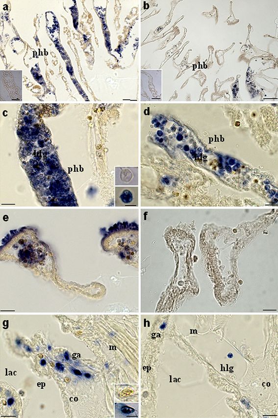
**PCell Tissue Res Fig. 6 ISH assay of transverse sections of the body wall of C. intestinalis. Histological sections from ascidians at 4-8 h after LPS inoculation. a Pharynx bars (phb). Bar 100 μm. Inset Section treated with sense probe. Bar 50 μm. c Pharynx bars (phb) and hemocyte with large granules (hlg). Bar 40 μm. Inset Hemocyte with large granules treated with sense probe (top) and with anti-sense probe (bottom). Bar 10 μm. e Stigmata. Bar 40 μm. g Connective tissue (co) under the epidermis (ep) with lacunae (lac) of the open circulatory system, granular amebocytes (ga), and muscles (m). Bar 40 μm. Inset Granular amebo- cyte treated with sense probe (top) and with anti-sense probe (bottom). Bar 10 μm. Histological sections from naive ascidians. b Pharynx bars (phb). Bar 100 μm. Inset Section treated with sense probe. Bar 50 μm. d Pharynx bars (phb) and hemocyte with large granules (hlg). Bar 40 μm. f Stigmata. Bar 40 μm. h Connective tissue (co) under the epidermis (ep) with lacunae (lac) of the open circulatory system, granular amebocytes (ga), hemocyte with large granules (hlg), and muscles (m). Bar 40 μm (Bonura et al. 2009; Vizzini et al. 2008). The meaning of the sponse to external signals that might explain the second second increase in the expression profiles of inflammatory wave of CiCAP upregulation at 24 h post-injection. molecules remains to be clarified. One possible explanation Moreover, we cannot exclude the possibility that the is a delayed inflammatory response by distinct tissues of the decrease in expression is attributable to cell death after LPS pharynx; this might include hemocytes inside the vessels and injection, whereas the subsequent increase might be attrib- epithelial tissues. On the other hand, we do not know the utable to hemocyte proliferation and the appearance of new regulatory effect of other inflammatory molecules in re- CiCAP-expressing circulating cells.
Cell Tissue Res

    Based on previously published papers (Bonura et al.                Gibbs GM, Lo JC, Nixon B, Jamsai D, O’Connor AE, Rijal S,
                                                                            Sanchez-Partida LG, Hearn MT, Bianco DM, O’Bryan MK
2009; Vizzini et al. 2008), ISH has been performed in order
                                                                            (2010) Glioma pathogenesis-related 1-like 1 is testis enriched,
to identify challenged hemocytes inside the pharynx vessels                 dynamically modified, and redistributed during male germ cell
at the time course corresponding to the main expression                     maturation and has a potential role in sperm-oocyte binding.
peak. At 4 h after inoculation, LPS induces a prompt                        Endocrinology 151:2331–2342
                                                                       Gilbert W (1978) Why genes in pieces? Nature 271:501
CiCAP upregulation in hemocytes with large granules
                                                                       Guo M, Teng M, Niu L, Liu Q, Huang Q, Hao Q (2005) Crystal
contained both in vessels and in associated with connective                 structure of the cysteine-rich secretory protein stecrisp reveals
tissue lining the hemolymph lacunae of the open circulatory                 that the cysteine-rich domain has a K+ channel inhibitor-like fold.
system. Although a cell count has not been performed, the                   J Biol Chem 280:12405–12412
                                                                       Hawdon JM, Hotez PJ (1996) Hookworm: developmental biology of
CiCAP-expressing hemocytes seem to be more abundant                         the infectious process. Curr Opin Genet Dev 6:618–623
than those found in naive ascidians. Interestingly, the                Kovalick GE, Griffin DL (2005) Characterization of the SCP/TAPS
epithelium of pharynx stigmata expresses CiCAP only after                   gene family in Drosophila melanogaster. Insect Biochem Mol
LPS challenge. This result is in agreement with a recent                    Biol 35:825–835
                                                                       Long M, Deutsch M, Wang W, Betran E, Brunet FG, Zhang J (2003)
paper on CiTNFα expression (Parrinello et al. 2010)
                                                                            Origin of new genes: evidence from experimental and computa-
disclosing that several regions of the pharynx epithelial                   tional analyses. Genetica 118:171–182
tissues are probably involved in the inflammatory response.            Moyle M, Foster DL, McGrath DE, Brown SM, Laroche Y, De
    In conclusion, we have described the isolation of a novel               Meutter J, Stanssens P, Bogowitz CA, Fried VA, Ely JA, et al
                                                                            (1994) A hookworm glycoprotein that inhibits neutrophil
CAP gene that is up-regulated after LPS injection in the
                                                                            function is a ligand of the integrin CD11b/CD18. J Biol Chem
body wall of C.intestinalis. The CiCAP expression profile                   269:10008–10015
together with studies on tissue localization suggest a                 Parrinello N, Patricolo E, Canicattì C (1984) Inflammatory-like
possible role for this protein in the ascidian innate immune                reaction in the tunic of Ciona intestinalis (Tunicata). Encapsula-
                                                                            tion and tissue injury I. Biol Bull 167:229–37
responses.                                                             Parrinello N, De Leo G, Di Bella MA (1990) Fine structural
                                                                            observations of the granulocytes involved in the tunic
                                                                            inflammatory-like reaction of Ciona intestinalis (Tunicata). J
References                                                                  Invertebr Pathol 56:181–189
                                                                       Parrinello N, Vizzini A, Arizza V, Salerno G, Parrinello D, Cammarata
                                                                            M, Giaramita FT, Vazzana M (2008) Enhanced expression of a
Azumi K, De Santis R, De Tomaso A, Rigoutsos I, Yoshizaki F, Pinto          cloned and sequenced Ciona intestinalis TNFalpha-like (CiTNF
    MR, Marino R, Shida K, Ikeda M, Ikeda M, Arai M, Inoue Y,               alpha) gene during the LPS-induced inflammatory response. Cell
    Shimizu T, Satoh N, Rokhsar DS, Du Pasquier L, Kasahara M,              Tissue Res 334:305–317
    Satake M, Nonaka M (2003) Genomic analysis of immunity in a        Parrinello N, Vizzini A, Salerno G, Sanfratello MA, Cammarata M,
    urochordate and the emergence of the vertebrate immune system:          Arizza V, Vazzana M, Parrinello D (2010) Inflamed adult
    "waiting for Godot". Immunogenetics 55:570–581                          pharynx tissues and swimming larva of Ciona intestinalis share
Bonura A, Vizzini A, Salerno G, Parrinello N, Longo V, Colombo P            CiTNFalpha-producing cells. Cell Tissue Res 341:299–311
    (2009) Isolation and expression of a novel MBL-like collectin      Ren C, Ren CH, Li L, Goltsov AA, Thompson TC (2006)
    cDNA enhanced by LPS injection in the body wall of the                  Identification and characterization of RTVP1/GLIPR1-like genes,
    ascidian Ciona intestinalis. Mol Immunol 46:2389–2394                   a novel p53 target gene cluster. Genomics 88:163–172
Cammarata M, Benenati G, Odom EW, Salerno G, Vizzini A, Vasta          Sato Y, Okamoto K, Kagami A, Yamamoto Y, Igarashi T, Kizaki H
    GR, Parrinello N (2007) Isolation and characterization of a fish        (2004) Streptococcus mutans strains harboring collagen-binding
    F-type lectin from gilt head bream (Sparus aurata) serum.               adhesin. J Dent Res 83:534–539
    Biochim Biophys Acta 1770:150–155                                  Shikamoto Y, Suto K, Yamazaki Y, Morita T, Mizuno H (2005)
Cammarata M, Arizza V, Cianciolo C, Parrinello D, Vazzana M,                Crystal structure of a CRISP family Ca2+-channel blocker
    Vizzini A, Salerno G, Parrinello N (2008) The prophenoloxidase          derived from snake venom. J Mol Biol 350:735–743
    system is activated during the tunic inflammatory reaction of      Swalla BJ, Cameron CB, Corley LS, Garey JR (2000) Urochordates
    Ciona intestinalis. Cell Tissue Res 333:481–492                         are monophyletic within the deuterostomes. Syst Biol 49:52–64
Del Valle A, Jones BF, Harrison LM, Chadderdon RC, Cappello M          Szyperski T, Fernandez C, Mumenthaler C, Wuthrich K (1998)
    (2003) Isolation and molecular cloning of a secreted hookworm           Structure comparison of human glioma pathogenesis-related
    platelet inhibitor from adult Ancylostoma caninum. Mol Biochem          protein GliPR and the plant pathogenesis-related protein P14a
    Parasitol 129:167–177                                                   indicates a functional link between the human immune system
Delsuc F, Brinkmann H, Chourrout D, Philippe H (2006) Tunicates             and a plant defense system. Proc Natl Acad Sci USA 95:2262–
    and not cephalochordates are the closest living relatives of            2266
    vertebrates. Nature 439:965–968                                    Thompson JD, Higgins DG, Gibson TJ (1994) CLUSTAL W:
Gibbs GM, Scanlon MJ, Swarbrick J, Curtis S, Gallant E, Dulhunty            improving the sensitivity of progressive multiple sequence
    AF, O’Bryan MK (2006) The cysteine-rich secretory protein               alignment through sequence weighting, position-specific gap
    domain of Tpx-1 is related to ion channel toxins and regulates          penalties and weight matrix choice. Nucleic Acids Res 22:
    ryanodine receptor Ca2+ signaling. J Biol Chem 281:4156–4163            4673–4680
Gibbs GM, Roelants K, O’Bryan MK (2008) The CAP superfamily:           Trexler M, Banyai L, Patthy L (2000) The LCCL module. Eur J
    cysteine-rich secretory proteins, antigen 5, and pathogenesis-          Biochem 267:5751–5757
    related 1 proteins—roles in reproduction, cancer, and immune       Urayama S, Harada Y, Nakagawa Y, Ban S, Akasaka M, Kawasaki N,
    defense. Endocr Rev 29:865–897                                          Sawada H (2008) Ascidian sperm glycosylphosphatidylinositol-
Cell Tissue Res
anchored CRISP-like protein as a binding partner for an epidermis during the inflammatory response of the ascidian
allorecognizable sperm receptor on the vitelline coat. J Biol Ciona intestinalis. Dev Comp Immunol 32:682–692
Chem 283:21725–21733 Yamazaki Y, Morita T (2004) Structure and function of snake venom
Vizzini A, Pergolizzi M, Vazzana M, Salerno G, Di Sano C, Macaluso cysteine-rich secretory proteins. Toxicon 44:227–231
P, Arizza V, Parrinello D, Cammarata M, Parrinello N (2008) Zelensky AN, Gready JE (2005) The C-type lectin-like domain
FACIT collagen (1alpha-chain) is expressed by hemocytes and superfamily. FEBS J 272:6179–6217You can also read