Mechanisms of aggregation and fibril formation of the amyloidogenic N-terminal fragment of apolipoprotein A-I
←
→
Page content transcription
If your browser does not render page correctly, please read the page content below
JBC Papers in Press. Published on July 24, 2019 as Manuscript RA119.008000
        The latest version is at http://www.jbc.org/cgi/doi/10.1074/jbc.RA119.008000

Mechanisms of aggregation and fibril formation of the amyloidogenic N-terminal fragment of
                                    apolipoprotein A-I

     Chiharu Mizuguchi1‡, Miho Nakagawa1, Norihiro Namba1, Misae Sakai1, Naoko Kurimitsu1,
    Ayane Suzuki1, Kaho Fujita1, Sayaka Horiuchi1, Teruhiko Baba2, Takashi Ohgita1, Kazuchika
                                  Nishitsuji3, and Hiroyuki Saito1*

From the 1Department of Biophysical Chemistry, Kyoto Pharmaceutical University, 5 Misasagi-
Nakauchi-cho, Yamashina-ku, Kyoto 607-8414, Japan, 2Biotechnology Research Institute for Drug
Discovery, National Institute of Advanced Industrial Science and Technology (AIST), Tsukuba
Central 5, 1-1-1 Higashi, Tsukuba 305-8565, Japan, and 3Department of Biochemistry, Wakayama
Medical University, 811-1 Kimiidera, Wakayama 641-8509, Japan

                   Running title: Aggregation and fibril formation mechanism of apoA-I
*
 To whom correspondence should be addressed: Hiroyuki Saito, Department of Biophysical Chemistry,
Kyoto Pharmaceutical University, 5 Misasagi-Nakauchi-cho, Yamashina-ku, Kyoto 607-8414, Japan,

                                                                                                               Downloaded from http://www.jbc.org/ by guest on October 31, 2019
Tel.: +81-75-595-4663; Fax.: +81-75-595-4762; E-mail: hsaito@mb.kyoto-phu.ac.jp
‡
    Present address: Division of Hygienic Chemistry, Osaka Institute of Public Health, Osaka 543-0026

Keywords: apolipoprotein A-I; amyloid; protein aggregation; fibril; activation energy; pyrene labeling;
hereditary systemic amyloidosis; G26R variant

Abstract                                                  Δ50‒58 variant was entropically unfavorable,
    The N-terminal (1‒83) fragment of the major           indicating that residues 50‒58 entropically
constituent of plasma high-density lipoprotein,           promote the nucleation step in fibril formation of
apolipoprotein A-I (apoA-I), strongly tends to            apoA-I 1‒83/G26R. Moreover, a residue-level
form amyloid fibrils, leading to systemic                 structural investigation of apoA-I 1‒83/G26R
amyloidosis. Here, using a series of deletion             fibrils with site-specific pyrene labeling
variants, we examined the roles of two major              indicated that the two amyloidogenic segments
amyloidogenic segments (residues 14‒22 and                are in close proximity to form an amyloid core
50‒58) in the aggregation and fibril formation of         structure, whereas the N- and C-terminal tail
an amyloidogenic G26R variant of the apoA-I 1‒            regions are excluded from the amyloid core.
83 fragment (apoA-I 1‒83/G26R). Thioflavin T              These results provide critical insights into the
fluorescence assays and atomic force                      aggregation mechanism and fibril structure of the
microscopy revealed that elimination of residues          amyloidogenic N-terminal fragment of apoA-I.
14‒22 completely inhibits fibril formation of
apoA-I 1‒83/G26R, whereas Δ32‒40 and Δ50‒
58 variants formed fibrils with markedly reduced                Apolipoprotein (apoA-I) is the major
nucleation and fibril growth rates. CD                    structural and functional constituent of plasma
measurements revealed structural transitions              high-density lipoprotein (HDL) that plays a
from random coil to β-sheet structures in all             critical role in the formation and metabolism of
deletion variants except for the Δ14‒22 variant,          HDL particles (1,2). Many naturally occurring
indicating that residues 14‒22 are critical for the       mutations in human apoA-I are associated with
β     transition     and     fibril     formation.        reduced plasma HDL levels and hereditary
Thermodynamic analysis of the kinetics of fibril          systemic       amyloidosis    (3).    To    date,
formation by apoA-I 1‒ 83/G26R indicated that             approximately 20 naturally occurring mutations
both nucleation and fibril growth are                     in human apoA-I associated with familial
enthalpically unfavorable, whereas entropically,          amyloid polyneuropathy have been reported
nucleation is favorable, but fibril growth is             (4,5), in which the majority of the amyloidogenic
unfavorable. Interestingly, the nucleation of the         mutations is clustered in two regions of the N-

                                                      1
terminal residues 26‒90 and 154‒178 (4,6,7).                    In the present study, we asked how each
Hereditary apoA-I amyloidosis is characterized            amyloidogenic segment in apoA-I modulates
by deposition of the N-terminal 80‒100-residue            aggregation and fibril formation of the N-
fragments of the variant protein as amyloid               terminal 1‒83 fragment of the G26R variant
fibrils in peripheral organs such as heart, liver,        using a series of deletion variants (Δ14‒22, Δ32‒
kidneys, or gastrointestinal tract, causing organ         40, Δ50‒58, and Δ68‒76) that lack different
damage (6,8,9). It has been hypothesized that the         amyloidogenic regions along the molecule. We
specific amyloidogenic mutations perturb the              also examined the intermolecular contacts of
native protein structure, increasing susceptibility       apoA-I 1‒83/G26R fibrils at a residue level by
to proteolysis and thereby releasing the N-               employing site-directed cysteine mutagenesis
terminal amyloidogenic fragment (4,10). The               and fluorescence labeling to gain structural
molecular basis for the onset and development of          insights into amyloid fibrils formed by the N-
apoA-I systemic amyloidosis is largely unknown.           terminal fragment of apoA-I G26R.
       Sequence-based analyses of the N-
terminal 1‒100 residues in apoA-I predict that            Results
there are two major aggregation-prone segments            Design of deletion or cysteine-substituted
(residues 14‒22 and 50‒58) together with minor            variants of apoA-I 1‒83/G26R
segment (residues 69‒72), in which the rank                    We designed two types of mutations in the
order of aggregation propensity is residues 14‒           N-terminal 1‒83 fragment of apoA-I G26R

                                                                                                                Downloaded from http://www.jbc.org/ by guest on October 31, 2019
22 > residues 50‒58 > residues 69‒72 (10,11). In          variant: deletion mutations that remove nine
agreement with this prediction, the N-terminal            amino acid residues in the amyloidogenic
1‒83 or 1‒93 fragments of apoA-I were shown               regions of the protein, and single amino acid
to have a strong propensity to form amyloid               mutations that introduce cysteine substitutions
fibrils (11,12). Studies of synthetic apoA-I              for labeling by pyrene maleimide at different
fragment peptides demonstrated that the peptides          positions along the molecule (Fig. 1A). The
containing either the first (residues 14‒22) or the       deletion variants, Δ14‒22, Δ50‒58, and Δ68‒76
second (residues 50‒58) amyloidogenic segment             lack the highly amyloidogenic segments of
have the ability to form amyloid-like fibrils with        residues 14‒22, 50‒58, and 69‒72, respectively
β-transition, whereas the peptide only containing         (10), whereas the Δ32‒40 variant lacks the
residues 69‒72 does not form fibrils at neutral           putative loop region between two β-strands from
pH (13,14). Interestingly, since the two major            residues 14‒31 to 41‒58 (23) (Fig. 1B). In the
amyloidogenic segments (residues 14‒22 and                cysteine variants, cysteine residues were
50‒58) overlap with the hydrophobic α-helix-              introduced into the highly amyloidogenic
forming regions upon lipid binding (15,16), lipid         segments together with the N- and C-terminal tail
membrane environments significantly affect the            regions that have low amyloid propensity (Fig.
fibril-forming properties of the N-terminal               1B). The Cys-substituted positions were
fragments or peptides of apoA-I (17-19).                  selectively labeled with pyrene maleimide to
       We previously reported that the Iowa               probe the polarity and intermolecular proximity
(G26R) point mutation, the first and most                 of different segments of apoA-I 1‒83/G26R
common amyloidogenic mutation found in                    fragment in the fibril form. We note that cysteine
apoA-I (20,21), greatly facilitates fibril                mutations and pyrene labeling did not
formation by the N-terminal 1‒83 fragment of              significantly inhibit the fibril-forming ability of
apoA-I in solution (12) as well as on lipid               the protein although several mutants appeared to
membranes (18,19). Secondary structure                    have somewhat enhanced fibril-forming
examinations of the G26R variant of full-length           propensity (Supplemental Fig. S1).
apoA-I by electron paramagnetic resonance
spectroscopy (21) and hydrogen‒deuterium                  Effect of deletion on fibril-forming property of
exchange mass spectroscopy (22) demonstrated              apoA-I 1‒83/G26R
that the G26R mutation induces widespread α-                     We first examined the fibril-forming
helix destabilization in the N-terminal helix             propensities of the deletion variants of apoA-I 1‒
bundle domain and promotes a transition of                83/G26R using the amyloid-sensitive fluorescent
residues 27‒56 to β-strand-rich structure.                dye, thioflavin T (ThT). As previously reported
However, structural information on amyloid                (12), apoA-I 1‒83/G26R exhibited large increase
fibrils formed by the N-terminal fragment of              in ThT fluorescence at neutral pH (Fig. 2A). In
apoA-I G26R variant is lacking.                           contrast, there was no increase in ThT

                                                      2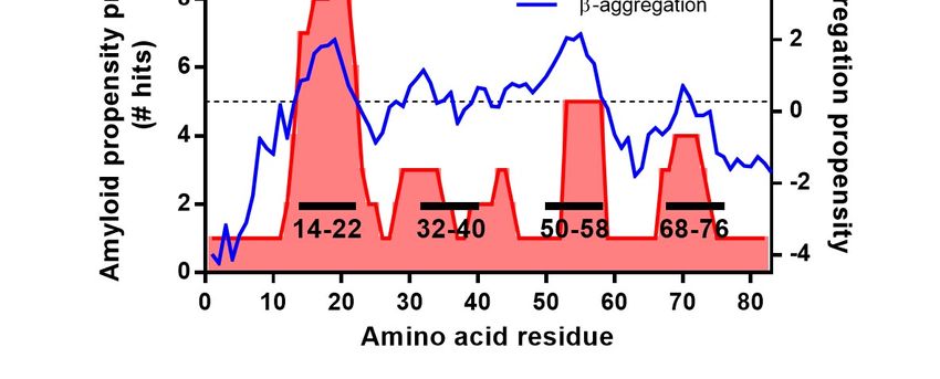
fluorescence for the Δ14‒22 variant, indicating            changes during incubation. As shown in Fig. 3,
that residues 14‒22 are crucial for fibril                 all spectra of the deletion variants except for the
formation of apoA-I 1‒83/G26R. Significantly               Δ14‒22 displayed a single minimum at around
delayed increases in ThT fluorescence for the              216 nm after incubation for 120 h, implying
Δ32‒40 and Δ50‒58 variants suggest that the                conversion from random coil to β-sheet-rich
segments spanning residues 32‒40 as well as 50‒            structure. Consistent with the delayed increase in
58 are important for nucleation in fibril                  ThT fluorescence (Fig. 2A), such apparent
formation of the 1‒83/G26R variant.                        transition to β-structure was not observed for the
Interestingly, the Δ68‒76 variant exhibited                Δ32‒40 and Δ50‒58 variants at shorter
greatly enhanced ThT fluorescence intensity (Fig.          incubation for 60 h (Supplemental Fig. S4A). No
2A, inset), suggesting the possibility that                secondary structural change occurred for the
residues 68‒76 inhibit the fibril-forming ability          Δ14‒22 variant during incubation.
of apoA-I 1‒83/G26R. However, precipitation                       Fig. 4 shows atomic force microscopy
analyses of fibrils formed by the 1‒83/G26R                (AFM) and transmission electron microscopy
deletion variants indicated that the fibril-forming        (TEM) images of the 1‒83/G26R deletion
ability of the Δ68‒76 variant is somewhat less             variants after 120 h incubation. AFM images
than the other apoA-I 1‒83/G26R variants except            (Fig. 4A) show that the 1‒83/G26R as well as
for Δ14‒22 (Supplemental Fig. S2). Such                    Δ32‒40, Δ50‒58, and Δ68‒76 variants formed
inhibited or delayed increase in ThT                       straight fibrils, whereas the Δ14‒22 did not form

                                                                                                                 Downloaded from http://www.jbc.org/ by guest on October 31, 2019
fluorescence for the Δ14‒22, Δ32‒40 or Δ50‒58              apparent fibrils but rather formed small spherical
variants and the enhanced ThT fluorescence for             aggregates. TEM observations also confirm the
the Δ68‒76 variant were also observed at                   formation of straight fibrils by the 1‒83/G26R as
different protein concentrations (Supplemental             well as the deletion variants except for the Δ14‒
Fig. S3).                                                  22, in which the Δ14‒22 variant rarely formed
       The kinetics of ThT fluorescence increase           fibrils (Fig. 4B). In contrast, apparent straight
were analyzed by the Finke-Watzky two-step                 fibrils were not observed for the Δ32‒40 and
model of a homogeneous nucleation followed by              Δ50‒58 variants at incubation for 60 h
an autocatalytic heterogeneous fibril growth,              (Supplemental Fig. S4B). These results indicate
which has been applied to a broad range of                 that the highly amyloidogenic segment spanning
protein     aggregation       kinetics      (24-27).       residues 14‒22 is necessary for the formation of
Comparison of rate constants of the nucleation             amyloid fibril structure by apoA-I 1‒83/G26R,
(k1) and fibril growth (k2) for fibril formation by        whereas the segments of residues 32‒40 and 50‒
the deletion variants (Figs. 2B and 2C) clearly            58 are not necessary for fibril formation but
demonstrates that the Δ32‒40 and Δ50‒58                    modulate the kinetics of nucleation and fibril
variants have significantly decreased rate                 formation. Consistently, the fibrils formed by the
constants of both nucleation and fibril growth,            deletion variants after 120 h incubation exhibited
whereas the Δ68‒76 exhibits similar kinetic                similar stability against urea denaturation to 1‒
parameters compared to the 1‒83/G26R. Kinetic              83/G26R (Supplemental Fig. S5). In addition,
parameters obtained by the empirical sigmoidal             the fibrils formed by the deletion variants
equation (Figs. 2D and 2E) provide similar                 induced strong cytotoxicity with HEK293 cells
conclusions: the Δ32‒40 and Δ50‒58 variants                similarly to 1‒83/G26R fibrils, whereas the Δ14‒
significantly increase the lag time and decrease           22 exhibited reduced cytotoxicity (Supplemental
the apparent rate constant for fibril growth. We           Fig. S6). This indicates that the formation of the
note that the Finke-Watzky model is based on               fibril structure is critical to the cytotoxicity of
two pseudoelementary reaction steps of                     apoA-I 1‒83/G26R variants (12).
nucleation and fibril growth without further
considerations on the nuclei size and fibril               Thermodynamic analysis of fibril formation by
propagation mechanisms such as secondary                   apoA-I 1‒83/G26R
pathways (27), although it has been proposed                     To understand the thermodynamic aspects
that secondary nucleation process occurs during            of the aggregation of apoA-I 1‒83/G26R into
fibril formation of amyloid β peptides and α-              amyloid fibrils, we explored the effect of
synuclein (28-30).                                         temperature on the kinetics of formation of
       We next performed circular dichroism                amyloid-like fibrils. We note that CD
(CD) measurements of the 1‒83/G26R deletion                measurements confirmed that there are no
variants to determine the secondary structural             secondary structural changes across the

                                                       3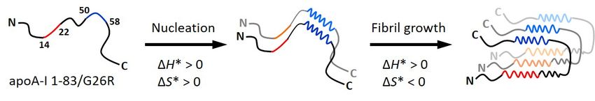
temperature range of 25–42 ºC (data not shown).            from the 1‒83/G26R is much smaller than that
Fig. 5A shows the time course of ThT                       for the Δ50‒58 (Table 1). Regarding the Δ68‒76
fluorescence intensity with the fitted curves by           variant, the thermodynamic parameters for
the Finke-Watzky two-step model at different               nucleation are similar, but the unfavorable
temperatures. Comparison of rate constants of              activation enthalpy of fibril growth is
the nucleation (k1) and fibril growth (k2) at each         significantly reduced compared to the 1‒
temperature indicates that both rate constants             83/G26R (Table 1). This indicates that residues
greatly increase with temperature (Fig. 5B).               68‒76 inhibit fibril growth of apoA-I 1‒83/G26R
From the linear relationship of ln (k1/T) or ln            by increasing the unfavorable activation
(k2/T) with 1/T based on the Eyring equation (Fig.         enthalpy.
5C), the activation enthalpy ΔH* and entropy ΔS*
for the nucleation and fibril growth steps in fibril       Determination of amyloid core regions in apoA-
formation by apoA-I 1‒83/G26R were obtained.               I 1‒83/G26R fibrils
Table 1 summarizes the ΔH* and ΔS* values and                     We      next    performed      fluorescence
the activation Gibbs free energy ΔG*. These                measurements of pyrene-labeled apoA-I 1‒
parameters demonstrated that although the                  83/G26R variants to map intermolecular
activation free energy barriers for nucleation and         proximities of different segments in the fibril
fibril growth are similar, the contributions of            form. Pyrene fluorescence emission spectra
activation enthalpy and entropy to the free                display unique features that give information on

                                                                                                                  Downloaded from http://www.jbc.org/ by guest on October 31, 2019
energy barrier are quite different: the nucleation         the spatial proximity and polarity of the
step is enthalpically unfavorable but entropically         environment. When two pyrene rings are within
favorable, whereas fibril growth is both                   10 Å of each other, a broad and red-shifted
enthalpically and entropically unfavorable. That           emission peak appears generally at around 470
is, the free energy barrier for nucleation is              nm, attributed to formation of an excited-state
entirely enthalpic, whereas that for fibril growth         dimer, excimer (31). Thus, the excimer ratio
consists of both enthalpic and entropic barriers.          (ratio of fluorescence intensity of excimer at 470
        We also performed thermodynamic                    nm to that of monomer at 385 nm) indicates the
analysis of fibril formation by the deletion               extent of excimer formation and, therefore, the
variants to gain insight into the role of each             intermolecular proximity of pyrene-labeled
residues in fibril formation by apoA-I 1‒                  positions of the protein in oligomers and fibrils
83/G26R (Fig. 5D and Supplemental Fig. S7).                (32-36). In addition, we also determined the
The Δ14‒22 variant did not form fibrils at any             pyrene scale as the intensity ratio between bands
temperatures examined (Supplemental Fig. S7A).             at 375 and 385 nm to evaluate the local
In contrast, the Δ50‒58 variant exhibited                  hydrophobicity around the probe at the labeled
temperature-dependent increases in ThT                     positions (33,37).
fluorescence (Supplemental Fig. S7C), and                         Figs. 6A and 6B show typical fluorescence
Eyring plots of rate constants of k1 and k2 (Fig.          emission spectra of pyrene-labeled apoA-I 1‒
5D) gave the thermodynamic parameters for                  83/G26R variants before and after incubation.
fibril formation of the Δ50‒58 variant (Table 1).          Before incubation, no excimer peaks at around
Despite similar activation free energy barriers            470 nm were observed for all pyrene-labeled
between apoA-I 1‒83/G26R and the Δ50‒58                    variants (all fluorescence emission spectra of
variant, a large reduction in the unfavorable              pyrene-labeled variants are shown in
activation enthalpy and the concomitant                    Supplemental Fig. S8). In fibrils, a large excimer
unfavorable activation entropy for nucleation in           peak appeared when the label was at position 22,
fibril formation of the Δ50‒58 are observed, in            whereas only a small excimer peak was seen with
sharp contrast to the favorable entropy of                 the label at position 81. Fig. 6C shows the profile
nucleation for the 1‒83/G26R. For fibril growth            of excimer ratios as well as pyrene scale in fibrils
of the Δ50‒58 variant, the activation enthalpy             as a function of residue number. Positions 22 and
and entropy are both unfavorable. These results            52 exhibit high excimer ratios, consistent with
indicate that residues 50‒58 entropically drive            two regions around the highly amyloidogenic
the nucleation in fibril formation by apoA-I 1‒            segments (residues 14‒22 and 50‒58) being in
83/G26R. Similar to the Δ50‒58, the Δ32‒40                 strong intermolecular contacts. In contrast, low
variant exhibits the unfavorable activation                excimer ratios at positions 2, 8, 72, and 81
enthalpy and entropy for nucleation, in which the          suggest sequestration of the N- and C-terminal
reduction in the unfavorable activation enthalpy           tail regions from intermolecular contacts. The

                                                       4
relatively low excimer ratio at position 36 agrees the deletion of residues 32‒40 on the kinetics of
with the prediction that residues 32‒40 form a nucleation and fibril growth (Fig. 2) is possibly
loop region between two β-strands (23). We note because the lack of residues 32‒40 may alter the
that pyrene scale values for all labeled positions appropriate intramolecular interaction between
are similarly low (Fig. 6C), indicating that two putative β-strands consisting of highly
pyrene molecules at all labeled positions in amyloidogenic residues of 14‒22 and 50‒58 in
fibrils are located in hydrophobic environments. fibril formation. Deletion of residues 68‒76 that
are rich in negatively charged amino acids has
Discussion small effects on the kinetics of fibril formation,
Sequence-based predictions of the β- but causes great increases in ThT fluorescence
aggregation (11) and amyloid propensity (10) intensity (Fig. 2). This indicates that the C-
indicate that there are two major aggregation- terminal tail region that is rich in negatively
prone segments of residues 14‒22 and 50‒58 in charged amino acids has inhibitory effects on
the N-terminal 1‒83 residues of apoA-I. Based aggregation and fibril formation of apoA-I 1‒
on the secondary structure examination of the 83/G26R as observed for α-synuclein (39).
aggregates of apoA-I G26R variant (21), The thermodynamic analyses of ThT
together with a finding that a peptide comprising fluorescence kinetics (Fig. 5 and Supplemental
residues 46‒59 of apoA-I forms amyloid-like Fig. S7) give further insights into the molecular
fibrils (13), it was considered that two segments mechanism of aggregation and fibril formation
Downloaded from http://www.jbc.org/ by guest on October 31, 2019
spanning residues 14‒31 and 46‒59 in apoA-I 1‒ of apoA-I 1‒83/G26R and deletion variants. In
83/G26R are highly amyloidogenic regions (Fig. fibril formation of apoA-I 1‒83/G26R, the
1B). Indeed, we previously demonstrated that nucleation process is enthalpically unfavorable
synthetic apoA-I fragment peptides containing but entropically favorable, whereas the fibril
these two amyloidogenic segments have the elongation process is enthalpically and
ability to form amyloid-like fibrils (14,18,38). entropically unfavorable (Table 1). The
However, details on the roles of each unfavorable activation enthalpies of nucleation
amyloidogenic segments on amyloid fibril and fibril growth are generally observed for
formation by apoA-I 1‒83/G26R are unknown. many amyloidogenic proteins (40-43), likely due
The present study demonstrated that the to the net unfavorable formation and breakage of
deletion of residues 14‒22 completely inhibits β- many weak interactions necessary to reach the
transition and fibril formation of apoA-I 1‒ transition state (42). In contrast, the favorable
83/G26R, whereas the deletion of residues 50‒58 activation entropies of nucleation observed for
markedly decreases the rate constants of the 1‒83/G26R (Table 1) as well as a fungal
nucleation and fibril elongation (Figs. 2B and prion-forming domain (40) and amyloid-β 42
2C). This information clearly indicates that the (43) peptides are thought to come from the
first amyloidogenic segment containing residues desolvation of hydrophobic regions of the
14‒22 is crucial for fibril formation of apoA-I 1‒ protein/peptide molecule in the transition state
83/G26R, whereas the second amyloidogenic (42,43). The unfavorable activation entropy of
segment containing residues 50‒58 is largely fibril growth for the 1‒83/G26R may arise
involved in the nucleation step of fibril formation. because re-solvation of the fibril surface
Indeed, a Y18P mutation that impairs formation dominates over desolvation of the existing
of β-sheet structure in residues 14‒22 strongly nucleus (43).
inhibited fibril formation by apoA-I 1‒83/G26R The activation energy parameters for
(18). Interestingly, the effects of deletions on the nucleation in fibril formation of the Δ50‒58
fibril-forming ability of apoA-I 1‒83/G26R variant are quite different from those of the 1‒
bound to lipid membranes are quite different 83/G26R (Table 1). Based on the X-ray crystal
from those in solution (Supplemental Fig. S9). structure of C-terminal truncated human apoA-I
Since α-helix formation of the amyloidogenic Δ185‒243 showing that residues 44‒55 are in an
segments upon lipid binding strongly inhibits β- extended nonhelical conformation (44), it was
transition and fibril formation by the N-terminal proposed that residues 44‒55 provide a template
fragment of apoA-I (18,19), it is likely that the for nucleation of intermolecular β-aggregation
second amyloidogenic segment containing (7). In this regard, it is possible that the favorable
residues 50‒58 is not available for fibril activation entropy of nucleation of apoA-I 1‒
formation of apoA-I 1‒83/G26R on the 83/G26R comes from desolvation of the exposed
membrane surface (19). The inhibitory effect of segment in residues 44‒55 in the nucleation
5process. Consistent with this idea, deletion of methionine oxidized full-length apoA-I revealed
residues 50‒58 causes the activation entropy of that several amino acids within residues 13‒22
nucleation to be highly negative (Table 1), and 50‒59 are detected as β-sheet structure (47).
indicating that the segment in residues 50‒58 The results of pyrene excimer fluorescence of
entropically promotes nucleation in fibril apoA-I 1‒83/G26R fibrils (Fig. 6) demonstrate
formation of the 1‒83/G26R molecule. In for the first time that the two segments with high
addition, a large reduction in the unfavorable amyloid-forming propensity (residues 14‒22 and
activation enthalpy of nucleation observed for 50‒58) are indeed in close proximity to form
the Δ50‒58 variant (Table 1) suggests that amyloid core structure, whereas the N-terminal
desolvation of the segment in residues 50‒58 (residues 1‒10) and C-terminal (residues 70‒83)
seems to contribute to the unfavorable activation tail regions as well as central (around residues
enthalpy in forming a nucleus (42). 32‒40) region are likely to be excluded from the
The Δ32‒40 variant also exhibits the amyloid core. Since the C-terminal 70‒83
unfavorable activation enthalpy and entropy for residues (EFWDNLEKETEGLR) are rich in
nucleation similar to the case of Δ50‒58 variant negatively charged amino acids at neutral pH,
(Table 1). However, the reduction in the electrostatic repulsions would inhibit the
unfavorable activation enthalpy from the 1‒ intermolecular aggregation of this region. In
83/G26R is much smaller than that for the Δ50‒ addition, the presence of proline residues at the
58. This may suggest that the deletion of residues extreme N-terminal region is likely to prevent the
Downloaded from http://www.jbc.org/ by guest on October 31, 2019
32‒40 inhibits the nucleation step possibly β-structure-rich aggregation (48,49). It should be
through altering the appropriate intermolecular noted that in amyloid fibrils, apoA-I molecules
interaction of the second amyloidogenic segment are proposed to be packed in a parallel, in-
containing residues 50‒58. In contrast, the register β-sheet structure (10,23).
thermodynamic parameters of nucleation for the In summary, we have demonstrated that
Δ68‒78 variant are similar to those of the 1‒ the two highly amyloidogenic segments of
83/G26R, but the unfavorable activation residues 14‒22 and 50‒58 play crucial roles in
enthalpy of fibril growth is significantly reduced aggregation and fibril formation of the G26R
(Table 1). This indicates that residues 68‒76 variant of the N-terminal 1‒83 fragment of
enthalpically inhibit fibril growth of apoA-I 1‒ apoA-I, in which residues 14‒22 are necessary
83/G26R and may explain why the Δ68‒78 for fibril formation whereas residues 50‒58
variant exhibits the great increases in ThT entropically drive the nucleation process. In
fluorescence intensity (Fig. 2). That is, the lower addition, structural investigation of apoA-I 1‒
unfavorable activation enthalpy of fibril growth 83/G26R fibrils indicates that these two
is likely to decrease the critical concentration at amyloidogenic segments are in close proximity
which fibrils appear from nuclei (45,46), leading to form amyloid core structure, whereas the N-
to the formation of a large number of fibrils. and C-terminal tail regions are excluded from the
Indeed, the great enhancement of ThT amyloid core. The present findings provide novel
fluorescence was also observed for the 1‒ molecular insights into the aggregation
83/G26R and the Δ50‒58 variant with increasing mechanism and fibril structure of the
concentration of protein (Supplemental Fig. S3). amyloidogenic N-terminal fragment of apoA-I as
The use of pyrene to probe the spatial summarized in Fig. 7.
proximity (< 10 Å) between pairs of labeled
residues in the proteins has been successfully Experimental procedures
employed to gain structural details of oligomers Preparation of recombinant apoA-I proteins
and fibrils formed by a yeast prion (32), α- The N-terminal fragment 183 of apoA-I
synuclein (35,36), and non-disease related with G26R substitution (apoA-I 1-83/G26R) and
HypF-N protein (34). Based on the fluorescence its engineered variants with deletions of Δ14‒22,
resonance energy transfer study, we previously Δ32‒40, Δ50‒58, and Δ68‒76, or cysteine
proposed a hypothetical model of fibril structure substitutions of E2C, W8C, L14C, L22C, S36C,
of the N-terminal fragment of apoA-I G26R S52C, S58C, L64C, W72C, and G81C were
variant, in which β-strands from residues 14‒31 expressed in E. coli as thioredoxin fusion
and 41‒58 form a self-complementary steric proteins and isolated and purified as described
zipper stabilized by van der Waals and (12,18). Cleavage of the thioredoxin fusion
hydrophobic interactions (23). In addition, solid protein with thrombin leaves the target apoA-I
state NMR analysis of aggregates formed by with two extra amino acids, Gly-Ser, at the N
6terminus. The apoA-I preparations were at least growth of fibrils and tm is the time to 50% of
95% pure as assessed by SDS-PAGE. These maximal fluorescence. The lag time is calculated
apoA-I variants were dialyzed from 6M GdnHCl as tm 2/k.
with or without 1% β-mercaptoethanol solution ThT fluorescence data were also analyzed
into the appropriate buffer before use. Labeling by the Finke‒Watzky two-step model equation
of cysteine-containing apoA-I 1-83/G26R for nucleation followed by autocatalytic growth
variants with N-(1-pyrene)maleimide (Thermo (24,41):
Fisher Scientific, Rockford, IL) was performed
as described (50,51). F F0 k1 k2[A]0
1 (2)
Fmax F0 k1 exp(k1 k2 [A]0 )t k2 [A]0
Preparation of small unilamellar vesicles
Small unilamellar vesicles were prepared where [A]0 is initial concentration of monomer
as described previously (18,52). Briefly, a dried protein, k1 and k2 are the rate constants
film of egg phosphatidylcholine (Kewpie, Tokyo, corresponding to the nucleation and growth of
Japan) was hydrated in 10 mM Tris-HCl buffer fibrils, respectively. Thermodynamic parameters
(150 mM NaCl, 0.02% NaN3, pH 7.4) and for nucleation and fibril growth were determined
sonicated on ice under nitrogen. After removing from the Eyring equations:
titanium debris, the samples were centrifuged in
k H * 1 S * k
a Beckman MLA-55 rotor for 1.5 h at 15 °C at ln ln B (3)
T R T R h
Downloaded from http://www.jbc.org/ by guest on October 31, 2019
40,000 rpm to separate any remaining large
vesicles. hk
G * RT ln (4)
CD spectroscopy kBT
Far-UV CD spectra were recorded from where kB and h are the Boltzmann and Planck
190 to 260 nm at 25 °C using a Jasco J-1500 constants, respectively. The activation enthalpy
spectropolarimeter (JASCO, Tokyo, Japan). The (ΔH*) and entropy (ΔS*) were obtained from the
apoA-I variants of 50 μg/ml in 10 mM Tris-HCl slope and y-intercept of the linear plot according
buffer (pH 7.4) were subjected to CD to equation 3, respectively. The activation Gibbs
measurements in a 1-mm quartz cuvette, and the free energy (ΔG*) was calculated from the rate
results were corrected by subtracting the buffer constant k according to equation 4 or from ΔH*
base line. and ΔS* according to ∆G* = ∆H* - T∆S*.
For proximity analysis, pyrene emission
Fluorescence measurements fluorescence of pyrene-labeled apoA-I 1‒
Fluorescence measurements were carried 83/G26R variants were monitored before and
out with F-2700 or F-7000 fluorescence after incubation. To obtain optimal pyrene
spectrophotometers (Hitachi High-Technologies, excimer signals, mixtures of 25 mol% of pyrene-
Tokyo, Japan) at 25 °C. Kinetics of formation of labeled and 75 mol% of unlabeled proteins were
amyloid-like structure were monitored using used (32,35). Pyrene emission fluorescence was
ThT. ApoA-I 1‒83/G26R variants (100 g/mL) recorded from 360 to 600 nm using a 342 nm
in 10 mM Tris-HCl buffer (150 mM NaCl, excitation wavelength. Pyrene scale was
0.02% NaN3, pH 7.4) were incubated at 37 ºC calculated as the ratio of fluorescence intensity at
with agitation on an orbital rotator in the 375 nm to that at 385 nm (33,37). Excimer to
presence of 10 μM ThT. ThT fluorescence was monomer ratio was calculated as the ratio of
recorded at 485 nm with an excitation fluorescence intensity at 470 nm (excimer signal)
wavelength of 445 nm. The time-dependent to that at 385 nm (monomer signal).
increase in ThT fluorescence intensity was fitted For chemical denaturation experiments,
to a sigmoidal equation (12,53): fibrils of apoA-I 1‒83/G26R deletion variants
Fmax F0 (50 g/ml) in 10 mM Tris-HCl buffer (150 mM
F F0 (1) NaCl, 0.02% NaN3, pH 7.4) were incubated
1 exp[kapp (tm t )] overnight at 4 °C with urea at various
where F is the fluorescence intensity, F0 is the concentrations in the presence of 10 M ThT.
initial base line during the lag phase, and Fmax is Denaturation of fibrils was monitored from the
the final base line after the growth phase has change in ThT fluorescence intensity.
ended. kapp is the apparent rate constant for the
7AFM TEM images were obtained at 120 kV on an FEI
Each sample solution in 10 mM Tris buffer Tecnai F20 transmission electron microscope.
was diluted to 10 µg/mL with distilled water, and
10 µL of the mixture was deposited on freshly Cytotoxicity assay
cleaved mica (The Nilaco Co., Tokyo, Japan). Cytotoxicity was measured using a 3-(4,5-
After washing three times with distilled water dimethylthiazol-2-yl)-2,5-diphenyltetrazolium
(20 µL), samples were imaged under ambient bromide (MTT) assay as described (12). Briefly,
conditions at room temperature using a HEK293 cells were plated on poly-L-lysine-
NanoScope® IIIa Tapping mode AFM (Veeco, coated 24-well plates in DMEM containing 2%
Plainview, NY) and Micro cantilever OMCL- FBS. After 24 h incubation, apoA-I 1‒83/G26R
AC160TS-R3 (Olympus, Tokyo, Japan). deletion variants in 10 mM phosphate-buffered
saline were added and cells were further
TEM incubated for 24 h. Cell viability was
A 5-μl droplet of the sample suspension quantitatively determined by reduction of MTT.
was placed on a glow-discharged Cu grid (300
mesh) coated with carbon, and then a 150-μl Statistical analysis
droplet of 2% phosphotungstic acid solution Data were analyzed via one-way analysis
(adjusted at pH 7.0 with NaOH) was added. After of variance followed by Dunnett’s test, by means
excess staining solution was blotted with a filter of Prism software (GraphPad Software, La Jolla,
Downloaded from http://www.jbc.org/ by guest on October 31, 2019
paper after 1 min incubation, the grid was dried CA). Results were regarded as significant for P
under illumination of an incandescent lamp. < 0.05.
Acknowledgements
TEM observations were performed by Ms. K. Yan and Dr. K. Hirose (EM group, Tsukuba Innovation
Arena, AIST, Tsukuba). The authors thank Drs. Michael C. Phillips and Sissel Lund-Katz (University
of Pennsylvania) and Dr. W. Sean Davidson (University of Cincinnati) for valuable advice.
Conflict of Interest
The authors declare that they have no conflicts of interest with the contents of this article.
Author Contributions
C.M. and H.S. designed the study; C.M., M.N., N.N., M.S., N.K., A.S., K.F, and S.H. performed
experiments; T.O. and T.B. recorded the AFM and TEM images, respectively; K.N. and H.S. analyzed
data; C.M. and H.S. wrote the paper. All authors reviewed the results and approved the final version of
the manuscript.
References
1. Phillips, M. C. (2013) New insights into the determination of HDL structure by apolipoproteins:
Thematic review series: high density lipoprotein structure, function, and metabolism. J. Lipid
Res. 54, 2034-2048
2. Rosenson, R. S., Brewer, H. B., Jr., Ansell, B. J., Barter, P., Chapman, M. J., Heinecke, J. W.,
Kontush, A., Tall, A. R., and Webb, N. R. (2016) Dysfunctional HDL and atherosclerotic
cardiovascular disease. Nat. Rev. Cardiol. 13, 48-60
3. Sorci-Thomas, M. G., and Thomas, M. J. (2002) The effects of altered apolipoprotein A-I
structure on plasma HDL concentration. Trends Cardiovasc. Med. 12, 121-128
4. Arciello, A., Piccoli, R., and Monti, D. M. (2016) Apolipoprotein A-I: the dual face of a protein.
FEBS Lett. 590, 4171-4179
5. Tougaard, B. G., Pedersen, K. V., Krag, S. R., Gilbertson, J. A., Rowczenio, D., Gillmore, J.
D., and Birn, H. (2016) A case report of hereditary apolipoprotein A-I amyloidosis associated
with a novel APOA1 mutation and variable phenotype. Eur. J. Med. Genet. 59, 474-477
6. Obici, L., Franceschini, G., Calabresi, L., Giorgetti, S., Stoppini, M., Merlini, G., and Bellotti,
V. (2006) Structure, function and amyloidogenic propensity of apolipoprotein A-I. Amyloid 13,
191-205
87. Gursky, O., Mei, X., and Atkinson, D. (2012) The crystal structure of the C-terminal truncated
apolipoprotein A-I sheds new light on amyloid formation by the N-terminal fragment.
Biochemistry 51, 10-18
8. Andreola, A., Bellotti, V., Giorgetti, S., Mangione, P., Obici, L., Stoppini, M., Torres, J.,
Monzani, E., Merlini, G., and Sunde, M. (2003) Conformational switching and fibrillogenesis
in the amyloidogenic fragment of apolipoprotein A-I. J. Biol. Chem. 278, 2444-2451
9. Joy, T., Wang, J., Hahn, A., and Hegele, R. A. (2003) APOA1 related amyloidosis: a case report
and literature review. Clin. Biochem. 36, 641-645
10. Das, M., Mei, X., Jayaraman, S., Atkinson, D., and Gursky, O. (2014) Amyloidogenic
mutations in human apolipoprotein A-I are not necessarily destabilizing - a common
mechanism of apolipoprotein A-I misfolding in familial amyloidosis and atherosclerosis. FEBS
J. 281, 2525-2542
11. Raimondi, S., Guglielmi, F., Giorgetti, S., Di Gaetano, S., Arciello, A., Monti, D. M., Relini,
A., Nichino, D., Doglia, S. M., Natalello, A., Pucci, P., Mangione, P., Obici, L., Merlini, G.,
Stoppini, M., Robustelli, P., Tartaglia, G. G., Vendruscolo, M., Dobson, C. M., Piccoli, R., and
Bellotti, V. (2011) Effects of the known pathogenic mutations on the aggregation pathway of
the amyloidogenic peptide of apolipoprotein A-I. J. Mol. Biol. 407, 465-476
12. Adachi, E., Nakajima, H., Mizuguchi, C., Dhanasekaran, P., Kawashima, H., Nagao, K., Akaji,
K., Lund-Katz, S., Phillips, M. C., and Saito, H. (2013) Dual role of an N-terminal
Downloaded from http://www.jbc.org/ by guest on October 31, 2019
amyloidogenic mutation in apolipoprotein A-I: destabilization of helix bundle and enhancement
of fibril formation. J. Biol. Chem. 288, 2848-2856
13. Wong, Y. Q., Binger, K. J., Howlett, G. J., and Griffin, M. D. (2012) Identification of an
amyloid fibril forming peptide comprising residues 46-59 of apolipoprotein A-I. FEBS Lett.
586, 1754-1758
14. Adachi, E., Kosaka, A., Tsuji, K., Mizuguchi, C., Kawashima, H., Shigenaga, A., Nagao, K.,
Akaji, K., Otaka, A., and Saito, H. (2014) The extreme N-terminal region of human
apolipoprotein A-I has a strong propensity to form amyloid fibrils. FEBS Lett. 588, 389-394
15. Saito, H., Lund-Katz, S., and Phillips, M. C. (2004) Contributions of domain structure and lipid
interaction to the functionality of exchangeable human apolipoproteins. Prog. Lipid Res. 43,
350-380
16. Chetty, P. S., Mayne, L., Lund-Katz, S., Stranz, D., Englander, S. W., and Phillips, M. C. (2009)
Helical structure and stability in human apolipoprotein A-I by hydrogen exchange and mass
spectrometry. Proc. Natl. Acad. Sci. U. S. A. 106, 19005-19010
17. Mendoza-Espinosa, P., Montalvan-Sorrosa, D., Garcia-Gonzalez, V., Moreno, A., Castillo, R.,
and Mas-Oliva, J. (2014) Microenvironmentally controlled secondary structure motifs of
apolipoprotein A-I derived peptides. Mol. Cell. Biochem. 393, 99-109
18. Mizuguchi, C., Ogata, F., Mikawa, S., Tsuji, K., Baba, T., Shigenaga, A., Shimanouchi, T.,
Okuhira, K., Otaka, A., and Saito, H. (2015) Amyloidogenic mutation promotes fibril formation
of the N-terminal apolipoprotein A-I on lipid membranes. J. Biol. Chem. 290, 20947-20959
19. Mizuguchi, C., Nakamura, M., Kurimitsu, N., Ohgita, T., Nishitsuji, K., Baba, T., Shigenaga,
A., Shimanouchi, T., Okuhira, K., Otaka, A., and Saito, H. (2018) Effect of Phosphatidylserine
and Cholesterol on Membrane-mediated Fibril Formation by the N-terminal Amyloidogenic
Fragment of Apolipoprotein A-I. Sci. Rep. 8, 5497
20. Nichols, W. C., Dwulet, F. E., Liepnieks, J., and Benson, M. D. (1988) Variant apolipoprotein
AI as a major constituent of a human hereditary amyloid. Biochem. Biophys. Res. Commun.
156, 762-768
21. Lagerstedt, J. O., Cavigiolio, G., Roberts, L. M., Hong, H. S., Jin, L. W., Fitzgerald, P. G., Oda,
M. N., and Voss, J. C. (2007) Mapping the structural transition in an amyloidogenic
apolipoprotein A-I. Biochemistry 46, 9693-9699
22. Chetty, P. S., Ohshiro, M., Saito, H., Dhanasekaran, P., Lund-Katz, S., Mayne, L., Englander,
W., and Phillips, M. C. (2012) Effects of the iowa and milano mutations on apolipoprotein A-
I structure and dynamics determined by hydrogen exchange and mass spectrometry.
Biochemistry 51, 8993-9001
23. Girych, M., Gorbenko, G., Trusova, V., Adachi, E., Mizuguchi, C., Nagao, K., Kawashima, H.,
Akaji, K., Lund-Katz, S., Phillips, M. C., and Saito, H. (2014) Interaction of thioflavin T with
9amyloid fibrils of apolipoprotein A-I N-terminal fragment: resonance energy transfer study. J.
Struct. Biol. 185, 116-124
24. Morris, A. M., Watzky, M. A., Agar, J. N., and Finke, R. G. (2008) Fitting neurological protein
aggregation kinetic data via a 2-step, minimal/"Ockham's razor" model: the Finke-Watzky
mechanism of nucleation followed by autocatalytic surface growth. Biochemistry 47, 2413-
2427
25. Watzky, M. A., Morris, A. M., Ross, E. D., and Finke, R. G. (2008) Fitting yeast and
mammalian prion aggregation kinetic data with the Finke-Watzky two-step model of nucleation
and autocatalytic growth. Biochemistry 47, 10790-10800
26. Nakajima, K., Ogi, H., Adachi, K., Noi, K., Hirao, M., Yagi, H., and Goto, Y. (2016) Nucleus
factory on cavitation bubble for amyloid beta fibril. Sci. Rep. 6, 22015
27. Iashchishyn, I. A., Sulskis, D., Nguyen Ngoc, M., Smirnovas, V., and Morozova-Roche, L. A.
(2017) Finke-Watzky Two-Step Nucleation-Autocatalysis Model of S100A9 Amyloid
Formation: Protein Misfolding as "Nucleation" Event. ACS Chem. Neurosci. 8, 2152-2158
28. Cohen, S. I., Linse, S., Luheshi, L. M., Hellstrand, E., White, D. A., Rajah, L., Otzen, D. E.,
Vendruscolo, M., Dobson, C. M., and Knowles, T. P. (2013) Proliferation of amyloid-beta42
aggregates occurs through a secondary nucleation mechanism. Proc. Natl. Acad. Sci. U. S. A.
110, 9758-9763
29. Meisl, G., Yang, X., Hellstrand, E., Frohm, B., Kirkegaard, J. B., Cohen, S. I., Dobson, C. M.,
Downloaded from http://www.jbc.org/ by guest on October 31, 2019
Linse, S., and Knowles, T. P. (2014) Differences in nucleation behavior underlie the contrasting
aggregation kinetics of the Abeta40 and Abeta42 peptides. Proc. Natl. Acad. Sci. U. S. A. 111,
9384-9389
30. Tornquist, M., Michaels, T. C. T., Sanagavarapu, K., Yang, X., Meisl, G., Cohen, S. I. A.,
Knowles, T. P. J., and Linse, S. (2018) Secondary nucleation in amyloid formation. Chem.
Commun. (Camb) 54, 8667-8684
31. Bains, G., Patel, A. B., and Narayanaswami, V. (2011) Pyrene: a probe to study protein
conformation and conformational changes. Molecules 16, 7909-7935
32. Krishnan, R., and Lindquist, S. L. (2005) Structural insights into a yeast prion illuminate
nucleation and strain diversity. Nature 435, 765-772
33. Thirunavukkuarasu, S., Jares-Erijman, E. A., and Jovin, T. M. (2008) Multiparametric
fluorescence detection of early stages in the amyloid protein aggregation of pyrene-labeled
alpha-synuclein. J. Mol. Biol. 378, 1064-1073
34. Campioni, S., Mannini, B., Zampagni, M., Pensalfini, A., Parrini, C., Evangelisti, E., Relini,
A., Stefani, M., Dobson, C. M., Cecchi, C., and Chiti, F. (2010) A causative link between the
structure of aberrant protein oligomers and their toxicity. Nat. Chem. Biol. 6, 140-147
35. Gallea, J. I., and Celej, M. S. (2014) Structural insights into amyloid oligomers of the Parkinson
disease-related protein alpha-synuclein. J. Biol. Chem. 289, 26733-26742
36. Haney, C. M., and Petersson, E. J. (2018) Fluorescence spectroscopy reveals N-terminal order
in fibrillar forms of alpha-synuclein. Chem. Commun. 54, 833-836
37. Tamamizu-Kato, S., Kosaraju, M. G., Kato, H., Raussens, V., Ruysschaert, J. M., and
Narayanaswami, V. (2006) Calcium-triggered membrane interaction of the alpha-synuclein
acidic tail. Biochemistry 45, 10947-10956
38. Mikawa, S., Mizuguchi, C., Nishitsuji, K., Baba, T., Shigenaga, A., Shimanouchi, T., Sakashita,
N., Otaka, A., Akaji, K., and Saito, H. (2016) Heparin promotes fibril formation by the N-
terminal fragment of amyloidogenic apolipoprotein A-I. FEBS Lett. 590, 3492-3500
39. Levitan, K., Chereau, D., Cohen, S. I., Knowles, T. P., Dobson, C. M., Fink, A. L., Anderson,
J. P., Goldstein, J. M., and Millhauser, G. L. (2011) Conserved C-terminal charge exerts a
profound influence on the aggregation rate of alpha-synuclein. J. Mol. Biol. 411, 329-333
40. Sabate, R., Castillo, V., Espargaro, A., Saupe, S. J., and Ventura, S. (2009) Energy barriers for
HET-s prion forming domain amyloid formation. FEBS J. 276, 5053-5064
41. Morris, A. M., and Finke, R. G. (2009) -synuclein aggregation variable temperature and
variable pH kinetic data: a re-analysis using the Finke-Watzky 2-step model of nucleation and
autocatalytic growth. Biophys. Chem. 140, 9-15
1042. Buell, A. K., Dhulesia, A., White, D. A., Knowles, T. P., Dobson, C. M., and Welland, M. E.
(2012) Detailed analysis of the energy barriers for amyloid fibril growth. Angew. Chem. Int. Ed.
51, 5247-5251
43. Cohen, S. I. A., Cukalevski, R., Michaels, T. C. T., Saric, A., Tornquist, M., Vendruscolo, M.,
Dobson, C. M., Buell, A. K., Knowles, T. P. J., and Linse, S. (2018) Distinct thermodynamic
signatures of oligomer generation in the aggregation of the amyloid-beta peptide. Nat. Chem.
10, 523-531
44. Mei, X., and Atkinson, D. (2011) Crystal structure of C-terminal truncated apolipoprotein A-I
reveals the assembly of high density lipoprotein (HDL) by dimerization. J. Biol. Chem. 286,
38570-38582
45. Hortschansky, P., Christopeit, T., Schroeckh, V., and Fandrich, M. (2005) Thermodynamic
analysis of the aggregation propensity of oxidized Alzheimer's -amyloid variants. Protein Sci.
14, 2915-2918
46. Arosio, P., Knowles, T. P., and Linse, S. (2015) On the lag phase in amyloid fibril formation.
Phys. Chem. Chem. Phys. 17, 7606-7618
47. Witkowski, A., Chan, G. K. L., Boatz, J. C., Li, N. J., Inoue, A. P., Wong, J. C., van der Wel,
P. C. A., and Cavigiolio, G. (2018) Methionine oxidized apolipoprotein A-I at the crossroads
of HDL biogenesis and amyloid formation. FASEB J. 32, 3149-3165
48. Williams, A. D., Portelius, E., Kheterpal, I., Guo, J. T., Cook, K. D., Xu, Y., and Wetzel, R.
Downloaded from http://www.jbc.org/ by guest on October 31, 2019
(2004) Mapping A amyloid fibril secondary structure using scanning proline mutagenesis. J.
Mol. Biol. 335, 833-842
49. Abedini, A., and Raleigh, D. P. (2006) Destabilization of human IAPP amyloid fibrils by
proline mutations outside of the putative amyloidogenic domain: is there a critical
amyloidogenic domain in human IAPP? J. Mol. Biol. 355, 274-281
50. Mizuguchi, C., Hata, M., Dhanasekaran, P., Nickel, M., Phillips, M. C., Lund-Katz, S., and
Saito, H. (2012) Fluorescence analysis of the lipid binding-induced conformational change of
apolipoprotein E4. Biochemistry 51, 5580-5588
51. Kono, M., Okumura, Y., Tanaka, M., Nguyen, D., Dhanasekaran, P., Lund-Katz, S., Phillips,
M. C., and Saito, H. (2008) Conformational flexibility of the N-terminal domain of
apolipoprotein A-I bound to spherical lipid particles. Biochemistry 47, 11340-11347
52. Saito, H., Dhanasekaran, P., Nguyen, D., Deridder, E., Holvoet, P., Lund-Katz, S., and Phillips,
M. C. (2004) -Helix formation is required for high affinity binding of human apolipoprotein
A-I to lipids. J. Biol. Chem. 279, 20974-20981
53. Nielsen, L., Khurana, R., Coats, A., Frokjaer, S., Brange, J., Vyas, S., Uversky, V. N., and Fink,
A. L. (2001) Effect of environmental factors on the kinetics of insulin fibril formation:
elucidation of the molecular mechanism. Biochemistry 40, 6036-6046
54. Tsolis, A. C., Papandreou, N. C., Iconomidou, V. A., and Hamodrakas, S. J. (2013) A consensus
method for the prediction of 'aggregation-prone' peptides in globular proteins. PLoS One 8,
e54175
55. Tartaglia, G. G., Pawar, A. P., Campioni, S., Dobson, C. M., Chiti, F., and Vendruscolo, M.
(2008) Prediction of aggregation-prone regions in structured proteins. J. Mol. Biol. 380, 425-
436
Footnotes
This work was partly supported by JSPS KAKENHI Grant Number JP17H03979 (H.S.).
The abbreviations used are: AFM, atomic force microscopy; apoA-I, apolipoprotein A-I; CD, circular
dichroism; HDL, high-density lipoprotein; ThT, thioflavin T.
11Table 1. Thermodynamic parameters for nucleation and fibril growth in fibril formation of apoA-I 1‒
83/G26R and deletion variantsa.
∆H* (kJ/mol)b ∆S* (J/mol K)b ∆G* (kJ/mol)c
ApoA-I 1‒83/G26R
Nucleation (k1) 126 ± 20 55 ± 14 109
Fibril growth (k2) 65 ± 6 ‒122 ± 33 103
∆32–40
Nucleation (k1) 77 ± 12 ‒122 ± 64 115
Downloaded from http://www.jbc.org/ by guest on October 31, 2019
Fibril growth (k2) 87 ± 22 ‒47 ± 14 102
∆50–58
Nucleation (k1) 22 ± 14 ‒281 ± 160 110
Fibril growth (k2) 98 ± 6 ‒20 ± 2 104
∆68-76
Nucleation (k1) 137 ± 16 70 ± 14 115
Fibril growth (k2) 47 ± 10 ‒170 ± 45 100
a
The data were from at least three independent experiments.
b
ΔH* and ΔS* were obtained from the slope and y-intercept of the linear plot, respectively, according
to equation 3.
c
ΔG* was calculated from ΔH* and ΔS* according to ∆G* = ∆H* – T∆S* at 37 °C.
12Downloaded from http://www.jbc.org/ by guest on October 31, 2019
Figure 1. Design of deletion and cysteine variants of apoA-I 1‒83/G26R. A, Primary sequence of
the N-terminal 1‒83 residues of human apoA-I G26R variant. Deleted regions in the deletion variants
are highlighted with shaded box. The amino acids substituted with cysteine for pyrene labeling are
underlined and shown in bold. B, Amyloid propensity prediction and β-aggregation propensity of the
N-terminal 1‒83 residues of apoA-I G26R variant. Amyloid propensity prediction was generated using
the consensus algorithm AmylPred2 (54). β-Aggregation propensity was calculated with the
Zyggregator method (55). Residues 14‒31 (21) and 46‒59 (13) predicted to have high amyloid-forming
propensity are shown as arrows.
13Downloaded from http://www.jbc.org/ by guest on October 31, 2019
k1 (h )
-1
2
0
8
6
R
2
0
8
6
R
-2
-4
-5
-7
-2
-4
-5
-7
26
26
14
32
50
68
14
32
50
68
/G
/G
83
83
1-
1-
Lag time (h)
2
0
8
6
R
-2
-4
-5
-7
26
14
32
50
68
/G
83
1-
Figure 2. Formation of amyloid-like structure was monitored by ThT fluorescence for apoA-I 1‒
83/G26R and deletion variants at 37 °C. A, 1‒83/G26R (▲); 1‒83/G26R Δ14‒22 (); 1‒83/G26R
Δ32‒40 (Δ); 1‒83/G26R Δ50‒58 (○); 1‒83/G26R Δ68‒76 (■). The inset shows comparison of 1‒
83/G26R and 1‒83/G26R Δ68‒76. ApoA-I 1‒83 variants were incubated at 37 ºC with agitation on an
orbital rotator in the presence of 10 μM ThT. The data were from at least three independent experiments.
The solid lines are the fitted curves by the Finke-Watzky two-step model. Protein concentration was
0.1 mg/ml. a. u., arbitrary units. B and C, Comparison of rate constants of nucleation (k1) and fibril
growth (k2) for fibril formation of 1‒83/G26R variants according to the Finke-Watzky equation 2. D
and E, Comparison of lag time (D) and apparent rate constant (E) for the growth of fibrils of apoA-I 1‒
83/G26R variants according to the sigmoidal equation 1. **, p < 0.01; ***, p < 0.001; ****, p < 0.0001
versus “1‒83/G26R”.
14Downloaded from http://www.jbc.org/ by guest on October 31, 2019
Figure 3. Far-UV CD spectra of apoA-I 1‒83/G26R and deletion variants before (0 h, dashed line)
and after incubation for 120 h (solid line). A, 1‒83/G26R; B, 1‒83/G26R Δ14‒22; C, 1‒83/G26R
Δ32‒40; D, 1‒83/G26R Δ50‒58; E, 1‒83/G26R Δ68‒76. Protein concentration was 50 g/ml.
15Downloaded from http://www.jbc.org/ by guest on October 31, 2019
Figure 4. AFM (A) and TEM (B) images of apoA-I 1‒83/G26R and deletion variants after 120 h
incubation. Scale bars represent 1 m (AFM) and 0.5 m or 100 nm (TEM), respectively.
16ThT fluorescence intensity (a.u.)
A B
k2 ( M h )
k1 (h )
-1
-1
-1
C D
Downloaded from http://www.jbc.org/ by guest on October 31, 2019
ln (k2/T (M s K ))
ln (k2/T (M s K ))
ln (k1/T (s K ))
ln (k1/T (s K ))
-1
-1
-1
-1
-1
-1
-1
-1
-1
-1
Figure 5. Thermodynamic analysis of amyloid fibril formation of apoA-I 1‒83/G26R variants. A,
Kinetics of formation of amyloid-like structure monitored by ThT fluorescence for apoA-I 1‒83/G26R
at different temperatures. The data were from at least four independent experiments. The dashed lines
are the fitted curves by the Finke-Watzky two-step model. Protein concentration was 50 g/ml. a. u.,
arbitrary units. B, Comparison of rate constants of nucleation (k1) and fibril growth (k2) for fibril
formation of 1‒83/G26R. C, Eyring plots of rate constants of k1 and k2 for fibril formation of 1‒83/G26R.
D, Eyring plots of rate constants of k1 and k2 for fibril formation of 1‒83/G26R Δ50‒58.
17Fluorescence intensity (a.u.)
Fluorescence intensity (a.u.)
Downloaded from http://www.jbc.org/ by guest on October 31, 2019
Figure 6. Mapping of amyloid core regions in apoA-I 1‒83/G26R fibrils. A and B, Fluorescence
emission spectra of pyrene-labeled apoA-I 1‒83/G26R variants before (0 h, dashed line) and after
incubation for 120 h (solid line). A, 1‒83/L22C-Py/G26R; B, 1‒83/G26R/G81C-Py. Protein
concentration was 10 g/ml. C, Excimer ratio profile for pyrene-labeled apoA-I 1-83/G26R variants in
amyloid fibrils. The ratio of excimer fluorescence to monomer fluorescence is plotted. The pyrene scale
as the ratio of pyrene fluorescence intensity at 375 nm to that at 385 nm is also plotted. Residues 14‒
31 and 46‒59 predicted to have high amyloid-forming propensity are shown as arrows.
18Downloaded from http://www.jbc.org/ by guest on October 31, 2019
Figure 7. Schematic representation of the aggregation mechanism of apoA-I 1‒83/G26R. In
aggregation and fibril formation of apoA-I 1–83/G26R, the two highly amyloidogenic segments of
residues 14‒22 (shown in red) and 50‒58 (shown in blue) play crucial roles: residues 14‒22 are
necessary for fibril formation whereas residues 50‒58 entropically drive the nucleation process. The
nucleation process is enthalpically unfavorable (activation enthalpy ΔH* > 0) but entropically favorable
(activation entropy ΔS* > 0), whereas the fibril elongation process is enthalpically and entropically
unfavorable (ΔH* > 0, ΔS* < 0). The favorable activation entropy of nucleation is thought to come from
desolvation of residues 50‒58. In a fibrillar state, the two amyloidogenic segments are in close
proximity to form amyloid core structure, whereas the N- and C-terminal tail regions are excluded from
the amyloid core. It has been proposed that apoA-I molecules are packed in a parallel, in-register β-
sheet structure in amyloid fibrils (10,23). See the text for more detail.
19Mechanisms of aggregation and fibril formation of the amyloidogenic N-terminal
fragment of apolipoprotein A-I
Chiharu Mizuguchi, Miho Nakagawa, Norihiro Namba, Misae Sakai, Naoko Kurimitsu,
Ayane Suzuki, Kaho Fujita, Sayaka Horiuchi, Teruhiko Baba, Takashi Ohgita,
Kazuchika Nishitsuji and Hiroyuki Saito
J. Biol. Chem. published online July 24, 2019
Access the most updated version of this article at doi: 10.1074/jbc.RA119.008000
Alerts:
• When this article is cited
• When a correction for this article is posted
Click here to choose from all of JBC's e-mail alerts
Downloaded from http://www.jbc.org/ by guest on October 31, 2019You can also read