Book Chapter Implication of Ultraviolet Irradiation in Photo-carcinogenesis
←
→
Page content transcription
If your browser does not render page correctly, please read the page content below
Immunology and Cancer Biology

Book Chapter

Implication of Ultraviolet Irradiation in
Photo-carcinogenesis
Farah Kobaisi1,2, Eric Sulpice1, Mohammad Fayyad-Kazan2, Ali
Nasrallah1, Xavier Gidrol1, Hussein Fayyad-Kazan2 and Walid
Rachidi1*
1
 University of Grenoble Alpes, CEA/IRIG/Biomics, France
2
 Laboratory of Cancer Biology and Molecular Immunology,
Faculty of Sciences I, Lebanese University, Lebanon

*Corresponding Author: Walid Rachidi, University of
Grenoble Alpes, CEA/IRIG/Biomics, 38000 Grenoble, France

Published February 25, 2021

How to cite this book chapter: Farah Kobaisi, Eric Sulpice,
Mohammad Fayyad-Kazan, Ali Nasrallah, Xavier Gidrol,
Hussein Fayyad-Kazan, Walid Rachidi. Implication of
Ultraviolet Irradiation in Photo-carcinogenesis. In: Hussein
Fayyad Kazan, editor. Immunology and Cancer Biology.
Hyderabad, India: Vide Leaf. 2021.

© The Author(s) 2021. This article is distributed under the terms
of the Creative Commons Attribution 4.0 International
License(http://creativecommons.org/licenses/by/4.0/),      which
permits unrestricted use, distribution, and reproduction in any
medium, provided the original work is properly cited.

Abstract
Human skin is exposed, on a daily basis, to various exogenous
threats including ultra violet (UV) solar rays. Long-term UV
exposure can lead to serious consequences such as photo ageing,
freckles as well as formation of either malignant or benign skin
tumors. Such exposure activates distinct signaling pathways and
triggers the formation of lesions whose regulation and repair are

1                                             www.videleaf.com
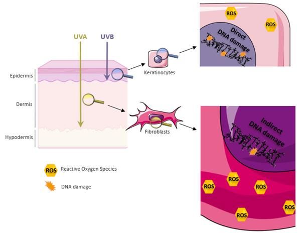
Immunology and Cancer Biology

fine-tuned and can determine the cell’s fate (survival, apoptosis
or carcinogenesis).

UV Radiation, Skin Penetration and Resulting
Damage
Solar UV radiation is sub-classified into UVA (320-400nm),
UVB (280-320nm) and UVC (200-280nm). UVC is absorbed by
the ozone layer rendering the UV spectrum reaching earth
restricted to 95% UVA and 5% UVB. These two solar rays allow
the induction of DNA lesions, thus playing a critical role in skin
carcinogenesis. The more energetic UVB can be absorbed by the
epidermis and superficial dermis; whereas the less energetic
more penetrating UVA can reach deep into the dermis.

Figure 1: UV skin penetration and molecular outcomes.
Out of solar rays, UVA and UVB can penetrate the skin to reach deep into the
dermis or be restricted to the epidermis respectively. UVB rays induce direct
damages to the DNA and the formation of reactive oxygen species (ROS).
UVA’s damage to the DNA is indirect via photosensitization reactions by ROS
produced at a high rate.

2                                                     www.videleaf.com
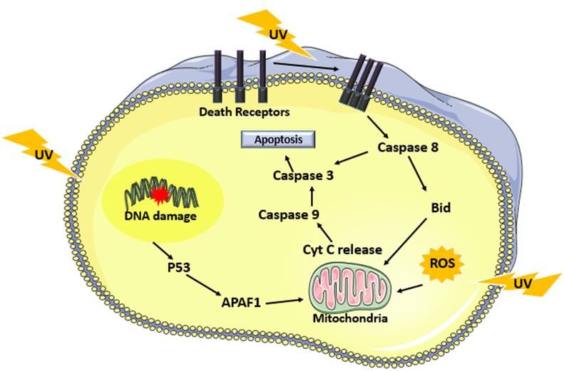
Immunology and Cancer Biology On one hand, UVB irradiation generates direct damages to the DNA in the form of dimeric pyrimidine photoproducts including Cyclobutane pyrimidine dimers (CPDs), 6-4 pyrimidine- pyrimidone photoproducts (6-4PPs) and Dewar isomers [3]. The latter are formed between two adjacent pyrimidine sites TT, CT, TC or CC. The 6-4 PPs are readily repaired in faster kinetics compared to CPDs where the TC and CC-CPD are the most mutagenic ones [4]. UVB also contributes to the generation of double-strand breaks via collapsing the replication forks at dimer sites and formation of reactive oxygen species (ROS) [5]. The generation of 8-hydroxy-2-deoxyguanosines (8-OHdG) was reported post UVB irradiation that leads to GT transversion. The majority of UVB-induced mutations are CT or CCTT transitions designated as the UVB signature mutation [6]. On the other hand, UVA damage to the DNA is indirect via reactive oxygen species that are generated at bigger amounts than UVB- induced ROS. The most common UVA-induced lesion is 8-oxo guanine. CPDs’ indirect formation post UVA are mediated by chemically generated excited electronic states [7]. In addition to DNA damage, ROS can also contribute to lipid peroxidation and protein oxidation (figure 1). UV-Induced Apoptosis Apoptosis is triggered in irradiated cells by three different mechanisms. UV-induced DNA damage favors the activation of ataxia telangiectasia and rad3 related (ATR), ataxia telangiectasia mutated (ATM) and DNA-PK kinases that ultimately activates p53 via checkpoint kinase ChK1/2 as part of the DNA damage response. p53 regulates the transcription of cell cycle protein p21 and several pro-apoptotic factors including APAF1, NOXA, PUMA, Bax and Bak. This contributes to the arrest of the cell cycle. If damage repair fails, P53 via downstream effectors the modulation of mitochondrial permeability to allow the release of cytochrome c. APAF1, procaspase 9 and cytochrome c favor the formation of the apoptosome leading to the activation of caspase 9 then caspase 3 [2]. Moreover, the production of reactive oxygen species post UV induces, in addition to DNA damage, lipid peroxidation, protein oxidation and the release of cytochrome c to mediate 3 www.videleaf.com
Immunology and Cancer Biology

another intrinsically triggered apoptotic pathway. One final
mechanism of apoptosis is mediated via the extrinsic effect of
UV in clustering death receptors, includng the tumor necrosis
factor (TNF) receptor superfamily members the CD95
(Fas/APO-1) or TRAIL, leading to the activation of a cascade of
caspases from caspase 8 till caspase 3 [8] (figure 2).

Figure 2: Mechanisms of UV induced apoptosis.
UV irradiation triggers cell apoptosis by three different mechanisms. The first
is mediated by p53 activation due to DNA damage that ultimately leads to the
activation of caspase 9 followed by caspase 3. Reactive oxygen species
generated by UV also induce apoptosis by enabling the release of cytochrome c
from the mitochondria. One final mechanism is via UV mediated clustering of
death receptors leading to the activation of caspase 8.

UV-Induced Signal Transduction
AKT Pathway

The full kinase activity of Akt is achieved on one hand by its
phosphorylation at two distinct sites, Thr308 phosphorylation via
receptor tyrosine kinase activated PI3K and Ser473 by mTOR
complex 2 (mTOR/Rictor; mTORC2) [9]. Its inhibition, on the
other hand, is achieved by the tumor suppressor PTEN [10]. UV
triggers tyrosine kinase receptors as well as the inhibition of
PTEN to enable Akt activation post-irradiation [11]. This further

4                                                       www.videleaf.com
Immunology and Cancer Biology

permits the activation of anti-apoptotic transcription factor NFκB
[10] and p53 negative regulator MDM2 [12]. The latter together
with Akt mediated inhibition of FOXO [13], pro-apoptotic
forkhead transcription factor, and apoptotic proteins Bad and
caspase 9 [14] infers an anti-apoptotic Akt mediated signal. Cell
cycle progression is also achieved via the inhibition of nuclear
translocation of cell cycle inhibitors p21 and p27 [15]. Akt also
increases metabolic activity via the phosphorylation and thus
inhibition of glycogen synthase kinase (GSK3) [16]. The
outcome involves the modulation of protein synthesis and
autophagy. The Akt-mediated inhibition of TSC2, tuberous
sclerosis protein 2, leads to activation of mTOR/Raptor
(mTORC1) that promotes translation via phosphorylation of
eukaryotic initiation factor 4E (eIF-4E) binding protein-1 (4E-
BP1) and p70/p85 S6 kinase (S6K) [17] (figure 3).

Figure 3: Physiological functions triggered by UV induced Akt signaling.
UV induced Akt activation can induce the activation of several downstream
cellular functions including inhibition of apoptosis, cell cycle progression,
metabolic activity inductions and translation. Detailed description available in
the text [2].

5                                                        www.videleaf.com

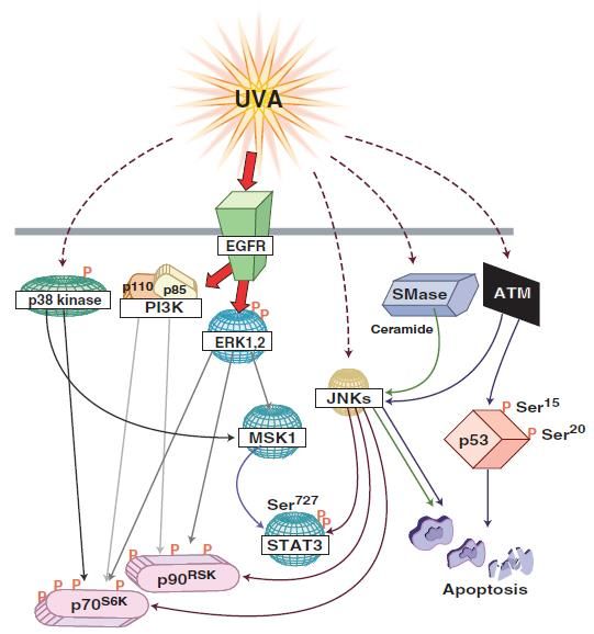
Immunology and Cancer Biology

MAPK Pathway

Another UV-activated pathway is the mitogen-activated protein
kinase (MAPK) pathway with final effectors including the c-
JUN NH2 terminal kinases (JNKs), the extracellular signal-
regulated kinases (ERKs) and p38 kinases that regulate the
activity of transcription factors NFκB and AP-1. The different
forms of UV irradiation (UVA, UVB, UVC) explicit different
modes of MAPK activation that is dose-dependent.

UVA induced MAPK signaling

UVA induces the activation of epidermal growth factor receptor
(EGFR) that then leads to the phosphorylation of the 40S
ribosomal protein S6 by p70S6K (70-kD ribosomal S6 kinase) and
p90RSK (90-Kd ribosomal S6 kinase, also known as MAPKAP-
K1) [18,19]. The activation of p70S6K, on one hand, is achieved
by different MAPK pathways phosphorylating four distinct sites.

PI3K activation induces the phosphorylation at Ser411, Thr421
and Ser424 while mTOR phosphorylates the Thr389 site. These
four sites are phosphorylated by ERK1/2. P38 phosphorylates
Thr389 and JNK added phosphate groups on both Ser411 and
Thr389 [20]. On the other hand, ERK and JNK but not p38
enable the phosphorylation/activation of p90RSK at Ser381 [21].
It should be noted that the inhibition of EGFR abrogated the UV
induced phosphorylation of ERK but not p38 and JNK implying
that EGFR mediated signaling to p90RSK and p70S6K is ERK
[22] and PI3K dependent. These ribosomal kinases regulate
various cellular functions like proliferation and differentiation.
Moreover, UVA irradiation can direct cellular decision towards
apoptosis triggered by p53 and JNK [23]. The latter being
activated by sphingomyelinase (SMase) and thus ceramide
hydrolysis [24] (figure 4).

6                                              www.videleaf.com
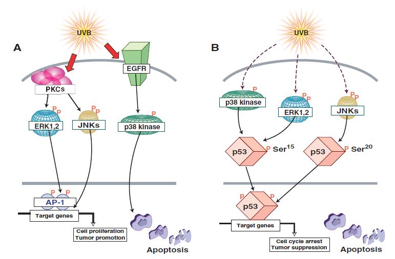
Immunology and Cancer Biology

Figure 4: UVA induced MAPK signaling.
UVA activates EGFR that leads to the phosphorylation/activation of several
downstream effectors including p70S6K and p90RSK. UVA mediated apoptosis is
mediated by the activation of JNK, ATM, or SMase [1].

UVB induced MAPK signaling
MAPK mediated UVB induced activation of AP-1 via PKC

UVB-induced activation of AP-1 implicates protein kinase C
(PKC) that favors activation of JNK and ERK [25]. ERK further
on mediates activation of AP-1 [26]. P38, however, was shown
to be activated in EGFR dependent mechanism post UVB and
that it further on led to apoptosis [27] (figure 5A).

7                                                    www.videleaf.com
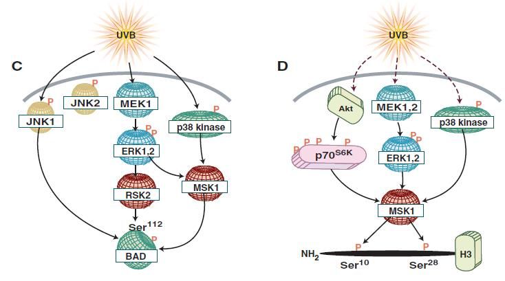
Immunology and Cancer Biology

MAPK-mediated UVB Induced Apoptosis

UVB-induced p53 dependent apoptosis is mediated via the
phosphorylation of the latter at Ser20 by JNK [28]. Nonetheless,
p38 and ERK phosphorylate p53 at Ser15 [29] (figure 5B).
Apoptosis is also mediated by the activation of the pro-apoptotic
protein BAD. The phosphorylation of BAD is carried out by
several kinases including JNK1, ERK downstream kinases RSK1
and MSK1 and by p38 kinase allowing BAD’s dissociation from
the anti-apoptotic Bcl-XL [30] (figure 5C).

MAPK mediated UVB Induced Chromatin Remodeling

The phosphorylation of nucleosome structural protein histone H3
regulates its role in enabling gene expression and chromatin
remodeling. Two phosphorylation sites are present on histone H3
which are the Ser10 and Ser28. Ser10 phosphorylation is
mediated via ERK and p38 [29] whereas Ser28’s
phosphorylation is mediated by JNK, ERK and p38 kinases
together with ERK and p38 downstream kinase MSK1 [31,32]
(figure 5D).

MAPK mediated UVB Induced Growth Control

Growth control regulation can be mediated at either the
transcriptional level via ribosomal kinases or at the translational
level by translation initiation factors. UVB activates PI3K
pathway triggering the phosphorylation of several downstream
effectors like Akt and p70S6K [33]. Akt UVB-induced
activation is mediated via MSK1 [34] (figure 5E). Moreover, the
p38 kinase through its downstream effector MSK1
phosphorylates eukaryotic initiation factor 4E (eIF-4E)-binding
protein (4E-BP1) allowing its dissociation from eIF-4F, relieving
the translational block and allowing cap-dependent translation
initiation [35] (figure 5F).

8                                              www.videleaf.com
Immunology and Cancer Biology

Figure 5: UVB induced MAPK signaling.
UVB can trigger different cellular functions via the activation of the diverse
MAPK effectors. (A) UVB can, on one hand, activate PKC leading to the
activation of AP-1 and, on the other hand, mediate the activation of p38 via
EGFR that leads to apoptosis. (B) P53 phosphorylation is achieved by both

9                                                      www.videleaf.com
Immunology and Cancer Biology ERK and p38 at Ser15 and by JNK on Ser15. (C) BAD phosphorylation at Ser112 is facilitated by JNK1, RSK2 and MSK1. (D) Histone H3 is phosphorylated at two sites: Ser10 by ERK and p38 kinase and at Ser28 by ERK, p38 and JNK. MSK1, a downstream effector of ERK and p38, also favors the phosphorylation at Ser28. (E) UVB mediated activation of Akt is PI3K dependent and MSK1 can also be involved in such activation. (F) 4EBP1 phosphorylation by the p38 kinase to MSK1 pathway favors its activation and dissociation from eIF-4E [1]. UV-Induced Mutations and Carcinogenesis p53 p53 plays a critical role in the cell cycle arrest post-stress induction. It activates cell cycle checkpoints halting cycle progression allowing enough time for DNA damage repair. If the repair was unsuccessful, p53 aids in the commitment of the cell to apoptosis by the expression of several pro-apoptotic proteins. UV-induced activation of p53 is mainly mediated via (ATR) that phosphorylates checkpoint kinases (chk1/2) ultimately leading to p53 phosphorylation at Ser15 and Ser20 [36]. Mutations in p53 can be mainly detected in squamous cell carcinoma and less in basal cell carcinoma. Such mutations carry the UV fingerprint C T transition [37]. Such mutations hinder the pro-apoptotic activity of p53 resulting in amplification of DNA damage accumulating cells. That combined with UVR-induced upregulation of heat shock proteins poses a combined effect on carcinogenesis evident via their co-localization with mutant p53 in squamous cell carcinoma [38]. PTEN Phosphate tensin homolog is a known inhibitor of the PI3K/Akt leading to the deregulation of cell proliferation and induction of apoptosis. Besides, PTEN also has a role in the regulation of DNA damage repair of NER and DSB. In the course of NER, Ming et al. showed that downregulation of PTEN expression in the epidermis of mice predisposes them to skin tumorigenesis upon UV irradiation [39]. One hypothesis involves the reduction of xeroderma pigmentosum C (XPC) expression upon PTEN downregulation via the Akt/p38 pathway. XPC is a DNA damage recognition protein in the nucleotide excision repair 10 www.videleaf.com
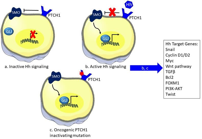
Immunology and Cancer Biology pathway required for the identification of the UV induced DNA damages. The decrease of XPC thus allows the accumulation of CPDs and 6-4PPs. In DSB, PTEN downregulation leads to decreased Rad51 expression [40]. PTCH PTCH gene encodes for a membrane receptor in the sonic hedgehog (Shh) pathway. The later plays an important role in regeneration and re-differentiation of adult tissue. The canonical signaling in the Shh pathway starts with the biding of the Shh ligand to the transmembrane receptor Patched (PTCH) inactivating it. This will relieve the PTCH-mediated inhibition of smoothened (SMO). SMO will further on activate the downstream signaling to result in the translocation of the Glioma zinc finger transcriptions factors (Gli) to the nucleus to initiate transcription [41]. Some of the downstream expressed genes are involved in tumorigenesis and they include BCL2 for apoptosis resistance, SNAIL for epithelial-mesenchymal transition, Wnt2 for cell stemness, and TGFβ for immune suppression among others [42]. PTCH mutations bear the UV signature CT or CCTT transitions. The latter allows the inactivation of PTCH and its role in the inhibition of smoothened (SMO) expression. Therefore, SMO can then mediate the activation of Gli1 transcriptional factor resulting in tumor formation [43]. Such deficiency was recorded in 85% of BCC [44]. Gorlin syndrome is characterized with an increased predisposition to basal cells carcinomas due to a mutation in PTCH1 tumor suppressor gene. Charazac et al. reported that fibroblasts from Gorlin patients exhibit high radio-sensitivity. These cells appeared to manifest low expression and activity of base excision repair pathway proteins thus linking Gorlin clinical manifestations to possible defects in DNA damage response or repair capacity [45]. NRAS and BRAF NRAS and BRAF play a major role in the mitogen-activated protein kinase (MAPK) pathway. The binding of growth factor to their respective tyrosine kinase receptor activates this pathway. RAS GTPase then conveys the intracellular signaling. 11 www.videleaf.com
Immunology and Cancer Biology Active Ras bound to GTP allows the recruitment of effectors including the RAF family of serine/threonine kinases that regulate cell proliferation. RAF further on activates MAP kinase kinase (MEK1/2) and then MAP kinase (ERK1/2). The latter promotes cell proliferation [46]. Activating mutations in these genes are mainly found in melanoma. Only a single mutation in either gene can exist at a time [47]. Mutations in either are of the activating type enhancing the MAPK signaling. BRAF most frequent mutation is a TA transversion.[48]. NRAS most frequent mutation is either a CA transversion or AG transitions in sun-exposed areas [49]. These mutations are not exactly a CT UV signature mutation, however, their comparison with the mutagenic profile in bacteria reveals that they may be derived from UVA mediated oxidative DNA damage [50]. Figure 6: The sonic hedgehog pathway and the effect of UV irradiation. The SHH pathway controls the expression of several downstream effectors. a) In the inactive state, PTCH receptor inhibits SMO and thus preventing the translocation of Gli to the nucleus blocking transcription of underlying genes. b) However, in the presence of the Hh ligand the PTCH receptor becomes inactive losing its inhibitory effect on SMO favoring the activity of Gli in transcription. c) Inactivating mutation of PTCH induced by UV irradiation relieve SMO inhibition and activates the pathway to express downstream genes via Gli. SHH: sonic hedgehog, PTCH: patched receptor, SMO: smoothened, Gli: glioma transcription factor 12 www.videleaf.com
Immunology and Cancer Biology RAC1 and PREX2 PREX2 is a GTP/GDP exchange factor that can promote tumorigenesis via the inhibition of PTEN, a known regulator of the PI3K signaling pathway [51]. PREX2 also mediates the activation of the GTPase RAC involved in cell migration [52]. Large scale cancer genomic profiling revealed the involvement of mutations in these two genes with the melanoma oncogenesis [53, 54]. These genes interact to promote the PI3K/Akt [52, 55]. Mutations in the RAC1 gene possess the typical UV signature CT transition mutation in sun-exposed areas that allows the activation of RAC1 and the underlying signaling pathways [56]. PREX2 mutations are also mainly found in sun-exposed areas [54] where it inhibits PTEN annulling its effect in the inhibition of the PI3K pathway. Although other genes have been recorded to be involved in skin carcinogenesis, they do not possess a UV signature mutation profile signifying a different origin other than UV as their carcinogenesis initiator. The latter includes mutations in the cell cycle genes found in both basal and squamous cell carcinoma [57]. In addition, loss of p16INK4A was also identified in melanoma leading to the enhancement of CDK4 activity promoting proliferation [58]. UV-Irradiation Induced Immunosuppression Genetic mutations alone do not represent the only initiators of skin tumorigenesis. An intricate signaling network interplays with such mutations leading ultimately to tumor generation one of which is the UV irradiation-induced immune suppression. The first form of suppression is mediated via the defective antigen presentation activity of Langerhans cells. UVR leads to the elevation of IL6, IL10 and TNFα levels thus inhibiting the skin immune response by downregulating Langerhans cells activity [59]. Moreover, UV irradiation was found to have a negative effect on the number of natural killer cells in a UV dose- dependent manner. The latter cells are implicated in anti-tumor and anti-viral infection [60]. UVB exposure also increases the expression of cyclooxygenase (COX-2) enzyme that catalyzes 13 www.videleaf.com
Immunology and Cancer Biology the first step for the conversion of arachidonic acid to active prostaglandin involved in the inflammatory response. COX-2 is implicated in the development of several types of tumors [61]. Finally, one last activator of immune suppression is urocanic acid (UCA), a histamine deamination product highly available in the skin [62]. The inhibition of UCA with an antibody against it enabled the delay of tumor formation in irradiated skin [63]. Conclusion Despite the filtration of the harmful UVC radiation by the ozone layer, the two remaining solar ultraviolet rays reaching the earth’s surface are proving to be hazardous. UVB radiation can directly damage the DNA of exposed cells while UVA can indirectly create such damage via the generation of reactive oxygen species. After such induced stress, the cells that fail to repair the damage can either be committed to apoptosis or persist where their damaged DNA progresses to the UV signature CT transition. These mutations have been identified in several genes correlated with skin carcinogenesis signifying a link between UV exposure and tumorigenesis. Moreover, UV irradiation activates several downstream signaling pathways including the Akt and MAPK pathways implicated in the regulation of cell proliferation, apoptosis and tumor promotion among many others. Finally, an additional instigator of tumor formation can be the UV-induced immune suppression where the inhibition of the action of some immune cells allows the cells’ harboring damage and tumors escape from the surveillance of the body. For that, possessing an active repair system is essential to eliminate such dangerous UV-induced lesions where any defect will give rise to diseases like Xeroderma Pigmenstosum and others with increased risks of skin cancers and neurodegeneration. 14 www.videleaf.com
Immunology and Cancer Biology

References
1. Bode A, AM, Z Dong. Mitogen-activated protein kinase
   activation in UV-induced signal transduction. Sci STKE.
   2003; 2003: Re2.
2. Strozyk E, D Kulms. The role of AKT/mTOR pathway in
   stress response to UV-irradiation: implication in skin
   carcinogenesis by regulation of apoptosis, autophagy and
   senescence. Int. J. Mol. Sci. 2013; 14: 15260-15285.
3. Ravanat JL, T Douki, J Cadet. Direct and indirect effects of
   UV radiation on DNA and its components. J Photochem
   Photobiol B. 2001; 63: 88-102.
4. Seebode C, J Lehmann, S Emmert. Photocarcinogenesis and
   Skin Cancer Prevention Strategies. Anticancer Res. 2016;
   36: 1371-1378.
5. Rajesh P Rastogi, Shailendra P Singh, Donat-P Häder,
   Rajeshwar P Sinha. Detection of reactive oxygen species
   (ROS)      by      the    oxidant-sensing     probe    2',7'-
   dichlorodihydrofluorescein diacetate in the cyanobacterium
   Anabaena variabilis PCC 7937. Biochem Biophys Res
   Commun. 2010; 397: 603-607.
6. Armstrong JD, BA Kunz. Site and strand specificity of UVB
   mutagenesis in the SUP4-o gene of yeast. Proc Natl Acad Sci
   U S A. 1990; 87: 9005-9009.
7. George J Delinasios, Mahsa Karbaschi, Marcus S Cooke,
   Antony R Young. Vitamin E inhibits the UVAI induction of
   "light" and "dark" cyclobutane pyrimidine dimers, and
   oxidatively generated DNA damage, in keratinocytes. Sci
   Rep, 2018; 8: 423.
8. Chih-Hung Lee, Shi-Bei Wu, Chien-Hui Hong, Hsin-Su Yu,
   Yau-Huei Wei. Molecular Mechanisms of UV-Induced
   Apoptosis and Its Effects on Skin Residential Cells: The
   Implication in UV-Based Phototherapy. Int J Mol Sci, 2013;
   14: 6414-6435.
9. Mihail S Iordanov, Remy J Choi, Olga P Ryabinina, Thanh-
   Hoai Dinh, Robert K Bright,et al. The UV (Ribotoxic) stress
   response of human keratinocytes involves the unexpected
   uncoupling of the Ras-extracellular signal-regulated kinase
   signaling cascade from the activated epidermal growth factor
   receptor. Mol Cell Biol, 2002; 22: 5380-5394.

15                                            www.videleaf.com
Immunology and Cancer Biology

10. Ichiro Yajima, Mayuko Y Kumasaka, Nguyen Dinh Thang,
    Yuji Goto, Kozue Takeda, et al. RAS/RAF/MEK/ERK and
    PI3K/PTEN/AKT Signaling in Malignant Melanoma
    Progression and Therapy. Dermatol Res Pract, 2012; 2012:
    354191.
11. M Ming, W Han, J Maddox, K Soltani, CR Shea, et al.
    UVB-induced ERK/AKT-dependent PTEN suppression
    promotes survival of epidermal keratinocytes. Oncogene.
    2010; 29: 492-502.
12. Tanya M Gottlieb, Juan Fernando Martinez Leal, Rony
    Seger, Yoichi Taya, Moshe Oren. Cross-talk between Akt,
    p53 and Mdm2: possible implications for the regulation of
    apoptosis. Oncogene. 2002; 21: 1299-1303.
13. Pascale F Dijkers, Kim U Birkenkamp, Eric W-F Lam, N
    Shaun B Thomas, Jan-Willem J Lammers, et al. FKHR-L1
    can act as a critical effector of cell death induced by cytokine
    withdrawal: protein kinase B-enhanced cell survival through
    maintenance of mitochondrial integrity. J Cell Biol. 2002;
    156: 531-542.
14. SR Datta, H Dudek, X Tao, S Masters, H Fu, et al. Akt
    phosphorylation of BAD couples survival signals to the cell-
    intrinsic death machinery. Cell. 1997; 91: 231-241.
15. Franke TF. PI3K/Akt: getting it right matters. Oncogene.
    2008; 27: 6473-6488.
16. Naihan Xu, Yuanzhi Lao, Yaou Zhang, David A Gillespie.
    Akt: a double-edged sword in cell proliferation and genome
    stability. J.Oncol. 2012; 2012: 951724.
17. Ma XM, J Blenis. Molecular mechanisms of mTOR-
    mediated translational control. Nat Rev Mol Cell Biol. 2009;
    10: 307-318.
18. Dufner A, G Thomas. Ribosomal S6 kinase signaling and the
    control of translation. Exp Cell Res. 1999; 253: 100-109.
19. Frodin M, S Gammeltoft. Role and regulation of 90 kDa
    ribosomal S6 kinase (RSK) in signal transduction. Mol Cell
    Endocrinol. 1999; 151: 65-77.
20. Y Zhang, Z Dong, M Nomura, S Zhong, N Chen, et al.
    Signal transduction pathways involved in phosphorylation
    and activation of p70S6K following exposure to UVA
    irradiation. J Biol Chem. 2001; 276: 20913-20923.

16                                               www.videleaf.com
Immunology and Cancer Biology
21. Y Zhang, S Zhong, Z Dong, N Chen, AM Bode, et al. UVA
induces Ser381 phosphorylation of p90RSK/MAPKAP-K1
via ERK and JNK pathways. J Biol Chem. 2001; 276:
14572-14580.
22. Y Zhang, Z Dong, AM Bode, WY Ma, N Chen, et al.
Induction of EGFR-dependent and EGFR-independent
signaling pathways by ultraviolet A irradiation. DNA Cell
Biol. 2001; 20: 769-779.
23. Yiguo Zhang, Wei-Ya Ma, Akira Kaji, Ann M Bode, Zigang
Dong. Requirement of ATM in UVA-induced signaling and
apoptosis. J Biol Chem. 2002; 277: 3124-3131.
24. Y Zhang, P Mattjus, PC Schmid, Z Dong, S Zhong, et al.
Involvement of the acid sphingomyelinase pathway in uva-
induced apoptosis. J Biol Chem, 2001; 276: 11775-11782.
25. N Chen, Wy Ma, C Huang, Z Dong. Translocation of protein
kinase Cepsilon and protein kinase Cdelta to membrane is
required for ultraviolet B-induced activation of mitogen-
activated protein kinases and apoptosis. J Biol Chem. 1999;
274: 15389-15394.
26. C Huang, J Li, N Chen, W Ma, GT Bowden, et al. Inhibition
of atypical PKC blocks ultraviolet-induced AP-1 activation
by specifically inhibiting ERKs activation. Mol Carcinog.
2000; 27: 65-75.
27. S Nakamura, H Takahashi, M Kinouchi, A Manabe, A
Ishida-Yamamoto, et al. Differential phosphorylation of
mitogen-activated protein kinase families by epidermal
growth factor and ultraviolet B irradiation in SV40-
transformed human keratinocytes. J Dermatol Sci. 2001; 25:
139-149.
28. She QB, WY Ma, Z Dong. Role of MAP kinases in UVB-
induced phosphorylation of p53 at serine 20. Oncogene.
2002; 21: 1580-1589.
29. She QB, N Chen, Z Dong. ERKs and p38 kinase
phosphorylate p53 protein at serine 15 in response to UV
radiation. J Biol Chem. 2000; 275: 20444-20449.
30. Qing-Bai She, Wei-Ya Ma, Shuping Zhong, Zigang Dong.
Activation of JNK1, RSK2, and MSK1 is involved in serine
112 phosphorylation of Bad by ultraviolet B radiation. J Biol
Chem. 2002; 277: 24039-24048.
17 www.videleaf.comImmunology and Cancer Biology
31. S Zhong, Y Zhang, C Jansen, H Goto, M Inagaki,et al. MAP
kinases mediate UVB-induced phosphorylation of histone
H3 at serine 28. J Biol Chem. 2001; 276: 12932-12937.
32. S Zhong, C Jansen, QB She, H Goto, M Inagaki,et al.
Ultraviolet B-induced phosphorylation of histone H3 at
serine 28 is mediated by MSK1. J Biol Chem. 2001; 276:
33213-33219.
33. M Nomura, A Kaji, Z He, WY Ma, K Miyamoto, et al.
Inhibitory mechanisms of tea polyphenols on the ultraviolet
B-activated phosphatidylinositol 3-kinase-dependent
pathway. J Biol Chem. 2001; 276: 46624-46631.
34. M Nomura, A Kaji, WY Ma, S Zhong, G Liu, et al. Mitogen-
and stress-activated protein kinase 1 mediates activation of
Akt by ultraviolet B irradiation. J Biol Chem. 2001; 276:
25558-25567.
35. Guangming Liu, Yiguo Zhang, Ann M Bode, Wei-Ya Ma,
Zigang Dong. Phosphorylation of 4E-BP1 is mediated by the
p38/MSK1 pathway in response to UVB irradiation. J Biol
Chem. 2002; 277: 8810-8816.
36. Masaoki Kawasumi, Bianca Lemos, James E Bradner, Renee
Thibodeau, Yong-son Kim, et al. Protection from UV-
induced skin carcinogenesis by genetic inhibition of the
ataxia telangiectasia and Rad3-related (ATR) kinase.
Proceedings of the National Academy of Sciences. 2011;
108: 13716-13721.
37. Kumar R, G Deep, R Agarwal. An Overview of Ultraviolet B
Radiation-Induced Skin Cancer Chemoprevention by
Silibinin. Curr Pharmacol Rep. 2015; 1: 206-215.
38. Ingela Kindas-Mügge, Constanze Rieder, Ilse Fröhlich,
Michael Micksche, Franz Trautinger. Characterization of
proteins associated with heat shock protein HSP27 in the
squamous cell carcinoma cell line A431. Cell Biology
International. 2002; 26: 109-116.
39. Mei Ming, Li Feng, Christopher R Shea, Keyoumars Soltani,
Baozhong Zhao, et al. PTEN positively regulates UVB-
induced DNA damage repair. Cancer Res. 2011; 71: 5287-
5295.
40. Wen Hong Shen, Adayabalam S Balajee, Jianli Wang, Hong
Wu, Charis Eng,et al. Essential role for nuclear PTEN in
18 www.videleaf.comImmunology and Cancer Biology
maintaining chromosomal integrity. Cell. 2007; 128: 157-
170.
41. Gabriela Basile Carballo, Jéssica Ribeiro Honorato, Giselle
Pinto Farias de Lopes, Tania Cristina Leite de Sampaio E
Spohr et al. A highlight on Sonic hedgehog pathway. Cell
Commun Signal. 2018; 16: 11.
42. Jia Y, Y Wang, J Xie. The Hedgehog pathway: role in cell
differentiation, polarity and proliferation. Arch Toxicol.
2015; 89: 179-191.
43. Fahimeh Rahnama, Takashi Shimokawa, Matthias Lauth,
Csaba Finta, Priit Kogerman,et al. Inhibition of GLI1 gene
activation by Patched1. Biochem J. 2006; 394: 19-26.
44. Terumi Mizuno, Shoji Tokuoka, Masao Kishikawa, Eiji
Nakashima, Kiyohiko Mabuchi, et al. Molecular basis of
basal cell carcinogenesis in the atomic-bomb survivor
population: p53 and PTCH gene alterations. Carcinogenesis.
2006; 27: 2286-2294.
45. Aurélie Charazac, Nour Fayyad, David Beal, Sandrine
Bourgoin-Voillard, Michel Seve, et al. Impairment of Base
Excision Repair in Dermal Fibroblasts Isolated From Nevoid
Basal Cell Carcinoma Patients. Front Oncol. 2020; 10: 1551.
46. Nicolas Dumaz, Fanélie Jouenne, Julie Delyon, Samia
Mourah, Armand Bensussan, et al. Atypical BRAF and
NRAS Mutations in Mucosal Melanoma. Cancers (Basel).
2019; 11.
47. Marcia S Brose, Patricia Volpe, Michael Feldman, Madhu
Kumar, Irum Rishi, et al. BRAF and RAS mutations in
human lung cancer and melanoma. Cancer Res. 2002; 62:
6997-7000.
48. John A Curtin, Jane Fridlyand, Toshiro Kageshita, Hetal N
Patel, Klaus J Busam, et al. Distinct sets of genetic
alterations in melanoma. N Engl J Med. 2005; 353: 2135-
2147.
49. Katarina Omholt, Anton Platz, Lena Kanter, Ulrik Ringborg,
Johan Hansson. NRAS and BRAF mutations arise early
during melanoma pathogenesis and are preserved throughout
tumor progression. Clin Cancer Res. 2003; 9: 6483-6488.
50. KC Cheng, DS Cahill, H Kasai, S Nishimura, LA Loeb. 8-
Hydroxyguanine, an abundant form of oxidative DNA
19 www.videleaf.comImmunology and Cancer Biology
damage, causes G----T and A----C substitutions. J Biol
Chem. 1992; 267: 166-172.
51. Barry Fine, Cindy Hodakoski, Susan Koujak, Tao Su, Lao H
Saal, et al. Activation of the PI3K pathway in cancer through
inhibition of PTEN by exchange factor P-REX2a. Science.
2009; 325: 1261-1265.
52. Sarah M Mense, Douglas Barrows, Cindy Hodakoski, Nicole
Steinbach, David Schoenfeld, et al. PTEN inhibits PREX2-
catalyzed activation of RAC1 to restrain tumor cell invasion.
Sci Signal. 2015; 8: ra32.
53. Eran Hodis, Ian R Watson, Gregory V Kryukov, Stefan T
Arold, Marcin Imielinski, et al. A landscape of driver
mutations in melanoma. Cell. 2012; 150: 251-263.
54. Michael F.Berger, Eran Hodis, Timothy P Heffernan,
Yonathan Lissanu Deribe, Michael S Lawrence, et al.
Melanoma genome sequencing reveals frequent PREX2
mutations. Nature. 2012; 485: 502-506.
55. Douglas Barrows, Sarah M Schoenfeld, Cindy Hodakoski,
Antonina Silkov, Barry Honig, et al. p21-activated Kinases
(PAKs) Mediate the Phosphorylation of PREX2 Protein to
Initiate Feedback Inhibition of Rac1 GTPase. J Biol Chem.
2015; 290: 28915-28931.
56. Halaban R. RAC1 and melanoma. Clin Ther. 2015; 37: 682-
685.
57. Isabelle Conscience, Nicolas Jovenin, Christelle Coissard,
Marianne Lorenzato, Anne Durlach, et al. P16 is
overexpressed in cutaneous carcinomas located on sun-
exposed areas. European Journal of Dermatology. 2006; 16:
518-522.
58. Amaya Viros, Berta Sanchez-Laorden, Malin Pedersen,
Simon J Furney, Joel Rae, et al. Ultraviolet radiation
accelerates BRAF-driven melanomagenesis by targeting
TP5. Nature. 2014; 511: 478-482.
59. C Petit-Frère, P H Clingen, M Grewe, J Krutmann, L Roza,
et al. Induction of interleukin-6 production by ultraviolet
radiation in normal human epidermal keratinocytes and in a
human keratinocyte cell line is mediated by DNA damage. J
Invest Dermatol. 1998; 111: 354-359.
60. Hiroko Miyauchi-Hashimoto, Akira Sugihara, Kiyoji
Tanaka, Takeshi Horio. Ultraviolet radiation-induced
20 www.videleaf.comImmunology and Cancer Biology
impairment of tumor rejection is enhanced in xeroderma
pigmentosum a gene-deficient mice. J Invest Dermatol.
2005; 124: 1313-1317.
61. M Athar, KP An, KD Morel, AL Kim, M Aszterbaum, et al.
Ultraviolet B(UVB)-induced cox-2 expression in murine
skin: an immunohistochemical study. Biochem Biophys Res
Commun. 2001; 280: 1042-1047.
62. De Fabo EC, FP Noonan. Mechanism of immune
suppression by ultraviolet irradiation in vivo. I. Evidence for
the existence of a unique photoreceptor in skin and its role in
photoimmunology. J Exp Med. 1983; 158: 84-98.
63. S Beissert, D Rühlemann, T Mohammad, S Grabbe, A El-
Ghorr, et al. IL-12 prevents the inhibitory effects of cis-
urocanic acid on tumor antigen presentation by Langerhans
cells: implications for photocarcinogenesis. J Immunol.
2001; 167: 6232-6238.
21 www.videleaf.comYou can also read