Computational design of transmembrane pores - Baker Lab
←
→
Page content transcription
If your browser does not render page correctly, please read the page content below
Article

Computational design of transmembrane
pores

https://doi.org/10.1038/s41586-020-2646-5                       Chunfu Xu1,2,3,12, Peilong Lu1,2,4,5,12 ✉, Tamer M. Gamal El-Din6, Xue Y. Pei7, Matthew C. Johnson2,
                                                                Atsuko Uyeda8, Matthew J. Bick1,2,10, Qi Xu4,5, Daohua Jiang6, Hua Bai1,2, Gabriella Reggiano1,2,
Received: 24 August 2019
                                                                Yang Hsia1,2, TJ Brunette1,2, Jiayi Dou1,2,11, Dan Ma4,5,9, Eric M. Lynch2, Scott E. Boyken1,2,10,
Accepted: 29 May 2020                                           Po-Ssu Huang1,2,11, Lance Stewart1, Frank DiMaio1,2, Justin M. Kollman2, Ben F. Luisi7,
                                                                Tomoaki Matsuura8, William A. Catterall6 ✉ & David Baker1,2,3 ✉
Published online: xx xx xxxx

    Check for updates
                                                                Transmembrane channels and pores have key roles in fundamental biological
                                                                processes1 and in biotechnological applications such as DNA nanopore sequencing2–4,
                                                                resulting in considerable interest in the design of pore-containing proteins. Synthetic
                                                                amphiphilic peptides have been found to form ion channels5,6, and there have been
                                                                recent advances in de novo membrane protein design7,8 and in redesigning naturally
                                                                occurring channel-containing proteins9,10. However, the de novo design of stable,
                                                                well-defined transmembrane protein pores that are capable of conducting ions
                                                                selectively or are large enough to enable the passage of small-molecule fluorophores
                                                                remains an outstanding challenge11,12. Here we report the computational design of
                                                                protein pores formed by two concentric rings of α-helices that are stable and
                                                                monodisperse in both their water-soluble and their transmembrane forms. Crystal
                                                                structures of the water-soluble forms of a 12-helical pore and a 16-helical pore closely
                                                                match the computational design models. Patch-clamp electrophysiology
                                                                experiments show that, when expressed in insect cells, the transmembrane form of
                                                                the 12-helix pore enables the passage of ions across the membrane with high
                                                                selectivity for potassium over sodium; ion passage is blocked by specific chemical
                                                                modification at the pore entrance. When incorporated into liposomes using in vitro
                                                                protein synthesis, the transmembrane form of the 16-helix pore—but not the 12-helix
                                                                pore—enables the passage of biotinylated Alexa Fluor 488. A cryo-electron
                                                                microscopy structure of the 16-helix transmembrane pore closely matches the design
                                                                model. The ability to produce structurally and functionally well-defined
                                                                transmembrane pores opens the door to the creation of designer channels and pores
                                                                for a wide variety of applications.

Recent work on de novo membrane protein design has focused on                                   membrane and because of the possibility of undesired interactions
compact bundles of helices without large central cavities7,8. De novo                           between the non-polar residues introduced to interact with lipids and
design of transmembrane proteins with large pores presents a more                               the residues at intersubunit interfaces. For example, the oligomeriza-
stringent thermodynamic challenge, because such proteins have a                                 tion state of coiled-coil assemblies has been shown to be very sensitive
larger surface-area-to-volume ratio and therefore a lower density of                            to the placement of non-polar residues14, and so non-polar residues
stabilizing interactions. A homo-oligomeric architecture in which a                             incorporated to interact with the bilayer could substantially alter the
cyclic arrangement of several identical copies of a single subunit sur-                         oligomeric state.
rounds the pore—as found in many natural protein pores—is attractive                               We set out to design transmembrane protein pores using a two-step
for protein design, because the subunit need not be a large protein.                            approach. We reasoned that protein pores formed from two concentric
Soluble single-ring coiled-coil assemblies of peptide helices that sur-                         rings of α-helices containing buried hydrogen-bond networks for struc-
round central pores have been designed13, but the transformation of                             tural specificity could be stable both in the water-soluble form and, after
soluble oligomeric protein pores into their membrane counterparts is                            resurfacing the lipid-exposed residues with membrane-compatible
challenging because of the altered thermodynamics of folding in the                             hydrophobic residues, in the transmembrane form. The increased

Institute for Protein Design, University of Washington, Seattle, WA, USA. 2Department of Biochemistry, University of Washington, Seattle, WA, USA. 3Howard Hughes Medical Institute,
1

University of Washington, Seattle, WA, USA. 4Zhejiang Provincial Laboratory of Life Sciences and Biomedicine, Key Laboratory of Structural Biology of Zhejiang Province, School of Life
Sciences, Westlake University, Hangzhou, China. 5Institute of Biology, Westlake Institute for Advanced Study, Hangzhou, China. 6Department of Pharmacology, University of Washington,
Seattle, WA, USA. 7Department of Biochemistry, University of Cambridge, Cambridge, UK. 8Department of Biotechnology, Graduate School of Engineering, Osaka University, Suita, Japan.
9
 Department of Biological Structure, University of Washington, Seattle, WA, USA. 10Present address: Lyell Immunopharma, Inc., Seattle, WA, USA. 11Present address: Department of
Bioengineering, Stanford University, Stanford, CA, USA. 12These authors contributed equally: Chunfu Xu, Peilong Lu. ✉e-mail: lupeilong@westlake.edu.cn; wcatt@uw.edu; dabaker@uw.edu

                                                                                                                                                     Nature | www.nature.com | 1
Article
interhelical interaction surfaces in such two-ring bundles could result       fraction with detergent, and purified by affinity chromatography and
in greater stability compared with single-ring structures—particularly        SEC (Extended Data Fig. 2c). TMHC6 eluted as a single peak during SEC,
for larger pore sizes—and, together with buried hydrogen-bond net-            and circular dichroism measurements showed that the structure is
works, provide greater structural robustness to conversion from               α-helical and has a high thermal stability, with a circular dichroism spec-
soluble to transmembrane forms. We chose to focus on the design of            trum at 95 °C similar to that at 25 °C (Fig. 1d). The protein sedimented as
homo-oligomeric cylindrical two-ring bundles consisting of a mini-            a hexamer in detergent solution in AUC experiments, consistent with
mum of 12 closely packed helices—as parametrically designed two-ring          the design model (Fig. 1e). Electron microscopy of negatively stained
bundles of ten or fewer helices generally do not contain pores large          samples showed populations of particles that had shapes and sizes
enough for solutes to traverse—using subunits containing between              consistent with the design model (Extended Data Fig. 3c).
2 and 4 helices to avoid having to build such large structures from a            To investigate ion conductance by TMHC6, we performed whole-cell
single polypeptide.                                                           patch-clamp experiments on Trichoplusia ni insect cells (Hi5) express-
                                                                              ing the designed transmembrane proteins. Using extracellular (bath)
                                                                              and intracellular (electrode) solutions containing 100 mM NaCl and
Designing an ion channel                                                      100 mM NaF, respectively, the TMHC6 construct exhibited a symmetric
We first explored the design of water-soluble pore containing helical         current–voltage relationship for inward and outward sodium-ion (Na+)
bundles with a two-ring, six-fold symmetric topology, formed from             current as a function of membrane potential (Fig. 1f). Gadolinium ions
monomeric subunits composed of an inner and an outer helix that               (Gd3+)—a potent blocker of cation channels—blocked TMHC6 from the
are bridged with a short loop. Backbones were generated by sampling           extracellular side, reducing the ion conductance to nearly the control
α-helical and superhelical parameters using a generalization of the           value for untransfected cells (Fig. 1f). To test the ion selectivity, Hi5 cells
Crick coiled-coil parameterization15–17 (see Supplementary Informa-           expressing TMHC6 were bathed in a solution containing 100 mM of
tion). After backbone generation and loop closure, we searched for            the chloride salt of the monovalent cations of potassium (K+), Na+ and
hydrogen-bond networks across the intermolecular interfaces using             caesium (Cs+), and the methylammonium ion (CH3NH3+). The patch
Rosetta HBNet18 and carried out combinatorial sequence optimiza-              pipettes contained the equivalent concentration of the fluoride salt of
tion for the remaining residue positions, keeping the polar networks          the same cation, except for CH3NH3+, for which the pipette contained
found by HBNet fixed. The interfacial core residues and backbone              100 mM caesium fluoride (CsF). TMHC6 showed a significantly higher
parameters of the inner helices were set to those of a previously char-       conductance for K+, with a current density of 600 pA pF−1 at +100 mV.
acterized ‘single-ring’ helical bundle13 to reduce the design space.          The selectivity order was K+ (600 pA pF−1) ≫ CH3NH3+ = Cs+ (170 pA pF−1) >
Rosetta ‘fold-and-dock’19 structure prediction calculations were used         Na+ (60 pA pF−1) ≈ Ba2+ (54 pA pF−1) (Fig. 1g). By comparison, a previously
to investigate the extent to which the designed sequences encode the          designed transmembrane protein (TMHC2) without a pore8 yielded a
design target topologies. Designs for which the lowest-energy sampled         current density for K+ of around 40 pA pF−1 at +100 mV, which was close
structures were close to the target design structures (Extended Data          to the background value (Extended Data Fig. 3d).
Fig. 1a) were selected for experimental characterization.                        In the design model, the extracellular ring of six Glu44 residues is the
   We obtained synthetic genes encoding the selected designs and              site of cation entry (Fig. 2a). We tested this hypothesis by site-directed
expressed them in Escherichia coli. All three hexameric designs selected      mutagenesis and chemical modification. Mutation of Glu44 to pheny-
for experimental validation were well expressed and soluble in E. coli,       lalanine (E44F) removed the negative charge at the extracellular entry
and could be efficiently purified using nickel-affinity chromatography        to the pore and reduced the conductance to 308 ± 81 pA pF−1 (mean ±
and size-exclusion chromatography (SEC). Multiangle light scattering          s.e.m.), 51.3% of the control value (Extended Data Fig. 3d). This reduc-
(MALS, Extended Data Fig. 2a) and analytical ultracentrifugation (AUC,        tion probably results from a direct effect on ion traversal through the
Extended Data Fig. 1c) indicated that one of the designs formed a single      pore: TMHC6(E44F) is expressed at a similar level to the TMHC6 parent
homogeneous species with the target oligomeric state. The circular            (Extended Data Fig. 3f), and the purified mutant protein has a similar
dichroism spectrum of this hexameric design (denoted WSHC6, for               solution behaviour to that of TMHC6 (Extended Data Fig. 2c). To further
water-soluble hairpin C6) showed that the structure was highly helical        test the importance of this site, we constructed the E44H mutant in
and stable to thermal denaturation up to 95 °C (Extended Data Fig. 1b).       which Glu44 is mutated to histidine—removing the negative charge in
The small-angle X-ray scattering (SAXS) profile of WSHC6 was close to         this position and adding a partial positive charge. The divalent cation
that computed from the design model, suggesting that WSHC6 folds              Cd2+ blocks the conductance of TMHC6, and threefold higher Cd2+ con-
into the desired shape in solution (Extended Data Fig. 1d). We deter-         centrations were required to block TMHC6(E44H), probably because of
mined the crystal structure of WSHC6 and found it to closely match            the reduced electrostatic attraction and/or disrupted metal coordina-
the computational design model (Fig. 1a) with a Cα root-mean-square           tion in the mutant protein (Fig. 2b). To enable a chemical modification
deviation (r.m.s.d.) of 0.89 Å. A chain of discrete water molecules           approach, we constructed the E44C mutant in which Glu44 is mutated
occupies the WSHC6 channel (Fig. 1b, Extended Data Fig. 1e, Extended          to cysteine; this removed the negative charge at the extracellular entry
Data Table 1); the narrowest constriction is at Leu51 with a diameter of      to the pore and reduced the conductance to 360 ± 36 pA pF−1, 60%
approximately 4 Å, as calculated by HOLE20.                                   of the control. We then took advantage of the chemical reactivity of
   We next sought to convert the stable and structurally well-defined         the substituted cysteine residue to test sulfhydryl reagents as pore
WSHC6 pore into a transmembrane hexameric pore (denoted TMHC6,                blockers. We found that perfusion of three different methanethiosul-
for transmembrane hairpin C6). We redesigned the lipid-exposed resi-          fonates—the negatively charged MTSES, the positively charged MTSET
dues (see Supplementary Information) and incorporated one ring of six         and the hydrophobic MTS-TBAE—all completely blocked the ion con-
glutamate residues (E-ring) and two rings of lysine residues (K-rings) at     ductance of TMHC6(E44C) within a few minutes under voltage-clamp
the openings of the central channel on the extracellular and intracel-        control (Fig. 2c, d). The lack of dependence on charge or hydrophilicity
lular side, respectively, to increase the polarity of the pore entrance       suggests that these compounds function by direct steric blocking of
and exit (Fig. 1c, Extended Data Fig. 3a, b). Similar rings are observed in   the pore. The blockage by these reagents is entirely dependent on the
the calcium channel Orai21,22. The narrowest regions of the TMHC6 pore        introduced cysteine residue: they had no effect on the original TMHC6
in the design model are the E-ring (3.3 Å), the K-rings (4.3 Å and 5.7 Å)     design that lacks the cysteine (Fig. 2c, d). To determine whether cova-
and two intervening rings of hydrophobic leucine residues (L-rings,           lent modification had any global effect on the folding or assembly of
3.6 Å and 4.3 Å) (Fig. 1c). A synthetic gene was obtained for TMHC6           the pore, we expressed and purified TMHC6(E44C) from E. coli and
and the protein was expressed in E. coli, extracted from the membrane         incubated it with MTSES. Covalent modification was observed by mass

2 | Nature | www.nature.com
TMHC6 TMHC6 + Gd3+
a c f
Radius (Å)
1.5 2.0 2.5 3.0 3.5 4.0
E44 50
2 nA
10 ms TMHC6
40 4
L51
Distance (Å)
2
Current (nA)
30
L58 0
TMHC6 + Gd3+
20 –2
K65
10 –4
K68
Cytoplasm 0 –60 –40 –20 0 20 40 60
Voltage (mV)
b d e g
40 18,000 rpm
K+
[T]220 (×10–3) (deg cm2 dmol–1)
0.8 22,000 rpm
30 TMHC6 CH3NH3+
[T]220 (×10–3)
–20
26,000 rpm 600 Cs+
0.6 OD
Current (pA pF–1)
20 –30
0.6 400 Na+
A230
10 20 40 60 80 200 Ba2+
Temperature (°C) 0.4 D: 58.2 kDa
E: 57.4 kDa
0
0
–200
0.2
–10 –400
Residuals
–20
25 °C 0.03 –600
95 °C
0.00
–30 Re25 °C
–0.03 –100 –50 0 50 100
200 210 220 230 240 250 6.65 6.75 6.85 6.95 7.05 7.15
Voltage (mV)
Wavelength (nm) Radius (cm)
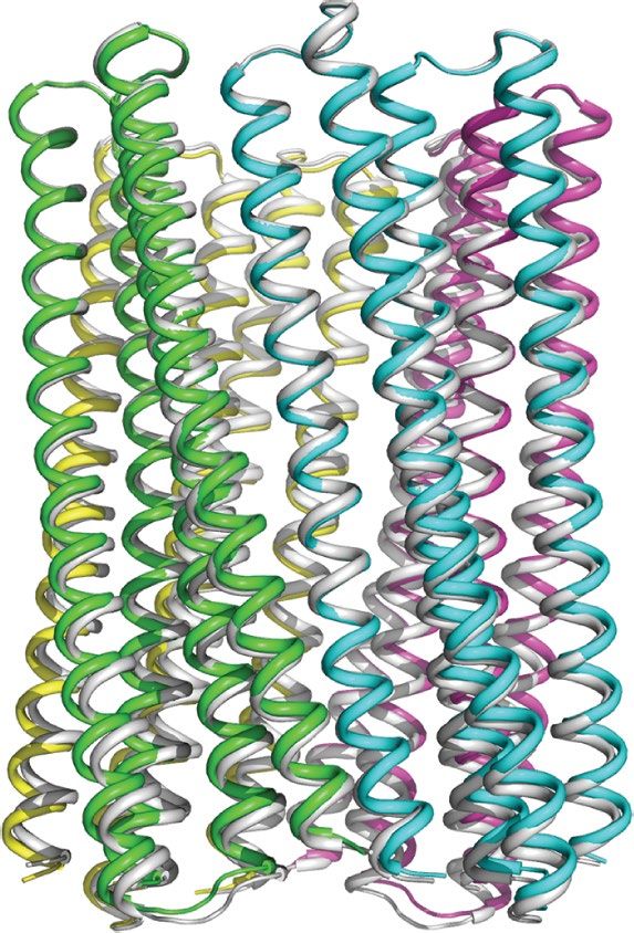
Fig. 1 | The X-ray crystal structure of the water-soluble hexameric WSHC6 Re25 °C was taken when the sample had cooled back to 25 °C after the thermal
and the ion conductivity of the 12-helix TMHC6 transmembrane channel. melt scan. [θ]220, molar ellipticity at 220 nm. e, AUC sedimentation–equilibrium
a, Superposition of the backbones of the crystal structure (blue) and the curves at three different rotor speeds for TMHC6 yield a measured molecular
design model (grey) of WSHC6. The Cα r.m.s.d. between the crystal structure mass of approximately 58 kDa, consistent with the designed hexamer. D and E
and the design model is 0.89 Å. The red sphere represents a water molecule. indicate the molecular mass of the oligomer design and the molecular mass
b, The cross-section of the WSHC6 channel. A chain of water molecules (red calculated from the experiment, respectively. A 230, absorbance (in arbitrary
spheres) occupies the central pore (Extended Data Fig. 1e). c, Model of the units (AU)) at 230 nm; OD, optical density. f, Conductivity in whole-cell
TMHC6 channel. The permeation path, calculated by HOLE 20, is illustrated by patch-clamp experiment on insect cells expressing TMHC6. The channel
the blue surface in the left panel. Constriction sites along the channel are the blocker Gd3+ diminished ion conductance to a level equivalent to that of
E-ring (E44), the K-rings (K65, K68), and two intervening L-rings (L51, L58). untransfected cells. g, TMHC6 has considerably higher conductance for K+
Right, the radius of the pore along the permeation path. d, Circular dichroism than for Na+, Cs+, CH3NH3+ and Ba2+. Ten cells were measured for each permeant
spectra and temperature melt curve (inset) of the TMHC6 channel. No ion species; data are mean ± s.e.m.
apparent unfolding transition is observed up to 95 °C. The spectrum labelled
spectrometry analysis (Extended Data Fig. 3e), but there was no change The ability to design transmembrane pores de novo lays the foundation
in pore assembly or solution behaviour more generally (Extended Data for broad exploration of the pore diameters and chemical interactions
Fig. 2d), further suggesting that chemical modification blocks ion that are required for selective conductance of a wide range of ions.
conductance by direct steric occlusion. This understanding should enable the design of channels that directly
Taken together, the high ion selectivity, specific inhibition by mul- modulate cell function. As a first step in this direction, we expressed
tivalent cations, and complete block by MTS reagents acting at the TMHC6 in a yeast strain that requires potassium for growth, and found
extracellular entry to the pore strongly suggest that ion passage occurs that the growth rate increased considerably in a K+-dependent manner
through the designed central transmembrane pore. Leak conductances (Extended Data Fig. 4).
would not be expected to have these properties. As is the case for some
naturally occurring channels, such as the physiologically important
store-operated calcium channel Orai23, single-channel recordings on Building a larger transmembrane nanopore
TMHC6 did not yield clear signals. The single-channel conductance To explore the generation of larger transmembrane pores that are
may be too low to measure reliably, perhaps because of the narrow capable of transporting organic molecules larger than single ions, we
diameter of the central non-polar lined portion of the pore. designed water-soluble helical bundles with eight-fold symmetry. Our
The residue composition and structure of the conductive channel approach was similar to that described above for WSHC6 except that a
of TMHC6 are reminiscent of calcium-selective Orai channels, but starting ‘single-ring’ template was not used; instead, the structure and
TMHC6 is selective for K+. This selectivity probably reflects the narrow sequence of the inner ring of helices were sampled de novo in paral-
3.3 Å constriction of the TMHC6 pore, which only allows conductance lel to those of the outer ring. We obtained synthetic genes encoding
of at least partially dehydrated K+. Natural potassium channels have selected octameric pore designs and expressed them in E. coli. Four
selectivity filters of similar size and conduct K+ through direct inter- out of five of the octameric designs were well expressed and soluble
actions with backbone carbonyls, without any intervening equatorial in E. coli, and could be purified by nickel-affinity chromatography and
waters of hydration24. By contrast, voltage-gated sodium and calcium SEC. However, none of the designs populated only the target octameric
channels—which conduct hydrated Na+ and Ca2+ (refs. 25,26)—have state. There are only small differences in interface geometry between
selectivity filters that are 4.6 Å2, well suited to accommodate hydrated C7, C8 and C9 assemblies (the angles between subunits are 51°, 45°
sodium and calcium ions25–27 but not hydrated potassium ions. and 40°, respectively), and mixtures were observed for most of the
Nature | www.nature.com | 3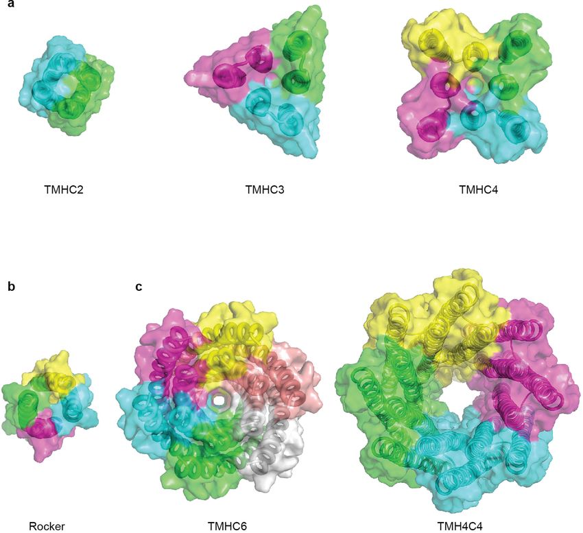
Article
a b a b c
1.0 TMHC6(E44H)
Fractional block
0.8
0.6 TMHC6
0.4
0.2
1 10 100
E-ring [Cd2+] (mM)
c d d e Liposome
100
Normalized current
Control 2.5 mM MTSES 80 TMHC6 or
4
TMH4C4 DNA
60 In vitro protein synthesis
40
20 tavidin
Streptavidin
0 (53 kDa)
Cyto
Cytoplasm
l
ES
T
E
tro
M TSE
BA
Alexa Fluor 488–biocytin (1 kDa)
TS
on
TMHC6 TMHC6(E44C)
-T
or
M
M
C
TS Alexa Fluor 488–poly(A)–biotin (4.6 kDa)
Fig. 2 | Blocking of the ion conductance of TMHC6 by chemical
modification at the pore entrance. a, The extracellular ring of six Glu44 f Alexa Fluor 488–biocytin (1 kDa) g Alexa Fluor 488–poly(A)–biotin (4.6 kDa)
0.08 0.08
AF488/OA647 (AU)
residues (E-ring, shown as sticks) is a likely site for cation entry. b, Blocking of
AF488/OA647 (AU)
0.07 Without DNA 0.07 Without DNA
0.06 TMHC6 0.06
the K+ conductance of TMHC6 and the E44H mutant by Cd2+. Threefold higher TMHC6
0.05 TMH4C4 0.05 TMH4C4
Cd2+ concentrations were required to block TMHC6(E44H) compared with 0.04 0.04
TMHC6, probably because of the reduced electrostatic attraction in the 0.03 * * 0.03
0.02 0.02
former. Three cells were measured for each concentration; data are mean ± 0.01 0.01
s.e.m. c, d, Blocking of the conductance of TMHC6(E44C) using cysteine 0 0
0 5 10 15 20 25 0 5 10 15 20 25
reactive reagents. In c, the y axis shows the current amplitude and the x axis Time (h) Time (h)
indicates the time scale. Negatively charged MTSES, positively charged MTSET,
and hydrophobic MTS-TBAE all completely blocked the ion conductance of Fig. 3 | The X-ray crystal structure of water-soluble WSHC8 and the
TMHC6(E44C) within a few minutes under voltage-clamp control, whereas they characterization of the 16-helix TMH4C4 transmembrane channel.
had no effect on TMHC6 in control experiments. Nine cells for the control and a, b, Superposition of the full octameric assemblies and the monomeric
three cells for each reagent were measured. In d, the bars represent the mean of subunits of the crystal structure (blue) and the design model (grey) of WSHC8.
the measurements and individual data points are shown. The Cα r.m.s.d. is 2.51 Å (octamer) and 0.97 Å (monomers). The larger deviation
for the octamer is caused by the slight tilting of the hairpin monomers along
the superhelical axis of the complex. c, The cross-section of the WSHC8
channel. d, Model of TMH4C4 with 16 transmembrane helices. The
designs. We reasoned that achieving a well-defined C8 state would
electrostatic surface of the neutral transmembrane regions is shown in grey.
require more precise interface definition; therefore, we carried out a
e, Liposome permeability assay. Membrane channels are generated by in vitro
second round of C8 designs with larger intersubunit interaction surface translation inside streptavidin-containing liposomes, biotin-labelled
areas (Extended Data Fig. 5a, b) and more hydrogen-bond networks fluorescent dyes are added to the surrounding buffer, and the amount of dye
across the interface. Ten out of fifteen of the second-round designs trapped inside the liposomes is measured by flow cytometry. f, g, TMH4C4
that passed the in silico test (Extended Data Fig. 1f) were expressed functions as a size-dependent transmembrane sieve. Incorporation of
and soluble in E. coli, and two were found to be monodisperse and TMH4C4 into liposomes enabled the accumulation of the 1 kDa but not the
octameric by MALS analysis (Extended Data Fig. 2b). One of the two 4.6 kDa fluorescent dye, whereas TMHC6 disallowed both. Shown are the time
was further confirmed by AUC (Extended Data Fig. 1h). The circular courses of the median values of the histogram of Alexa Fluor 488/Ovalbumin
dichroism spectrum of the AUC-verified octameric design (denoted conjugated to Alexa Fluor 647 (AF488/OA647) fluorescence (Extended Data
WSHC8, for water-soluble hairpin C8) showed that the structure was Fig. 6e), which represents the concentration of the Alexa Fluor 488 inside the
highly helical and had a melting temperature of 85 °C (Extended Data liposome. n = 7; data are mean (of the obtained median values) ± s.e.m.
*P = 0.0128 (TMH4C4 vs control) and 0.0220 (TMH4C4 vs TMHC6); two-sided
Fig. 1g). We again found excellent agreement between the experimental
Student’s paired t-test. In control experiments performed with α-haemolysin
and calculated SAXS profiles, indicating that WSHC8 folds into the
from Staphylococcus aureus, a well-studied channel-forming protein33 with a
desired shape in solution (Extended Data Fig. 1i).
pore constriction of approximately 15 Å, only the smaller dye accumulated
We determined the crystal structure of WSHC8 (Fig. 3a, b, Extended
(Extended Data Fig. 6f, g), suggesting that—as intended—the assay measures
Data Table 1) and found that the Cα r.m.s.d. values between the crystal solute passage through the transmembrane pores.
structure and the design model for the monomeric subunit and the full
octameric pore were 0.97 Å and 2.51 Å, respectively. The larger devia-
tion in this case compared with WSHC6 is caused by the slight tilting expressed in E. coli, and the membrane fraction purified and solubilized
of the hairpin monomers along the superhelical axis of the complex in detergent. After affinity chromatography, the protein eluted as a
(Extended Data Fig. 1j). As in the design model, the crystal structure monodisperse peak during SEC (Extended Data Fig. 2e). Circular dichro-
contains a long and continuous central channel with an inner diameter ism measurements showed that TMHC8 had the expected α-helical
of approximately 10 Å as calculated by HOLE. We converted WSHC8 into secondary structure with a melting temperature of 75 °C (Extended
an octameric membrane-spanning pore (TMHC8) by redesigning the Data Fig. 5e). AUC experiments showed that TMHC8 formed complexes
membrane-exposed and pore-facing residues of the crystal structure with a molecular mass of 98.9 kDa, which lies in between the masses of
(see Supplementary Information, Extended Data Fig. 5c, d). The design a 14-helix heptamer and a 16-helix octamer (Extended Data Fig. 5g). To
model has a central pore with a diameter of 10 Å and a transmembrane resolve this ambiguity, we linked two monomers of TMHC8 together
span of 31 Å. A gene encoding TMHC8 was synthesized, the protein using a short loop to create a four-helix subunit. This redesign, denoted
4 | Nature | www.nature.com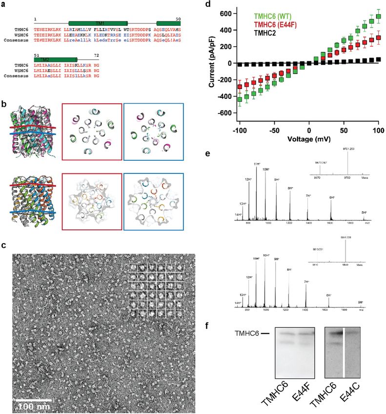
TMH4C4 (Fig. 3d), was purified to homogeneity using nickel-affinity a
chromatography and SEC (Extended Data Fig. 2f). Circular dichro-
ism spectroscopy showed that TMH4C4 was fully α-helical and was
thermally stable up to 95 °C (Extended Data Fig. 5f). AUC experiments
showed that TMH4C4 sedimented as a tetramer in detergent solution, 90°°
consistent with the 16-helix design model (Extended Data Fig. 5h).
Expression of the designed larger transmembrane nanopore in insect
cells resulted in cell death, probably because of induced cell permeabil-
ity; we were therefore unable to assess the channel activity in these cells.
Instead, we used a liposome-based assay coupled to in vitro protein Cytoplasm
ytoplasm
synthesis28–30. TMHC6 and TMH4C4 were produced inside liposomes
b c
containing streptavidin, and biotinylated Alexa Fluor 488 (which has
a molecular mass of around 1 kDa) was added outside (Fig. 3e). SEC
analysis revealed that the in vitro-synthesized TMHC6 and TMH4C4
had similar elution volumes to the bacterially expressed and purified
90°
proteins (Extended Data Fig. 6a). Consistent with the much larger pore
radius of TMH4C4 compared with TMHC6, we observed considerably
more accumulation of dye within proteoliposomes containing only
TMH4C4 than in proteoliposomes containing an equivalent amount
of TMHC6, or in empty liposomes (Fig. 3f, Extended Data Fig. 6b, c,
e). The narrowest dimension of the head group of the fluorophore
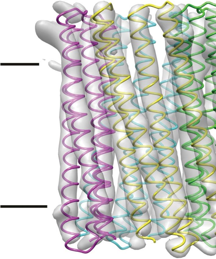
Fig. 4 | Cryo-EM structures of the 16-helix TMH4C4 transmembrane
is approximately 12 Å (Extended Data Fig. 6d), whereas the nominal channel. a, Cryo-EM density (grey surface) and structure model (coloured
diameter of the constriction region of the TMH4C4 pore is estimated ribbon) of the 16-helix TMH4C4 protein. Electron microscopy maps, generated
to be 10 Å by HOLE; thermal fluctuations of the side chains and back- in Chimera34, are shown in two perpendicular views. b, Superposition of the
bone probably allow for permeation by the fluorophore. By contrast, cryo-EM structure (coloured) and design model based on the crystal structure
the 3.3 Å constriction of TMHC6 is far too narrow to allow the passage of the soluble form (grey) of TMH4C4. c, Structure alignment of the four
of fluorophores. Increasing the size of the fluorescent conjugate to protomers in one tetrameric cryo-EM structure of TMH4C4.
4.6 kDa—by inserting polyadenine oligo DNA (A11) between Alexa Fluor
488 and biotin—blocked passage through both the TMHC6 and the
TMH4C4 pores (Fig. 3g), consistent with the estimated hydrodynamic of the substantial buried surface area and polarity of the interaction
diameter of this compound of 30 Å31. surfaces makes these structures robust to changes in the surrounding
In seeking to obtain an overall structure of the 16-helix transmem- environment and reduces confounding effects that result from inter-
brane channel, we used cryogenic electron microscopy (cryo-EM) actions between the hydrophobic lipid-facing residues during fold-
to determine the three-dimensional (3D) structure of TMH4C4. The ing. This approach enables the construction of substantial pores with
protein was concentrated to around 6 mg ml−1, applied to EM grids, environments very different from that of the surrounding lipids: the
and cryo-EM images were collected and processed following standard TMHC6 pore clearly shows selective transmembrane ion conductance,
protocols (Extended Data Fig. 7a–e). To avoid potential bias, C1 sym- and the 10 Å-diameter TMH4C4 pore—clearly evident in the cryo-EM
metry was applied for automatic image processing and classification; structure—is lined with polar residues and provides passage to solutes
this yielded a dominant 16-helix form containing about 40% of all the as large as biotinylated Alexa Fluor 488.
3D classified particles with the most continuous and intact map among Our strategy—first designing soluble pore-containing structures
all classes. Further classification and refinement focused on this set of and then converting the stable designs into transmembrane proteins
particles resulted in a 5.9 Å resolution map from 64,739 (out of a total after determination of the crystal structure—leverages the consider-
of 1,559,110 for 3D classification) selected particles (Fig. 4a, Extended ably more straightforward structural characterization of soluble pro-
Data Fig. 7d, e, Extended Data Table 2). The cryo-EM structure clearly teins as a key step towards building complex transmembrane proteins
shows the formation of a 16-helix assembly with a large central pore, with a high success rate (see Supplementary Information). Building on
consistent with the design model built from the crystal structure of the channels described here, custom design now provides a platform
the soluble form (Fig. 4a). Density encircling the membrane-spanning through which to understand the underlying chemistry and physics of
region probably originates from surrounding detergent molecules solute permeation and selectivity by modulating pore structures and
(Extended Data Fig. 7e, f). A structure model of TMH4C4 built on the selectivity filters in ways that are not possible with native channels,
basis of the EM map (Fig. 4a, b) showed some deviation among the and also enables a wide range of applications. Among many possibili-
four protomers, so the tetramer is not perfectly symmetric (Fig. 4c); ties, custom-designed pores could provide new routes to generating
however, the central-pore-containing 16-helix structure corresponding membranes with selective permeabilities, sensing molecules in the
to the original TMH4C4 design is clearly resolved, and aligns well with environment, and controlling cellular behaviour.
the design model (Fig. 4b).
Online content
Any methods, additional references, Nature Research reporting sum-
Conclusion maries, source data, extended data, supplementary information,
Our advances in designing structurally well-defined transmembrane acknowledgements, peer review information; details of author con-
pores (for comparison to previous de novo membrane protein designs, tributions and competing interests; and statements of data and code
see Extended Data Fig. 8), like advances in protein design generally8,10,32, availability are available at.https://doi.org/10.1038/s41586-020-2646-5.
both inform our understanding of general principles of protein bio-
physics and open the door for a wide range of applications. From the
1. Gilbert, R. J. C., Bayley, H. & Anderluh, G. Membrane pores: from structure and
perspective of membrane-protein folding, our success in designing
assembly, to medicine and technology. Phil. Trans. R. Soc. Lond. B 372, 20160208
transmembrane homo-oligomeric structures with subunit interfaces (2017).
that contain hydrogen-bond networks suggests that the combination 2. Eisenstein, M. An ace in the hole for DNA sequencing. Nature 550, 285–288 (2017).
Nature | www.nature.com | 5
Article
3.    Kasianowicz, J. J., Brandin, E., Branton, D. & Deamer, D. W. Characterization of individual   20. Smart, O. S., Neduvelil, J. G., Wang, X., Wallace, B. A. & Sansom, M. S. P. HOLE: a program
      polynucleotide molecules using a membrane channel. Proc. Natl Acad. Sci. USA 93,                  for the analysis of the pore dimensions of ion channel structural models. J. Mol. Graph.
      13770–13773 (1996).                                                                               14, 354–360 (1996).
4.    Clarke, J. et al. Continuous base identification for single-molecule nanopore DNA             21. Hou, X., Pedi, L., Diver, M. M. & Long, S. B. Crystal structure of the calcium
      sequencing. Nat. Nanotechnol. 4, 265–270 (2009).                                                  release-activated calcium channel Orai. Science 338, 1308–1313 (2012).
5.    Lear, J. D., Wasserman, Z. R. & DeGrado, W. F. Synthetic amphiphilic peptide models for       22. Hou, X., Burstein, S. R. & Long, S. B. Structures reveal opening of the store-operated
      protein ion channels. Science 240, 1177–1181 (1988).                                              calcium channel Orai. eLife 7, e36758 (2018).
6.    Akerfeldt, K. S., Lear, J. D., Wasserman, Z. R., Chung, L. A. & DeGrado, W. F. Synthetic      23. Dynes, J. L., Amcheslavsky, A. & Cahalan, M. D. Genetically targeted single-channel
      peptides as models for ion channel proteins. Acc. Chem. Res. 26, 191–197 (1993).                  optical recording reveals multiple Orai1 gating states and oscillations in calcium influx.
7.    Joh, N. H. et al. De novo design of a transmembrane Zn2+-transporting four-helix bundle.          Proc. Natl Acad. Sci. USA 113, 440–445 (2016).
      Science 346, 1520–1524 (2014).                                                                24. Jiang, Y. et al. X-ray structure of a voltage-dependent K+ channel. Nature 423, 33–41 (2003).
8.    Lu, P. et al. Accurate computational design of multipass transmembrane proteins.              25. Payandeh, J., Scheuer, T., Zheng, N. & Catterall, W. A. The crystal structure of a
      Science 359, 1042–1046 (2018).                                                                    voltage-gated sodium channel. Nature 475, 353–358 (2011).
9.    Mahendran, K. R. et al. A monodisperse transmembrane α-helical peptide barrel.                26. Tang, L. et al. Structural basis for Ca2+ selectivity of a voltage-gated calcium channel.
      Nat. Chem. 9, 411–419 (2017).                                                                     Nature 505, 56–61 (2014).
10.   Mravic, M. et al. Packing of apolar side chains enables accurate design of highly stable      27. Pan, X. et al. Structure of the human voltage-gated sodium channel NaV1.4 in complex
      membrane proteins. Science 363, 1418–1423 (2019).                                                 with β1. Science 362, eaau2486 (2018).
11.   Joh, N. H., Grigoryan, G., Wu, Y. & DeGrado, W. F. Design of self-assembling                  28. Fujii, S. et al. Liposome display for in vitro selection and evolution of membrane proteins.
      transmembrane helical bundles to elucidate principles required for membrane protein               Nat. Protoc. 9, 1578–1591 (2014).
      folding and ion transport. Phil. Trans. R. Soc. Lond. B 372, 20160214 (2017).                 29. Fujii, S., Matsuura, T., Sunami, T., Kazuta, Y. & Yomo, T. In vitro evolution of α-hemolysin
12.   Niitsu, A., Heal, J. W., Fauland, K., Thomson, A. R. & Woolfson, D. N. Membrane-spanning          using a liposome display. Proc. Natl Acad. Sci. USA 110, 16796–16801 (2013).
      α-helical barrels as tractable protein-design targets. Phil. Trans. R. Soc. Lond. B 372,      30. Dwidar, M. et al. Programmable artificial cells using histamine-responsive synthetic
      20160213 (2017).                                                                                  riboswitch. J. Am. Chem. Soc. 141, 11103–11114 (2019).
13.   Thomson, A. R. et al. Computational design of water-soluble α-helical barrels. Science        31. Sim, A. Y. L., Lipfert, J., Herschlag, D. & Doniach, S. Salt dependence of the radius of
      346, 485–488 (2014).                                                                              gyration and flexibility of single-stranded DNA in solution probed by small-angle X-ray
14.   Rhys, G. G. et al. Maintaining and breaking symmetry in homomeric coiled-coil                     scattering. Phys. Rev. E 86, 021901 (2012).
      assemblies. Nat. Commun. 9, 4132 (2018).                                                      32. Huang, P.-S., Boyken, S. E. & Baker, D. The coming of age of de novo protein design.
15.   Crick, F. H. C. The Fourier transform of a coiled-coil. Acta Crystallogr. 6, 685–689              Nature 537, 320–327 (2016).
      (1953).                                                                                       33. Song, L. et al. Structure of staphylococcal α-hemolysin, a heptameric transmembrane
16.   Grigoryan, G. & Degrado, W. F. Probing designability via a generalized model of helical           pore. Science 274, 1859–1865 (1996).
      bundle geometry. J. Mol. Biol. 405, 1079–1100 (2011).                                         34. Pettersen, E. F. et al. UCSF Chimera—a visualization system for exploratory research and
17.   Huang, P. S. et al. High thermodynamic stability of parametrically designed helical               analysis. J. Comput. Chem. 25, 1605–1612 (2004).
      bundles. Science 346, 481–485 (2014).
18.   Boyken, S. E. et al. De novo design of protein homo-oligomers with modular                    Publisher’s note Springer Nature remains neutral with regard to jurisdictional claims in
      hydrogen-bond network-mediated specificity. Science 352, 680–687 (2016).                      published maps and institutional affiliations.
19.   Das, R. et al. Simultaneous prediction of protein folding and docking at high resolution.
      Proc. Natl Acad. Sci. USA 106, 18978–18983 (2009).                                            © The Author(s), under exclusive licence to Springer Nature Limited 2020

6 | Nature | www.nature.com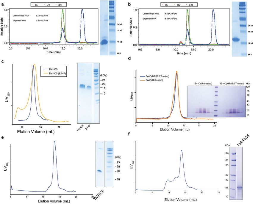
Westlake University for providing computation support, and the Mass Spectrometry and
                                                                                                  Metabolomics Core Facility of Westlake University for mass spectrometry analysis. This work
Reporting summary
                                                                                                  was supported by the Howard Hughes Medical Institute, the Air Force Office of Scientific
Further information on research design is available in the Nature                                 Research (FA9550-18-1-0297) and NSF grant CHE 1629214. W.A.C. was supported by NIH
Research Reporting Summary linked to this paper.                                                  research grant R35 NS111573. P.L. was supported by NSFC Project 31901054, Tencent
                                                                                                  Foundation and the foundation of Westlake University. SAXS data collection at SIBYLS is
                                                                                                  funded through DOE BER Integrated Diffraction Analysis Technologies (IDAT) program and
                                                                                                  NIGMS grant P30 GM124169-01, ALS-ENABLE. T.M. was in part supported by KAKENHI grants
Data availability                                                                                 17H00888 from JSPS. B.F.L. and X.Y.P. are supported by a Wellcome Trust Investigator award
                                                                                                  (200873/Z/16/Z). The ALS-ENABLE beamlines are supported in part by the National Institutes
Coordinates and structure files have been deposited to the Protein                                of Health, National Institute of General Medical Sciences, grant P30 GM124169-01 and the
Data Bank with accession codes 6TJ1 (WSHC6, P21221), 6TMS (WSHC6,                                 Howard Hughes Medical Institute. The Advanced Light Source is a Department of Energy
P1) and 6O35 (WSHC8). An electron microscopy map of TMH4C4 and                                    Office of Science User Facility under contract number DE-AC02-05CH11231.

the associated structure model have been deposited in the Electron
                                                                                                  Author contributions C.X., P.L., T.M.G., L.S., T.M., W.A.C. and D.B. planned the research and
Microscopy Data Bank and Protein Data Bank with accession codes
                                                                                                  designed experiments. C.X., P.L. and D.B. designed the proteins. C.X., P.L., T.M.G., J.D., T.M.,
EMD-30126 and 6M6Z, respectively. Source data are provided with                                   W.A.C. and D.B. wrote the manuscript. C.X. and P.L. carried out biophysical characterizations.
this paper.                                                                                       T.M.G. and W.A.C. performed patch–clamp experiments and analysed data. A.U. and T.M.
                                                                                                  performed liposome permeability assays and analysed data. X.Y.P., M.J.B. and B.F.L. solved
                                                                                                  crystal structures. P.L., Q.X., M.C.J. and J.M.K. determined the cryo-EM structure. G.R. and F.D.
                                                                                                  refined the cryo-EM structure. J.D. carried out the yeast growth assay. D.J. and D.M. prepared
Code availability                                                                                 samples for patch–clamp experiments. E.M.L. took negatively stained electron micrographs.
                                                                                                  H.B., Y.H., T.B., S.E.B. and P.-S.H. helped with design calculations. All authors discussed the
All program code is in Rosetta distribution (https://www.rosettacom-                              results and commented on the manuscript.
mons.org). Example design protocols are provided in the Supplemen-
tary Information.                                                                                 Competing interests The University of Washington has submitted a US provisional patent
                                                                                                  application (63/017,810) that covers the computational design of transmembrane pores
                                                                                                  described in this paper, listing C.X., P.L., T.M.G., W.A.C. and D.B. as inventors.
Acknowledgements We thank A. Kang for assistance with crystallization; B. Sankaran of the
Berkeley Center for Structural Biology at the Advanced Light Source for help with X-ray
diffraction data collection; J. Sumida for AUC support; L. Carter for assistance with SEC–MALS;   Additional information
U. Nattermann, Q. Zhou and H. Shen for assistance with EM; SIBYLS mail-in SAXS program at         Supplementary information is available for this paper at https://doi.org/10.1038/s41586-020-
the Advanced Light Source (ALS) for SAXS data collection; L. Jan, T. Cheng and R. Zhou from       2646-5.
UCSF for sharing yeast strain SGY1528; and Rosetta@Home volunteers for contributing               Correspondence and requests for materials should be addressed to P.L., W.A.C. or D.B.
computing resources. The cryo-EM work was performed at the Cryo-EM Facility of Westlake           Peer review information Nature thanks Eric Gouaux and the other, anonymous, reviewer(s) for
University. We thank the Arnold and Mabel Beckman Cryo-EM Center at the University of             their contribution to the peer review of this work.
Washington for access to electron microscopes, the Information Technology Center of               Reprints and permissions information is available at http://www.nature.com/reprints.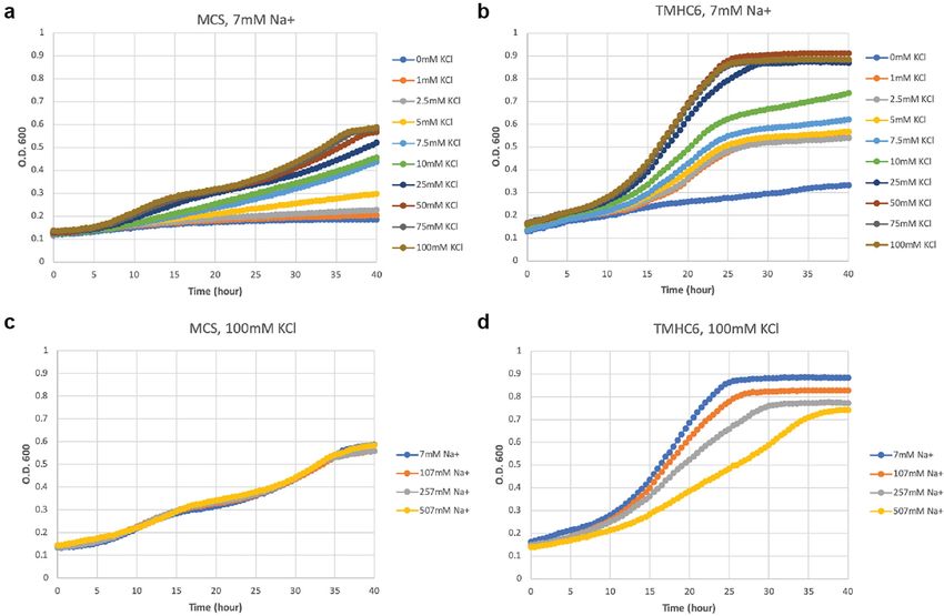
Article

Extended Data Fig. 1 | Design and characterization of water-soluble pores.
a, f, Design models (insets) and energy versus r.m.s.d. plots generated from
Rosetta ‘fold-and-dock’ structure prediction calculations. The predicted
structures converge on the design models with r.m.s.d. values less than 2.0 Å.
Structures in the alternative energy minima at large r.m.s.d. positions also
recapitulate the design models but with chain identities in the r.m.s.d.
calculations reversed. b, g, Wavelength-scan and temperature-scan (insets)
circular dichroism spectra. WSHC6 does not melt up to 95 °C, while WSHC8 has
a melting temperature of 85 °C. The overlap of the pre- and post-annealing
circular dichroism spectra indicates that the thermal denaturation is reversible.
c, h, Representative analytical ultracentrifugation sedimentation-equilibrium
curves at three different rotor speeds for WSHC6 and WSHC8, 0.2 OD230 and
0.3 OD230 in PBS (pH 7.4), respectively. The determined oligomeric states
match those of the design models. d, i, Small-angle X-ray scattering (SAXS)
characterization. The experimental scattering profiles (black) are similar to
scattering profiles computed from the design models (red). e, The chain of
water molecules in the pore of WSHC6 crystal structure (red spheres) is verified
by processing the data and refining the structure in the P1 space group.
j, Overlay of the crystal structure (blue) and the design model (grey) of WSHC8.
Helices are more tilted in the crystal structure than in the design model.
Extended Data Fig. 2 | Representative SEC and SDS–PAGE of the designed   treated or untreated with MTSES. e, TMHC8. f, TMH4C4. These experiments
proteins. a, b, WSHC6 and WSHC8. Molecular masses are determined by      were repeated twice with similar results.
coupling SEC with MALS. c, TMHC6 and the E44F mutant. d, TMHC6(E44C)
Article

Extended Data Fig. 3 | Comparisons between WSHC6 and TMHC6 and                     E44F single mutant reduced the K+ current to half that of TMHC6. TMHC2, a
additional characterizations of TMHC6 and mutants. a, Sequence                     previously designed transmembrane protein without a pore, does not conduct
alignment of TMHC6 with WSHC6. b, Pore-lining residues in WSHC6 and                ions across the membrane. Three cells were measured for each protein; data
TMHC6. Top row, overlay of the crystal structure (colours) and the design          are mean ± s.e.m. e, The covalent modification of TMHC6(E44C) by MTSES.
model (grey) of WSHC6. The pore is lined with alternating leucine (red layer)      Mass spectrometry analysis that there is a 140 Da increase in molecular mass
and isoleucine (blue layer) residues. Bottom row, the TMHC6 pore is lined with     for the mutant after MTSES treatment, in agreement with the predicted value.
E44 ring (red layer) and K65 ring (blue layer) at the extracellular and            f, Expression of TMHC6 and mutants in insect cells for the whole-cell
intracellular sides, respectively. c, Negative stain EM for TMHC6 in amphipols.    patch-clamp experiments. The same amount of cells were loaded into the gel
Protein particles on the EM grid showed round shape and size consistent with       and the expression levels for two variants were examined by western blot. The
the design model (scale bar at the bottom left, 100 nm). Inset, close-up view of   E44F mutant had a similar, if not higher, expression level to TMHC6. The E44C
representative particles; each side of the particle frames represent 12.8 nm.      mutant was expressed at a slightly lower level compared to TMHC6. These
d, Disrupting mutation in the TMHC6 pore entrance reduces the current. The         experiments were repeated three times with similar results.
Extended Data Fig. 4 | The expression of TMHC6 complements a yeast strain        with the insensitive growth rates of MCS yeast (c). These results suggest that
defective in K+ uptake. a, SGY1528 with an empty vector MCS grew slowly when     TMHC6 conducts K+ and complements the defective K+ uptake in strain SGY1528;
K+ concentration was lower than 5 mM. The observed growth rate showed            this rescuing effect is sensitive to extracellular Na+ concentrations indicating an
dependency on extracellular K+ concentration. b, SGY1528 supplemented with the   increased Na+ permeability. Detailed methods are described in the Supplementary
TMHC6 gene rescued the yeast growth at lower extracellular K+ concentrations     Information. The minimal medium and the seeding process are carefully designed
(1 mM–5 mM) and showed increased growth rates at higher extracellular K+         to not contain or bring in potassium. The background K+ concentration should be
concentrations (7.5 mM–100 mM). c, d, With increasing concentrations of          low, which is suggested by the sharp difference between curves for 0 and 1 mM K+
extracellular Na+, TMHC6 yeast showed decreased growth rate (d) in comparison    in the case of TMHC6.
Article

Extended Data Fig. 5 | Design and additional characterizations of the               TMH4C4 corresponding to the three layers in the top row. Glu69 and Glu87 are
designed 16-helix pores. a, b, Design models from the first and second rounds       replaced with glutamine and leucine, respectively. e, f, Circular dichroism
of water-soluble designs. The monomers of the first round designs (a, 70 amino      spectra and thermal stability of 16-helix transmembrane pores. An unfolding
acids) are considerably smaller than those of the second round (b, 100 amino        transition is observed at around 75 °C for TMHC8 (e). TMH4C4 (f) is thermally
acids). c, Sequence alignment of TMHC8 with WSHC8. d, Pore-lining residues in       stable up until 95 °C. g, h, Representative AUC sedimentation-equilibrium
WSHC8 and TMH4C4. The crystal and cryo-EM structures are in colours.                curves of 16-helix transmembrane nanopores. By fitting the data globally as a
The design models are in grey. Top row, the lumen of WSHC8 pore is freely           single ideal species in solution, TMHC8 is shown to form complexes with a
water-accessible, so the residues inside the pore are all polar. Shown in the       molecular mass of 98.9 kDa, which is in between the masses of a heptamer and
figure are three representative layers of the pore-lining residues in the crystal   an octamer. The molecular mass of TMH4C4 is determined to be 98.1 kDa, very
structure, Glu69 ring (red), Lys80 ring (blue), and Glu87 (orange). The missing     close to that of a tetramer. ‘MW (D)’ refers to the molecular mass of the
heavy atoms of these flexible residues are built using Rosetta with backbone        oligomer design and ‘MW (E)’ refers to the molecular mass determined in the
constraints. Bottom row, three pore-lining layers in the cryo-EM structure of       experiment.
Extended Data Fig. 6 | See next page for caption.
Article
Extended Data Fig. 6 | In vitro protein synthesis and characterizations of      **P < 0.01; from left to right P = 1.65 × 10 −6, 0.0357, 0.0040, 0.0024). d, The
TMHC6 and TMH4C4. a, SEC analyses of TMHC6 and TMH4C4 purified from             narrowest dimension of the head group of Alexa Fluor 488–biocytin is
E. coli (top) and synthesized in vitro (middle and bottom). b, The in vitro     approximately 12 Å. The van der Waals radius of nitrogen atoms is 1.55 Å.
synthesized products were analysed by SDS–PAGE and autoradiography. The         e, Representative original data for Fig. 3f. Data of approximately 15,000 to
means of three independent experiments are shown. The error bars indicate       20,000 particles are shown. Similar results were obtained with 7 independent
s.e.m. c, EmrE, one of the E. coli-derived membrane proteins, showed strong     experiments. f, Flow cytometry data of the liposomes with pores made of
interaction with LUV, whereas GusA, a soluble enzyme, did not. For TMHC6 and    α-haemolysin (AH). Time courses of the median values of the histogram of
TMH4C4, the fraction interacting with LUV was found to be 25.9% and 17.6%,      AF488/OA647 fluorescence, which represents the concentration of the Alexa
respectively, among synthesized, indicating that the fraction associated with   Fluor 488 inside the liposome, are shown. The means of three independent
the membrane is similar between them. The mean of four independent              experiments are plotted with the error bars indicating the s.e.m.
experiments are shown. The error bars indicate s.e.m. Student’s paired t-test   g, Representative original data of f. Similar results were obtained with 3
with a two-sided distribution was used to calculate the P values (*P < 0.05,    independent experiments.
Extended Data Fig. 7 | Cryo-EM resolution estimation and data processing.           and refinement, a final data set containing 64,739 particles was used for 3D
a, Exemplary cryo-EM micrograph of purified TMH4C4 after drift correction           auto-refinement within RELION 3.0. Local resolution of TMH4C4 was
and dose-weighting. All the micrographs showed similar results. b, Class            determined within RELION 3.0. Coloured full views (lower lane) from two
averages after the final round of 2D classification sorted in descending order      different orientations illustrate the resolution of different regions in the
by the number of particles in each class. The white scale bar in the bottom right   protein. The low resolution ‘belt’ in the right panel indicates the density for
panel indicates 10 nm. c, Angular distribution plot for the final reconstruction    detergents. f, EM density from 213,654 particles. An EM map (grey) from the
from two different views. d, The gold-standard Fourier shell correlation (FSC)      second round of 3D classification from 213,654 particles (e) is shown in three
curves for the 3D reconstruction. Deriving map resolution from FSC = 0.143 is       perpendicular views. Superposition of the cryo-EM structure of TMH4C4
indicated. e, Processing of 2,166 EM micrographs resulted in a total number of      (cyan) to the map shows a good fit.
2,146,524 TMH4C4 particles. After a 2D sub-classification, 3D classifications
Article

Extended Data Fig. 8 | Advances in membrane protein design, from             view of designer transmembrane pores described in this study. Central pores
compact helical bundles to transmembrane pores. a, b, Surface view of        with different sizes are visible.
previously reported de novo designed transmembrane proteins7,8. c, Surface
Extended Data Table 1 | Crystallographic statistics for WSHC6 and WSHC8

*Data for WSHC6 and WSHC8 structures were collected from a single crystal. Values in parentheses are for the highest-resolution shell.
#I/σ(I) (along a*, b* and c* axes).
Article
Extended Data Table 2 | Cryo-EM image processing statistics for TMH4C4
nature research | reporting summary
                                                                                                      Corresponding author(s): David Baker
                                                                                                      Last updated by author(s): Apr 23, 2020

Reporting Summary
Nature Research wishes to improve the reproducibility of the work that we publish. This form provides structure for consistency and transparency
in reporting. For further information on Nature Research policies, see Authors & Referees and the Editorial Policy Checklist.

Statistics
For all statistical analyses, confirm that the following items are present in the figure legend, table legend, main text, or Methods section.
n/a Confirmed
           The exact sample size (n) for each experimental group/condition, given as a discrete number and unit of measurement
           A statement on whether measurements were taken from distinct samples or whether the same sample was measured repeatedly
           The statistical test(s) used AND whether they are one- or two-sided
           Only common tests should be described solely by name; describe more complex techniques in the Methods section.

           A description of all covariates tested
           A description of any assumptions or corrections, such as tests of normality and adjustment for multiple comparisons
           A full description of the statistical parameters including central tendency (e.g. means) or other basic estimates (e.g. regression coefficient)
           AND variation (e.g. standard deviation) or associated estimates of uncertainty (e.g. confidence intervals)

           For null hypothesis testing, the test statistic (e.g. F, t, r) with confidence intervals, effect sizes, degrees of freedom and P value noted
           Give P values as exact values whenever suitable.

           For Bayesian analysis, information on the choice of priors and Markov chain Monte Carlo settings
           For hierarchical and complex designs, identification of the appropriate level for tests and full reporting of outcomes
           Estimates of effect sizes (e.g. Cohen's d, Pearson's r), indicating how they were calculated
                                                 Our web collection on statistics for biologists contains articles on many of the points above.

Software and code
Policy information about availability of computer code
  Data collection              Rosetta software suite 3 was used for protein design calculations. AVIV Data Collection software v3.45 was used for CD. ProteomeLab XL-
                               I for AUC. AutoEMation for cryoEM. Igor Pro 6.37 software for patch-clamp experiments. BD FACSuite v1.0.5.3841 for FACS.

  Data analysis                Sedfit 15.1b, Sednterp 20120828 beta and Sedphat 14.0 were used for AUC data analysis. CRYSOL and SASPLOT in ATSAS 2.7.2 for SAXS
                               data analysis. XDS version Jan 26, 2018, Phaser, version 2.8.2., Phenix.refine version dev_3112, Phenix version 1.11.1-2575, and iMosflm
                               from CCP4 V7 for X-ray data analysis. RELION 3.0.6 and Chimera 1.13.1 for cryoEM data processing and refinement. FlowJo v10.6.2 for
                               FACS analysis. Protein structures and models were visualized using PyMOL 1.7.6.5 HOLE 2.0 for calculating designed pore radius.
                               TMHMM server 2.0 for predicting membrane compatibility of designed proteins.
For manuscripts utilizing custom algorithms or software that are central to the research but not yet described in published literature, software must be made available to editors/reviewers.
We strongly encourage code deposition in a community repository (e.g. GitHub). See the Nature Research guidelines for submitting code & software for further information.

Data
Policy information about availability of data
 All manuscripts must include a data availability statement. This statement should provide the following information, where applicable:
     - Accession codes, unique identifiers, or web links for publicly available datasets
                                                                                                                                                                                                October 2018

     - A list of figures that have associated raw data
     - A description of any restrictions on data availability

Coordinates and structure files have been deposited to the Protein Data Bank with accession codes 6TJ1 and 6TMS (WSHC6) and 6O35 (WSHC8). EM structure of
TMH4C4 and the associated atomic model have been deposited in the Electron Microscopy Data Bank and Protein Data Bank with the following accession codes:
EMD-30126 and PDB 6M6Z. AUC, CD, patch-clamp, and FACS experiments in the main figures have associated raw data.

                                                                                                                                                                                                      1
You can also read