Crystal structures of influenza nucleoprotein complexed with nucleic acid provide insights into the mechanism of RNA interaction
←
→
Page content transcription
If your browser does not render page correctly, please read the page content below
4144–4154 Nucleic Acids Research, 2021, Vol. 49, No. 7                                                                Published online 30 March 2021
doi: 10.1093/nar/gkab203

Crystal structures of influenza nucleoprotein
complexed with nucleic acid provide insights into the
mechanism of RNA interaction
Yun-Sang Tang1 , Shutong Xu2,3 , Yu-Wai Chen4 , Jia-Huai Wang2,* and Pang-Chui Shaw                                                                    1,5,*

1
 Centre for Protein Science and Crystallography, School of Life Sciences, The Chinese University of Hong Kong,
Hong Kong SAR, China, 2 Department of Medical Oncology and Department of Cancer Biology, Dana-Farber Cancer
Institute, Harvard Medical School, Boston, MA 02115, USA, 3 College of Life Science and Technology, Huazhong
Agricultural University, Wuhan, China, 4 Department of Applied Biology and Chemical Technology, The Hong Kong

                                                                                                                                                                  Downloaded from https://academic.oup.com/nar/article/49/7/4144/6204651 by guest on 29 October 2021
Polytechnic University, Hong Kong SAR, China and 5 Li Dak Sum Yip Yio Chin R & D Centre for Chinese Medicine,
The Chinese University of Hong Kong, Hong Kong SAR, China

Received February 02, 2021; Revised March 09, 2021; Editorial Decision March 11, 2021; Accepted March 29, 2021

ABSTRACT                                                                          ture. The structure of RNP is maintained by NP-NP homo-
                                                                                  oligomerization, which is mediated by the insertion of the
The nucleoprotein (NP) of influenza virus is the core                             tail-loop from one NP protomer into the groove of its neigh-
component of the ribonucleoprotein (RNP) and per-                                 bour (1,2). Genomic viral RNA (vRNA) is predicted to
forms multiple structural and functional roles. Struc-                            wrap along a helical path constituted by positive residues
tures of the influenza A, B and D NP molecules have                               on NP (3–5). During viral replication when a positive-sense
been solved previously, but structural information on                             cRNA is synthesized, NP is recruited to stabilize nascent
how NP interacts with RNA remains elusive. Here we                                cRNA and forms a replicative intermediate named comple-
present the crystal structure of an obligate monomer                              mentary RNP (cRNP) (6,7). In the vRNA synthesis stage
of H5N1 NP in complex with RNA nucleotides to 2.3                                 that follows, NP is again recruited for its encapsidation and
Å, and a C-terminal truncation of this mutant, also                              formation of new progeny vRNP.
in complex with RNA nucleotides, to 3 Å. In both                                    The crystal structures of H1N1 and H5N1 NP have been
                                                                                  solved previously (1,2). The NP molecule shows a crescent
structures, three nucleotides were identified near two
                                                                                  shape with a head domain (residues 150–276 and 429–452)
positive grooves of NP suggested to be important                                  and a body domain (residues 1–149 and 277–386) com-
for RNA binding. Structural evidence supports that                                prised of mainly helices and beta strands. The tail-loop
conformational changes of flexible loops and the C-                               structure spans from residues 402 – 428 and protrudes out-
terminal tail both play important roles in the binding                            wards the head domain. It demonstrates considerable de-
of RNA. Based on the structure, we propose a mech-                                gree of flexibility so that tail-loop insertion at different an-
anism by which NP captures RNA by flexible loops                                  gles is possible, leading to the formation of oligomers of
and transfers it onto the positive binding grooves.                               various sizes. Later, monomeric structures of H1N1 NP
Binding of RNA by NP is a crucial step for template                               harbouring R416A mutation, or with the tail-loop struc-
re-encapsidation during transcription and replication                             ture deleted, have also been resolved (8,9). Crystal struc-
and cRNP formation. Our structures thus provide in-                               tures of influenza B and D NP both show conserved overall
                                                                                  fold highly similar to influenza A NP (10,11). NP is a basic
sights into the molecular virology of the influenza
                                                                                  molecule with pI of about 9.4. Conserved positive residues
virus.                                                                            have been identified on the surface of NP. By mutagenesis,
                                                                                  contribution of these residues towards RNA binding has
                                                                                  been analysed. By Surface Plasmon Resonance assay, Ng
INTRODUCTION
                                                                                  et al. (2) have found that mutation of the positive groove
The ribonucleoprotein (RNP) of influenza virus is the struc-                      formed by R74, R75, R174, R175, R221 (NP-G1) com-
tural unit where the negative-sense genome segments are                           pletely abolished RNA binding, while mutation of a nearby
encompassed. Each RNP contains the heterotrimeric poly-                           groove formed by R150, R152, R156, R162 (NP-G2) led
merase complex at one end, and multiple copies of nucleo-                         to a 4.7-fold decrease in RNA affinity. Also implicated in
protein (NP) forming a double helical rod shape architec-

* To
   whom correspondence should be addressed. Tel: +852 39431363; Fax: +852 26037246; Email: pcshaw@cuhk.edu.hk
Correspondence may also be addressed to Jia-Huai Wang. Tel: +1 617 632 3983; Fax: +1 617 632 4393; Email: jwang@crystal.harvard.edu

C The Author(s) 2021. Published by Oxford University Press on behalf of Nucleic Acids Research.
This is an Open Access article distributed under the terms of the Creative Commons Attribution-NonCommercial License
(http://creativecommons.org/licenses/by-nc/4.0/), which permits non-commercial re-use, distribution, and reproduction in any medium, provided the original work
is properly cited. For commercial re-use, please contact journals.permissions@oup.com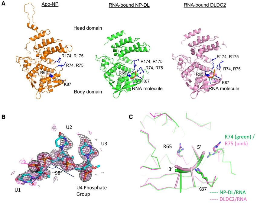
Nucleic Acids Research, 2021, Vol. 49, No. 7 4145
RNA-binding is a flexible loop spanning E73-K91, deletion were eluted with NP elution buffer (20 mM sodium phos-
of which rendered a 6.4-fold drop in RNA affinity. phate pH7, 150 mM NaCl, 500 mM imidazole). Eluted pro-
NP binds RNA without sequence specificity, although teins were further purified with a heparin column and eluted
it has higher affinity towards C and U (12,13). Each NP with 1.5 M NaCl in 20 mM sodium phosphate pH 7. For
roughly binds 24 nucleotides (14), while more recent evi- proteins used for crystallization screenings, an additional
dence supports the presence of RNA secondary structures step of gel filtration was performed with Superdex 200 (GE
which remain unbound by NP, and is proposed for RNA– Healthcare) in 20 mM 3-(N-morpholino)propanesulfonic
RNA interaction during genome packaging (15,16). The acid (MOPS) pH 7, 150 mM NaCl. NP mutant proteins
affinity of NP towards RNA is dependent on RNA length. used in this study is described in Table 1.
Yamanaka et al. (17) have showed that NP binds RNA of
at least 15 nucleotides in length. RNA binding to NP is me-
Crystallization of NP-RNA complexes
diated by arginines on NP (18), consistent with recent find-
ings that RNA binding affinity is also dependent on salt- NP from gel filtration was pooled and concentrated to
Downloaded from https://academic.oup.com/nar/article/49/7/4144/6204651 by guest on 29 October 2021
concentration (19). 8–10 mg/ml using Amicon-ultra centrifugal filter unit at
Here, by solving the crystal structures H5N1 NP–RNA MWCO 10,000 kDa (Millipore). NP was mixed with 2 -O-
complexes, we specify the importance of R65 and K87 methylated RNA oligos (Ribobio) at molar ratio of 1:1.5
during the capture of RNA molecule. Our structures not and incubated in 16 ◦ C for 1 h or overnight. Protein-
only demonstrate previously uncharacterized amino acids RNA mixture was centrifuged to remove possible pre-
in close contact with incoming RNA, but also describe cipitation before used for crystallization screening. Ini-
residues reported to be important for RNA binding. We tial crystallization conditions were picked after surveying
also demonstrate the regulatory role of the C-terminal through commercially available crystallization kits. RNA
residues of NP on RNA binding. Based on crystallographic with length of 5, 8, 9 and 24 nucleotides were tested, but
and microscale thermophoresis data, we propose a mecha- only the 8-mer and 9-mer oligoes led to diffractable crys-
nism by which RNA is recruited to NP molecules. tals. Crystals for NP tail-loop deletion mutant (NP-DL)
and a 9-mer poly-uridine RNA were obtained in 0.1 M
MATERIALS AND METHODS 2-(N-morpholino)ethanesulfonic acid (MES) pH 6.0, 0.1
M NaCl and 11% (w/v) PEG2000. Crystals for NP with
Plasmids and reagents two extreme C-terminal residues truncated (NP-DLDC2)
cDNA of A/HK/483/97 (H5N1) NP was described pre- and an 8-mer poly-uridine RNA were obtained in 0.1 M 4-
viously (2). pET28a vector was obtained from Invitrogen. (2-hydroxyethyl)-1-piperazineethanesulfonic acid (HEPES)
RNA oligos for crystallization and Kd determination were pH 7.5 and 5% propan-2-ol.
obtained from Ribobio, Guangzhou, China. C-terminal
peptide of NP was obtained from GL Biochem, Shanghai, Structure determination, processing and refinement
China.
Crystals were harvested into a 1:1 mixture of reservoir
buffer and sucrose (final concentration 17% as cryo-
Protein expression and purification
protectant) and then flash-frozen in liquid nitrogen. All
NP was subcloned onto pET28a vector and mutations datasets were collected at Shanghai Synchrotron Radia-
were introduced where appropriate by overlapping exten- tion Facility, Shanghai (SSRF, Shanghai, China) beamline
sion PCR. NP and variant inserts were double-digested BL17U1. Data reduction was performed within the CCP4
with EcoRI and HindIII and ligated using T4 DNA lig- suite (20,21). Structure of NP-DL/RNA was determined
ase (ThermoFisher). NP and variant proteins were over- by molecular replacement using an unpublished H5N1
expressed in Escherichia coli BL21(DE3)pLysS cells as re- NP tail-loop deletion mutant as search model (available
combinant proteins tagged with hexahistidine at the N- upon request). Structure of NP-DLDC2/RNA was deter-
terminus. Cells were grown in LB medium supplemented mined by molecular replacement using NP-DL/RNA com-
with kanamycin and chloramphenicol. Isopropyl -D-1- plex with RNA moiety manually removed as search model.
thiogalactopyranoside (IPTG) at a final concentration of All molecular replacement calculations were performed in
0.4 mM was added at OD600 = 0.6–0.8 to induce protein Phaser-MR (22). Model building was carried out in Coot
expression. Cells were cultured at 21◦ C with agitation at 210 (23) with the RNA nucleotides built and fit manually. Struc-
rpm for 12–16 h and harvested by centrifugation. Cells were tures were iteratively refined with Refmac/CCP4 suite (24).
then resuspended and sonicated in cold NP lysis buffer [20 Structures were validated with MolProbity (25). Protein
mM sodium phosphate pH7, 1.5 M sodium chloride, 1 mM structures were visualized in PyMOL (DeLano 2002, The
phenylmethylsulfonyl fluoride (PMSF)]. Whole cell lysate PyMOL Molecular Graphics System, Schrödinger, LLC).
was cleared by centrifugation at 21 000 g for 1 h at 4 ◦ C. Su- Protein-RNA interaction was analyzed by PISA (26) and
pernatant was loaded onto a nickel affinity column. After LigPlot+ (27).
initial washing with NP lysis buffer, ribonuclease A (RNase
A) from bovine pancreas (Sigma) was added at 1 U/ml to
Dissociation constant determination using microscale ther-
remove residual endogenous RNA from E. coli. The nickel
mophoresis
affinity beads were incubated at room temperature for 1
h with shaking and then washed extensively with NP ly- 9-nucleotide poly-uridine RNA oligos were obtained from
sis buffer supplemented with 50 mM imidazole. Proteins Ribobio with a Cy5 tag at the 5 end and were methy-
4146 Nucleic Acids Research, 2021, Vol. 49, No. 7
Table 1. Influenza A NP mutants generated
NPA mutant Mutations NPA mutant Mutations
NP-DL 402–428 DL87 402–428, K87E
DLDC2 402–428, 497–498 DL174 402–428, R174E
DLDC3 402–428, 496–498 DL87–74 402–428, R74E, K87E
DLDC4 402–428, 495–498 DL87–75 402–428, R75E, K87E
DLDC8 402–428, 491–498 DL87–65 402–428, R65E, K87E
DL65 402–428, R65E DL65–75 402–428, R65E, R75E
DL72 402–428, D72K DL65–174 402–428, R65E, R174E
DL74 402–428, R74E DL3A 402–428, Y148A, R152A, R156A
DL75 402–428, R75E
lated at the 2 -O positions. NPA or variants were concen- trant to crystallization. Eventually, we were able to co-
Downloaded from https://academic.oup.com/nar/article/49/7/4144/6204651 by guest on 29 October 2021
trated to concentrations as indicated in the text or figures crystallize the obligated monomeric mutant of NP, NP-
and buffer-exchanged to MST buffer (PBS pH 7.4, 0.1% DL, with a 9-mer RNA. Crystal structure of NP-DL/9-
Tween-20). MST experiments were performed on a Mono- mer RNA complex was solved to 2.3 Å in P1 space group.
lith NT.115 machine (Nanotemper, Munich, Germany). 10 There were two NP molecules in an asymmetric unit, while
l of RNA at 50 nM was titrated against a serial dilution of three nucleotides could be modelled to chain B. We also se-
proteins so that each sample was at a total volume of 20 L. rially truncated NP-DL at the C-terminus to give DLDC2,
RNA-protein mixtures were briefly centrifuged before load- DLDC3, DLDC4 and DLDC8 variants. Nevertheless, only
ing into Nanotemper’s Standard-Treated capillaries. Mea- DLDC2 produced diffraction quality crystals when co-
surements were made in 23◦ C using 15% LED power and crystallized with RNA nucleotides. Hence we also solved
20% MST power. the crystal structure of DLDC2/8-mer RNA complex and
refined it to 3 Å in P1 space group, with also two NP
molecules in one asymmetric unit. Data collection and re-
Competition assay finement statistics for both complexes are detailed in Table
9-Nucleotide poly-uridine RNA oligos methylated at the 2 - 2.
O positions was also obtained from Ribobio. The DLDC8 The overall fold of the protein core of NP-DL was highly
variant was concentrated to 2.2 uM and buffer-exchanged similar to the wild-type apo-NP (PDB 2Q06), indicating
to MST buffer (PBS pH 7.4, 0.1% Tween-20). A short that the structure was not significantly distorted upon dele-
peptide, corresponding to the C-terminal eight residues of tion of the tail-loop (Figure 1A). The root-mean-square de-
NPA, with the sequence DNAEEYDN, was mixed with viation (RMSD) between NP-DL and apo-NP (PDB 2Q06)
DLDC8 at a molar ratio of 1:20. MST experiments were was 0.78 Å (over 386 C␣ atoms in chain A). Residues 23–
performed on a Monolith NT.LabelFree machine (Nan- 75, 87–201, 206–207, 211–388, 438–452, 462–497 of chain
otemper, Munich, Germany). 10 l of protein/peptide mix- A and 22–78, 85–388, 437–453, 458–497 of chain B were
ture was titrated against a serial dilution of 9-mer RNA modelled unambiguously. Deletion of the tail-loop (S402–
so that each sample was at 20 l. RNA-protein mixtures A428) renders the flanking N- and C- terminal sequences
were briefly centrifuged before loading into Nanotemper’s flexible. Also, only part of region I204–R214 could be mod-
Standard-Treated capillaries for LabelFree machine. Mea- elled in our structures. This region encompasses a large por-
surements were made in 23◦ C using 10% LED power and tion of a nuclear localization signal (K198–R216) on NP
40% MST power. and is exposed on the surface. On the other hand, residues
K91–K112 in our structures formed a well-structured beta-
sheet motif which was more ordered when compared to
Microscale thermophoresis data analysis wild-type NP. In the DLDC2 structure, residues 23–77, 87–
Data analysis was performed on Nanotemper’s 200, 213–388, 438–452, 462–496 of chain A and 21–76, 85–
MO.Affinity V2.3 software using thermophoresis sig- 202, 204–208, 215–389, 437–453, 459–496 of chain B were
nal at MST-on-time = 2.5 s (for NT115 machine) or modeled. RMSD between NP-DL and DLDC2 were 0.46
1.5 s (for LabelFree machine). Curves of Fnorm against (over 387 C␣ atoms in chain A), indicating high degree of
concentration were fitted with Kd model, assuming 1:1 resemblance between the two variants.
binding, using data points from at least three independent
measurements and with target concentration fixed. Fnorm Structure of RNA bound on NP. 9-mer and 8-mer RNA
is defined as (Fhot /Fcold ) × 1000. Obvious outlier points oligo was used, respectively, in co-crystallization with NP-
due to protein aggregation or capillary contamination were DL and DLDC2, while three nucleotides could be modelled
removed. Error bars represent mean ± standard deviation. to chain B at 1.0 in each structure (Figure 1B). Only resid-
ual density which was too weak to be assigned was observed
at the equivalent position at the chain A. These nucleotides
RESULTS were bound to a cavity on the NP surface at the body do-
main defined by positive residues R65, R74/75 and K87
Structure of NP bound with RNA
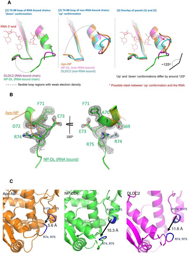
(Figure 1C). The interacting interface spanned an area of
Structure of NP. Despite various NP structures are avail- 235 and 255 Å2 , respectively, between the RNA and the pro-
able by crystallography, NP–RNA complexes are recalci- tein core in NP-DL and DLDC2 complexes. The position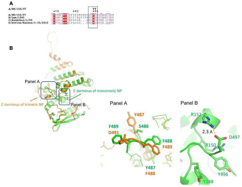
Nucleic Acids Research, 2021, Vol. 49, No. 7 4147
Table 2. Data collection and refinement statistics for NP–RNA complexes. Values in brackets are for the highest-resolution shell. RMSD: root-mean-
square-deviation from ideal values. Ramachandran analysis was done by MolProbity (25)
NP-DL-RNA DLDC2-RNA
0.1 M MES, pH 6.0, 0.1 M NaCl and
Crystallization condition 11% PEG2000 0.1 M HEPES, pH 7.5 and 5% propan-2-ol
Ligand 9-mer poly-uridine RNA 8-mer poly-uridine RNA
Beamline SSRF BL17U1 SSRF BL17U1
Data collection
Space Group P1 P1
Cell dimensions a, b, c (Å) ␣, , ␥ 54.0, 60.6, 82.7 106.7, 109.0, 96.6 53.8, 60.1, 81.4 107.3, 106.7, 96.1
(degrees)
Resolution (Å) 25.52–2.30 (2.42–2.30) 38.58–3.00 (3.18–3.00)
Rmerge 10.2% (25.3%) 16.8% (17.1%)
I/I 7.1 (2.5) 5.8 (3.6)
Downloaded from https://academic.oup.com/nar/article/49/7/4144/6204651 by guest on 29 October 2021
Completeness 90.4 (91.6) 86.1 (88.2)
Multiplicity 1.9 (1.9) 1.9 (2.0)
Total observations 69 016 (10198) 30 372 (5137)
Total unique 37106 (5475) 15710 (2610)
Refinement
No. of atoms 6727 (overall), 6498 (macromolecules), 6421 (overall), 6309 (macromolecules), 63
74 (ligands), 155 (waters) (ligands), 49 (waters)
Resolution (Å) 24.50–2.30 36.62–3.00
Rwork /Rfree 0.193/0.248 0.289/0.332
RMSD bond length (Å) 0.0102 0.007
RMSD angle (◦ ) 1.710 1.312
MolProbity score 2.23 2.17
Ramachandran Favored 96.6%, allowed 2.1%, outlier Favored 96.7%, allowed 3.2%, outlier
1.25% 0.13%
and orientation of the three nucleotides were both highly had been suggested to capture RNA from ambient environ-
similar in the NP-DL-RNA and DLDC2-RNA structures, ment while the R174, R175 had been proposed to be impor-
suggesting the interaction is reproducible crystallographi- tant for correct positioning of the RNA (2). The changes in
cally. These nucleotides were in close proximity to the flexi- local interaction environment should be implicated in the
ble 74–88 loop previously proposed for RNA capture (2). mechanism of RNA binding.
The RNA backbone adopted a special conformation so
that in the NP-DL complex, the 5 (U1) and the middle Microscale thermophoresis identified important elements for
(U2) nucleotides were twisted to give an angle of 98o (Fig- RNA binding. Single-point and double-point mutants on
ure 1B). Similarly, a backbone twist of 96o was also ob- NP-DL were designed based on the two NP structures. We
served for the three equivalent nucleotides in the DLDC2 then utilized microscale thermophoresis (MST) to deter-
complex. In fact, conformations of these three nucleotides mine the dissociation constants (Kd ) of NP variants against
in the two structures were very similar (Figure 1C). All three a Cy5-tagged 9-mer RNA oligo (Table 4 and Supplemen-
nucleotides adopted the anti- conformation. The two uracil tary Figures S2 and S3). NP-DL bound to 9-mer RNA
bases of U2 and the 3 nucleotide (U3) were almost co- with Kd of 4.2 M. DLDC2 bound to this RNA with a
planar to one another, as a result base stacking between similar affinity, with Kd of 4.5 M. Single-point variants
them was only minimal. The RNA molecule was mainly DL65, DL74 and DL87 each decreased affinity to RNA for
held by polar contacts (Table 3 and Supplementary figure about 2-fold, with Kd of 9.2 M, 10.5 M and 10.9 M re-
S1). Extensive hydrogen bonding was identified between spectively. Effect of mutation on R75 was more prominent,
phosphate groups and NP side chains. U1 phosphate was where a decrease in affinity of about 4-fold was observed (Kd
in contact with R74, U2 phosphate was held by S69 and = 17.6 M). We considered these fold-changes reasonable
T92, while U3 phosphate interacted with K87 (Table 3 and taking into account that the RNA relied on multiple contact
Figure 2A). points for binding. Double mutants DL87–65, DL87–75,
To sustain the characteristic conformation of the bases, DL65–75 and DL87–74 were then generated which showed
side chain of R65 was within less than 3 Å to O2 of U1 around 5- to 6-fold decrease in RNA-binding affinity. Taken
uracil base. R175 side chain was also in close proximity to together, the MST results were consistent with the bind-
O4 position of U1 base at around 4 Å. O4 of U2 uracil ing models observed in the two NP structures. DL174 only
base interacted with main chain nitrogen of D88. O3 of demonstrated a Kd of ∼52 M, which was more than 10-
U3 ribose was held by side chain of S367. These polar con- fold decrease than NP-DL. The DL65–174 double mutant
tacts were conserved in the DLDC2 complex, except that interacted with RNA very weakly at a Kd of ∼92 M, equiv-
phosphate of U1, which interacted with R74 in the NP-DL alent to a >20-fold drop.
complex, formed hydrogen bond with R75 instead (Figure
2B and Supplementary figure S1). In both structures, a salt Structural analysis of the NP-RNA complexes. By compar-
bridge between D72 and R175 was possible. R74, R75 and ing our structures with the wild-type H5 NP (PDB 2Q06),
R174, R175 formed a positive groove and were located re- we observed different conformations within the loop re-
spectively on two flexible loops (Figure 1). The 74–88 loop gions of NP. As shown in Figure 3A and 3B, the 74–88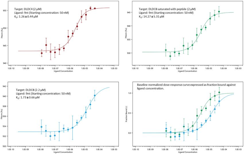
4148 Nucleic Acids Research, 2021, Vol. 49, No. 7
Downloaded from https://academic.oup.com/nar/article/49/7/4144/6204651 by guest on 29 October 2021
Figure 1. (A) Apo-NP (PDB 2Q06) and the obligate monomeric NP-DL and DLDC2 shown in the same orientation. Three RNA nucleotides are modeled
onto the body domain near the 74–88 loop. The proteins share highly similar tertiary folds demonstrating neither RNA binding nor tail-loop deletion
distorts the overall structure. G1 residues (R74, R75, R174, R175), R65 and K87 are marked in blue in the structures. (B) 2Fo – Fc omit maps were
generated for the nucleotides modeled on NP-DL (black: simple omit map; pink: simulated-annealing omit map) and contoured at 1 as shown. (C)
The RNA moieties on NP-DL and DLDC2 are situated in a cavity defined by R65, R74/75 and K87. The conformations of two RNA moieties do not
deviate significantly. Protein cores are shown as ribbons (NP-DL: green; DLDC2: magenta). RNA is shown as cartoon in green (NP-DL/RNA) and pink
(DLDC2/RNA).
loop adopted two major orientations. The C-terminal end, the ‘down’ conformation can accommodate the RNA, be-
roughly after K87 of the loop, remained at about the same cause side-chains of N76, R77 and Y78 would clash with
orientation in all protomers. The N-terminal portion (from the 5 -end of the RNA moiety if arranged in ‘up’ conforma-
D72 onwards) in the RNA-bound chains adopted a ‘down’ tion. Taken together, we conclude that during the capture of
conformation whereas the non-RNA-bound chains had 74– RNA, flexibility of the 74–88 loop leads to dynamic changes
88 loops resembling wild-type NP, adopting an ‘up’ con- of local loop conformations. The overall result of this flexi-
formation. The two conformations differ by about 125o . It bility is the opening and closure of positive G1 groove (de-
should however be mentioned that also because of loop flex- fined by R74, R75, R174 and R175) (Figure 3C).
ibility, electron density for the tip portion of the loop was Consistent with the H1 NP tail-loop deletion and R416A
weak despite effort on map sharpening. As a result, a com- monomer structures, the G2 binding site (defined by R150,
plete loop could not be modeled. The ‘down’ conforma- R152, R156 and R162) was blocked by the C-terminal
tion could be stabilized by the salt bridge between R175 residues of NP protein. We confirmed the contribution of
and D72. By MST analysis, a D72K NP variant (DL72) G2 groove to RNA binding by demonstrating a drop of
recorded a drop in RNA affinity by ∼3-fold. We also sur- RNA affinity to the DL3A variant, harboring triple muta-
veyed all influenza A NP structures in the Protein Data tions Y148A–R152A–R156A, by >4-fold. Our structures
Bank and observed both conformations in the RNA-free showed that the C-terminal tail in H5 NP made more ex-
state. However, it was obvious from our structures that only tensive polar contacts with the protein core, compared withNucleic Acids Research, 2021, Vol. 49, No. 7 4149
Table 3. Interactions between RNA and the protein core identified in the Table 4. Kd values of NP mutants against 9-mer RNA measured by MST
NP-RNA complexes
Fold-change
Interacting RNA moiety Interacting RNA moiety Kd value (relative to
Side-chain on NP-DL (shortest on DLDC2 (shortest NP variants (M) NP-DL)
interactions distance in Å) distance in Å)
NP-DL (402–428) 4.17 ± 0.26 1
R65 U1/O2 (2.96) U1/O2 (3.41) DL65 (402–428, R65E) 9.22 ± 0.89 –2.2
R74 U1/PO4 (2.51) Not applicable DL72 (402–428, D72K) 11.96 ± 1.08 –2.9
R75 Not applicable U1/PO4 (4.16) DL74 (402–428, R74E) 10.49 ± 0.76 –2.5
K87 U3/PO4 (3.33) U2/PO4 (3.64), U3/PO4 DL75 (402–428, R75E) 17.64 ± 1.07 –4.2
(3.52) DL87 (402–428, K87E) 10.94 ± 1.03 –2.6
S69 U2/PO4 (2.50) U2/PO4 (3.03) DL174 (402–428, R174E) 52.53 ± 4.08 –12.6
T92 U2/PO4 (2.63) U2/PO4 (3.05) DL87–65 (402–428, R65E, K87E) 26.92 ± 1.83 –6.5
R175 U1/O4 (3.26) U1/O4 (3.98) DL65–75 (402–428, R65E, R75E) 21.57 ± 1.98 –5.2
S367 U3/O3 (3.33) U3/O3 (3.29) DL75–87 (402–428, R75E, K87E) 19.16 ± 0.74 –4.6
DL74–87 (402–428, R74E, K87E) 27.32 ± 2.63 –6.5
Downloaded from https://academic.oup.com/nar/article/49/7/4144/6204651 by guest on 29 October 2021
Main-chain Interacting RNA Interacting RNA DL65–174 (402–428, R65E, R174E) 91.86 ± 15.46 –22.0
interactions moiety on NP-DL moiety on DLDC2 DLDC2 (402–428, 497–498) 4.54 ± 0.40 –1.1
(shortest distance in Å) (shortest distance in Å) DL-3A (402–428, Y148A, R152A, 18.47 ± 1.23 –4.4
R156A)
D88/N U2/O4 (2.66) U2/O4 (3.00)
the H1 NP. We identified three pairs of salt bridges, namely
D491–R355, E494–R361 and D497–R152. The phenyl ring
of Y496 was adjacent to aromatic Y148 with the two rings
roughly perpendicular to each other. R150 was also4150 Nucleic Acids Research, 2021, Vol. 49, No. 7
Downloaded from https://academic.oup.com/nar/article/49/7/4144/6204651 by guest on 29 October 2021
Figure 3. (A) Here we align the 74–88 loops on all RNA-bound chains in panel (1) and non-RNA bound chains from our structures to apo-NP (PDB
2Q06) in panel (2). Peptides are colored according to colors of labels. When bound with RNA, the loop adopts a ‘down’ conformation, in contrast to the
‘up’ conformation when RNA-free. Due to the weak density, part of the loops could not be modeled confidently and is indicated by dashes here. Panel (3)
is an overlay of panels (1) and (2) and the possible clash between the loop in ‘up’ conformation with the 5 RNA is indicated. (B) 2mFo – DFc map (black,
contoured at 1) for residues F71-R75 (green) on NP-DL complex is shown. The peptide is overlaid with the corresponding residues from apo-NP (PDB
2Q06, orange). The two peptides align with each other until D72 where they deviate to give two different conformations. (C) The 74–88 loop from apo-NP
(PDB 2Q06), NP-DL and DLDC2 are shown with positive residues R74, R75, R174 and R175 marked blue. The ‘down’ conformation of the 74–88 loop
in NP-DL and DLDC2 lead to opening up of G1 groove as indicated by increased distances between R174 to R74.Nucleic Acids Research, 2021, Vol. 49, No. 7 4151
Downloaded from https://academic.oup.com/nar/article/49/7/4144/6204651 by guest on 29 October 2021
Figure 4. (A) Alignment of NP sequences near the C-terminal region extracted from influenza A, B, C and D nucleoproteins. Conserved YFF motif is
highlighted in box. (B) In H5 NP, the C-terminal tail of obligate monomers flips onto the protein core to cover the RNA-groove and as such Y496 is
brought to vicinity to both Y148 and R150 of the positive G1 groove. D497-R152 salt-bridge is one of the stabilizing forces for this conformation. Shown
in Figure 4B is an overlay of wild-type NP (orange) and the obligate monomer NP-DL (green). Panel A highlights the rearrangement of the YFF motif
during this conformation change while panel B shows the local environment around the D497-R152 salt-bridge when NP is in monomeric state.
lar excess of peptide, affinity of DLDC8 to 9-mer RNA was NPA, NPB, NPC and NPD, further signifying its func-
significantly decreased by around 8-fold (Figure 5). Taken tional importance. We also demonstrate that flexibility of
together, the tail peptide played a role in regulating RNA 74–88 loop allows it to adopt at least two major confor-
binding. mations, with only one of them (the ‘down’ conformation)
allowing it to capture an RNA molecule. The presence of
a highly conserved aspartic acid at position 72, and the
DISCUSSION
fact that its side chain being spatially close to the side
We present here two complex structures formed between chains of R174/175 tempted us to postulate that the flex-
RNA oligo and H5 NP. Previously, we demonstrated that ibility of the 74–88 loop is regulated by the two positively
the Kd of a 24-mer RNA to NP monomers was within the charged arginine residues at 174 and 175. Previous studies
range of 0.4 to 1 M (28) which represents around 17- to by Li et al. (29) could not rescue D72A mutant virus, high-
43-fold weaker binding compared to wild-type trimeric NP lighting the biological relevance of this residue. Changes
with Kd of 23 nM (2). Here, with an RNA oligo which in loop conformations are therefore crucial to RNA bind-
was around one-third in length, the Kd between monomeric ing. Our MST results show that deleting four or more C-
NP-DL versus a 9-mer RNA was further decreased to terminal residues are sufficient to increase RNA affinity
4 M. Based on structural analysis, we have confirmed for 2- to 3-fold. Hence, deletions on the tail likely open up
residues R74, R75, R175 are important for RNA bind- an additional binding pocket on the protein. Blockage of
ing, and showed R65 and K87, two previously unchar- RNA-binding site by the tail likely accounts for the discrep-
acterized residues, to be important for RNA binding. In- ancy in RNA affinity between trimeric and monomeric NP.
deed, the positive charge at position 65 is conserved among Therefore, we have also provided additional evidence for4152 Nucleic Acids Research, 2021, Vol. 49, No. 7
Downloaded from https://academic.oup.com/nar/article/49/7/4144/6204651 by guest on 29 October 2021
Figure 5. Label-free MST was performed to determine the affinity between DLDC4 and DLDC8 NP variants towards 9-mer RNA (top-left and bottom-
left panels, respectively). By saturating DLDC8 mutant with a synthetic tail peptide, RNA binding was attenuated by 8-fold. MST curve is expressed as
Fnorm (0/00) versus concentration. Data represent mean values from at least three independent measurements. Error bars represent standard deviation. In
the bottom right panel, for comparison, MST curves were expressed as fraction of protein bound vs ligand (RNA) concentration (Blue: DLDC8; Green:
peptide-saturated DLDC8).
the role of NP loops and tail elements in regulating RNA Based on our current structural and biochemical data, we
binding. can propose a mechanism by which NP binds RNA (Sup-
Further the above evidence, following the electron den- plementary Figure S7). In the first instance, the G1 groove,
sity of phosphorous atoms we can attempt to extend the formed mainly by R74/75 and R174/175 on two flexible
nucleotide chain and trace the RNA phosphate backbone loops, is able to open and close. This movement is dependent
based on the DLDC2 complex which allows two more nu- on the N-terminal portion of the 74–88 loop. The opening
cleotides to be modelled at the 5 end. The nucleotides thus and closure of this region packages RNA to the site near
built would resemble a distorted hook with the 5 end right R65, K87, as shown in our structures. The C-terminal tail
next to the G2 groove (Supplementary figure S6A). These must displace from its current position. Currently there is
two extra nucleotides exhibit nice interaction with the pro- no direct evidence as to whether this displacement is depen-
tein. (Supplementary figure S6B and Supplementary table dent of NP-NP oligomerization. However, based on the re-
S1). Displacement of the C-terminal tail would likely facil- arrangement near the YFF motif, we postulate that NP-NP
itate the correct positioning of these nucleotides. oligomerization should aid this movement. Finally, the 5 -
In our structures, positive arginine or lysine residues are end of the RNA shifts into the G2 groove which has pos-
highly relevant for RNA binding. Intriguingly, base stack- itive charges exposed after the departure of the acidic tail
ing is minimal in our NP-RNA model, in contrast to the residues. This model is in line with our biochemical analysis
extensive base stacking in vesicular stomatitis virus (VSV) in this and our previous work (2) which showed the impor-
and rabies NP. Only 3–5 nucleotides could be recorded tance of G1 and G2 residues. Besides, molecular dynamics
in our NP-RNA structures, despite that 9-mer or 8-mer (32) had suggested several loop regions including 73–90 and
RNAs were used during crystallization. The remaining nu- 360–373 for RNA capture. We have now showcased the im-
cleotides likely hover over the protein core flexibly, implying portance of loop 74–88 on RNA-binding. Also, as observed
that significant degree of RNA disorder can be tolerated in our structure, S367 interacts with 3 end of the RNA and
in influenza NP, which is not the case in VSV and rabies R361 forms a salt bridge with E494 at the C-terminal tail.
where RNA is encapsidated in the interior of a ring of NP The importance of loop 360–373 on RNA binding is thus
oligomers (30,31). substantiated.Nucleic Acids Research, 2021, Vol. 49, No. 7 4153
Our work here not only deepens the understanding on 9. Ye,Q., Guu,T.S.Y., Mata,D.A., Kuo,R.L., Smith,B., Krug,R.M. and
the molecular basis of influenza NP-RNA binding but also Tao,Y.J. (2012) Biochemical and structural evidence in support of a
coherent model for the formation of the double-helical influenza A
offers new insights to RNA binding mechanism by NP. We virus ribonucleoprotein. MBio, 4, e00467–12.
propose that the tail peptide could be an attractive RNA 10. Ng,A.K.L., Lam,M.K.H., Zhang,H., Liu,J., Au,S.W.N., Chan,P.K.S.,
binding inhibitor to be tested further. Wang,J. and Shaw,P.C. (2012) Structural basis for RNA binding and
homo-oligomer formation by influenza B virus nucleoprotein. J.
Virol., 86, 6758–6767.
DATA AVAILABILITY 11. Donchet,A., Oliva,J., Labaronne,A., Tengo,L., Miloudi,M.C.A.,
Gerard,F., Mas,C., Schoehn,G.W.H., Ruigrok,R., Ducatez,M. et al.
CCP4 suite is downloaded under academic licenses at https: (2019) The structure of the nucleoprotein of influenza D shows that
//www.ccp4.ac.uk/download/. all Orthomyxoviridae nucleoproteins have a similar NPCORE, with
Crystal structures of NP-DL and NP-DLDC2 com- or without a NPTAIL for nuclear transport. Sci. Rep., 9, 600.
plexed with RNA are deposited with the Protein Data bank 12. Baudin,F., Bach,C., Cusack,S. and Ruigrok,R.W. (1994) Structure of
influenza virus RNP. I. Influenza virus nucleoprotein melts secondary
under accession number 7DXP and 7DKG, respectively. structure in panhandle RNA and exposes the bases to the solvent.
Downloaded from https://academic.oup.com/nar/article/49/7/4144/6204651 by guest on 29 October 2021
EMBO J., 13, 3158–3165.
13. Albo,C., Valencia,A. and Portela,A. (1995) Identification of an RNA
SUPPLEMENTARY DATA binding region within the N-terminal third of the influenza A virus
Supplementary Data are available at NAR Online. nucleoprotein. J. Virol., 69, 3799–3806.
14. Ortega,J., Martı́n-Benito,J., Zürcher,T., Valpuesta,J.M.,
Carrascosa,J.L. and Ortı́n,J. (2000) Ultrastructural and functional
ACKNOWLEDGEMENTS analyses of recombinant influenza virus ribonucleoproteins suggest
dimerization of nucleoprotein during virus amplification. J. Virol., 74,
We thank Prof Paul KS Chan (Department of Microbi- 156–163.
ology, The Chinese University of Hong Kong) for the in- 15. Lee,N., Le Sage,V., Nanni,A.V, Snyder,D.J., Cooper,V.S. and
Lakdawala,S.S. (2017) Genome-wide analysis of influenza viral RNA
fluenza H5N1 NP cDNA. We also thank Dr Andy KL Ng and nucleoprotein association. Nucleic Acids Res., 45, 8968–8977.
and Dr Edwin CY Lo for useful discussions on experimen- 16. Dadonaite,B., Gilbertson,B., Knight,M.L., Trifkovic,S., Rockman,S.,
tal design and the manuscript. Laederach,A., Brown,L.E., Fodor,E. and Bauer,D.L.V. (2019) The
structure of the influenza A virus genome. Nat. Microbiol., 4,
1781–1789.
FUNDING 17. Yamanaka,K., Ishihama,A. and Nagata,K. (1990) Reconstitution of
influenza virus RNA-nucleoprotein complexes structurally
Theme-based Research Grant under Hong Kong Re- resembling native viral ribonucleoprotein cores. J. Biol. Chem., 265,
search Grant Council (HKRGC) [T11-705/14N to 11151–11155.
P.C.S.]; NIH [HL103526 to J.H.W.]; Claudia Adams Barr 18. Elton,D., Medcalf,L., Bishop,K., Harrison,D. and Digard,P. (1999)
Award [to J.H.W.]. Funding for open access charge: The Identification of amino acid residues of influenza virus nucleoprotein
essential for RNA binding. J. Virol., 73, 7357–7367.
Chinese University of Hong Kong. 19. Labaronne,A., Swale,C., Monod,A., Schoehn,G., Crépin,T. and
Conflict of interest statement. None declared. Ruigrok,R.W.H. (2016) Binding of RNA by the nucleoproteins of
influenza viruses A and B. Viruses, 8, 247.
20. Battye,T.G.G., Kontogiannis,L., Johnson,O., Powell,H.R. and
REFERENCES Leslie,A.G.W. (2011) iMOSFLM: a new graphical interface for
1. Ye,Q., Krug,R.M. and Tao,Y.J. (2006) The mechanism by which diffraction-image processing with MOSFLM. Acta Crystallogr. D.
influenza A virus nucleoprotein forms oligomers and binds RNA. Biol. Crystallogr., 67, 271–281.
Nature, 444, 1078–1082. 21. Evans,P.R. and Murshudov,G.N. (2013) How good are my data and
2. Ng,A.K.L., Zhang,H., Tan,K., Li,Z., Liu,J.H., Chan,P.K.S., Li,S.M., what is the resolution? Acta Crystallogr. D. Biol. Crystallogr., 69,
Chan,W.Y., Au,S.W.N., Joachimiak,A. et al. (2008) Structure of the 1204–1214.
influenza virus A H5N1 nucleoprotein: implications for RNA 22. McCoy,A.J., Grosse-Kunstleve,R.W., Adams,P.D., Winn,M.D.,
binding, oligomerization, and vaccine design. FASEB J., 22, Storoni,L.C. and Read,R.J. (2007) Phaser crystallographic software.
3638–3647. J. Appl. Crystallogr., 40, 658–674.
3. Coloma,R., Valpuesta,J.M., Arranz,R., Carrascosa,J.L., Ortı́n,J. and 23. Emsley,P., Lohkamp,B., Scott,W.G. and Cowtan,K. (2010) Features
Martı́n-Benito,J. (2009) The structure of a biologically active and development of Coot. Acta Crystallogr. Sect. D Biol. Crystallogr.,
influenza virus ribonucleoprotein complex. PLoS Pathog., 5, 66, 486–501.
e1000491. 24. Kovalevskiy,O., Nicholls,R.A., Long,F., Carlon,A. and
4. Arranz,R., Coloma,R., Chichon,F.J., Conesa,J.J., Carrascosa,J.L., Murshudov,G.N. (2018) Overview of refinement procedures within
Valpuesta,J.M., Ortin,J. and Martin-Benito,J. (2012) The structure of REFMAC 5: Utilizing data from different sources. Acta Crystallogr.
native influenza virion ribonucleoproteins. Science, 338, 1634–1637. Sect. D Struct. Biol., 74, 215–227.
5. Moeller,A., Kirchdoerfer,R.N., Potter,C.S., Carragher,B. and 25. Williams,C.J., Headd,J.J., Moriarty,N.W., Prisant,M.G., Videau,L.L.,
Wilson,I.A. (2012) Organization of the influenza virus replication Deis,L.N., Verma,V., Keedy,D.A., Hintze,B.J., Chen,V.B. et al. (2018)
machinery. Science, 338, 1631–1634. MolProbity: more and better reference data for improved all-atom
6. Vreede,F.T., Ng,A.K.L., Shaw,P.C. and Fodor,E. (2011) Stabilization structure validation. Protein Sci., 27, 293–315.
of influenza virus replication intermediates is dependent on the 26. Krissinel,E. and Henrick,K. (2007) Inference of macromolecular
RNA-binding but not the homo-oligomerization activity of the viral assemblies from crystalline state. J. Mol. Biol., 372, 774–797.
nucleoprotein. J. Virol., 85, 12073–12078. 27. Wallace,A.C., Laskowski,R.A. and Thornton,J.M. (1995) LIGPLOT:
7. York,A., Hengrung,N., Vreede,F.T., Huiskonen,J.T. and Fodor,E. a program to generate schematic diagrams of protein-ligand
(2013) Isolation and characterization of the positive-sense replicative interactions. Protein. Eng., 8, 127–134.
intermediate of a negative-strand RNA virus. Proc. Natl. Acad. Sci. 28. Chan,W.H., Ng,A.K.L., Robb,N.C., Lam,M.K.H., Chan,P.K.S.,
U.S.A., 110, E4238–E4245. Au,S.W.N., Wang,J.H., Fodor,E. and Shaw,P.C. (2010) Functional
8. Chenavas,S., Crépin,T., Delmas,B., Ruigrok,R.W. and analysis of the influenza virus H5N1 nucleoprotein tail loop reveals
Slama-Schwok,A. (2013) Influenza virus nucleoprotein: structure, amino acids that are crucial for oligomerization and
RNA binding, oligomerization and antiviral drug target. Future ribonucleoprotein activities. J. Virol., 84, 7337–7345.
Microbiol., 8, 1537–1545.4154 Nucleic Acids Research, 2021, Vol. 49, No. 7
29. Li,Z., Watanabe,T., Hatta,M., Watanabe,S., Nanbo,A., Ozawa,M., (2006) Crystal structure of the rabies virus nucleoprotein-RNA
Kakugawa,S., Shimojima,M., Yamada,S., Neumann,G. et al. (2009) complex. Science, 313, 360–363.
Mutational analysis of conserved amino acids in the influenza A virus 32. Tarus,B., Chevalier,C., Richard,C.A., Delmas,B., Di Primo,C. and
nucleoprotein. J. Virol., 83, 4153–4162. Slama-Schwok,A. (2012) Molecular dynamics studies of the
30. Green,T.J., Zhang,X., Wartz,G.W. and Luo,M. (2006) Structure of nucleoprotein of influenza A virus: role of the protein flexibility in
the vesicular stomatitis virus nucleoprotein-RNA complex. Science, RNA binding. PLoS One, 7, e30038.
313, 357–360.
31. Albertini,A.A.V., Wernimont,A.K., Muziol,T., Ravelli,R.B.G.,
Clapier,C.R., Schoehn,G., Weissenhorn,W. and Ruigrok,R.W.H.
Downloaded from https://academic.oup.com/nar/article/49/7/4144/6204651 by guest on 29 October 2021You can also read