DNA hybridisation kinetics using single-molecule fluorescence imaging
←
→
Page content transcription
If your browser does not render page correctly, please read the page content below
Essays in Biochemistry (2021) 65 27–36
https://doi.org/10.1042/EBC20200040

Review Article

DNA hybridisation kinetics using single-molecule
fluorescence imaging
    Rebecca Andrews
Gene Machines Laboratory, Biological Physics Research Group, Clarendon Laboratory, Department of Physics, University of Oxford, Oxford, U.K.
Correspondence: Rebecca Andrews (rebecca.andrews@physics.ox.ac.uk)

                                                                                                                                                                                            Downloaded from http://portlandpress.com/essaysbiochem/article-pdf/65/1/27/908153/ebc-2020-0040c.pdf by guest on 29 April 2021
                                            Deoxyribonucleic acid (DNA) hybridisation plays a key role in many biological processes
                                            and nucleic acid biotechnologies, yet surprisingly there are many aspects about the pro-
                                            cess which are still unknown. Prior to the invention of single-molecule microscopy, DNA
                                            hybridisation experiments were conducted at the ensemble level, and thus it was impossi-
                                            ble to directly observe individual hybridisation events and understand fully the kinetics of
                                            DNA hybridisation. In this mini-review, recent single-molecule fluorescence-based studies
                                            of DNA hybridisation are discussed, particularly for short nucleic acids, to gain more in-
                                            sight into the kinetics of DNA hybridisation. As well as looking at single-molecule studies
                                            of intrinsic and extrinsic factors affecting DNA hybridisation kinetics, the influence of the
                                            methods used to detect hybridisation of single DNAs is considered. Understanding the ki-
                                            netics of DNA hybridisation not only gives insight into an important biological process but
                                            also allows for further advancements in the growing field of nucleic acid biotechnology.

                                          Background
                                          Deoxyribonucleic acid (DNA) hybridisation, especially of short DNAs, is an essential process in bi-
                                          ology, however, much is still unknown about the exact process of hybridisation and its kinetics. As
                                          DNA hybridisation is also used across a range of biological and biotechnological applications such as
                                          hybridisation-based next-generation DNA sequencing [1], fluorescence-based in situ imaging [2], and
                                          super-resolution imaging [3,4], it is essential to achieve a much more complete understanding of the ki-
                                          netics of hybridisation.
                                             DNA hybridisation is not a necessarily permanent reaction with DNA able to undergo many re-
                                          versible hybridisation events known as transient hybridisation. Transient DNA hybridisation occurs when
                                          a double-stranded DNA (dsDNA), see Figure 1A, is formed temporarily (for milliseconds to seconds)
                                          via base-pairing of two complementary strands; the dsDNA subsequently thermally dissociates into
                                          single-stranded DNAs (ssDNAs), see Figure 1B. Ensemble measurements have long been established to
                                          study DNA hybridisation [5] but lack the capability to directly observe heterogeneity of single DNAs.
                                             Single-molecule fluorescence imaging excites only a small volume of a sample in order to reduce the dif-
                                          fuse background fluorescence, enabling single DNA molecules to be imaged. In general, single-molecule
                                          microscopy has many key advantages over ensemble microscopy such as the ability to independently
                                          measure the association rate and dissociation rate of a reaction and also the ability to observe static and
                                          dynamic heterogeneity within samples otherwise lost to averaging in ensemble measurements. A com-
                                          mon single-molecule fluorescence microscopy technique is total internal reflection fluorescence (TIRF)
                                          microscopy (Figure 1C), where the excitation light is totally internally reflected at the boundary to a
Received: 25 September 2020               glass slide-mounted sample [6]. The resulting non-propagating exponentially decaying evanescent wave
Revised: 17 December 2020                 penetrates the sample 100–200 nm creating a small illumination volume, key for single-molecule mi-
Accepted: 21 December 2020                croscopy. TIRF microscopy restricts spatially where biomolecules can be observed limiting reactions to,
Version of Record published:              or just above, the surface of the glass slide, lending itself well to experiments with surface-immobilised
16 April 2021                             molecules. A modification that can be made to traditional TIRF microscopes to improve the imaging of

© 2021 The Author(s). This is an open access article published by Portland Press Limited on behalf of the Biochemical Society and distributed under the Creative Commons Attribution   27
License 4.0 (CC BY).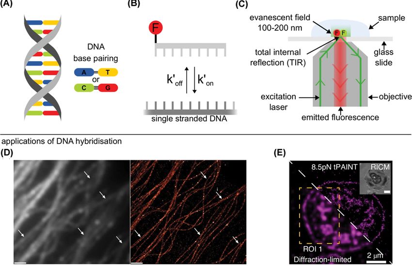
Essays in Biochemistry (2021) 65 27–36
                                                                                                                                         https://doi.org/10.1042/EBC20200040

                                                                                                                                                                                          Downloaded from http://portlandpress.com/essaysbiochem/article-pdf/65/1/27/908153/ebc-2020-0040c.pdf by guest on 29 April 2021
         Figure 1. Single-molecule applications of transient DNA hybridisation
         (A) DNA exists as a double helix with DNA base pairing rules: adenine (A) - thymine (T) and cytosine (C) - guanine (G), two and three
         hydrogen bonds between pairs respectively (black lines). (B) Transient hybridisation can be characterised by the association rate
         (kon ) as the DNA binds and the dissociation rate (koff ) as the DNA unbinds. Top strand is fluorescently labelled (F). (C) Fluorophores
         are excited and fluorescence is collected for a single-molecule total internal reflection fluorescence (TIRF) microscope. Evanescent
         field, created by total internal reflection of incident laser beam, excites fluorophores close to the surface (100–200 nm). (D) Left:
         Diffraction limited image of microtubules imaged using the ensemble fluorescence produced by the transient hybridisation of la-
         belled DNAs. Right: Super-resolved image of microtubules imaged using the transient hybridisation of single-labelled DNAs. White
         arrows indicated areas that are significantly enhanced by super-resolution imaging. Scale bar: 1 μm. (Reprinted from Jungmann
         et al., Nat. Methods, 2014; used with permission). (E) Bottom-half: diffraction limited image, top-half: strain-free tension-PAINT
         (sf-tPAINT) image of 8.5 pN integrin forces during platelet activation. Inset is a reflection interference contrast microscopy (RICM)
         image (Reprinted from Brockman et al., Nat. Methods, 2020: used with permission).

         single-molecule surface-based DNA hybridisation is changing the intensity profile of the incident laser beam to a
         flat-top profile instead of a gaussian for even illumination of the field of view [7].
            Single-molecule fluorescence microscopy allows individual DNA hybridisation events to be observed directly,
         therefore, enabling the investigation of the kinetics of such reactions. Commonly in surface-based studies, that use
         TIRF microscopy, ssDNAs are labelled with fluorescent dyes and recorded for seconds to minutes as they bind to a
         complementary ssDNAs appropriately spaced on a solid support. Transient hybridisation can be quantified as two
         separate processes, with binding of the nucleic acids described by an association rate, kon , and the separation of the
         double-stranded nucleic acid described by a dissociation rate, koff , see Figure 1B. For the reaction shown in Figure 1B,
         the association rate, kon , is dependent on the concentration of the fluorescently labelled ssDNAs available for bind-
         ing. Another way the binding can be quantified, taking into consideration the concentration of the ssDNAs, is by the
         association rate constant k on (M−1 s−1 ). For labelled ssDNA concentrations below 300 nM, one can assume that there
         are no reactions between the labelled ssDNAs, making the dissociation rate, koff , equivalent to the dissociation rate
         constant, k off (s−1 ) [8]. Further, under the assumption of no photobleaching of the fluorescent dyes, the association
                                                         −1
         rate constant, k on , can be written as kon = τunbound [ssDNA]−1 and the dissociation rate constant, k off , can be written
                    −1
         as koff = τbound , where τunbound is the average time between hybridisation events and τbound is the average time of a
         hybridisation event [9].

28             © 2021 The Author(s). This is an open access article published by Portland Press Limited on behalf of the Biochemical Society and distributed under the Creative Commons
     Attribution License 4.0 (CC BY).
Essays in Biochemistry (2021) 65 27–36
https://doi.org/10.1042/EBC20200040
Using various single-molecule fluorescence microscopy techniques, the transient binding of fluorescently labelled
DNAs has been utilised to create biotechnologies such as DNA points accumulation for imaging in nanoscale topog-
raphy (DNA-PAINT) [3]. DNA-PAINT uses repeated transient hybridisation of short fluorescently labelled DNA to
an immobilised complementary DNA to create a super-resolved image, see Figure 1D, with the method being quanti-
tative [10] and having the ability to create multicolour images [11,12], even to create 124-plex images within minutes
[13]. Since its invention, DNA-PAINT has been implemented alongside many different super-resolution microscopy
techniques in order to image targets inside cells, such as structured illumination microscopy (SIM) [14], stimu-
lated emission depletion (STED) microscopy [4], stochastic optical reconstruction microscopy (STORM) [15,16]
and spinning disk confocal (SDC) microscopy [17]. DNA-PAINT so far has a wide range of biological applications
such as imaging synaptic proteins [18], imaging forces inside live cells [19] (Figure 1E), creating 3D images of inter-
nal cell structures [11,16] and immunostaining of neuronal cells, tissues and microtubules [4,14] (Figure 1D). For
DNA-PAINT, and other biotechnologies, a greater understanding of the kinetics of DNA hybridisation will lead to
the optimisation of hybridisation assays making these technologies more powerful.
Downloaded from http://portlandpress.com/essaysbiochem/article-pdf/65/1/27/908153/ebc-2020-0040c.pdf by guest on 29 April 2021
Single-molecule fluorescence microscopy techniques and
hybridisation kinetics
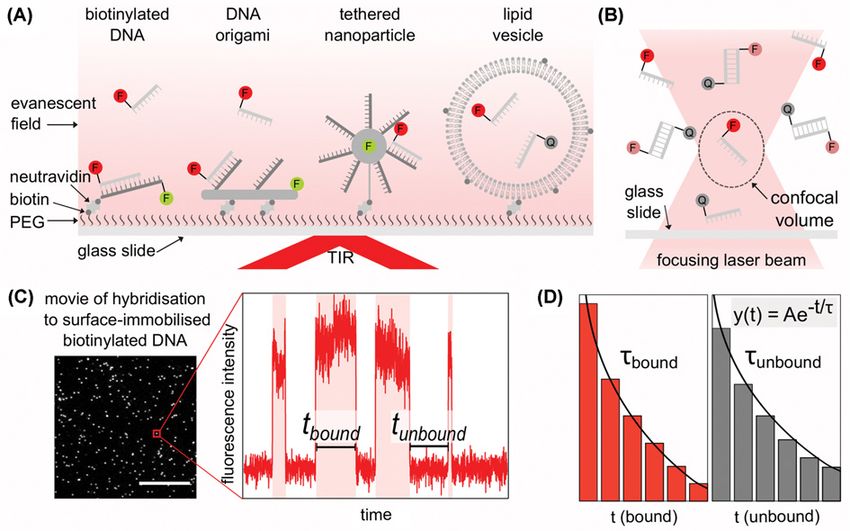
Surface-immobilised single-molecule DNA hybridisation
Commonly, surface-immobilised DNA hybridisation experiments are conducted using TIRF microscopy, where la-
belled or unlabelled ssDNAs (probes) hybridise with ssDNAs immobilised on a passivated glass surface. Immobilisa-
tion is usually achieved through a biotin–neutravidin linker; however, there are alternative ways to immobilise DNA
on the surface, such as by using ssDNAs immobilised on DNA origami [20], on surface-tethered nanoparticles [21]
or on long tethers [22], see Figure 2A. A large advantage of studying DNA hybridisation with immobilised DNA, is
the ability to observe the same DNA molecule during multiple hybridisation events; however, the local environment
for surface-immobilised DNA is drastically different.
From single-molecule fluorescence movies of surface-immobilised DNA the kinetics of hybridisation can be cal-
culated through the changes in the intensity of fluorescence measured over time – the exact changes depend on the
experimental fluorophore design. For example, a fluorophore can be used for localisation of a binding site either on the
immobilised DNA, or in close proximity, and a differently coloured fluorophore can be directly excited on the hybri-
dising DNA with a recorded increase in fluorescence upon hybridisation. A similar design uses a pair of fluorophores,
one on each ssDNA, to observe binding through the use of Förster resonance energy transfer (FRET) between the
two fluorophores in close proximity. Another method of fluorophore labelling, is labelling one set of the ssDNAs with
a fluorescence quencher, a non-fluorescent molecule that absorbs energy, and when the quencher strand hybridises
with a fluorescently labelled strand, the reduction/absence of fluorescence indicates binding. Figure 2C shows a field
of view from a movie of labelled ssDNAs being directly excited when binding to surface-immobilised ssDNAs that
were previously localised using a different coloured fluorophore, first example in Figure 2A. For each single-molecule
a fluorescence intensity vs. time trace can be plotted for the length of the movie. By measuring the duration of bind-
ing events (tbound ) and the time between binding events (tunbound ) frequency histograms can be created and fitted by
a decaying exponential function. From the decaying exponential fit, the average time of hybridisation (τbound ) can be
calculated from the tbound histogram and the average time between hybridisation events (τunbound ) can be calculated
from the tunbound histogram, as shown in Figure 2D.
Compared with solution-based experiments, ssDNAs that hybridise with surface-immobilised DNAs experience
a greater repulsive electrostatic force [23–25]. When modelled, the electrostatic repulsion from surface-immobilised
ssDNAs is the main reason for reduced k on when compared with solution-based models [26]. Crowding of the DNA
at high densities [27] and non-specific interactions of the probes with the surface [28] also contribute to a reduction
in k on . Intriguingly, single-molecule measurements have shown that after non-specific binding, probes perform a
search process on the surface but the result is a low yield of hybridisation with the immobilised DNA, therefore, there
is no increase in k on [27,29]. On the other side of the reaction, single-molecule surface-based studies are able to show
k off is a combination of multiple distinct dissociation rates, corresponding to different dissociation behaviours, rather
than a single average rate as seen in ensemble studies [29,30]. Interestingly, the type of surface used can also affect
k off with hydrophilic surfaces decreasing the average k off when compared with hydrophobic surfaces [31].
There are alternative single-molecule fluorescence methods for surface-based experiments which do not directly
tether the DNA, such as trapping or through confinement of the hybridising DNAs. Surface-tethered lipid vesicles
(∼100 nm in diameter) which contain fluorescently labelled biomolecules are an example of confinement [32]; within
each vesicle, the DNAs can freely diffuse and interact without being modified or hindered by tethering allowing DNA
© 2021 The Author(s). This is an open access article published by Portland Press Limited on behalf of the Biochemical Society and distributed under the Creative Commons Attribution 29
License 4.0 (CC BY).Essays in Biochemistry (2021) 65 27–36
https://doi.org/10.1042/EBC20200040
Downloaded from http://portlandpress.com/essaysbiochem/article-pdf/65/1/27/908153/ebc-2020-0040c.pdf by guest on 29 April 2021
Figure 2. Single-molecule methods to measure DNA hybridisation kinetics
(A) Methods for surface-immobilised single-molecule DNA hybridisation measurements. From left to right: single stranded DNA
(ssDNA) immobilised via biotin/neutravidin, ssDNAs immobilised via DNA origami, ssDNAs immobilised via a tethered nanopar-
ticle, ssDNAs confined via immobilised lipid vesicle. Polyethylene glycol (PEG) on surface of glass slide to allow immobilisation
via biotin and neutravidin. In the sample, ssDNA labelled with fluorophores (F) or fluorescence quenchers (Q) are excited by an
evanescent field from total internal reflection (TIR) of the incident laser beam. (B) Confocal microscopy for single-molecule imaging
of hybridisation between fluorescently labelled ssDNAs and ssDNAs labelled with a quencher within the confocal volume (dashed
ellipse). (C) Example of field of view from movie of hybridisation of fluorescently labelled ssDNAs to surface-immobilised DNA. Scale
bar 10 μm. Red box: Example fluorescence intensity vs. time trace for a single unlabelled surface-immobilised DNA undergoing
transient hybridisation with fluorescently labelled ssDNAs. A hybridisation event is characterised as a rise in fluorescence. The time
for hybridisation can be measured as tbound and the time between binding events measured as tunbound . (D) Histograms of tbound
and tunbound , fitted with a decaying exponential function (y(t) = Aexp(−t/τ), where A is a constant, τ is the calculated average time)
to calculate the average bound time, τbound and the average unbound time τunbound .
hybridisation observed [33], shown in Figure 2A. Since the membrane of lipid vesicles acts as a barrier to exchange
molecules with the exterior of the vesicles, considerable effort has been taken to make them porous [34–36]. Even with
such alternatives, there is no universal method of surface immobilisation which allows DNA hybridisation kinetics
to be observed as they would be free in solution.
Single-molecule DNA hybridisation in solution
Traditionally, ensemble fluorescence experiments of DNA hybridisation were conducted in solution; currently, this
is also a method that makes this possible at the single-molecule level. Through the use of a confocal microscope
(Figure 2B), a small volume of a sample (∼0.2 fl) [37] can be fluorescently illuminated, enabling imaging of single
molecules as they diffuse in solution. However, standard confocal microscopy only provides a brief snapshot (∼1 ms)
as each molecule randomly diffuses through the excitation volume. Therefore, standard confocal microscopy is not
well suited to observing multiple hybridisation events on a single molecule over long time periods, which is needed
to gather data on hybridisation kinetics.
A new confocal microscopy method, 3D single-molecule tracking (3D-SMT), has been developed to observe tran-
sient DNA hybridisation in solution over longer time periods (∼1 s) [38]. As the total maximum acquisition time
30 © 2021 The Author(s). This is an open access article published by Portland Press Limited on behalf of the Biochemical Society and distributed under the Creative Commons
Attribution License 4.0 (CC BY).Essays in Biochemistry (2021) 65 27–36
https://doi.org/10.1042/EBC20200040
is short, only fast transient hybridisation events between very short DNAs (8 nucleotides or less) can be recorded.
Another novel approach for longer observations of DNA hybridisation in solution uses fluorescence to monitor the
position of a labelled DNA whilst in the presence of an electric field. The changes in the DNA diffusion and drift re-
veals the kinetics of hybridisation [39]. Clearly, measuring DNA hybridisation in solution would avoid the changes in
the kinetic rates seen for surface immobilised assays, but currently there is no solution-based method able to provide
as higher throughput on hybridisation kinetics over longer periods of time as surface-immobilised assays.
Effect of fluorophores on hybridisation kinetics
DNA hybridisation can simply be modelled as the interaction of two ssDNAs, however, in fluorescence microscopy
one or both of the DNAs is modified for the inclusion of a small-molecule fluorescent dye. Fluorescent dyes are usually
chosen specifically for their optical properties with less consideration for how the fluorophore interacts locally. When
using an assay with fluorescently labelled DNAs, dye–DNA and dye–dye interactions during DNA hybridisation can
Downloaded from http://portlandpress.com/essaysbiochem/article-pdf/65/1/27/908153/ebc-2020-0040c.pdf by guest on 29 April 2021
potentially alter hybridisation kinetics.
At the ensemble level, dye–dye or dye–quencher interactions, where a quencher is a non-fluorescent molecule that
absorbs energy, between labelled strands have been shown to reduce k off , therefore, creating a stabilising effect for
hybridisation [40,41]. As well as dye–dye interactions, dyes attached at the end of a DNA, also known as terminal
dyes, reduce k off due to stacking interactions with the adjacent end DNA bases [42].
Single-molecule fluorescence measurements have been able to look further at the changes in hybridisation kinet-
ics due to the presence of fluorophores. DNA hybridisation between two fluorescently labelled ssDNAs, or within a
fluorescently labelled DNA hairpin, found dye–dye interactions to increase the stability of the hybridisation and re-
duce k off [33,43]. Similarly, single-molecule experiments show terminal labelling with a cyanine fluorescent dye (Cy3)
stabilises hybridisation by stacking with terminal bases, reducing k off , but also leads to an increase in k on of oligonu-
cleotides [33,44,45]. Terminal dyes not only stabilise hybridisation through stacking but electrostatically interact with
the DNA in dye–DNA interactions, with positively charged dyes showing a larger stabilising effect than negatively
charged dyes [43]. Fluorescently labelling DNA clearly shows stabilising effects during hybridisation leading to a de-
creased k off but can also increase k on in terminally labelled DNA. Therefore, the transient binding of fluorescently
labelled ssDNAs needs to be considered a modified interaction compared with unlabelled DNAs when analysing
hybridisation kinetics but still acts as an incredibly powerful and useful tool for biotechnological applications.
Intrinsic factors affecting hybridisation kinetics
DNA sequence
A DNA duplex exists as two ssDNAs bound together through complementary base binding via hydrogen bonds and
stacking interactions between bases. For base-pairing, adenine (A)–thymine (T) binding consists of two hydrogen
bonds and is, therefore, weaker than guanine (G)–cytosine (C) binding which forms three hydrogen bonds, see Figure
1A. It is well known that a DNA sequence with a higher G/C content will hybridise more stably than a sequence
containing mostly A/T bases, consequently, playing a determining factor in the dissociation rate of transient hybridi-
sation. Intriguingly, modelling of sequence-dependent effects on DNA hybridisation shows that DNA sequence can,
in fact, affect both the association and dissociation rates [46]. At the single-molecule level, where DNA hybridisation
kinetics can be directly imaged, the DNA sequence used for multiple binding sites has been seen to affect the k on of
individual probes. If multiple side-by-side binding sequences are used, there is a linear increase in the total number
of binding events to that entire binding site compared with a single binding sequence, as would be logically expected.
If the binding sequence is designed as periodic sequence motifs, such as a repeating TCC sequence, rather than the
whole binding sequence repeating side-by-side, the same linear increase in k on with an increase in the number of
binding sites is observed for the entire binding area [47]. Compared with side-by-side repeats, periodic sequence
motifs provide the same number of individual binding sites, however, they are not all available at the same time due
to the overlapping nature of the binding sites. Therefore, periodic sequence motifs increase k on for individual probes
as predicted by theory, as there are more ways to correct a hybridisation misalignment before hybridisation of the
entire probe [46].
Number of bases
The actual sequence of bases is important in the hybridisation kinetics but also is the number of bases involved
in hybridisation. From ensemble experiments, it is well established that long complementary ssDNAs bind more
stably than short complementary ssDNAs during DNA hybridisation. Single-molecule experiments, are able to give
greater insight into the exact changes in the kinetics of DNA hybridisation when the length of the DNA is changed.
© 2021 The Author(s). This is an open access article published by Portland Press Limited on behalf of the Biochemical Society and distributed under the Creative Commons Attribution 31
License 4.0 (CC BY).Essays in Biochemistry (2021) 65 27–36
https://doi.org/10.1042/EBC20200040
For short DNAs (7–12 nucleotides), an extra base in length can dramatically change the duration of time for which
the ssDNA hybridises before it thermally dissociates [8,48,49], with k off drastically decreasing with increasing length
[3,8,50,51]. In fact, there is a negative single exponential relationship between k off and the length of DNA [8]. However,
during such hybridisation events, k on does not show any noticeable change when the ssDNA is free of secondary
structures. Understanding the changing kinetics of DNA hybridisation at the level of an additional base is essential
for the development of biotechnologies which depend on the repeated binding of short ssDNAs.
Single base-pair mismatch
Although the length of a DNA can be an indication of the stability of its binding during hybridisation it is neces-
sary to know the DNA sequence and its complementarity with the other DNA involved in hybridisation. Base-pair
mismatches have a destabilising effect on DNA hybridisation, and the shorter the DNA the larger the effect of
even a single base mismatch on the stability. Single-molecule experiments are able to show clearly that a single
Downloaded from http://portlandpress.com/essaysbiochem/article-pdf/65/1/27/908153/ebc-2020-0040c.pdf by guest on 29 April 2021
base-pair mismatch can dramatically decrease the time a DNA is hybridised due to increases in k off [48,52–55]. Com-
monly, single-molecule fluorescence experiments use a central mismatch for discrimination from the complementary
[52–54,56], with a minority of experiments placing mismatches away from the centre [48,55]. Further, the type of
mismatch can also affect the change in the dissociation rate; e.g. C–C mismatches are more destabilising than G–G
mismatches [33]. The effect of mismatches on k on is much less clear. Multiple studies have found that k on decreases in
the case of a single-base mismatch [33,52–54]; however, other studies claim there to be no change [55], or in fact, an
increase [33]. When looking specifically at k on of short DNAs, seven contiguous bases are necessary for the smallest
decrease in k on , indicating terminal mismatches have the least impact on k on [33].
Secondary structure of ssDNA
Formation of a secondary structure due to transient hybridisation within a single DNA strand can occur when it
diffuses in solution or when it is tethered on a solid support; such secondary structures can affect hybridisation
kinetics. Specifically, both ensemble and simulated DNA hybridisation experiments showed that secondary structures
in DNAs can drastically reduce k on during hybridisation [57–61]. Single-molecule experiments, which are capable
of measuring small distances such as between two points on a DNA, are well suited to study the negative effect of
secondary structures on k on . A reduction in k on is observed for secondary structures due to the structure of the
hybridising strand, such as non-competing duplex regions within the strand [62], and also due to strands internally
forming hairpins. When comparing DNAs of the same hybridising sequence those which formed hairpin secondary
structures had a reduced k on compared with unstructured DNAs of a similar length [31]. This decrease in k on , due
to secondary structures, can even be seen for short DNAs [22,63]. For technological applications, where fast repeated
hybridisation is desired such as DNA PAINT [3], a reduced k on is unfavourable. To avoid this, a DNA sequence with
no internal complementary bases can be used to eliminate most secondary structures that reduce k on [64].
External factors affecting hybridisation kinetics
Salts
The overall negative charge that nucleic acids carry, due to the negatively charged phosphate groups in the DNA
backbone, results in repulsive electrostatic forces during DNA hybridisation which must be overcome for successful
hybridisation. This repulsion applies to nucleic acids in solution but even more so to a situation where a local field is
produced, such as nucleic acids immobilised on a surface – a key method used for single-molecule fluorescence DNA
hybridisation studies.
A way to shield (or ‘screen’) the electrostatic forces, and consequently increase the rate of hybridisation, is to add
cations in the form of salts to the local environment. Monovalent and divalent cations, such as Na+ and Mg2+ , can be
used to shield the electrostatic repulsion between two ssDNAs, hence, facilitating and stabilising hybridisation. At the
ensemble level, which cation or combination of cations is the most effective is debated [25,65,66] but it is universally
recognised that cations play a large role in shielding the electrostatic repulsion between ssDNAs, hence, encouraging
duplex formation.
As one of the key controllable variables in single-molecule DNA hybridisation, the effects of salt type and concen-
tration have been examined extensively. It is generally seen that k on positively correlates with the salt concentration
in solution independent of which salt cation [8,22,31,33,64]. As an alternative, there are also other molecules that
can provide electrostatic shielding such as cationic conjugated polymers (CCPs) [54]. In terms of hybridisation, the
increased k on in the presence of cations is due to increasing the proportion of successful binding events [31] as the
cations alter the structure of the ssDNAs making hybridisation more favourable [8]. This trend in increasing k on ,
32 © 2021 The Author(s). This is an open access article published by Portland Press Limited on behalf of the Biochemical Society and distributed under the Creative Commons
Attribution License 4.0 (CC BY).Essays in Biochemistry (2021) 65 27–36
https://doi.org/10.1042/EBC20200040
however, is not seen for structured ssDNAs such as hairpins [31], as the opening rate of hairpins slows with increas-
ing salt concentrations due to stabilisation of the secondary structure [67]. Although it is clear that an increase in
salt concentration has a positive effect on k on for unstructured ssDNAs, any correlation with k off is not. Some studies
report a slight decrease in k off with an increase in the salt concentration [8]; however, more commonly there is very
little or no change in k off with increasing salt concentrations [22,31,33].
Ethylene carbonate
Another way to affect hybridisation kinetics is by introducing ethylene carbonate (EC) an aprotic solvent thought to
increase the solubility of the DNA bases [68]. Studied at the single-molecule level, in the presence of 0–15% EC, there
is a five to tenfold decrease in k off in the transient binding of a 9-nucleotide probe. Intriguingly, there is no reduction
in k on across the range of EC concentrations.
Downloaded from http://portlandpress.com/essaysbiochem/article-pdf/65/1/27/908153/ebc-2020-0040c.pdf by guest on 29 April 2021
Presence of DNA-binding proteins
Rapid transient binding of ssDNAs in biotechnologies that implement DNA transient hybridisation is key, for exam-
ple, in creating super-resolution images using DNA PAINT [3]. A novel way to increase k on for such reactions is the
use of Argonaute proteins that bind the ssDNA probes, providing them with a helical structure, before the probes
hybridise [69]. The helical structure of the probes allows DNA hybridisation to occur at a faster rate – an order of
magnitude increase in k on . Also, the presence of the protein is shown to stabilise binding decreasing k off significantly
when compared with similar probes in the absence of Argonaute proteins.
Concentration
An ever-present external factor in DNA hybridisation is the concentration of the ssDNAs in solution. Single-molecule
studies show that the association rate, kon , linearly increases with increasing concentration of hybridising ssDNAs in
solution [3,8,44,63,70]. On the other side of the reaction, there is no known dependence between the dissociation rate
koff and the concentration of the same ssDNAs in solution [3,63]. The maximum concentration of labelled ssDNAs
in single-molecule TIRF microscopy measurements is limited to 50–100 nM, due to the fluorescence background
produced by unbound DNA which if too high can prevent the detection of single molecules.
Temperature
Another external factor during DNA hybridisation is the temperature, and as transient hybridisation is thermally
driven, it is obvious that the temperature of the measurement will affect the kinetics DNA hybridisation. A high
temperature provides short DNAs with more thermal energy to escape a hybridised state, as shown in the observed
increase in k off in single-molecule imaging experiments [3], and when investigated further with single molecules the
behaviour can described by Eyring transition state theory [71]. Interestingly, k on has been seen to slightly decrease
with increasing temperature [3], possibly due to increased events where the ssDNA dissociates whilst in the process
of trying to hybridise – known as abortive hybridisation.
Concluding remarks and the future
Single-molecule fluorescence studies of DNA hybridisation, mainly of short DNAs (7–12 nucleotides), show that
the association rate constant of hybridisation, k on , is clearly influenced by external factors such as salt concen-
tration, DNA-binding proteins and temperature but also by secondary structures in the ssDNA. On the other
hand, the dissociation rate constant of hybridisation, k off , is more clearly influenced by the length of the ssDNA
and complementarity of the ssDNA. Further, one cannot disregard that the hybridisation kinetics measured using
single-molecule fluorescence microscopy methods are affected by the methods needed to image single molecules,
such as surface-immobilisation and labelling the DNAs with fluorophores. Studies on surface-immobilised molecules
show an altered k on due to electrostatic repulsion and crowding at the surface; further, the presence of fluorescent dyes
on labelled DNA decreases k off due to dye interactions that in turn increase the stability of the dsDNA formed. Such
a complex dependency on many factors complicates the accurate prediction of the exact rates of hybridisation for
chosen DNAs. With the ever popular and increasing use of nucleic acid hybridisation for biotechnologies, a greater
insight into the kinetics of DNA hybridisation will not only allow for improvements of existing technologies but also
the invention of many more.
© 2021 The Author(s). This is an open access article published by Portland Press Limited on behalf of the Biochemical Society and distributed under the Creative Commons Attribution 33
License 4.0 (CC BY).Essays in Biochemistry (2021) 65 27–36
https://doi.org/10.1042/EBC20200040
Summary
• DNA hybridisation is a key for biological functions but also in many biotechnologies.
• Single-molecule fluorescence experiments allow hybridisation kinetics to be directly imaged.
• Although not fully understood, there are many intrinsic and extrinsic factors that can affect DNA
hybridisation kinetics.
• A more informed picture of the kinetics of DNA hybridisation will allow for greater advancements in
nucleic acid-based biotechnologies.
Downloaded from http://portlandpress.com/essaysbiochem/article-pdf/65/1/27/908153/ebc-2020-0040c.pdf by guest on 29 April 2021
Competing Interests
R. Andrews holds a patent application: Andrews, Kapanidis. PCT/GB2018/052598. 2018.
Funding
This work was supported by the U.K. Engineering and Physical Sciences Research Council Studentship [grant number
EP/N509711/1].
Open Access
Open access for this article was enabled by the participation of University of Oxford in an all-inclusive Read & Publish pilot with
Portland Press and the Biochemical Society under a transformative agreement with JISC.
Acknowledgements
Many thanks to Achillefs N. Kapanidis for valuable discussions.
Abbreviations
DNA, deoxyribonucleic acid; dsDNA, double-stranded DNA; DNA-PAINT, DNA Points Accumulation for Imaging in Nanoscale
Topography; EC, ethylene carbonate; kon , association rate; koff , dissociation rate; kon , association rate constant; koff , dissoci-
ation rate constant; ssDNA, single-stranded DNA; TIRF, total internal reflection fluorescence.
References
1 Gaudin, M. and Desnues, C. (2018) Hybrid capture-based next generation sequencing and its application to human infectious diseases. Front. Microbiol.
9, 2924, https://doi.org/10.3389/fmicb.2018.02924
2 Huber, D., Voith von Voithenberg, L. and Kaigala, G.V. (2018) Fluorescence in situ hybridization (FISH): History, limitations and what to expect from
micro-scale FISH? Micro Nano Eng. 1, 15–24, https://doi.org/10.1016/j.mne.2018.10.006
3 Jungmann, R., Steinhauer, C., Scheible, M., Kuzyk, A., Tinnefeld, P. and Simmel, F.C. (2010) Single-molecule kinetics and super-resolution microscopy
by fluorescence imaging of transient binding on DNA origami. Nano Lett. 10, 4756–4761, https://doi.org/10.1021/nl103427w
4 Spahn, C., Hurter, F., Glaesmann, M., Karathanasis, C., Lampe, M. and Heilemann, M. (2019) Protein-specific, multicolor and 3D STED imaging in cells
with DNA-labeled antibodies. Angew. Chem. Int. Ed. 58, 18835–18838, https://doi.org/10.1002/anie.201910115
5 Wetmur, J.G. (1991) DNA probes: applications of the principles of nucleic acid hybridization. Crit. Rev. Biochem. Mol. Biol. 26, 227–259,
https://doi.org/10.3109/10409239109114069
6 Martin-Fernandez, M.L., Tynan, C.J. and Webb, S.E.D. (2013) A ‘pocket guide’ to total internal reflection fluorescence. J. Microsc. 252, 16–22,
https://doi.org/10.1111/jmi.12070
7 Stehr, F., Stein, J., Schueder, F., Schwille, P. and Jungmann, R. (2019) Flat-top TIRF illumination boosts DNA-PAINT imaging and quantification. Nat.
Commun. 10, 1268, https://doi.org/10.1038/s41467-019-09064-6
8 Dupuis, N.F., Holmstrom, E.D. and Nesbitt, D.J. (2013) Single-molecule kinetics reveal cation-promoted DNA duplex formation through ordering of
single-stranded helices. Biophys. J. 105, 756–766, https://doi.org/10.1016/j.bpj.2013.05.061
9 Yang, H., Li, H. and Liu, T. (2019) Photobleaching statistics in single-molecule on-/off-time distributions. J. Chem. Phys. 151, 174101,
https://doi.org/10.1063/1.5126500
10 Jungmann, R., Avendaño, M.S., Dai, M., Woehrstein, J.B., Agasti, S.S., Feiger, Z. et al. (2016) Quantitative super-resolution imaging with qPAINT. Nat.
Methods 13, 439–442, https://doi.org/10.1038/nmeth.3804
11 Jungmann, R., Avendaño, M.S., Woehrstein, J.B., Dai, M., Shih, W.M. and Yin, P. (2014) Multiplexed 3D cellular super-resolution imaging with
DNA-PAINT and Exchange-PAINT. Nat. Methods 11, 313–318, https://doi.org/10.1038/nmeth.2835
34 © 2021 The Author(s). This is an open access article published by Portland Press Limited on behalf of the Biochemical Society and distributed under the Creative Commons
Attribution License 4.0 (CC BY).Essays in Biochemistry (2021) 65 27–36
https://doi.org/10.1042/EBC20200040
12 Gómez-Garcı́a, P.A., Garbacik, E.T., Otterstrom, J.J., Garcia-Parajo, M.F. and Lakadamyali, M. (2018) Excitation-multiplexed multicolor superresolution
imaging with fm-STORM and fm-DNA-PAINT. Proc. Natl. Acad. Sci. U.S.A. 115, 12991–12996, https://doi.org/10.1073/pnas.1804725115
13 Wade, O.K., Woehrstein, J.B., Nickels, P.C., Strauss, S., Stehr, F., Stein, J. et al. (2019) 124-Color super-resolution imaging by engineering DNA-PAINT
blinking kinetics. Nano Lett. 19, 2641–2646, https://doi.org/10.1021/acs.nanolett.9b00508
14 Wang, Y., Woehrstein, J.B., Donoghue, N., Dai, M., Avendaño, M.S., Schackmann, R.C.J. et al. (2017) Rapid sequential in situ multiplexing with DNA
exchange imaging in neuronal cells and tissues. Nano Lett. 17, 6131–6139, https://doi.org/10.1021/acs.nanolett.7b02716
15 Schueder, F., Strauss, M.T., Hoerl, D., Schnitzbauer, J., Schlichthaerle, T., Strauss, S. et al. (2017) Universal super-resolution multiplexing by DNA
exchange. Angew. Chem. Int. Ed. 56, 4052–4055, https://doi.org/10.1002/anie.201611729
16 Lin, D., Gagnon, L.A., Howard, M.D., Halpern, A.R. and Vaughan, J.C. (2018) Extended-depth 3D super-resolution imaging using Probe-Refresh STORM.
Biophys. J. 114, 1980–1987, https://doi.org/10.1016/j.bpj.2018.03.023
17 Schueder, F., Lara-Gutiérrez, J., Beliveau, B.J., Saka, S.K., Sasaki, H.M., Woehrstein, J.B. et al. (2017) Multiplexed 3D super-resolution imaging of
whole cells using spinning disk confocal microscopy and DNA-PAINT. Nat. Commun. 8, 1–9, https://doi.org/10.1038/s41467-017-02028-8
18 Böger, C., Hafner, A.-S., Schlichthärle, T., Strauss, M.T., Malkusch, S., Endesfelder, U. et al. (2019) Super-resolution imaging and estimation of protein
copy numbers at single synapses with DNA-point accumulation for imaging in nanoscale topography. Neurophotonics 6, 1–6,
Downloaded from http://portlandpress.com/essaysbiochem/article-pdf/65/1/27/908153/ebc-2020-0040c.pdf by guest on 29 April 2021
https://doi.org/10.1117/1.NPh.6.3.035008
19 Brockman, J.M., Su, H., Blanchard, A.T., Duan, Y., Meyer, T., Quach, M.E. et al. (2020) Live-cell super-resolved PAINT imaging of piconewton cellular
traction forces. Nat. Methods 17, 1018–1024, https://doi.org/10.1038/s41592-020-0929-2
20 Gietl, A., Holzmeister, P., Grohmann, D. and Tinnefeld, P. (2012) DNA origami as biocompatible surface to match single-molecule and ensemble
experiments. Nucleic Acids Res. 40, e110–e110, https://doi.org/10.1093/nar/gks326
21 Melnychuk, N., Egloff, S., Runser, A., Reisch, A. and Klymchenko, A.S. (2020) Light-harvesting nanoparticle probes for FRET-based detection of
oligonucleotides with single-molecule sensitivity. Angew. Chem. Int. Ed. 59, 6811–6818, https://doi.org/10.1002/anie.201913804
22 Schickinger, M., Zacharias, M. and Dietz, H. (2018) Tethered multifluorophore motion reveals equilibrium transition kinetics of single DNA double
helices. Proc. Natl. Acad. Sci. U.S.A. 115, E7512–E7521, https://doi.org/10.1073/pnas.1800585115
23 Peterson, A.W., Heaton, R.J. and Georgiadis, R.M. (2001) The effect of surface probe density on DNA hybridization. Nucleic Acids Res. 29, 5163–5168,
https://doi.org/10.1093/nar/29.24.5163
24 Qiao, W., Chiang, H.C., Xie, H. and Levicky, R. (2015) Surface vs. solution hybridization: effects of salt, temperature, and probe type. Chem. Commun.
51, 17245–17248, https://doi.org/10.1039/C5CC06674C
25 Springer, T., Sipova, H., Vaisocherova, H., Stepanek, J. and Homola, J. (2010) Shielding effect of monovalent and divalent cations on solid-phase DNA
hybridization: surface plasmon resonance biosensor study. Nucleic Acids Res. 38, 7343–7351
26 Vainrub, A. and Pettitt, B.M. (2002) Coulomb blockage of hybridization in two-dimensional DNA arrays. Phys. Rev. E 66, 041905,
https://doi.org/10.1103/PhysRevE.66.041905
27 Johnson-Buck, A., Nangreave, J., Jiang, S., Yan, H. and Walter, N.G. (2013) Multifactorial modulation of binding and dissociation kinetics on
two-dimensional DNA nanostructures. Nano Lett. 13, 2754–2759, https://doi.org/10.1021/nl400976s
28 Peterson, E.M., Manhart, M.W. and Harris, J.M. (2016) Single-molecule fluorescence imaging of interfacial DNA hybridization kinetics at selective
capture surfaces. Anal. Chem. 88, 1345–1354, https://doi.org/10.1021/acs.analchem.5b03832
29 Monserud, J.H. and Schwartz, D.K. (2014) Mechanisms of surface-mediated DNA hybridization. ACS Nano 8, 4488–4499,
https://doi.org/10.1021/nn4064874
30 Yazawa, K. and Furusawa, H. (2018) Probing multiple binding modes of DNA hybridization: a comparison between single-molecule observations and
ensemble measurements. ACS Omega 3, 2084–2092, https://doi.org/10.1021/acsomega.8b00135
31 Traeger, J.C. and Schwartz, D.K. (2017) Surface-mediated DNA hybridization: effects of DNA conformation, surface chemistry, and electrostatics.
Langmuir 33, 12651–12659, https://doi.org/10.1021/acs.langmuir.7b02675
32 Boukobza, E., Sonnenfeld, A. and Haran, G. (2001) Immobilization in surface-tethered lipid vesicles as a new tool for single biomolecule spectroscopy.
J. Phys. Chem. B 105, 12165–12170, https://doi.org/10.1021/jp012016x
33 Cisse, I.I., Kim, H. and Ha, T. (2012) A rule of seven in Watson-Crick base-pairing of mismatched sequences. Nat. Struct. Mol. Biol. 19, 623–627,
https://doi.org/10.1038/nsmb.2294
34 Ishitsuka, Y., Okumus, B., Arslan, S., Chen, K.H. and Ha, T. (2010) Temperature-independent porous nanocontainers for single-molecule fluorescence
studies. Anal. Chem. 82, 9694–9701, https://doi.org/10.1021/ac101714u
35 Cisse, I., Okumus, B., Joo, C. and Ha, T. (2007) Fueling protein–DNA interactions inside porous nanocontainers. 104, 12646–12650
36 Okumus, B., Arslan, S., Fengler, S.M., Myong, S. and Ha, T. (2009) Single molecule nanocontainers made porous using a bacterial toxin. J. Am. Chem.
Soc. 131, 14844–14849, https://doi.org/10.1021/ja9042356
37 Jonkman, J. and Brown, C.M. (2015) Any way you slice it-a comparison of confocal microscopy techniques. J. Biomol. Tech. 26, 54–65,
https://doi.org/10.7171/jbt.15-2602-003
38 Liu, C., Obliosca, J.M., Liu, Y.-L., Chen, Y.-A., Jiang, N. and Yeh, H.-C. (2017) 3D single-molecule tracking enables direct hybridization kinetics
measurement in solution. Nanoscale 9, 5664–5670, https://doi.org/10.1039/C7NR01369H
39 Wang, Q. and Moerner, W.E. (2014) Single-molecule motions enable direct visualization of biomolecular interactions in solution. Nat. Methods 11,
555–558, https://doi.org/10.1038/nmeth.2882
40 Zimmers, Z.A., Adams, N.M., Gabella, W.E. and Haselton, F.R. (2019) Fluorophore-quencher interactions effect on hybridization characteristics of
complementary oligonucleotides. Anal. Methods 11, 2862–2867, https://doi.org/10.1039/C9AY00584F
41 You, Y., Tataurov, A.V. and Owczarzy, R. (2011) Measuring thermodynamic details of DNA hybridization using fluorescence. Biopolymers 95, 472–486,
https://doi.org/10.1002/bip.21615
© 2021 The Author(s). This is an open access article published by Portland Press Limited on behalf of the Biochemical Society and distributed under the Creative Commons 35
Attribution License 4.0 (CC BY).Essays in Biochemistry (2021) 65 27–36
https://doi.org/10.1042/EBC20200040
42 Moreira, B.G., You, Y. and Owczarzy, R. (2015) Cy3 and Cy5 dyes attached to oligonucleotide terminus stabilize DNA duplexes: predictive
thermodynamic model. Biophys. Chem. 198, 36–44, https://doi.org/10.1016/j.bpc.2015.01.001
43 Hartmann, A., Krainer, G. and Schlierf, M. (2014) Different fluorophore labeling strategies and designs affect millisecond kinetics of DNA hairpins.
Molecules 19, 13735–13754, https://doi.org/10.3390/molecules190913735
44 Sobek, J., Rehrauer, H., Schauer, S., Fischer, D., Patrignani, A. and Landgraf, S. (2016) Single-molecule DNA hybridisation studied by using a modified
DNA sequencer: a comparison with surface plasmon resonance data. Methods Appl. Fluoresc. 4, 15002,
https://doi.org/10.1088/2050-6120/4/1/015002
45 Peterson, E.M., Manhart, M.W. and Harris, J.M. (2016) Competitive assays of label-free DNA hybridization with single-molecule fluorescence imaging
detection. Anal. Chem. 88, 6410–6417, https://doi.org/10.1021/acs.analchem.6b00992
46 Ouldridge, T.E., Sulc, P., Romano, F., Doye, J.P.K. and Louis, A.A. (2013) DNA hybridization kinetics: zippering, internal displacement and sequence
dependence. Nucleic Acids Res. 41, 8886–8895, https://doi.org/10.1093/nar/gkt687
47 Strauss, S. and Jungmann, R. (2020) Up to 100-fold speed-up and multiplexing in optimized DNA-PAINT. Nat. Methods 17, 789–791,
https://doi.org/10.1038/s41592-020-0869-x
48 Tang, J., Sun, Y., Pang, S. and Han, K.Y. (2017) Spatially encoded fast single-molecule fluorescence spectroscopy with full field-of-view. Sci. Rep. 7,
Downloaded from http://portlandpress.com/essaysbiochem/article-pdf/65/1/27/908153/ebc-2020-0040c.pdf by guest on 29 April 2021
10945, https://doi.org/10.1038/s41598-017-10837-6
49 Deußner-Helfmann, N.S., Auer, A., Strauss, M.T., Malkusch, S., Dietz, M.S., Barth, H.-D. et al. (2018) Correlative single-molecule FRET and DNA-PAINT
imaging. Nano Lett. 18, 4626–4630, https://doi.org/10.1021/acs.nanolett.8b02185
50 Whitley, K.D., Comstock, M.J. and Chemla, Y.R. (2017) Elasticity of the transition state for oligonucleotide hybridization. Nucleic Acids Res. 45,
547–555, https://doi.org/10.1093/nar/gkw1173
51 Auer, A., Strauss, M.T., Schlichthaerle, T. and Jungmann, R. (2017) Fast, background-free DNA-PAINT imaging using FRET-based probes. Nano Lett.
17, 6428–6434, https://doi.org/10.1021/acs.nanolett.7b03425
52 Su, X., Li, L., Wang, S., Hao, D., Wang, L. and Yu, C. (2017) Single-molecule counting of point mutations by transient DNA binding. Sci. Rep. 7, 1–9
53 Li, L., Yu, Y., Wang, C., Han, Q. and Su, X. (2019) Transient hybridization directed nanoflare for single-molecule miRNA Imaging. Anal. Chem. 91,
11122–11128, https://doi.org/10.1021/acs.analchem.9b01766
54 Li, Z., Zhou, X., Li, L., Liu, S., Wang, C., Li, L. et al. (2018) Probing DNA hybridization equilibrium by cationic conjugated polymer for highly selective
detection and imaging of single-nucleotide mutation. Anal. Chem. 90, 6804–6810, https://doi.org/10.1021/acs.analchem.8b00870
55 Johnson-Buck, A., Su, X., Giraldez, M.D., Zhao, M., Tewari, M. and Walter, N.G. (2015) Kinetic fingerprinting to identify and count single nucleic acids.
Nat. Biotechnol. 33, 1–4, https://doi.org/10.1038/nbt.3246
56 Hayward, S.L., Lund, P.E., Kang, Q., Johnson-Buck, A., Tewari, M. and Walter, N.G. (2018) Ultraspecific and amplification-free quantification of mutant
DNA by single-molecule kinetic fingerprinting. J. Am. Chem. Soc. 140, 11755–11762, https://doi.org/10.1021/jacs.8b06685
57 Zhang, J.X., Fang, J.Z., Duan, W., Wu, L.R., Zhang, A.W., Dalchau, N. et al. (2018) Predicting DNA hybridization kinetics from sequence. Nat. Chem. 10,
91–98, https://doi.org/10.1038/nchem.2877
58 Gao, Y., Wolf, L.K. and Georgiadis, R.M. (2006) Secondary structure effects on DNA hybridization kinetics: a solution versus surface comparison. Nucleic
Acids Res. 34, 3370–3377, https://doi.org/10.1093/nar/gkl422
59 Ratushna, V.G., Weller, J.W. and Gibas, C.J. (2005) Secondary structure in the target as a confounding factor in synthetic oligomer microarray design.
BMC Genomics 6, 31, https://doi.org/10.1186/1471-2164-6-31
60 Jo, J.J., Kim, M.J., Son, J.T., Kim, J. and Shin, J.S. (2009) Single-fluorophore monitoring of DNA hybridization for investigating the effect of secondary
structure on the nucleation step. Biochem. Biophys. Res. Commun. 385, 88–93, https://doi.org/10.1016/j.bbrc.2009.04.140
61 Chen, C., Wang, W., Wang, Z., Wei, F. and Zhao, X.S. (2007) Influence of secondary structure on kinetics and reaction mechanism of DNA hybridization.
Nucleic Acids Res. 35, 2875–2884, https://doi.org/10.1093/nar/gkm177
62 Blumhardt, P., Stein, J., Mücksch, J., Stehr, F., Bauer, J., Jungmann, R. et al. (2018) Photo-induced depletion of binding sites in DNA-PAINT microscopy.
Molecules 23, 3165, https://doi.org/10.3390/molecules23123165
63 Lackey, H.H., Chen, Z., Harris, J.M., Peterson, E.M. and Heemstra, J.M. (2020) Single-molecule kinetics show DNA pyrimidine content strongly affects
RNA:DNA and TNA:DNA heteroduplex dissociation rates. ACS Synth. Biol. 9, 249–253, https://doi.org/10.1021/acssynbio.9b00471
64 Schueder, F., Stein, J., Stehr, F., Auer, A., Sperl, B., Strauss, M.T. et al. (2019) An order of magnitude faster DNA-PAINT imaging by optimized sequence
design and buffer conditions. Nat. Methods 16, 1101–1104, https://doi.org/10.1038/s41592-019-0584-7
65 Owczarzy, R., Moreira, B.G., You, Y., Behlke, M.A. and Walder, J.A. (2008) Predicting stability of DNA duplexes in solutions containing magnesium and
monovalent cations. Biochemistry 47, 5336–5353, https://doi.org/10.1021/bi702363u
66 Nakano, S. (1999) Nucleic acid duplex stability: influence of base composition on cation effects. Nucleic Acids Res. 27, 2957–2965,
https://doi.org/10.1093/nar/27.14.2957
67 Mitchell, M.L., Leveille, M.P., Solecki, R.S., Tran, T. and Cannon, B. (2018) Sequence-dependent effects of monovalent cations on the structural
dynamics of trinucleotide-repeat DNA hairpins. J. Phys. Chem. B 122, 11841–11851, https://doi.org/10.1021/acs.jpcb.8b07994
68 Civitci, F., Shangguan, J., Zheng, T., Tao, K., Rames, M., Kenison, J. et al. (2020) Fast and multiplexed superresolution imaging with DNA-PAINT-ERS.
Nat. Commun. 11, 1–8, https://doi.org/10.1038/s41467-020-18181-6
69 Filius, M., Cui, T.J., Ananth, A.N., Docter, M.W., Hegge, J.W., Van Der Oost, J. et al. (2020) High-speed super-resolution imaging using protein-assisted
DNA-PAINT. Nano Lett. 20, 2264–2270, https://doi.org/10.1021/acs.nanolett.9b04277
70 Lee, J., Park, S., Kang, W. and Hohng, S. (2017) Accelerated super-resolution imaging with FRET-PAINT. Mol. Brain 10, 63,
https://doi.org/10.1186/s13041-017-0344-5
71 Holmstrom, E.D., Dupuis, N.F. and Nesbitt, D.J. (2014) Pulsed IR heating studies of single-molecule DNA duplex dissociation kinetics and
thermodynamics. Biophys. J. 106, 220–231, https://doi.org/10.1016/j.bpj.2013.11.008
36 © 2021 The Author(s). This is an open access article published by Portland Press Limited on behalf of the Biochemical Society and distributed under the Creative Commons
Attribution License 4.0 (CC BY).You can also read


