IOX1 activity as sepsis therapy and an antibiotic against multidrug resistant bacteria - Nature
←
→
Page content transcription
If your browser does not render page correctly, please read the page content below
www.nature.com/scientificreports
OPEN IOX1 activity as sepsis
therapy and an antibiotic
against multidrug‑resistant
bacteria
Su Jin Lee1,6, Jueng Soo You2,6, Amal Gharbi3,6, Yong Joo Kim3, Mi Suk Lee3,
Dong Hwan Kim4,5, Keun Woo Lee4,5, In Duk Jung3* & Yeong Min Park1,3*
Sepsis is caused by organ dysfunction initiated by an unrestrained host immune response to infection.
The emergence of antibiotic-resistant bacteria has rapidly increased in the last decades and has
stimulated a firm research platform to combat infections caused by antibiotic-resistant bacteria
that cannot be eradicated with conventional antibiotics. Strategies like epigenetic regulators such
as lysine demethylase (Kdm) has received attention as a new target. Thus, we sought to investigate
the epigenetic mechanisms in sepsis pathophysiology with the aim of discovering new concepts for
treatment. A transcriptome analysis of dendritic cells during their inflammatory state identified Kdm
as a critical molecule in sepsis regulation. Next, 8-hydroxyquinoline-5-carboxylic acid (IOX1) ability to
control endotoxemia induced by Lipopolysaccharide and bacterial sepsis was demonstrated. IOX1 has
been shown to regulate endotoxemia and sepsis caused by Escherichia coli and carbapenem-resistant
Acinetobacter baumannii and has also contributed to the suppression of multidrug-resistant bacterial
growth through the inhibition of DNA Gyrase. These findings show that IOX1 could be a component
agent against bacterial sepsis by functioning as a broad-spectrum antibiotic with dual effects.
Despite advancement in medical care, sepsis remains the leading cause of death from infection due to the emer-
gence of antibiotic resistance and the absence of novel treatments for s epsis1. A pioneering review highlighted that
the mechanisms of epigenetic regulation may be the major lead to immune suppression by averting the prolifera-
tion and function of effector immune cells during sepsis progression2,3. Histone lysine methylation is a revers-
ible epigenetic modification dependent on the activities of histone lysine methyltransferases and histone lysine
demethylases (Kdms)4,5. Among them, Kdm4A (JMJD2A) and Kdm6B (JMJD3) have been reported to play an
important role in modifying the expression of the central genes of the inflammatory signaling p athway6,7. How-
ever, with regard to Kdm regulation in sepsis induced by bacteria or endotoxins, basic molecular mechanisms
have not been reported. Equally important, most bacteria have two unique type II topoisomerases (topos), DNA
gyrase and topoisomerase IV, which catalyze the essential processes for DNA replication and cell survival8–11. Of
these, DNA gyrase has been an important antimicrobial drug t arget12,13. Thus, quinolones and fluoroquinolones
interfering with gyrase functions14–16 have attracted recognition as antibacterial agents displaying minimal side
effects17. It has been reported that 8-hydroxyquinoline-5-carboxylic acid (IOX1) does not require a prodrug
form during application and has a broad spectrum of cell permeability, suggesting that it could be an effective
Kdm inhibitor18,19. Thus, suggests that it might be used as a therapeutic intervention for diseases such as ane-
mia, inflammation and cancer. Therefore, we tested IOX1 in the treatment of sepsis, and we propose it as a new
promising broad-spectrum therapeutic agent for sepsis.
1
Department of Immunology, Laboratory of Dendritic Cell Differentiation and Regulation, School of Medicine,
Konkuk University, Chungju, Seoul 380‑701, Republic of Korea. 2Department of Biochemistry, School of Medicine,
Konkuk University, Chungju, Seoul 380‑701, Republic of Korea. 3Dandi Bioscience Inc, 6Th Floor of Real Company,
66, Acha San‑ro, Seongdong‑gu, Seoul, Republic of Korea. 4Division of Life Science, Research Institute of Natural
Science (RINS), Gyeongsang National University (GNU), 501 Jinju‑daero, Jinju 52828, Republic of Korea. 5Division
of Applied Life Science (BK21 Plus), Research Institute of Natural Science (RINS), Gyeongsang National University
(GNU), 501 Jinju‑daero, Jinju 52828, Republic of Korea. 6These authors contributed equally: Su Jin Lee, Jueng Soo
You and Amal Gharbi. *email: jungid@dandibio.com; immun3023@kku.ac.kr
Scientific Reports | (2021) 11:2942 | https://doi.org/10.1038/s41598-021-82377-z 1
Vol.:(0123456789)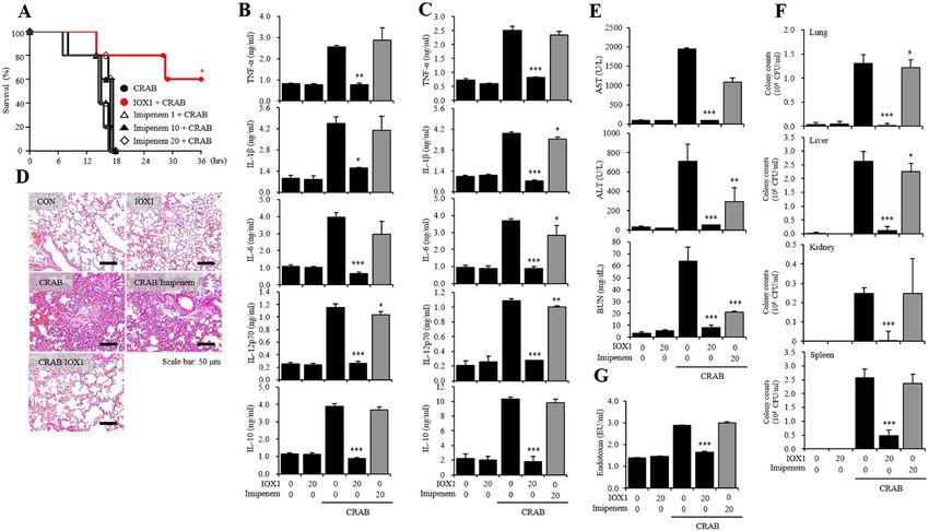
www.nature.com/scientificreports/
Figure 1. IOX1 suppresses the inflammatory response in LPS-induced DC maturation. (A) Chemical structure
of IOX1. (B) Mouse bone marrow-derived DCs were treated with the indicated concentrations of IOX1, DMSO
or H2O2 (negative control) overnight. The cytotoxicity of IOX1 in DCs was analyzed by a Luminescent Cell
Viability Kit. (C) DCs treated with IOX1 (50 μM) before or after LPS stimulation (50 ng/ml) at the indicated
times. Culture medium was collected, and the TNF-α, IL-1β, IL-6, IL-12p70 and IL-10 levels in the medium
were determined by ELISA. (D) BMDCs were pretreated for 30 min with the indicated concentrations of IOX1
before stimulation with LPS (50 ng/ml) overnight. The surface molecule expression of BMDCs was analyzed
by flow cytometry. The results of one representative experiment out of three experiments are shown. Data are
presented as the means ± SEMs. *P < 0.05; **P < 0.01; and ***P < 0.001 compared to DCs treated with LPS. n.s. no
significance.
Results
IOX1 inhibits the immune response via DC maturation induced by LPS. To investigate whether
histone lysine methylation is a targetable therapeutic point, we checked the expression change of enzymes that
are responsible for histone methylation and found that Kdm4a and Kdm6b are the most upregulated enzymes
upon LPS treatment in dendritic cells (DCs) (Fig. S1). Next, we searched for specific inhibitors of Kdm4a and
Kdm6b. Given the advantages of IOX1, we selected IOX1 from among a variety of Kdm inhibitors. The structure
of IOX1 (8-hydroxyquinoline-5-carboxylic acid) is shown in Fig. 1A. The cytotoxicity of IOX1 was measured
after treatment of dendritic cells with the indicated concentration of IOX1 in the same amount of DMSO and
a positive control, H2O2. In the case of DMSO, up to 0.5% of the volume of DMSO used to dissolve IOX1 was
found to be noncytotoxic, and in the case of IOX1, no cytotoxicity was observed below 200 μM (Fig. 1B). There-
fore, the concentration of IOX1 for DC function analysis in the future should be less than 200 μM. To analyze
the effect of IOX1 on LPS-induced DC activation, we investigated the changes in inflammatory cytokine mRNA
expression and secretion (Fig. S2 and Fig. 1C) and the expression of surface molecules as maturation markers
(Fig. 1D). IOX1 significantly reduced the secretion of TNF-α, IL-1β, IL-6, IL-12p70 and IL-10 induced by LPS
(Fig. 1C). The expression of surface molecules, such as CD80, CD86, MHC-I and MHC-II, induced by LPS was
also significantly reduced (Fig. 1D). These results indicate that IOX1 can effectively regulate the immune activity
of bone marrow DCs induced by LPS.
IOX1 suppresses the immune response by inhibiting Kdm4a. To determine whether the effect of
IOX1 is caused by inhibition of Kdm, we performed immunoprecipitation with histone H3 and Kdm4a, followed
by Western blot. In correlation with the transcriptome data, Kdm4a was highly upregulated upon LPS addition
Scientific Reports | (2021) 11:2942 | https://doi.org/10.1038/s41598-021-82377-z 2
Vol:.(1234567890)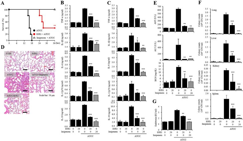
www.nature.com/scientificreports/

                                  Figure 2.  IOX1 reverses the downregulation of histone methylation via Kdm4a modification by LPS in vitro
                                  and in vivo. DCs from C57BL/6 mice were stimulated with 50 ng/ml LPS in the presence or absence of 50 μM
                                  IOX1 overnight. Lungs from BALB/c mice were injected with 10 mg/kg LPS in the presence or absence of
                                  20 mg/kg IOX1 overnight. Total protein extracts from DCs and lungs treated with IOX1 were used to detect
                                  Kdm4a (A,C), H3K9me3 and H3K36me3 (B,D) by Western blotting, Histone H3 was used as a loading control
                                  (full-length blots are presented in Figs. S10–S13); Immunoprecipitation (IP) assays were performed on DCs
                                  treated in the presence or absence of IOX1, using specific antibodies to Kdm4a and histone H3 as controls (full-
                                  length blots are presented in Figs. S10 and S12).

                                  in BMDCs, and the target modifications H3K36me3 and H3K9me3 levels decreased as Kdm4a levels increased
                                  (Fig. 2A,B). IOX1 significantly suppressed Kdm4a upregulation and restored H3K36me3 and H3K9me3 levels
                                  (Fig. 2A,B). Changes in Kdm4a levels and target histone methylation induced by LPS and IOX1 were mimicked
                                  in the lungs of mice (Fig. 2C,D). These results suggest that the inhibitory effect of IOX1 on the immune response
                                  occurs via a mechanism of Kdm inhibition.

                                  IOX1 anti‑inflammatory effect on an LPS‑induced endotoxicity mouse model.                        We initiated an
                                  in vivo study by validating the anti-inflammatory effect of IOX1. A 30-min pretreatment with IOX1 (20 mg/
                                  kg, i.p.) prior to LPS (20 mg/kg, i.p.) injection ameliorated the survival of endotoxemia-induced mice. The sur-
                                  vival rate increased within 18 h from 0% in mice injected solely with LPS to 40% in mice pretreated with IOX1
                                  (Fig. 3A). In mouse serum, the increased levels of TNF-α, IL-1β and IL-6 induced by LPS were significantly
                                  reduced by IOX1 administration (Fig. 3B). In lung tissues, the expression of TNF-α, IL-1β and IL-6 was increased
                                  by LPS injection, and infiltration of PMNs (polymorphonuclear leukocytes) was also significantly reduced by
                                  IOX1 administration (Fig. 3C,D). Furthermore, the levels of AST, ALT, BUN and creatinine, biomarkers of organ
                                  failure, that were enhanced by LPS injection were reduced by IOX1 administration (Fig. 3E). These results con-
                                  firmed that IOX1 has an anti-inflammatory effect on the LPS-induced endotoxicity mouse model.

                                  IOX1 antiseptic effect on an A. baumannii‑inoculated septic mouse model. Furthermore, an
                                  enhanced survival rate was observed in an A. baumannii-inoculated septic mouse model. Using the carbapenem
                                  antibiotic imipenem (1 mg/kg) as a control drug (100% survival rate), intraperitoneal administration of a clinical
                                  standard strain of A. baumannii (1.9 × 10⁴ CFU/mouse) 30 min after IOX1 administration resulted in reduced
                                  sepsis incidence within 18 h, with a 40% increase in the survival rate in the group pretreated with IOX1 (20 mg/
                                  kg) compared with that in the group injected only with A. baumannii (Fig. 4A). The levels of the proinflam-
                                  matory cytokines TNF-α, IL-1β, IL-6, IL-12p70, and IL-10 in the serum (Fig. 4B) and in the lungs (Fig. 4C)
                                  of the A. baumannii-derived sepsis mouse model treated with IOX1 were significantly reduced. H&E staining
                                  demonstrated that IOX1 could repress PMN (polymorphonuclear leukocyte) infiltration in the tissue of the cor-
                                  responding sepsis mouse model (Fig. 4D) and could also mitigate AST, ALT, BUN and creatinine levels in the
                                  lungs of the same mice (Fig. 4E). Interestingly, IOX1 showed an effective inhibition of colony growth (CFU) of
                                  A. baumannii in the major organs. A total repression of colony numbers was observed in the lung, kidney and
                                  spleen, and a significant reduction was reported in the liver (Fig. 4F). Additionally, the endotoxin levels in mouse
                                  serum were also significantly reduced (Fig. 4G). Notably, IOX1 effects were reproducible in an E. coli K1 sepsis
                                  mouse model, with a survival rate of 60% (Fig. S3). These results show that IOX1 can act as an endotoxin removal
                                  agent and an effective inhibitory molecule against bacterial colony growth, resulting in effective treatment of A.
                                  baumannii-induced sepsis.

Scientific Reports |   (2021) 11:2942 |                https://doi.org/10.1038/s41598-021-82377-z                                                   3

                                                                                                                                               Vol.:(0123456789)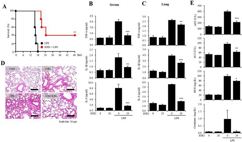
www.nature.com/scientificreports/
Figure 3. IOX1 anti-inflammatory effects on an LPS-induced endotoxemia mouse model. Six-week-old female
BALB/c mice were intraperitoneally injected with IOX1 (20 mg/kg). After 30 min, the mice were i.p. injected
with LPS. (A) The survival rates of IOX1- and LPS (20 mg/kg)-injected mice were monitored for 48 h. (B) The
serum of IOX1- and LPS (20 mg/kg)-injected mice were harvested 2 h after LPS injection. The levels of serum
proinflammatory cytokines (TNF-α, IL-1β and IL-6) were measured by sandwich ELISA kits. (C–E) After
overnight incubation, IOX1- and LPS (10 mg/kg)-injected mice were sacrificed for experiments. (C) The mouse
lungs were homogenized by stainless steel beads. The lung levels of proinflammatory cytokines (TNF-α, IL-1β
and IL-6) were measured by sandwich ELISA kits. (D) The PMN infiltrations in the lung were stained using the
hematoxylin and eosin standard staining method. (E) The levels of serum AST, ALT, BUN and creatinine were
measured by a laboratory medicine system.
IOX1 antibiotic effect on gram‑negative bacteria. Next, we sought to elaborate on the data obtained
above (Fig. 4F) by investigating the potential antibacterial activity exerted by IOX1 against bacteria. First, we
analyzed the time-kill kinetics profile of IOX1 against E. coli (DH5α) and A. baumannii following different addi-
tion of concentrations of 12.5, 25, 50 and 100 μg/ml at various checkpoint times of 0, 4, 6, 8, 10 and 12 h. IOX1
completely inhibited the growth of E. coli (DH5α) at up to 12 h at a concentration of 25 μg/ml, the same con-
centration for which antibiotic activity was shown for the positive control imipenem (Fig. 5A), and inhibited A.
baumannii at a concentration of 50 μg/ml (Fig. 5B). IOX1 exhibited inhibitory growth effects on different types
of gram-negative bacterium, S.enteritidis (Fig. S4A), S.typhimurium (Fig. S4B), K.pneumoniae (Fig. S4C), and
P.aeruginosa (Fig. S4D), with complete bacterial growth inhibition at a concentration of 50 μg/ml at up to 12 h.
The antibacterial effect of IOX1 was also confirmed for the gram-positive bacterium S. aureus (Fig. S5A) and
colistin-resistant AB (Fig. S5B) in a concentration-dependent manner, with complete bacterial growth inhibi-
tion at a concentration of 50 μg/ml at up to 12 h and 8 h, respectively. The antibacterial activity against not only
gram-negative bacteria but also gram-positive bacteria was shown. This antibacterial effect against different
types of bacteria was further proven in a cecal content injection (CCI) mouse model (Fig. S6). These results
indicate that IOX1 exhibits a bactericidal effect that led to a significant reduction in the bacterial colony count
and endotoxin levels.
IOX1 inhibits DNA gyrase activity similarly to quinolone antibiotics. To further understand the
mode of action (MOA) of IOX1, we tested whether JIB-04, a well-known cell-permeable pan-Jumonji histone
demethylase inhibitor, could also exhibit the same antimicrobial activity against E. coli (DH5α). However, unlike
IOX1, JIB-04 showed a negligible effect (Fig. S7). These data suggest that the antimicrobial activity of IOX1
was not due to the cell permeability of histone demethylase inhibitors but probably to the ability of IOX1 to
inhibit DNA gyrase, a mechanism feature of quinolone antibiotics (2), as IOX1 is structurally similar to this
family of antibiotics (Fig. 1A). Therefore, a molecular docking study was performed to investigate the binding
modes between IOX1 and E. coli DNA gyrase. The 3D structure of E. coli DNA gyrase was obtained from the
Protein Data Bank (http://www.rcsb.org, PDB ID: 6RKW). IOX1 was docked into the active site of E. coli DNA
gyrase using GOLD software20,21. The information for the initial IOX1-binding site in E. coli DNA gyrase for
Scientific Reports | (2021) 11:2942 | https://doi.org/10.1038/s41598-021-82377-z 4
Vol:.(1234567890)
www.nature.com/scientificreports/
Figure 4. IOX1 antiseptic effect on an A. baumannii-inoculated septic mouse model. Six-week-old female
BALB/c mice were intraperitoneally injected with IOX1 (20 mg/kg). After 30 min, the mice were i.p. injected
with A. baumannii (KUMC ATCC 19606, 1.9 × 10⁴ CFU/mouse). (A) The survival rates of IOX1- and A.
baumannii-injected mice were monitored for 48 h. (B) The serum of mice was harvested 2 h after A. baumannii
(9.5 × 10³ CFU/mouse) injection in the presence or absence of IOX1. The levels of serum inflammatory
cytokines (TNF-α, IL-1β, IL-6, IL-12p70 and IL-10) were measured by sandwich ELISA kits. (C–G) After
overnight incubation, IOX1- and A. baumannii (9.5 × 10³ CFU/mouse)-injected mice were sacrificed for
experiments. (C) The mouse lungs were homogenized by stainless steel beads. The levels of lung inflammatory
cytokines (TNF-α, IL-1β, IL-6, IL-12p70 and IL-10) were measured by sandwich ELISA kits. (D) PMN
infiltrations in the lung were stained using the H&E standard staining method. (E) The serum levels of AST,
ALT, BUN and creatinine were measured by a laboratory medicine system. (F) The mouse lungs, livers, kidneys
and spleens were homogenized by stainless steel beads. The lysates were diluted with PBS and incubated on LB
agar plates overnight. (G) The serum levels of endotoxin were determined by the LAL method and measured at
405 nm.
Figure 5. IOX1 antibiotic effect on gram-negative bacteria. (A) Bactericidal kinetics of IOX1 against susceptible
DH5a (4.03 × 10⁴ CFU/ml) and (B) A. baumannii KUMC.2015.sus (4.3 × 10⁴ CFU/ml).
the docking simulation was provided from the recently published 3D structure for the complex system of the
ligand and DNA gyrase22. The best docked conformation was selected based on cluster analysis and the GOLD
fitness score (Fig. 6A,B). The best cluster number from 100 trials for IOX1 was 80, and the GOLD fitness score
was 43.2 (Table S1). Our molecular docking results showed that IOX1 formed hydrogen bonds with the D1-T2
and D2-T2 bases of both DNA strands (Fig. 6C,D). IOX1 also bound with the D1-A3 and D2-A3 bases of the
Scientific Reports | (2021) 11:2942 | https://doi.org/10.1038/s41598-021-82377-z 5
Vol.:(0123456789)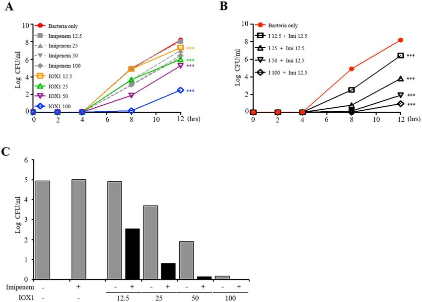
www.nature.com/scientificreports/
Figure 6. Mode of action of IOX1 as an antibiotic against bacteria. (A) Front views of the 3D structure of E. coli
DNA gyrase. DNA chains 1 and 2 are shown in cyan and blue, and subunits A and B are shown in gray and dark
gray, respectively. The active site is shown in the red cycle, and the IOX1 surface is shown in red. (B) Close-up
view of the active site of E. coli DNA gyrase after 90° rotation for a better view. (C) Close-up view of the active
site in E. coli DNA gyrase. The stick models of IOX1, DNA chain 1 and DNA chain 2 are shown in yellow, cyan,
and blue, respectively. Hydrogen bond, hydrophobic, and electrostatic interactions are shown as green, pink,
and orange, respectively. (D) Close-up view after 90° rotation showing a different angle view. (E) The mobility
of supercoiled bacterial DNA following IOX1 treatment processed by relative quantitation via ImageJ software.
(F) Enzymatic evaluation of ATPase activity of E. coli DNA gyrase in the presence of IOX1 from 0.5 to 10 μM
compared with that of the negative control (no enzyme) and the positive control (no inhibitor).
DNA strands via hydrophobic interactions and electrostatic interactions. Interestingly, the models showed that
IOX1 interacted mainly with the DNA and that the interaction with the DNA gyrase protein seemed to be very
weak or minor. In conclusion, the molecular docking study clearly confirmed that IOX1 interacted mainly with
the cleft of double-stranded DNA, which is bound to DNA gyrase, rather than with DNA gyrase itself. The
overall binding pattern of IOX1 is in good agreement with the experimental data22. Furthermore, we performed
a gyrase inhibition assay, and the relaxed circular PBR322 plasmid, which usually forms a supercoil structure
in the presence of gyrase activity, was used as a substrate. Supercoils and relaxed plasmids were determined by
agarose gel electrophoresis (Fig. 6E). Inhibition of gyrase activity increased the relaxed plasmid morphology by
regulating the activity of subunit A in DNA gyrase16. In addition, it has been suggested that antibacterial activity
is potentially demonstrated through inhibition of both subunit A and subunit B of DNA gyrase12; moreover, our
binding test confirmed the binding of IOX1 to the whole DNA gyrase (Fig. S8A) and precisely its binding to the
DNA Gyrase Subunit B (Fig. S8B). Therefore; we tried to determine whether IOX1 directly regulates the activity
of DNA Gyrase Subunit B by performing a DNA gyrase ATPase binding assay. This method is known to measure
the conversion of NADH to N AD+ by ATP hydrolysis and assess N AD+ production via a 340 nm s pectrogram19.
DNA gyrase ATPase activity was reduced in the presence of IOX1 compared to that in the negative control
(Fig. 6F). These results demonstrate that IOX1 is involved in both the A and B subunits of DNA gyrase, inhibit-
ing overall gyrase activity.
IOX1 increases the bactericidal effect against carbapenem‑resistant A. baumannii. The anti-
microbial activity of IOX1 was also tested against resistant strains of bacteria. Carbapenem-resistant A. bauman-
nii (CRAB) was found to proliferate in the presence of high concentrations of imipenem; in contrast, IOX1 suc-
ceeded in inhibiting bacterial growth in a concentration-dependent manner at up to 8 h (Fig. 7A). Additionally,
analysis of time-kill kinetics profiles of imipenem (carbapenem antibiotic), ciprofloxacin (quinolone antibiotic)
and IOX1 showed that, unlike IOX1, imipenem and ciprofloxacin exhibited no inhibitory growth effects on
multidrug (carbapenem and quinolone)-resistant (MDR) A. baumannii (Fig. S9A–D). Interestingly, IOX1 treat-
ment with imipenem (12.5 μg/ml) suggested a synergetic effect on CRAB growth (Fig. 7B). This observation
was further demonstrated when enhanced antimicrobial effects were observed by combining imipenem with
IOX1 (12.5, 25, 50 and 100 μg/ml) (Fig. 7C). This result shows that IOX1 exerts a promising antibacterial effect
on MDR bacteria.
IOX1 antiseptic effect on a carbapenem‑resistant A. baumannii‑inoculated septic mouse
model. We sought to study the antiseptic effect of IOX1 in vivo. For this aim, we used a CRAB-inoculated
septic mouse model. After 30 min of pretreatment with IOX1, mice were injected with CRAB (1.4 × 10⁴ CFU/
Scientific Reports | (2021) 11:2942 | https://doi.org/10.1038/s41598-021-82377-z 6
Vol:.(1234567890)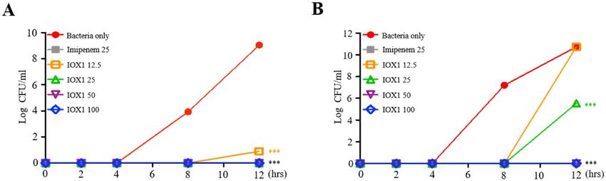
www.nature.com/scientificreports/
Figure 7. IOX1 enhances the killing of carbapenem-resistant A. baumannii. (A) Bactericidal kinetics of
imipenem against resistant and susceptible CRAB 90 (4.66 × 10⁴ CFU/ml) compared to those of IOX1. (B)
Synergistic antibacterial effect of a low concentration combination of IOX1 and imipenem on CRAB 90
(4.66 × 10⁴ CFU/ml). (C) The effect of IOX1 on bacterial growth in the presence or absence of imipenem at 8 h
of culture.
mouse). Within 18 h, the survival rates of mice injected solely with CRAB and mice treated with imipenem were
0%. However, IOX1 was able to increase the survival rate to 80% and to maintain it at 60% for 36 h (Fig. 8A).
Additionally, unlike imipenem, which showed no inhibitory effect, the levels of inflammatory cytokines, includ-
ing IL-10, TNF-α, IL-1β, IL-6 and IL-12p70, in serum (Fig. 8B) and in the lung (Fig. 8C) were reduced by
IOX1 administration. IOX1 was also found to be effective in reducing lung PMN infiltration (Fig. 8D) and in
attenuating organ damage, as reflected by a reduction in the levels of AST, ALT, BUN and creatinine (Fig. 8E).
IOX1-mediated bacterial clearance was also investigated, and the data showed an abrogated number of remain-
ing bacteria in the liver, lungs, kidneys and spleen (Fig. 8F). Furthermore, IOX1 reduced the endotoxin levels
in mouse serum that were originally induced by CRAB inoculation (Fig. 8G). These results show the promising
therapeutic effects of IOX1 on sepsis triggered by carbapenem-resistant bacteria.
Discussion
Unlike other histone modifications, histone methylation was discovered relatively late, and study of the reversible
nderway23. However, recent histone demethylase studies have been actively conducted in several
reaction is still u
fields; for example, Kdm6b is involved in the expression of comprehensive inflammatory genes in macrophages24
and Kdm4a is markedly expressed in neuroectodermal stem cells to induce tumor development25. The most
widely used 2OG analogs N-oxalyl glycine (NOG) and pancreatic ductal adenocarcinoma (PDAC), general inhib-
itors of 2OG oxygenase, have poor cell permeability26, requiring the use of prodrug diester derivatives, whereas
IOX1 can be transferred to the cytoplasm and nucleus without ester m odification27. Herein, we confirmed that
IOX1 inhibited the induction of proinflammatory cytokines in LPS-stimulated DCs and, in parallel, inhibited
the activity and expression of Kdm4a and Kdm6b induced by LPS (Figs. 1 and 2). In addition, IOX1 showed
significant therapeutic effects on bacterial-induced mouse sepsis models, including those with antibiotic-resistant
bacteria. In particular, the bacterial count in each organ caused by bacterial infection was demonstrated to be
reduced (Figs. 4F, 8F, and S3F). Similar to the other derivatives, IOX1, one of the small molecules with improved
physicochemical properties among 8-hydroxyquinolines, displayed direct antimicrobial activity. Remarkably, it
showed broad antimicrobial effects on E. coli, Acinetobacter baumannii (AB), double antibiotic-resistant (MDR)
Scientific Reports | (2021) 11:2942 | https://doi.org/10.1038/s41598-021-82377-z 7
Vol.:(0123456789)www.nature.com/scientificreports/
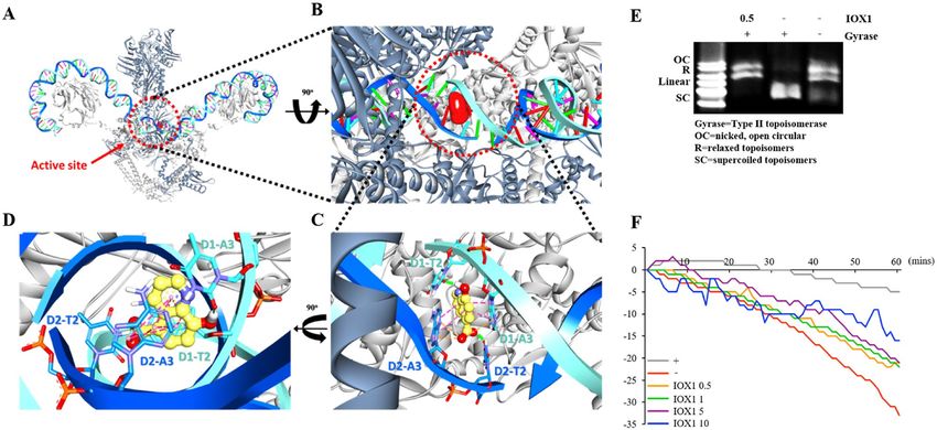
Figure 8. IOX1 antiseptic effect on a carbapenem resistant A. baumannii-inoculated septic mouse model.
Six-week-old female BALB/c mice were intraperitoneally injected with IOX1 (20 mg/kg). After 30 min, the mice
were i.p. injected with CRAB. (A) The survival rate of IOX1- and CRAB (1.4 × 10⁴ CFU/mouse)-injected mice
was monitored for 48 h. (B) The serum of IOX1- and CRAB (7 × 10³ CFU/mouse)-injected mice was harvested
2 h after LPS injection. The serum levels of inflammatory cytokines (TNF-α, IL-1β, IL-6, IL-12p70 and IL-10)
were measured by sandwich ELISA kits. (C–G) After overnight incubation, IOX1- and CRAB (7 × 10³ CFU/
mouse)-injected mice were sacrificed for experiments. (C) The mouse lungs were homogenized by stainless steel
beads. The lung levels of inflammatory cytokines (TNF-α, IL-1β, IL-6, IL-12p70 and IL-10) were measured by
sandwich ELISA kits. (D) The PMN infiltrations in the lung were stained by hematoxylin and eosin based on
the standard H&E staining method. (E) The serum levels of AST, ALT, BUN and creatinine were measured by
a laboratory medicine system. (F) The mouse lungs, livers, kidneys and spleens were homogenized by stainless
steel beads. The lysates were diluted with PBS and incubated on LB agar plates overnight. (G) The serum levels
of endotoxin were determined by the LAL method and measured at 405 nm.
bacterium (quinolone and carbapenem-resistant AB), colistin-resistant AB and gram-positive bacteria, thus
demonstrating excellent antimicrobial performance. We elucidated the mechanism of action that modulates the
broad-spectrum antimicrobial activity of IOX1 (Fig. 6). In addition to immunosuppressive activation of IOX1
through Kdms, we found that nonquinolone antibiotics rarely inhibit bacterial DNA gyrase, resulting in bacte-
rial antimicrobial activity and anti-inflammatory responses. As a demonstration of dual function, we presented
molecular modeling based on structural analysis that can be directly linked as direct and indirect evidence (Fig. 6
and Table S1) and showed that IOX1 simultaneously binds to bacterial DNA gyrase subunit B through isothermal
titration calorimetry (ITC) analysis (Fig. S8B). Its mode of action is unique in that it directly interacts with DNA
gyrase to prevent its binding to DNA. This study provides the first structural explanation of the Escherichia coli
DNA gyrase-targeting mechanism of IOX1. Developing sepsis drugs has been limited by the availability of a
mouse model for drug screening, which is currently regarded as a challenge, comprehending that inflammatory
conditions, such as trauma, burns, and infections, in mice are significantly different from those of the human
immune system. Moreover, most studies are based on the host’s immune system; therefore, it is very likely that
the compounds that show efficiency in mice will not be as effective in humans because of the variation in the
immune system response and the whole physiological system between the two species. Extensive studies on the
introduction of Kdm or topoisomerase inhibitors have recently been initiated, but none have been shown to
simultaneously inhibit Kdm and DNA gyrase, as IOX1 does. These results demonstrate that IOX1 can be used as a
broad-spectrum antibiotic, with the possibility of expanding the opportunity to develop and improve new drugs.
Materials and methods
Animals. Six-week-old female C57BL/6 mice (H-2 Kb and I-Ab) and 6-week-old Female BALB/c mice
(20 mg) were purchased from Orient Bio (Daejeon, South Korea). All experiments were performed in accord-
ance with relevant guidelines and regulations, and all procedures were monitored and approved by the Insti-
tutional Animal Care and Use Committee (IACUC) of Konkuk University (IACUC number: KU17044-2)30,31.
Scientific Reports | (2021) 11:2942 | https://doi.org/10.1038/s41598-021-82377-z 8
Vol:.(1234567890)www.nature.com/scientificreports/
Bacteria. E. coli DH5α (ATCC PTA-4750), Acinetobacter baumannii (KUMC ATCC 19606, used in vivo),
Acinetobacter baumannii (KUMC.2015.sus, used in time kill assays) and Staphylococcus aureus USA:300
(ATCC BAA-1556) was obtained from the American Type Culture Collection (Manassas, VA, USA). The E. coli
K1 strain RS218 (O18:K1:H7) used in the E. coli K1-induced sepsis mouse model was kindly gifted by Dr. Jang-
Won Yoon of Kangwon National University (Gangwon, South Korea). Carbapenem-resistant Acinetobacter bau-
mannii was obtained from a patient with informed consent following the approved protocol of the Institutional
Review Board for Human Study at Korea University Anam Hospital (ED14326/AN14326-001). All methods
were carried out in accordance with relevant guidelines and regulations. Multi drug resistant (MDR)-AB strain
15–20, 15–21, K-YYK-21 and K-YYK-22 used in the bacterial sepsis mouse model were kindly gifted by Dr.
Min‐Goo Lee of Korea University College of Medicine (Seoul, South Korea)30,31.
Reagents and antibodies. Recombinant mouse granulocyte–macrophage colony stimulating factor (GM-
CSF) was purchased from BioLegend (San Diego, CA, USA). To maintain DCs, RPMI 1640 medium, fetal bovine
serum (FBS), and penicillin–streptomycin solution were used, purchased from Biowest (Nouaille, France). E.
coli O111:B4 LPS used in the in vitro experiments was purchased from Invivogen (San Diego, CA, USA) and E.
coli O127:B8 LPS used for the in vivo experiments was purchased from Sigma-Aldrich (St. Louis, MO, USA).
Quinolone antibiotics ciprofloxacin was purchased from Sigma-Aldrich (St. Louis, MO, USA). IOX1 and JIB-04
were purchased from Selleckchem (Houston, TX, USA). An MTT Cell viability kit was purchased from Pro-
mega (Madison, WI, USA). For western blotting, Kdm4A, H3K9me3, H3K36me3 and H3 were purchased from
Abcam (Cambridge, MA, USA). Protease inhibitor cocktail Luria–Bertani (LB) broth powder and LB agar pow-
der were both purchased from Biobasic (Amherst, NY, USA) and a LAL Endotoxin Detection Kit was purchased
from Lonza (Basel, Switzerland)30,31.
Generation of murine bone marrow DCs (BMDCs). Bone marrow from the tibias and femurs was
used to isolate DCs after euthanasia of C57BL/6 mice following IACUC guidelines and in compliance with
ARRIVE guidelines. All methods were carried out in accordance with relevant guidelines and regulations. Red
blood cells were removed from the bone marrow using red blood cell lysis buffer, leaving only progenitor cells.
These progenitor cells were placed into cell culture plates with RPMI 1640 (containing 10% FBS, 1% penicillin/
streptomycin, and 10 ng/ml of GM-CSF) and incubated for 6 d at 37 °C under a 5% CO2 atmosphere. After six
days, precursor cells had differentiated into immature DCs. To produce mature DCs, immature DC were treated
with 50 ng/ml LPS and incubated overnight at 37 °C under 5% CO230,31.
Cell viability assay. The BMDCs were seeded at 2 × 10⁶ cells/well in a 96-well microplate and incubated
with IOX1 (10, 50, 100, 200, and 500 μM) for overnight. Cell Viability Assay is assessed the ATP present, an
indicator of metabolically active cells using Luminescent cell viability kit (Promega, Madison, WI, USA). The
ATP present into the cell culture supernatant was quantified using a L uminometer30,31.
Cytokine enzyme‑linked immunosorbent assays (ELISAs). The levels of various pro-inflammatory
cytokines, including tumor necrosis factor-α (TNF), interleukin 1β (IL-1β), interleukin 6 (IL-6), and interleukin
12p70 (IL-12p70) and anti-inflammatory cytokine, interleukin 10 (IL-10) were measured by sandwich ELISA
(eBioscience, San Diego, CA, USA). Optical density at 450 nm was measured using a Sunrise Spectrophotometer
(TECAN, Männedorf, Switzerland)30,31.
Surface staining. The BMDCs were stained by FITC anti-CD11c antibody (Clone: N418), PE anti-CD86
antibody (Clone: GL-1), PE anti-H-2Kd/H-2Dd antibody (Clone: 34–1-2S) and PE anti-I-A/I-E PE antibody
(Clone: M5/114.15.2) (BioLegend). The expressions of CD80, CD86, MHC class I and MHC class II on the
BMDC were analyzed by flow cytometry (FACSCalibur, BD).
Real time‑polymerase chain reaction (RT‑PCR). Total RNA was extracted using TRIzol reagent
(Thermo Fisher Scientific, San Jose, CA, USA), digested with DNase I (Biobasic, Amherst, NY, USA), and
reverse transcribed using a High-Capacity cDNA Reverse Transcription Kit (Applied Biosystems, Foster City,
CA, USA). Amplification of the cDNA was performed using a LightCycler 480 II (Roche, Basel, Switzerland) and
LightCycler 480 SYBR Green I Master mix (Roche, Basel, Switzerland), according to the manufacturer’s recom-
mended conditions30,31. The PCR primer sequences are given in Table S2.
Detection of AST, ALT, BUN and creatinine in mouse serum. Serum levels of AST (aspartate ami-
notransferase), ALT (alanine aminotransferase), BUN (blood urea nitrogen) and Creatinine were analyzed using
total laboratory automation (Hitachi, Japan) and TBA-200FR NEO (Toshiba, Japan) systems at the Konkuk
University Medical Center30,31.
Hematoxylin and Eosin (H&E) staining. Lungs were extracted from mice, fixed in 4% paraformalde-
hyde (PFA) solution, and dehydrated to prepare paraffin blocks. The paraffin blocks were then cut to a thick-
ness of 5 μm to prepare tissue slides. Any remaining paraffin was removed with xylene. Tissue slides were then
hydrated, stained with H&E, and dehydrated. After mounting the tissue slides, images were captured under a
microscope30,31.
Scientific Reports | (2021) 11:2942 | https://doi.org/10.1038/s41598-021-82377-z 9
Vol.:(0123456789)www.nature.com/scientificreports/
Western blotting and immunoprecipitation (IP). Proteins (a total of 25 μg per sample) were sepa-
rated by 10% sodium dodecyl sulfate polyacrylamide gel electrophoresis (SDS-PAGE) and transferred to poly-
vinylidene fluoride membranes. After blocking the membranes with 5% skim milk in Tris-buffered saline con-
taining 0.05% Tween-20 (TBS-T), they were incubated with the indicated antibodies. After washing with TBS-T,
the membranes were incubated with a secondary antibody conjugated to horseradish peroxidase and visualized
using an enhanced chemiluminescence kit (Merck, Darmstadt, Germany). For immunoprecipitations (IP), the
corresponding Ab was added to 1 ml each of the cellular extracts and incubated at 4 °C for 1 h on a rotator. Fifty
microliters of a 50% slurry of prewashed protein A-agarose beads was then added to each sample, followed by
incubation for an additional 12 h at 4 °C. The samples were washed four times in lysis buffer and subjected to
Western blot analyses as described above30,31.
Determination of bacterial counts in organ tissues. At the time of sacrifice, the lungs, liver, and kid-
neys were removed aseptically and placed separately in 1 ml of sterile PBS. The tissues were then homogenized
on ice using a tissue homogenizer under a vented hood. The lung, liver, and kidney homogenates were diluted
with PBS to 1:1000. After plating 10 μL of each diluted sample onto LB agar, the plates were then incubated
at 37 °C for 24 h. The numbers of colonies were then counted and used to assess the relative abundances of
bacteria30,31.
Detection of endotoxin in mouse serum. The levels of endotoxin in mouse sera were measured using
a LAL chromogenic end point assay (Lonza Group Ltd., Allendale, NJ, USA), according to the manufacturer’s
recommendations. Mouse sera were diluted tenfold in endotoxin-free PBS before the assay. After subtracting
background levels, the results were calculated relative to an E. coli endotoxin standard provided with the assay
kit. The relative amounts of endotoxin in each sample are expressed as EU/ml30,31.
LPS‑induced mouse endotoxemia model. The 6-week-old female BALB/c mice were intraperitoneally
injected 20 mg/kg of IOX1 (Sellectchem). For survival rate analysis, after the 30 min, IOX1-injected mice were
i.p. injected 20 mg/kg of LPS (E. coli O127:B8, Sigma-Aldrich, St. Louis, MO) (5 mice/group). The survival rate
was observed for 48 h. For other analysis, After the 2 h, IOX1-injected mice were i.p. injected 10 mg/kg of LPS (5
mice/group). After the 2 h, the serum was harvested for pro-inflammatory cytokine ELISA. Next day, these mice
were sacrificed depending on animal ethics. The half of the lung was fixed by paraformaldehyde. And then, these
lungs were sliced for making tissue slide and that slides were stained by hematoxylin and eosin. The other side
of lung was homogenized using a Bullet Blender homogenizer (Next Advance, NY, USA). The pro-inflammatory
cytokines in lung were measured by ELISA. The AST, ALT, BUN and creatinine levels in the serum were meas-
ured using total laboratory automation (Hitachi, Japan) and TBA-200FR NEO (Toshiba, Japan) systems. The
study was carried out in compliance with the ARRIVE guidelines.
A. baumannii (KUMC ATCC 19606)‑induced mouse sepsis model. The 6-week-old female BALB/c
mice were intraperitoneally injected 20 mg/kg of IOX1 (Selleckchem). For survival rate analysis, after the 30 min,
IOX1-injected mice were i.p. injected 1.9 × 10⁴ CFU/mouse of A. baumannii (5 mice/group). The survival rate
was observed for 36 h. For other analysis, after 30 min, IOX1-injected mice were i.p. injected 9.5 × 10³ CFU/
mouse of A. baumannii (5 mice/group). After the 2 h, the serum was harvested for pro-inflammatory cytokine
ELISA. Next day, these mice were sacrificed depending on animal ethics. The half of the lung was fixed by para-
formaldehyde. And then, these lungs were sliced for making tissue slide and that slides were stained by hema-
toxylin and eosin. The other side of lung, liver, kidneys and spleens were homogenized using a Bullet Blender
homogenizer (Next Advance, NY, USA). The pro-inflammatory cytokines in lung lysates were measured by
ELISA. The remained bacteria in tissue lysates were diluted by PBS and incubated at LB agar plate during over-
night. The AST, ALT, BUN and creatinine levels in the serum were measured using total laboratory automation
(Hitachi, Japan) and TBA-200FR NEO (Toshiba, Japan) systems. The levels of serum endotoxin were conducted
by limulus amebocytes lysate (LAL) assay method and the color reaction were measured at 405 nm. The study
was carried out in compliance with the ARRIVE guidelines.
Carbapenem‑resistant A. baumannii (CRAB)‑induced mouse sepsis model. The 6-week-old
female BALB/c mice were intraperitoneally injected 20 mg/kg of IOX1 (Selleckchem). For survival rate analy-
sis, after the 30 min, IOX1-injected mice were i.p. injected 1.4 × 10⁴ CFU/mouse of CRAB (5 mice/group). The
survival rate was observed for 48 h. For other analysis, after the 30 min, IOX1-injected mice were i.p. injected
7 × 10³ CFU/mouse of CRAB (5 mice/group). After the 2 h, the serum was harvested for pro-inflammatory
cytokine ELISA. Next day, these mice were sacrificed depending on animal ethics. The half of the lung was fixed
by paraformaldehyde. And then, these lungs were sliced for making tissue slide and that slides were stained
by hematoxylin and eosin. The other side of lung, liver, kidneys and spleens were homogenized using a Bullet
Blender homogenizer (Next Advance, NY, USA). The pro-inflammatory cytokines in lung lysates were measured
by ELISA. The remained bacteria in tissue lysates were diluted by PBS and incubated at LB agar plate during
overnight. The AST, ALT, BUN and creatinine levels in the serum were measured using total laboratory auto-
mation (Hitachi, Japan) and TBA-200FR NEO (Toshiba, Japan) systems. The levels of serum endotoxin were
conducted by limulus amebocytes lysate (LAL) assay method and the color reaction were measured at 405 nm.
The study was carried out in compliance with the ARRIVE guidelines.
Scientific Reports | (2021) 11:2942 | https://doi.org/10.1038/s41598-021-82377-z 10
Vol:.(1234567890)www.nature.com/scientificreports/
Time‑kill kinetics assay. The bactericidal activities of IOX1 were performed using a modified time-
kill kinetics assay28. DH5α (4.03 × 10⁴ CFU/ml), A. baumannii KUMC.2015.sus (4.3 × 10⁴ CFU/ml), CRAB
(4.66 × 10⁴ CFU/ml), MDR-A.baumannii AB 15–20 (1.7 × 10⁴ CFU/ml), 15–21 (1.5 × 10⁴ CFU/ml), K-YYK-21
(1.8 × 10⁴ CFU/ml), K-YYK-22 (3.1 × 10⁴ CFU/ml), S. aureus (9.0 × 10⁴ CFU/ml) and Colistin-resistant AB 2
(8.1 × 10⁴ CFU/ml) were incubated with IOX1 at different concentration (0, 12.5, 25, 50, 100 μg/ml), in MH
broth at 37 °C. Imipenem and quinolone were used as positive control, and the assay was performed in triplicate.
10 μl of bacterial suspensions was removed at various time intervals (0, 2, 4, 6, 8, and 12 h), serially diluted in
MH broth and plated onto LB agar overnight at 37 °C in the presence of 5% CO2 to obtain viable colonies. Amp
was used as positive control and the assay was performed in triplicate.
Molecular docking study. The molecular docking was performed by GOLD 5.2 (Genetic Optimization
for Ligand Docking) and the standard function GOLD Scorewas used for s coring26,27. Discovery Studio 2018
(DS) was used for preparation of SD file for the ligand IOX1 to utilize GOLD. All the active site residues within
15 Å radius spheres of the center were included for the calculation. Other parameters set as their default and the
number of GOLD runs set to 100. Top-ranked docking conformation was defined on the largest GOLD fitness
score value and clustering processed in keeping with RMSD.
Agarose gel DNA gyrase assays. Conditions to measure the supercoiling of a relaxed, closed-circular
plasmid substrate and cleavage complex stabilization in agarose gels were adapted from the literature and carried
out as described previously (29).
Enzymatic assays. The DNA gyrase ATPase linked assay was performed using an E. coli gyrase ATPase
linked assay kit according to the manufacturer’s instructions (Insiparlis cod. ATPG001). Reaction mixtures
(100 μl) contained 40 mM HEPES–KOH (pH 7.6), 10 mM magnesium acetate, 10 mM DTT, 2 mM ATP, 500 mM
potassium glutamate, 0.05 mg/ml albumin, 3 μg of relaxed pBR322 DNA, 80 mM PEP, stock pyruvate kinase/
lactate dehydrogenase (reported concentration in the manufacturer’s instructions), 20 mM NADH, 50 nM DNA
gyrase, and either 50, 25, 10, or 5 μM UVI5008 dissolved in 10% DMSO or 30 μM ciprofloxacin dissolved in
0.01 N HCl. The negative and positive controls were represented by the absence and presence of the enzyme in
10% DMSO, respectively. Absorbance at 340 nm was measured for 10 min at 25 °C. The reaction began after the
addition of 30 mM ATP. Absorbance at 340 nm was then monitored for 60 min at 25 °C30,31.
Statistical analysis. All experiments were repeated at least three times with consistent results. Unless oth-
erwise stated, data are expressed as means ± SEM. A Student’s t-test was performed to compare experimental
groups and controls and a Tukey’s multiple comparison test using Prism v3.0 (GraphPad Software, La Jolla, CA,
USA) was used to compare multiple groups. Kaplan–Meier curves for survival rates were analyzed using a log
rank test. The threshold of statistical significance was set at P < 0.0530,31.
Received: 5 October 2020; Accepted: 14 January 2021
References
1. Singer, M. et al. The third international consensus definitions for sepsis and septic shock (Sepsis-3). JAMA 315, 801–810 (2016).
2. Aldred, K. J., Kerns, R. J. & Osheroff, N. Mechanism of quinolone action and resistance. Biochemistry 53, 1565–1574 (2014).
3. Carson, W. F., Cavassani, K. A., Dou, Y. & Kunkel, S. L. Epigenetic regulation of immune cell functions during post-septic immu-
nosuppression. Epigenetics 6, 273–283 (2011).
4. Maes, T., Carceller, E., Salas, J., Ortega, A. & Buesa, C. Advances in the development of histone lysine demethylase inhibitors. Curr.
Opin. Pharmacol. 23, 52–60 (2015).
5. Rose, N. R. et al. Selective inhibitors of the JMJD2 histone demethylases: Combined nondenaturing mass spectrometric screening
and crystallographic approaches. J. Med. Chem. 53, 1810–1818 (2010).
6. Neelamegam, R. et al. Brain-penetrant LSD1 inhibitors can block memory consolidation. ACS Chem. Neurosci. 3, 120–128 (2012).
7. Thinnes, C. C. et al. Targeting histone lysine demethylases—Progress, challenges, and the future. Biochim. Biophys. Acta 1839,
1416–1432 (2014).
8. Black, M. T. et al. Mechanism of action of the antibiotic NXL101, a novel nonfluoroquinolone inhibitor of bacterial type II topoi-
somerases. Antimicrob. Agents Chemother. 52, 3339–3349 (2008).
9. Brown, P. O. & Cozzarelli, N. R. A sign inversion mechanism for enzymatic supercoiling of DNA. Science 206, 1081–1083 (1979).
10. Mizuuchi, K., Fisher, L. M., O’Dea, M. H. & Gellert, M. DNA Gyrase Action Involves the Introduction of Transient Double-Strand
Breaks Into DNA. Proc Natl Acad Sci USA 77, 1847–1851 (1980).
11. Wang, J. C. DNA topoisomerases. Annu. Rev. Biochem. 6, 635–392 (1996).
12. Barnard, F. M. & Maxwell, A. Interaction between DNA gyrase and quinolones: effects of alanine mutations at GyrA subunit
residues Ser83 and Asp87. Antimicrob. Agents Chemother. 7, 1994–2000 (2001).
13. Bates, A. D., Maxwell, A. & Maxwell, T. DNA topology (United Kingdom, Oxford, 1993).
14. Gellert, M., Mizuuchi, K., O’Dea, M. H. & Nash, H. A. DNA gyrase: An enzyme that introduces superhelical turns into DNA. Proc.
Natl. Acad. Sci. USA 73, 3872–3876 (1976).
15. Maxwell, A. & Lawson, D. M. The ATP binding site of type II topoisomerases as a target of antibacterial drugs. Curr. Top. Med.
Chem. 3, 283–303 (2003).
16. Collin, F., Karkare, S. & Maxwell, A. Exploiting bacterial DNA gyrase as a drug target: current state and perspectives. Appl. Micro-
biol. Biotechnol. 92, 479–497 (2011).
17. Andersson, M. I. & MacGowan, A. P. Development of the quinolones. J. Antimicrob. Chemother. 51(Suppl 1), 1–11 (2003).
Scientific Reports | (2021) 11:2942 | https://doi.org/10.1038/s41598-021-82377-z 11
Vol.:(0123456789)www.nature.com/scientificreports/
18. Hopkinson, R. J. et al. 5-Carboxy-8-hydroxyquinoline is a broad spectrum 2-oxoglutarate oxygenase inhibitor which causes iron
translocation. Chem. Sci. 4, 3110–3117 (2013).
19. Oblak, M., Kotnik, M. & Solmajer, T. Discovery and development of ATPase inhibitors of DNA gyrase as antibacterial agents.
Curr. Med. Chem. 14, 2033–2047 (2007).
20. Jones, G., Willett, P., Glen, R. C., Leach, A. R. & Taylor, R. Development and validation of a genetic algorithm for flexible docking.
J. Mol. Biol. 267, 727–748 (1997).
21. Verdonk, M. L., Cole, J. C., Hartshorn, M. J., Murray, C. W. & Taylor, R. D. Improved protein–ligand docking using GOLD. Proteins
52, 609–623 (2003).
22. Broeck, A. V., Lotz, C., Ortiz, J. & Lamour, V. Cryo-EM structure of the complete E. coli DNA gyrase nucleoprotein complex. Nat.
Commun. 10, 1–12 (2019).
23. Cloos, P. A. C., Christensen, J., Agger, K. & Helin, K. Erasing the methyl mark: histone demethylases at the center of cellular dif-
ferentiation and disease. Genes Dev. 22, 1115–1140 (2008).
24. Agger, K. et al. UTX and JMJD3 are histone H3K27 demethylases involved in HOX gene regulation and development. Nature 449,
731–734 (2007).
25. Shi, Y. & Whetstine, J. R. Dynamic regulation of histone lysine methylation by demethylases. Mol. Cell 25, 1–14 (2007).
26. Rose, N. R., McDonough, M. A., King, O. N. F., Kawamura, A. & Schofield, C. J. Schofield Inhibition of 2-oxoglutarate dependent
oxygenases. Chem. Soc. Rev. 40, 4364–4397 (2011).
27. King, O. N. et al. Quantitative high-throughput screening identifies 8-hydroxyquinolines as cell-active histone demethylase inhibi-
tors. PLoS ONE 5, e15535 (2010).
28. Tsuji, B. T. et al. In vitro pharmacodynamics of novel rifamycin ABI-0043 against Staphylococcus aureus. J. Antimicrob. Chemother
62, 156–150 (2008).
29. Walker, S. S. et al. Antibacterial small molecules targeting the conserved TOPRIM domain of DNA gyrase. PLoS ONE 12, e0180965
(2017).
30. Lee, S. J. et al. Drug repositioning of TANK-binding kinase 1 inhibitor CYT387 as an alternative for the treatment of Gram-negative
bacterial sepsis. Int. Immunopharmacol. 73, 482–490 (2019).
31. Park, H. J. et al. Tamarixetin exhibits anti-inflammatory activity and prevents bacterial sepsis by increasing IL-10 production. J.
Nat. Prod. 81, 1435–1443 (2018).
Acknowledgements
This research was supported by the Basic Science Research Program through the National Research Foun-
dation of Korea (NRF) and was funded by the Ministry of Education, Science and Technology (grants:
2016R1A5A2012284).
Author contributions
S.J.L., J.S.Y., and A.G. performed experiments, analyzed data, and wrote the paper. Y.J.K. analyzed data and wrote
the paper. M.S.L. performed ITC assay for the interaction of IOX1 and DNA gyrase. I.D.J. and Y.M.P. designed
experiments, analyzed data, and wrote paper. All authors read and revised the manuscript.
Competing interests
The authors declare no competing interests.
Additional information
Supplementary Information The online version contains supplementary material available at https://doi.
org/10.1038/s41598-021-82377-z.
Correspondence and requests for materials should be addressed to I.D.J. or Y.M.P.
Reprints and permissions information is available at www.nature.com/reprints.
Publisher’s note Springer Nature remains neutral with regard to jurisdictional claims in published maps and
institutional affiliations.
Open Access This article is licensed under a Creative Commons Attribution 4.0 International
License, which permits use, sharing, adaptation, distribution and reproduction in any medium or
format, as long as you give appropriate credit to the original author(s) and the source, provide a link to the
Creative Commons licence, and indicate if changes were made. The images or other third party material in this
article are included in the article’s Creative Commons licence, unless indicated otherwise in a credit line to the
material. If material is not included in the article’s Creative Commons licence and your intended use is not
permitted by statutory regulation or exceeds the permitted use, you will need to obtain permission directly from
the copyright holder. To view a copy of this licence, visit http://creativecommons.org/licenses/by/4.0/.
© The Author(s) 2021
Scientific Reports | (2021) 11:2942 | https://doi.org/10.1038/s41598-021-82377-z 12
Vol:.(1234567890)You can also read